This is a repository copy of Herpes simplex virus interferes with amyloid precursor protein
processing .
White Rose Research Online URL for this paper:
http://eprints.whiterose.ac.uk/1060/
Article:
Shipley, S.J., Parkin, E.T., Itzhaki, R.F. et al. (1 more author) (2005) Herpes simplex virus
interferes with amyloid precursor protein processing. BMC Microbiology, 5 (48). ISSN
1471-2180
https://doi.org/10.1186/1471-2180-5-48
[email protected] https://eprints.whiterose.ac.uk/
Reuse
Unless indicated otherwise, fulltext items are protected by copyright with all rights reserved. The copyright exception in section 29 of the Copyright, Designs and Patents Act 1988 allows the making of a single copy solely for the purpose of non-commercial research or private study within the limits of fair dealing. The publisher or other rights-holder may allow further reproduction and re-use of this version - refer to the White Rose Research Online record for this item. Where records identify the publisher as the copyright holder, users can verify any specific terms of use on the publisher’s website.
Takedown
If you consider content in White Rose Research Online to be in breach of UK law, please notify us by
Open Access
Research article
Herpes simplex virus interferes with amyloid precursor protein
processing
Suzanne J Shipley
1, Edward T Parkin
2, Ruth F Itzhaki
1and Curtis B Dobson*
1Address: 1Faculty of Life Sciences, Moffat Building, University of Manchester, Manchester, M60 1QD, UK and 2School of Biochemistry and
Molecular Biology, University of Leeds, Leeds, W. Yorks, LS2 9JT, UK
Email: Suzanne J Shipley - [email protected]; Edward T Parkin - [email protected]; Ruth F Itzhaki - [email protected]; Curtis B Dobson* - [email protected]
* Corresponding author
Abstract
Background: The early events underlying Alzheimer's disease (AD) remain uncertain, although environmental factors may be involved. Work in this laboratory has shown that the combination of herpes simplex virus type 1 (HSV1) in brain and carriage of the APOE-ε4 allele of the APOE gene strongly increases the risk of developing AD. The development of AD is thought to involve abnormal aggregation or deposition of a 39–43 amino acid protein – β amyloid (Aβ) – within the brain. This is cleaved from the much larger transmembranal protein 'amyloid precursor protein' (APP). Any agent able to interfere directly with Aβ or APP metabolism may therefore have the capacity to contribute towards AD. One recent report showed that certain HSV1 glycoprotein peptides may aggregate like Aβ; a second study described a role for APP in transport of virus in squid axons. However to date the effects of acute herpesvirus infection on metabolism of APP in human neuronal-type cells have not been investigated. In order to find if HSV1 directly affects APP and its degradation, we have examined this protein from human neuroblastoma cells (normal and transfected with APP 695) infected with the virus, using Western blotting.
Results: We have found that acute HSV1 (and also HSV2) infection rapidly reduces full length APP levels – as might be expected – yet surprisingly markedly increases levels of a novel C-terminal fragment of APP of about 55 kDa. This band was not increased in cells treated with the protein synthesis inhibitor cycloheximide
Conclusion: Herpes virus infection leads to rapid loss of full length APP from cells, yet also causes increased levels of a novel 55 kDa C-terminal APP fragment. These data suggest that infection can directly alter the processing of a transmembranal protein intimately linked to the aetiology of AD.
Background
The key events which initiate Alzheimer's disease (AD) remain unclear, though environmental factors have been shown to be involved [1]. The two main neuropathologi-cal features of AD – senile plaques (SP) and neurofibril-lary tangles – occur also in the normal elderly. AD may be
triggered when the numbers of these features increase to abnormal levels. Although there is much evidence sup-porting the involvement of one or both of these structures in the disease process, it is unclear whether they are involved directly, what processes underlie their forma-tion, and why their numbers rise during the development Published: 18 August 2005
BMC Microbiology 2005, 5:48 doi:10.1186/1471-2180-5-48
Received: 09 May 2005 Accepted: 18 August 2005
This article is available from: http://www.biomedcentral.com/1471-2180/5/48
© 2005 Shipley et al; licensee BioMed Central Ltd.
BMC Microbiology 2005, 5:48 http://www.biomedcentral.com/1471-2180/5/48
of AD. However, any environmental stimulus capable of leading to production of the abnormal proteins which make up these structures might thereby contribute to the occurrence of AD.
β-amyloid protein (Aβ) is the major proteinaceous com-ponent of senile plaques, and the abnormal deposition of aggregates of this protein is thought to give rise to SP for-mation. Aβ comprises a sequence of 39–43 amino acids and is formed by cleavage from the much longer amyloid precursor protein (APP), first by β-secretase then by γ -secretase. Generation of Aβ may lead eventually to the development of neurotoxic SPs, which themselves cause further tissue damage and SP generation, or alternatively to the production of small neurotoxic Aβ assemblies, which may be the most damaging form of Aβ [2]. What-ever the mechanism leading to neuronal damage after Aβ generation, any agent which interferes with amyloid sys-tems may thereby contribute to the development of AD.
Studies of the possible role of pathogens in AD were made possible in the early 1990s by the development of PCR, allowing detection of low levels of viral or bacterial DNA within human brain tissues. However very few investiga-tors have undertaken such studies, and most work has focused on herpes simplex virus type 1 (HSV1). A causal role for this virus in triggering AD was suggested by Itzhaki et al., who found that the risk of developing AD associated with carriage of an APOE-ε4 allele depends on the presence of latent HSV1 in the brain [3,4]. Coupled with the finding here of a higher APOE-ε4 allele frequency amongst individuals who suffer damage after HSV1 reac-tivation in the periphery – seen as cold sores – this indi-rectly supports a role for HSV1, perhaps acting with other pathogens [5], as a key environmental contributor to AD [3,4].
Recent epidemiological studies support the involvement of a pathogen in AD: cognitive function in AD patients declines for at least 2 months after a systemic infection [6]; cognitive decline in elderly cardiovascular patients correlates with viral burden [7]. These could be explained by systemic infection causing brain inflammation which, in turn, leads to reactivation of latent HSV1 – and conse-quent further damage in the CNS. Consistently, in mid-dle-aged controls, very few of whom would harbour HSV1 in brain [8], no cognitive decline occurs after systemic infection [9]. Enhancement of damage in brain, due to the presence of an infectious agent, is supported by the find-ing that inflammation in brain caused by lipopolysaccha-ride is augmented in pre-clinical prion-infected mice [10].
One mechanism by which HSV1 might contribute to AD was suggested by the detection of a sequence homology between Aβ and the HSV1 glycoprotein B (gB), a viral coat
protein which is involved in attachment of the virus to cells, and by the finding that synthetic peptides derived from gB can give rise to Aβ-like aggregates. The authors suggested that such viral proteins might act as a seed for amyloid plaque formation [11]. Another way in which infection might affect amyloid biochemistry is suggested by studies demonstrating that Aβ or APP are upregulated in response to a wide range of injurious stimuli, including head injury [12], stroke [13] or HIV infection [14]. Increased expression of APP – though not of the related protein amyloid precursor-like protein 2 (APLP2) – has been reported in cutaneous wound repair [15]. APP foci colocalise with sites of opportunistic infection in HIV dementia patients, including sites of herpes virus (cytomegalovirus) infection [16]. Another type of APP-HSV1 interaction has been demonstrated in axonal trans-port: APP was found to be present in HSV1 particles and it was suggested that this could lead to alterations in loca-tion and processing of APP at the nerve terminal [17] causing synaptic and neuronal dysfunction.
No studies have been made on HSV1 effects on APP or Aβ in cells in culture. Here we describe the first to investigate whether levels of APP and its metabolites are affected by HSV1 infection, using human neuronal-type cells in culture.
Results and discussion
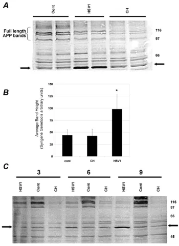
We carried out initial experiments on SHSY5Y cells to determine the level of HSV1 needed to ensure that on inoculation most cells were infected. We then examined levels of APP in lysates prepared from the cells 6 h after infection with this dose of HSV1. Western blots stained with anti C-terminal APP antibody (Fig. 1A) revealed, as expected, various full-length APP bands in uninfected cells corresponding to the three main isoforms in multiple glycosylation states. Deglycosylation experiments have shown that the lower single band (100 kDa) is immature APP695, that the doublet above this (approx molecular weight 115 kDa) is mature APP695 and immature APP751/APP770 and that the highest molecular weight group (approx. 125 to 135 kDa) comprises a mixture of mature APP751 and mature APP770 (Parkin et al., unpub-lished observations).
The intensity of these bands decreased appreciably in cells harvested only 3 h after inoculation with HSV1, as did those from cells treated with the protein synthesis inhibi-tor cycloheximide. This decline after infection may reflect loss of ability of the cells to generate new APP due simply to virus-induced shut-down of protein synthesis [18].
Effect of HSV1 infection on APP processing in SHSY5Y cells Figure 1
BMC Microbiology 2005, 5:48 http://www.biomedcentral.com/1471-2180/5/48
cycloheximide-treated cells, but had far greater intensity after HSV1 infection. This increase in HSV1-infected cells alone was highly reproducible, occurring in many experi-ments, each carried out independently, from the infection to the blotting stage. Band intensity for cells treated for 6 hours was quantified using Syngene Genetools software and averaged over five such experiments is shown in Fig-ure 1B; value for HSV1-infected cells was significantly dif-ferent from control, being elevated by 124% (95%CI 101 – 148%; p < 0.002), whereas that for cycloheximide-treated cells was similar to the control value.
To exclude the possibility that the band reflected synthesis of a viral protein which cross-reacts non-specifically with the antibody, we immunostained blots produced from gels of viral proteins from our HSV1 preparations. No evi-dence for any cross-reaction of the Sigma C-terminal APP antibody with these proteins could be found, suggesting that the strengthened band was cell-derived (results not shown). This result does not preclude any cross-reactivity with an ICP, as the latter would not be present within vir-ions, but the presence of the 55 kDa protein in uninfected cells, and also, at a higher level, in APP-transfected cells (see below), strongly supports a cellular, non-viral origin of the protein. Interestingly, APP has been detected in HSV1 virions by Western blotting [17], but this probably reflects the authors' usage of a much more concentrated and purified virus preparation than that of the inoculate we use for infecting cells.
We next examined the influence of duration of infection on these viral-induced changes (Fig. 1C). As anticipated, with increasing duration before harvesting, the depletion of APP became more pronounced (this was found also with cycloheximide treatment); in contrast, the intensity of the C-terminal 55 kDa fragment seen in the infected cells was not higher than in uninfected cells at 3 hr, but increased at 6 hr and 9 hr. However there was minimal further change as incubation increased to 24 hr (not shown). This result also was found to be reproducible on repeating the experiment several times from infection to blotting. It suggests that infection prevents synthesis of APP, leading to its gradual depletion, but causes also the abnormal accumulation of the 55 kDa fragment due, pre-sumably, to a decrease in degradation of the latter. How-ever, for longer incubation times, we cannot exclude the possibility of sequestration of some APP within newly synthesised viral particles, as reported elsewhere [17], which might reduce the amount of APP in the virus-free cell lysates used for the preparation of cell proteins.
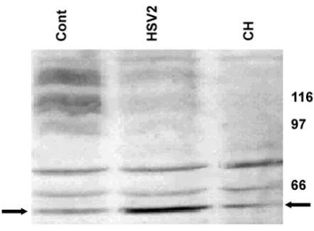
We repeated these experiments with herpes simplex virus type 2 (HSV2), to see whether these effects were specific to HSV1. Fig. 2 shows that very similar findings were obtained, with 55 kDa band intensity for HSV2-infected
cells 9 hr after infection being 101% greater than that for control cells (95%CI 94 – 107%; p < 0.005) suggesting that this phenomenon is not uniquely associated with HSV1 infection.
On the assumption that a corresponding N-terminal frag-ment of approximately similar size to that of the 55 kDa C-terminal fragment would be produced as a result of this abnormal processing (the size of APP being 110 kDa to 135 kDa), we probed our blots with the Sigma N-terminal antibody (Fig 3A). However we found no clear evidence for an increased amount of an N-terminal fragment of around this size after infection. Possibly, such a fragment would be either degraded or released into the cell culture medium, thus precluding its detection here.
To confirm further that the C-terminal fragment is derived from APP (and not for example from an APLP, i.e. of a type of protein related to APP, though with a less clear involvement in AD) we obtained human SYSY5Y cells that had been transfected with a human APP gene (APP695), and which over-express APP. We stained blots of proteins from non-transfected, APP transfected, and mock-transfected (i.e. with vector alone) cells with the C-terminal antibody, and found the 55 kDa band in all three cases, though its intensity was greater in the APP trans-fected cells alone (see Fig. 3B). This indicates that the 55 kDa fragment is indeed an APP product.
[image:5.612.325.546.89.252.2]Effect of HSV2 infection on APP processing in SHSY5Y cells Figure 2
Absence of N-terminal fragment and confirmation that 55 kDa fragment is derived from APP Figure 3
BMC Microbiology 2005, 5:48 http://www.biomedcentral.com/1471-2180/5/48
Conclusion
Infection of neuronal cells by HSV1 is known to cause rapid shutoff of protein synthesis prior to viral replication [19]. This is mediated by the virion host shut-off (vhs) protein, which destabilises cellular mRNAs causing them to be degraded. Consequently, and unsurprisingly, tran-scription of the gene(s) for APP would cease, as would its synthesis and that of other proteins. Our finding that lev-els of full length APP decline rapidly in infected cells and in cells treated with cycloheximide, suggest that in both cases turnover of full length APP is rapid.
The surprising increase in amount of the 55 kDa C-termi-nal APP fragment in infected cells shows that processing of APP as well as its synthesis is affected by HSV1, that this occurs within 6 h of infection, and that the level increases with time after infection up to at least 9 h. The size of this fragment and its staining with C-terminal antibody indi-cate that it includes the region of APP which contains Aβ.
The possibility that the 55 kDa band derives from an APLP or merely reflects a non-specific reaction of the APP anti-body with a viral protein or ICP is unlikely. Our viral prep-arations did not cross-react with this antibody, and at the time when this band begins to increase in intensity (6 h) the majority of ICPs would have already appeared. Fur-thermore the 55 kDa band is present (at lower levels) in cells not infected with virus. Also, cells transfected with APP (though not mock-transfected cells) have a higher level of this 55 kDa fragment than do untransfected unin-fected cells.
The fact that the mock-transfected and cycloheximide-treated cells do not show any intensification of the 55 kDa band suggests that this phenomenon is not due merely to a non-specific stress response, but occurs in response to infection, although the increase in the fragment after HSV2 infection shows the effect is not specific for HSV1 infection alone. Interestingly, infection of human brain microvascular endothelial cells with Chlamydia pneumonia (Cpn) may increase the intensity of a similar 55 kDa C-ter-minal APP band (personal communication, Professor Brian Balin, PA, USA). Thus, increased production of this fragment may be a general response to infection.
Currently HSV1 is the only virus that has been shown to be present in brain of most elderly humans [20] – and may be present as a whole functional genome [21]. Active infection of brain cells with other agents might lead to the same APP effects, but until or unless they are shown to be present, they can not be proposed as possible factors in AD. The situation regarding presence or absence of the bacterium Cpn in brain appears unresolved, although a direct role for Cpn in amyloid systems is supported by recent studies in mice showing amyloid deposition in
animals intracerebrally infected with Cpn [22]. Confirma-tion of Cpn presence would support the possibility that, as with HSV1, the increased level of the 55 kDa fragment that it causes might contribute to AD.
The low levels of Aβ secreted by SHSY5Y cells preclude our examining at present the effects of HSV1 infection on lev-els of Aβ in cell culture models. Eventual identification of those components of Aβ protein systems which are altered by active viral infection will clarify whether pathogens such as HSV1 can contribute directly to the development of AD neuropathology.
We report here that levels of full length APP rapidly decline in human neuronal type cells acutely infected with either HSV1 or HSV2. Also, the amount of a C-terminal 55 kDa APP fragment which contains the Aβ sequence appears to increase rapidly in infected cells. The fragment level is greater in (non-virally infected) SHSY5Y cells transfected with APP695, suggesting that it is almost cer-tainly derived from APP rather than from APLP. The fragment may increase as part of a host defence mecha-nism, and/or it might lead to increased generation of Aβ. We are now investigating whether the latter possibility is correct.
Methods
Cell culture
Human neuroblastoma (SHSY5Y) cells were maintained in Eagle's minimum essential medium (EMEM) supple-mented with 10% (v/v) foetal bovine serum (FBS), 2% (v/ v) glutamine and 1% (v/v) penicillin/streptomycin (10% medium), hereafter referred to as growth medium. Cells were incubated at 37°C in a humidified atmosphere of 5% carbon dioxide.
SHSY5Y neuroblastoma cells over-expressing APP695 were prepared by double blunt end ligation of the human APP695 sequence into the BstXI site of pIREShyg (BD Bio-sciences Clontech, California, USA). For stable transfec-tions 30 µg of DNA was introduced to cells by electroporation and selection was performed in normal growth medium containing 100 µg ml-1 hygromycin B selection antibiotic (Gibco BRL, Paisley, UK). 'Mock-transfected' cells were stably transfected with the empty pIREShyg vector.
plaque-forming units (pfu) per cell) with virus suspended in growth medium containing only 1% FBS. Once cyto-pathic end point was reached (after about two days) virus was harvested from medium and from cells disrupted by low power sonication. Cellular debris was removed by low speed centrifugation (1000 g, 10 min), and virus was isolated by high speed centrifugation of the supernatant (10000 g, 2 h, 4°C) using a Sorvall SS34 rotor. Virus-con-taining pellets were suspended in PBS, and stored in aliq-uots at -85°C; their infectivity was assessed by plaque assay of serial dilutions. The virus preparations were checked for bacterial sterility by inoculation into beef heart agar plates and confirming the absence of bacterial growth after incubation for several days at 37°C. For both viruses three sequential passages were prepared, and only passage 3 stocks used here.
SHSY5Y cells were seeded at a concentration of 8 million cells per flask (T175) and incubated overnight. Prior to infection, growth medium was discarded and cells were then washed briefly 10 ml of PBS at 37°C. HSV1 (or in some experiments HSV2) was introduced in 10 ml of growth medium (containing only 0.5% serum), at 3 pfu/ cell. For controls, either 0.5% serum containing growth medium alone or the latter containing cycloheximide at 10 µg/ml was used. After 1 hr incubation, the inoculating medium (or control treatment) was removed, and 10 ml of fresh 0.5% serum containing growth medium was added, followed by further incubation for various times.
Protein extraction and Western blotting
Cells were harvested by removing medium, washing twice with 10 ml PBS, and incubated in 1 mM EDTA (pH 7.4) (in PBS) at room temperature for 10 min. The cell suspen-sion was centrifuged (500 g, 5 min, 4°C), and the cell pel-let was resuspended in 400 µl of homogenisation buffer (0.5% Triton X-100 in PBS; 2 mM phenylmethylsulphlflu-oride (PMSF); 100 µg/ml of aprotinin and 100 µg/ml leu-peptin). Cell lysis was completed by sonication (MSE sonicator, 4°C, 10 µm amplitude, 6 × 10 s).
After measuring the protein concentration of each lysate (BCA protein assay; Pierce), samples were prepared for polyacrylamide gel electrophoresis (PAGE) by mixing 60 µg of protein with 0.25 volume of ×5 Laemmli sample buffer containing 25% β-mercaptoethanol, and boiling for 5 min. Samples were subjected to electrophoresis on 10% SDS-PAGE gels and the proteins transferred to PVDF membranes (Immobilin-P, Millipore) by Western blotting.
Membranes were blocked for 1 h using 8% skimmed-powdered milk in 0.5% Tween in TBS (TBST). The mem-brane was then washed (this and subsequent washes involved 5 separate 5 min washes in TBS) and then
incu-bated with primary antibody, diluted 1:4000 with TBS (for 1 1/2 h). Primary antibodies used were anti-C-termi-nal APP (Sigma; A8717), and the anti-N-termianti-C-termi-nal APP antibody 22C11. After a further wash, membranes were incubated with secondary antibody conjugated with per-oxidase (Pierce) which had been diluted 1:1250 with 8% milk in TBS for 1 h. After a final wash the membrane was incubated with Supersignal West Pico Chemiluminescent Substrate Kit (Pierce) for 10 min, and proteins were then visualised by chemiluminescence using a gel documenta-tion system (Syngene).
Authors' contributions
SJS carried out virology experiments and Western blotting studies, and helped to draft the manuscript. ETP prepared the APP-transfected cells, and participated in the study design. RFI participated in the study design and helped to draft the manuscript. CBD conceived of the study, coordi-nated it, and drafted the manuscript. All authors read and approved the final manuscript.
Acknowledgements
We are grateful to the Humane Research Trust, the Wellcome Trust, the Nuffield Foundation, and the Manchester Alzheimer's Research Trust Net-work for financial support. We thank also Edward Tsao who carried out some preliminary experiments and Prof Nigel Hooper (University of Leeds) for helpful discussion.
References
1. Raiha I, Kaprio J, Koskenvuo M, Rajala T, Sourander L: Alzheimer's disease in Finnish twins. Lancet 1996, 347:573-578.
2. Selkoe DJ: Deciphering the genesis and fate of amyloid beta-protein yields novel therapies for Alzheimer disease. J Clin Invest 2002, 110:1375-1381.
3. Itzhaki RF, Lin WR, Shang D, Wilcock GK, Faragher B, Jamieson GA:
Herpes simplex virus type 1 in brain and risk of Alzheimer's disease. Lancet 1997, 349:241-244.
4. Lin WR, Graham J, MacGowan SM, Wilcock GK, Itzhaki RF: Alzhe-imer's disease, herpes virus in brain, apolipoprotein E4 and herpes labialis. Alzheimer's Reports 1998, 1:173-178.
5. Dobson CB, Wozniak MA, Itzhaki RF: Do infectious agents play a role in dementia? Trends Microbiol 2003, 11:312-317.
6. Holmes C, El-Okl M, Williams AL, Cunningham C, Wilcockson D, Perry VH: Systemic infection, interleukin 1beta, and cognitive decline in Alzheimer's disease. J Neurol Neurosurg Psychiatry 2003,
74:788-789.
7. Strandberg TE, Pitkala KH, Linnavuori KH, Tilvis RS: Impact of viral and bacterial burden on cognitive impairment in elderly per-sons with cardiovascular diseases. Stroke 2003, 34:2126-2131. 8. Jamieson GA, Maitland NJ, Wilcock GK, Yates CM, Itzhaki RF:
Her-pes simplex virus type 1 DNA is present in specific regions of brain from aged people with and without senile dementia of the Alzheimer type. J Pathol 1992, 167:365-368.
9. Dickerson FB, Boronow JJ, Stallings C, Origoni AE, Cole S, Krivogor-sky B, Yolken RH: Infection with herpes simplex virus type 1 is associated with cognitive deficits in bipolar disorder. Biol Psychiatry 2004, 55:588-593.
10. Combrinck MI, Perry VH, Cunningham C: Peripheral infection evokes exaggerated sickness behaviour in pre-clinical murine prion disease. Neuroscience 2002, 112:7-11.
11. Cribbs DH, Azizeh BY, Cotman CW, LaFerla FM: Fibril formation and neurotoxicity by a herpes simplex virus glycoprotein B fragment with homology to the Alzheimer's A beta peptide.
Publish with BioMed Central and every scientist can read your work free of charge
"BioMed Central will be the most significant development for disseminating the results of biomedical researc h in our lifetime."
Sir Paul Nurse, Cancer Research UK
Your research papers will be:
available free of charge to the entire biomedical community
peer reviewed and published immediately upon acceptance
cited in PubMed and archived on PubMed Central
yours — you keep the copyright
Submit your manuscript here:
http://www.biomedcentral.com/info/publishing_adv.asp
BioMedcentral
BMC Microbiology 2005, 5:48 http://www.biomedcentral.com/1471-2180/5/48
12. Roberts GW, Gentleman SM, Lynch A, Graham DI: beta A4 amy-loid protein deposition in brain after head trauma. Lancet
1991, 338:1422-1423.
13. Popa-Wagner A, Schroder E, Walker LC, Kessler C: beta-Amyloid precursor protein and ss-amyloid peptide immunoreactivity in the rat brain after middle cerebral artery occlusion: effect of age. Stroke 1998, 29:2196-2202.
14. Esiri MM, Biddolph SC, Morris CS: Prevalence of Alzheimer plaques in AIDS. J Neurol Neurosurg Psychiatry 1998, 65:29-33. 15. Kummer C, Wehner S, Quast T, Werner S, Herzog V: Expression
and potential function of beta-amyloid precursor proteins during cutaneous wound repair. Exp Cell Res 2002, 280:222-232. 16. Wiley CA, Achim CL, Hammond R, Love S, Masliah E, Radhakrishnan L, Sanders V, Wang G: Damage and repair of DNA in HIV encephalitis. J Neuropathol Exp Neurol 2000, 59:955-965. 17. Satpute-Krishnan P, DeGiorgis JA, Bearer EL: Fast anterograde
transport of herpes simplex virus: role for the amyloid pre-cursor protein of alzheimer's disease. Aging Cell 2003,
2:305-318.
18. Matis J, Kudelova M: Early shutoff of host protein synthesis in cells infected with herpes simplex viruses. Acta Virol 2001,
45:269-277.
19. Roizman B, Sears A: Herpes simplex viruses and their replica-tion. In Fundamental virology Edited by: Fields B, Knipe D and Howley P. Philadelphia, Lippincott-Raven; 1995:1043-1107.
20. Lin WR, Wozniak MA, Cooper RJ, Wilcock GK, Itzhaki RF: Herpes-viruses in brain and Alzheimer's disease. J Pathol 2002,
197:395-402.
21. Wozniak MA, Shipley SJ, Combrinck M, Wilcock GK, Itzhaki RF: Pro-ductive herpes simplex virus in brain of elderly normal sub-jects and Alzheimer's disease patients. J Med Virol 2005,
75:300-306.