Copyright © 1997, American Society for Microbiology
The Herpes Simplex Virus Type 1 Latency-Associated Transcript Gene
Regulates the Establishment of Latency
RICHARD L. THOMPSON1ANDN. M. SAWTELL2*
Department of Molecular Genetics, Biochemistry and Microbiology, University of Cincinnati School of Medicine,1
and Division of Infectious Diseases, Children’s Hospital Medical Center,2Cincinnati, Ohio
Received 4 February 1997/Accepted 17 April 1997
Herpes simplex virus type 1 establishes latent infections in sensory neurons. During latency only one locus, the latency-associated transcript (LAT), is abundantly transcribed. Several lines of evidence suggest that this locus is required for the efficient reactivation from latency in experimental models. However, it is not yet clear whether this is a direct effect on the reactivation process per se or, as we have suggested, an indirect effect resulting from a decreased efficiency of establishment of latent infections. In this report wild-type and genetically engineered viral mutants were analyzed in a mouse model using a recently developed approach to precisely quantify latently infected neurons. It was found that strain KOS/M established latent infections, as defined by the presence of the viral genome, in about 30% of the neurons. Thirty-three percent of the mice with this latent viral burden reactivated in vivo following hyperthermic stress. In contrast, mutants in which either
the basal LAT promoter or the 5*end of the LAT gene was deleted established latency in only 10% of trigeminal
neurons (P < 0.00001), and these mice were impaired for reactivation. Repair of the locus resulted in wild-type levels of establishment and reactivation, mapping this function to the LAT region. Finer mapping demon-strated that a 2.3-kb fragment that contains the major LAT transcripts was sufficient to promote efficient establishment and subsequent reactivation when expressed in the context of a foreign gene. Hyperthermic stress applied during the first 3 days postinfection resulted in greatly increased numbers of neurons harboring the latent viral genome. This approach was found to increase the level of establishment of LAT-null mutants to that normally achieved by wild-type KOS/M. These establishment-repaired mice reactivated with wild-type efficiency. Thus, the LAT gene serves to increase the number of neurons in which latency is established, and no direct role for the LAT locus in reactivation could be demonstrated.
Viruses that persist in the human population at epidemic levels have evolved mechanisms to establish persistent or la-tent infections that can serve as a long-lived reservoir of infec-tious virus. The molecular mechanisms that regulate these altered states of infection are difficult to study because they occur rarely, often in a minor cell type in complex tissues, and in the context of the intact host. Herpes simplex virus type 1 (HSV-1) infects at the body surface and establishes latent infections in innervating sensory neurons of the trigeminal ganglia (TG) (1, 5, 36, 37). Stressful stimuli can induce the latent virus to reactivate and reenter the lytic phase, resulting in infectious lesions at the body surface. This lifelong cycle of latency and recurrent disease results in infection of;80% of the human population. Reactivation of HSV-1 is a leading cause of sporadic fatal viral encephalitis in the United States, and recurrent herpetic keratitis is the most important infec-tious cause of blindness in many parts of the world (1).
HSV-1 expresses.75 genes during lytic replication, and the expression of all but one of these, the latency-associated tran-script (LAT) gene, is strongly suppressed when latent infec-tions are established (1, 5, 36). Transcripts mapping to other regions of the viral genome have recently been detected with sensitive PCR techniques, suggesting that latency may be a more dynamic process than previously thought (15). A key question is to determine the role of the LAT gene in the natural history of herpetic disease. The fact that it is the only abundant viral transcript present during latency suggests that it
is likely to function as a viral-latency regulatory factor (5, 20, 36, 42). Analyses of LAT-null mutants have demonstrated that they replicate normally but are defective in their ability to reactivate from latent infections in a variety of animal models (2, 3, 6, 11, 24, 32, 39). In one report equivalent amounts of total viral DNA were detected in TG latently infected with wild-type strain KOS and a LAT deletion mutant, suggesting that there was no defect in establishment (15). Two other reports found a reduced amount of viral DNA in ganglia in-fected with LAT mutants (7, 32).
The consistent association of LAT-null mutants with an im-paired reactivation phenotype has resulted in the consensus that this gene functions to promote reactivation (2, 3, 11, 23–25, 39). A mechanism for this has not been forthcoming. Two hypotheses that are not mutually exclusive have been suggested. First, the LAT gene may function directly to pro-mote viral reactivation. Support for this hypothesis includes the observation that the LAT gene is actively transcribed in neurons during the latent state, suggesting a role in mainte-nance of latency or reactivation (5, 36, 42). However, recent studies have suggested that these transcript-positive cells may represent only a subset of latently infected neurons (10, 19), and whether they constitute the pool of neurons that reactivate is not clear. The second hypothesis is that the LAT gene functions to increase the establishment of latent infections and this increase is responsible for efficient reactivation (32). When assayed with promoter reporter viruses, the LAT promoter was most active in both neuron number and intensity of staining during the acute stage of infection. At the end of the acute replication phase of infection in TG (5 to 8 days postinfection [p.i.]) LAT promoter activity was found in hundreds of neu-rons per ganglion, and this number decreased through time. At * Corresponding author. Mailing address: Children’s Hospital
Med-ical Center, Division of Infectious Diseases, 3333 Burnet Ave., Cin-cinnati, OH 45229-3039.
5432
on November 9, 2019 by guest
http://jvi.asm.org/
30 days p.i. only about 30 neurons were positive. The fact that peak expression of this locus is coincident with the end of lytic replication in neurons in vivo suggests a role in the establish-ment of latency (32).
Recently, in situ PCR approaches have been employed to quantify neurons containing the viral genome (17, 19). In a comparative study one LAT mutant that reactivated with re-duced kinetics in vitro was shown to establish latency in fewer neurons (17). However, this mutant (DN/H) contains a dele-tion that affects at least three known viral transcripts.DN/H makes small plaques in culture, is defective in egress from the infected cell, and while not tested, is almost certainly defective for replication in vivo (2, 17). It is unclear if the in vivo phe-notypes observed are due to the mutation in the LAT gene or the second-site mutation(s) that adversely affects viral replica-tion in general in a neighboring gene(s).
Reported here is the application of contextual analysis of DNA (CXA-D) to quantify HSV-1 latent infections. With CXA, the cell types, the number of cells containing the viral genome, and the number of viral genomes within individual latently infected cells can be determined (30). Analysis of la-tent infections established by wild-type strain KOS/M and ge-netically engineered LAT mutants revealed a statistically sig-nificant reduction in the number of neurons that harbor the viral genome in the TG of mice latently infected with LAT-null mutants. The establishment phenotype was restored by rescue of the mutant to wild type or by expression of a 2.3-kb fragment containing the major LAT sequences in the context of a for-eign gene. It was found that both LAT1and LAT-null viruses could reactivate efficiently in vivo when establishment levels were also efficient.
MATERIALS AND METHODS
Cells and viruses.Rabbit skin cells (RSC) were cultured as previously de-scribed (38). The wild-type HSV-1 strain KOS/M was obtained from M. Levine of the University of Michigan, Ann Arbor. The derivation and history of this isolate have been previously described (32). Virus stocks were generated by routine propagation in RSC monolayers. Infected cells were harvested and son-icated, and the titer of the stock was determined by serial dilution plaque assay on RSC monolayers as previously described (38).
Construction of viral mutants.KOS/62 and KOS/29 (kindly provided by L. T. Feldman, University of California at Los Angeles) were constructed from the parental strain KOS/M and have been described previously (32). Briefly, KOS/62 was generated by deleting 1.6 kb (bp 118845 to 120468 in the inverted long repeat [IRL]) from both copies of the 59end of the HSV-1 LAT gene and replacing it with the Escherichia colib-galactosidase gene (b-Gal) and simian virus 40 (SV40) polyadenylation sequences derived from PCH110 (See Fig. 1). This virus ex-pressesb-gal in at least some neurons for up to 6 months (32). About 30 bp of the 59end of the primary LAT remains intact, but transcription downstream of the insert is disrupted by the SV40 polyadenylation sequences (see below). KOS/62 was genomically restored to wild type by recombination with a cloned fragment spanning bp 118640 to 120902 to generate KOS/62R by previously described methods (38). In KOS/29 the basal LAT promoter including the TATA box and RNA start site was removed by deletion of bp 118663 to 118866, and this mutant does not express the LATs in vivo after the acute stage of infection (21, 32). KOS/54 was constructed by deleting bp 204 to 2221 of theb-Gal (LacZ) gene in PCH110 and inserting a 2.3-kb ApaI fragment (bp 119270 to 121568). The ApaI fragment contains the sequences that are present in the 2.0- and 1.5-kb LATs (see Fig. 1). This construct was introduced into the KOS/62 genome by homologous recombination, replacing theb-Gal gene at the LAT locus as de-scribed elsewhere (38). In this construct the translational start and stop sites as well as the transcriptional stop and polyadenylation sequences of theb-Gal gene remain intact.
Recombinant viruses were identified by dot blot or Southern blot hybridization as described elsewhere (38) with appropriate probes radiolabeled with [32P]dCTP (Redi-Prime; Amersham) and then plaque purified to homogeneity
by limiting dilution. Genomic structures were confirmed by restriction fragment length polymorphism Southern blot analysis as previously described (38). All restriction enzyme sites and base pair numbering correspond to the published HSV-1 sequence of strain 17syn1(18, 26).
Inoculation of mice.Male outbred Swiss Webster mice (Charles River Breed-ing Laboratories, KBreed-ingston, N.Y., or Harlan Laboratories), 4 to 5 weeks of age, were used throughout these studies. Animals were housed in American
Associ-ation for Laboratory Animal Care-approved quarters with unlimited access to food and water. Mice were anesthetized by intraperitoneal injection of sodium pentobarbital (Nembutal) (50 mg/kg of body weight). Both corneas were scari-fied, and both sides of the snout were shaved and abraded to expose a 5-mm surface area as described elsewhere (31, 32). A total inoculum of 53106PFU
of KOS/M or the mutant was applied to the cornea and snout.
In vitro and in vivo acute-replication kinetics.Mice were inoculated as de-scribed above. At the indicated times p.i., the animals (three per time point) were sacrificed, and the appropriate tissues were removed, snap frozen, and stored at
280°C. These tissues were homogenized as 10% suspensions in minimal essen-tial medium clarified at 5,0003g for 5 min, and assayed for infectious-virus titer
on RSC monolayers.
Single-step and multistep analyses of replication kinetics were performed on slightly subconfluent RSC monolayers following infection at a high multiplicity of infection (MOI) (10 PFU/cell) or low MOI (0.01 PFU/cell). Cells and media were harvested at 4, 8, 12, 18, and 24 h p.i. (high MOI) or 4, 24, 48, and 72 h p.i. (low MOI) and subjected to three freeze/thaw cycles. Virus titers were deter-mined as described above.
Induced reactivation in vivo.Latently infected mice were subjected to the transient-hyperthermia induction procedure as described elsewhere (31). Briefly, mice were inoculated as detailed above and maintained for at least 30 days. Animals were then subjected to 10 min of hyperthermia, and 22 h posttreatment, TG pairs were removed from sacrificed animals, homogenized, and plated on RSC monolayers to detect infectious virus as described elsewhere (31). DNA was purified from positive cultures, and the genomic structure of the reactivating virus was confirmed by Southern blot restriction fragment length polymorphism analysis as above. In all cases the reactivated virus had a genomic structure indistinguishable from that of the infecting strain or mutant (Fig. 8 and data not shown).
Analysis of LAT expression.Mice were inoculated as above and at various times p.i. the TG from three animals were removed and snap frozen in liquid nitrogen. The tissues were homogenized in Ultraspec RNA, and total RNA was obtained as recommended by the manufacturer (Biotecx Laboratories). Ten micrograms of RNA was glyoxylated, electrophoresed, transferred to nylon membranes (GeneScreen), and probed as described elsewhere (29).
Analysis of transcription of the downstream region of the LAT gene.Mice were inoculated with 33106PFU of KOS/62, KOS/54, or KOS/62R onto
scarified corneas. At 6 days p.i., four mice from each group were sacrificed and TG were rapidly removed, snap frozen in liquid nitrogen, and stored at270°C. Frozen ganglia were homogenized in Ultraspec RNA reagent, and total RNA was isolated according to the manufacturer’s protocol.
cDNA was synthesized as follows. One-fifth of the RNA isolated from four ganglionic pairs was treated with 10 U of RNase-free DNase (Boehringer Mann-heim) for 45 min at 37°C. Following heat inactivation of DNase at 70°C for 10 min, the RNA was divided into two tubes, 5 pmol of a primer (59CATTAGA GCTGCATCCTTGTTGG 39) for the ubiquitously expressed mouse gene SKD3 (22) was added to one tube, and 5 pmol of a primer (59AGCAGGGGGGCA GGACTTTG 39) for a region downstream of the LAT intron splice acceptor site and complementary to the LAT strand at bp 123379 on the viral genome was added to the other. RNA and primers were incubated at 70°C for 10 min and placed on ice. Each primed RNA sample was divided into two tubes. Both tubes were handled identically with the exception that the reverse transcriptase was omitted from one tube. Reverse transcription was carried out in a 10-ml volume at 50°C for 45 min with Supersript II (Gibco BRL) in buffer conditions recom-mended by the supplier.
PCR amplification was carried out as follows. One-half of the sample (5ml) from each tube was placed in a 200-ml PCR tube, and 44.75ml of the PCR mix was added so that the final composition was 2.5 mM MgCl2–20 mM Tris-HCl
(pH 8.4)–50 mM KCl–200mM deoxynucleoside triphosphate mix–and 12.5 pmol of each amplification primer (see below). The samples were placed in Gene Amp PCR 2400 (Perkin-Elmer Cetus) and heated to 96°C for 7 min. The temperature was dropped to 63°C, at which time 1.25 U (0.25ml) of Taq DNA polymerase (Gibco BRL) was added to each tube. The reaction (melting at 95°C for 30 s, annealing at 60°C for 30 s, and extension at 72°C for 30 s) was performed 35 times with a final 7-min 72°C extension period. Oligonucleotides employed were (i) reverse SKD3 (same as the cDNA primer), (ii) forward SKD3, 59AGAAC GCACCGAGATCCCATAC 39, and (iii) probe SKD3 product, 59TCCTAGC CCCGAAGAAACACTC 39, for detection of the 193-bp product and (i) reverse LAT downstream primer (same as the cDNA primer), (ii) forward LAT down-stream primer, 59GGTCTCGGTCGCAGGGAAAC 39, and (iii) probe down-stream product, 59 CGTCAATCAGCACCCACGAG 39, for detection of a 152-bp product.
PCR products were analyzed as follows. One-tenth of each amplification product was electrophoresed through a 9% polyacrylamide gel and transferred to a GeneScreen Plus membrane (NEN) according to the manufacturer’s suggested alkaline capillary transfer protocol. The blots were incubated for 2 h at 50°C in a prehybridization mix, the32P-end-labeled oligonucleotide internal to the
am-plification primer probe was then added, and incubation continued at 50°C for an additional 4 h as previously described (30). The blots were washed (three times for 15 min) in 63SSPE (13SSPE is 0.18 M NaCl, 10 mM NaH2PO4, and 1 mM
EDTA [pH 7.7]) containing 1% sodium dodecyl sulfate (SDS) at 50°C and
on November 9, 2019 by guest
http://jvi.asm.org/
exposed to a storage phosphor screen (Molecular Dynamics) for 8 h. Plates were analyzed with the STORM 860 phosphorimaging system.
CXA of latency.Mice were infected as above and maintained for at least 30 days p.i. Enriched neuron populations were obtained exactly as described else-where (30). Briefly, animals were anesthetized with sodium pentobarbital and perfused with Streck’s tissue fixative. Fixed TG were dissociated into single-cell suspensions with collagenase, and neurons were purified on Percoll (Pharmacia) gradients. Neurons were stained with Ponceau-S and aliquoted into 200-ml PCR tubes. The tube contents were visualized microscopically, and only tubes con-taining a single neuron were employed. Following treatment with DNase, intra-cellular DNA was liberated with proteinase K and analyzed for the presence of the HSV-1 genome by the quantitative PCR assay developed by Katz et al. (13). Products were electrophoresed, probed with an internal32P-labeled
oligonucle-otide, and quantified on a PhosphorImager (Molecular Dynamics) using Image-Quant software.
RESULTS
Virus construction and replication properties. A series of
defined engineered mutants was generated and examined to address the function of the LAT gene. A schematic represen-tation of the mutants employed is shown in Fig. 1. KOS/M is wild type; KOS/62 and KOS/29 have been previously described (32). KOS/62 was genomically restored to wild type by recom-bination with a cloned fragment spanning bp 118640 to 120902 to generate KOS/62R (see Materials and Methods). KOS/54 was constructed by deleting bp 204 to 2221 of theb-Gal gene in PCH110 and inserting the 2.3-kb ApaI fragment (bp 119270 to 121568). This construct was introduced into the resident
b-Gal gene in KOS/62 by homologous recombination. In this virus the translational start and stop sites as well as the tran-scriptional stop and polyadenylation sequences of the b-Gal gene remain intact.
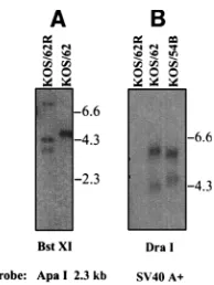
The genomic structures of the mutants were analyzed with at least five different restriction endonucleases and appropriate probes as described elsewhere (38). Representative blots are shown in Fig. 2. All mutations were shown to be diploid, and
for clarity only the IRL region is discussed. For the blot in Fig. 2A, viral DNA was cleaved with BstXI and probed with the 2.3-kb ApaI fragment. The deletion in KOS/62 eliminated two BstXI sites at bp 120216 and 120405, within the probed region, and theb-Gal insert contains two novel BstXI sites, the rele-vant one being at bp 3115 of theb-Gal gene. Only a single 4.8-kb band generated by cleavage at this site and the BstXI site at bp 127674 on the viral genome should hybridize to the probe. Repair of this locus should replace the deleted BstXI sites, resulting in two wild-type-probed bands of 4.2 and 3.6 kb. For the blot in Fig. 2B, viral DNA was digested with DraI and probed with a 450-bp BamHI-to-EcoRI fragment from PCH110 that contains the SV40 polyadenylation signals as well as a DraI site unique to theb-Gal gene insert. If both copies of the b-Gal gene were replaced in KOS/62R, no hybridizing fragments would be expected, and none were observed. KOS/62 should contain two hybridizing fragments, one from theb-Gal DraI site to bp 125987 within the “a” sequences at the joint between the IRL and the inverted short repeat and a
;5.8-kb fragment whose size can vary depending upon the copy number of reiterated sequences (26). A second predicted 4,247-bp fragment from the DraI site at bp 118002 to theb-Gal site should also hybridize. KOS/54 should retain the ;5.8-kb fragment, but the lower band should be shifted to 4,528 bp due to the deletion of;2 kb of theb-Gal gene and insertion of the 2.3-kb ApaI fragment (Fig. 1). The weak signals from the smaller fragments is expected, as they contain only;90 bp of the probe sequences.
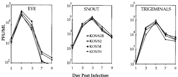
Experiments on replication kinetics were performed to en-sure that all viruses employed in this study replicated equiva-lently to the parental strain KOS/M. Groups of mice were infected and tissues were assayed as described above. As shown in Fig. 3, no differences in the ability to replicate were detected in vivo in the eye, snout, or TG. The virus titers peaked in the TG on day 5 p.i., ranging from 7.23104PFU (KOS/62R) to 1.23105PFU (KOS/62), a difference of less than twofold. In separate experiments KOS/29 yielded similar titers and kinet-ics (32; also, data not shown). Replication kinetkinet-ics experiments performed in vitro in RSC cultures under single-step (MOI, 10) or multistep (MOI, 0.01) conditions also did not reveal any differences between the viruses (not shown). Therefore, any latency phenotypes exhibited by the engineered strains could not be attributed to differences in general replication or dif-FIG. 1. Genomic structures. At the top is a schematic of the HSV-1 genome
with the unique long (UL), unique short (US), terminal long and short (TRL and TRS), and internal (IRL and IRS) repeats indicated. The region encoding the 59
[image:3.612.61.299.70.254.2]end of the LAT gene has been expanded, showing the location of the LAT promoter (Prom) (checkered box), the 2.3-kb ApaI fragment (open box), and the 2.0- and 1.5-kb stable LAT RNAs (LAT introns). This region of the genome is diploid, but for simplicity only the IRL region is shown. The LAT-null mutants KOS/62 and KOS/29 have been described previously (32). Striped boxes,b-Gal (LacZ) sequences; the solid boxes, SV40 RNA processing and polyadenylation sequences. The genome of KOS/62 was restored to wild type to generate KOS/ 62R, and KOS/54 was derived from KOS/62 by deleting part of theb-Gal gene present in KOS/62 and inserting the 2.3-kb ApaI fragment as described in Ma-terials and Methods.
FIG. 2. Analysis of genomic structures. The genomic structures of the mu-tants were analyzed with at least five different restriction endonucleases and appropriate probes as described in Materials and Methods (38). (A) Viral DNA was cleaved with BstXI and probed with the 2.3-kb ApaI fragment. Molecular size markers are indicated on the right. The largest band in the KOS/62R lane is the result of a partial digestion. Predicted results are described in the text. (B) Viral DNAs were cleaved with DraI and probed with a 450-bp fragment con-taining the SV40 poly(A)1sequences.
on November 9, 2019 by guest
http://jvi.asm.org/
[image:3.612.390.487.531.660.2]ferences in replication at the body surface or within the TG during the acute-stage infection.
LAT RNAs are expressed in vivo in the context of a foreign
gene.We next examined the expression of the LAT RNAs of
the mutant strains in vivo at 4 and 15 days p.i. Total RNA was extracted from six infected mouse TG, and 10mg was Northern blotted and probed with a 850-bp subfragment of the 2.3-kb ApaI fragment as described above. A representative blot is shown in Fig. 4. As expected, the 2.0-kb form of LAT RNA was first consistently detected at 4 days p.i. in mice infected with KOS/M. By 15 days p.i. both the 2.0- and the 1.5-kb LATs were readily detectable. The delayed expression of the 1.5-kb form of LAT is consistent with temporal-expression analyses previ-ously reported by Wagner and colleagues (40, 41). KOS/62R expressed both forms with abundance and kinetics similar to those of the parent KOS/M (not shown). As expected from previous reports (40, 42), no LATs were detected in RNAs from mice infected with KOS/62 (Fig. 4). With KOS/29 a reduced amount of the 2.0-kb LAT was occasionally evident at 4 days p.i. during the acute stage of infection, but not at day 15, and the 1.5-kb form was never detected (not shown). This finding was consistent with a report on this mutant by Nicosia et al., who suggested that an alternate or cryptic promoter may be utilized during active viral replication (21). Glorioso and colleagues have mapped an alternate promoter, or promoter element, to the region immediately 59of the start of the major LATs and suggested that it may be most important for expres-sion of the LAT region during the acute stage of infection in vitro and in vivo (4).
Interesting new findings came from the analysis of KOS/54.
Two independent isolates (A and B) expressed the 2.0- and 1.5-kb LAT RNAs with abundance and kinetics similar to those of KOS/M (Fig. 4). Farrell et al. have supplied solid evidence for the intron nature of the 2.0-kb RNA in transient transfection experiments (9). In vitro the 2.0-kb LAT RNA is appropriately spliced out of a foreign gene and had a negative impact on the transactivation of viral promoters by a cotrans-fected ICP0-containing plasmid. Others have suggested that the LATs are primary transcripts in vivo in neurons (35). A nonlinear conformation of the 2.0- and 1.5-kb LATs has re-cently been demonstrated, and so it is clear that they possess unusual physical properties (28, 43). The fact that the 2.0- and 1.5-kb LATs are produced in the context of a foreign transcrip-tion unit in vivo (KOS/54 in Fig. 4) supports the intron nature of both these RNAs. We have not, however, ruled out the possibility that these transcripts originate from a viral pro-moter located within the first;100 bp of the ApaI fragment or from a cryptic promoter generated by the construct.
Analysis of LAT gene transcription downstream of the
b-Gal insert in KOS/62 and KOS/54.Since the ApaI fragment
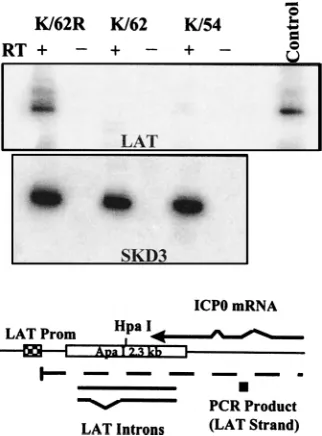
inserted into the b-Gal gene in KOS/54 contains the splice donor and acceptor sequences for the major LATs, it was possible that some spliced RNA containing downstream se-quences might be produced by this mutant. Transcription of the LAT strand downstream of the LAT splice acceptor site was examined by reverse transcriptase PCR to determine if this were the case. Groups of mice were infected as above with KOS/62, KOS/62R, or KOS/54, and RNAs isolated from gan-glia were analyzed as described in Materials and Methods. To control for the quality and quantity of RNA, a mouse gene (SKD3) that is actively transcribed in TG was similarly ana-lyzed. As shown in Fig. 5, all samples were positive for SKD3 and the signal was present only in the lanes that contained RT. A LAT-related product of the predicted size (152 bp) was detected by a labeled internal oligonucleotide probe in samples derived from mice infected with KOS/62R. Similar findings were seen with RNAs derived from mice infected with the parental strain KOS/M (not shown). This product was not detected in samples derived from KOS/62- or KOS/54-infected mice (Fig. 5). These findings demonstrate that transcription of the LAT locus downstream of the b-Gal gene insertion in KOS/62 and KOS/54 is absent or greatly reduced compared to that in the rescued isolate KOS/62R. Further they suggest that the only LAT sequences transcribed in KOS/54 reside within the ApaI 2.3-kb fragment inserted into theb-Gal gene. FIG. 3. Viral replication in vivo. Mice were infected in both eyes and on both sides of the snout as described in Materials and Methods. At the indicated times p.i. three mice per group were sacrificed and tissues were assayed for infectious HSV-1 on RSC monolayers. Although the x axis begins at 102PFU per g for clarity, no
[image:4.612.150.463.69.205.2]virus was found in TG at 1 day p.i. with any virus.
FIG. 4. Expression of the LAT RNAs in vivo in mouse TG. Expression of the LAT RNAs was examined at 4 (d4) and 15 (d15) days p.i. Total RNA extracted from infected mouse TG (10mg/lane) was electrophoresed and probed with a 850-bp subfragment of the 2.3-kb ApaI fragment. RNA size standards are indi-cated. No LAT were detected in KOS/62-infected mice. Two independent iso-lates of KOS/54 (A and B) expressed the 2.0- and 1.5-kb LAT RNAs with abundance and kinetics similar to those of the wild type.
on November 9, 2019 by guest
http://jvi.asm.org/
In vivo reactivation phenotype. Groups of mice were in-fected as above and maintained for at least 30 days p.i. The mice were then subjected to the hyperthermic stress (HS) reactivation protocol. This procedure, which consists of raising the core body temperature of the mouse to 43°C over a period of 10 min, results in the induced in vivo reactivation of HSV-1 in a significant percentage of the mice within 14 to 24 h post-HS (31). As expected, KOS/M reactivated in 30% of the animals (31, 32) by 22 h post-HS (Table 1). The LAT-null mutant KOS/62 did not reactive (P , 0.00001). In in vitro cocultivation assays LAT mutants reactivate with delayed ki-netics (7, 31). Additional mice latently infected with KOS/62 were analyzed 48 and 78 h postinduction, and again no reac-tivation was detected (0 of 12 at each time point), suggesting that the failure to detect reactivation of this mutant was not the result of a delay in reactivation post-HS. Repair of the deletion
in KOS/62 resulted in a wild-type reactivation frequency for KOS/62R (35%). KOS/29, which produces some 2.0-kb LAT at early times p.i. but not during latency, reactivated in a low percentage of the animals (5%). This result was consistent with prior reports by us (31) and others (7) that deletion of 203 bp of the basal LAT promoter results in viruses that reactivate inefficiently.
In stark contrast to mice latently infected with the LAT-null virus KOS/62 or the null “leaky” virus KOS/29, the KOS/54-infected mice reactivated efficiently (35%). Clearly, the ability of the viruses to reactivate in vivo correlated with their ability to express the LAT gene RNAs (see above). The rescue of the phenotype of KOS/62 by either repair of the genome or ex-pression of the 2.3-kb ApaI region in the context of theb-Gal gene demonstrated that the 1.2-kb region between bp 119279 and 120468 was essential for efficient reactivation and strongly suggested that the transcription of the 2.3-kb ApaI fragment was sufficient.
The percentage of neurons in which latency is established
correlates with ability to reactivate. A new approach, CXA,
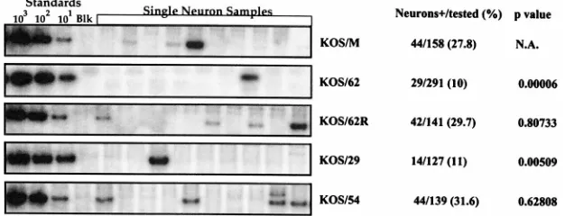
was employed to quantify the percentage of neurons that har-bored the viral genome in latently infected TG neurons. Mice were infected as above, tissues were fixed in situ, TG were removed and dissociated into single-cell populations, and neu-rons were purified on Percoll gradients as described above. Six TG from each group were pooled, so that approximately 120,000 randomized neurons were sampled (30). Neurons were aliquoted into PCR tubes and visualized with the aid of Pon-ceau-S staining to ensure that only one neuron was present as described elsewhere (30). Single-neuron samples were treated with DNase immobilized on beads and subsequently assayed by quantitative PCR for the presence of the HSV-1 genome as described elsewhere (30). PCRs were performed in groups of 24, which included standards of known quantities of HSV-1 DNA and three blank controls consisting of cell-free buffer that was processed along with the test samples. At least four separate experiments were performed on each test group, and in all cases the percentages of positive neurons detected were similar (not shown) (30). Representative blots and compiled results are shown in Fig. 6.
The LAT1viruses KOS/M and KOS/62R established latency in;30% of the mouse TG neurons, and there was no signif-icant difference between these viruses. Thus, latent infections were detected;30-fold more frequently than by in situ hybrid-ization for the LATs and;7-fold more frequently than esti-mated by in situ PCR (2, 19). In contrast, both LAT-null mutants KOS/62 and KOS/29 established latency in only about 10% of the neurons, a statistically significant difference from the percentage for the wild type (P50.00003) (Fig. 6). KOS/ 54, in which the LAT gene has been disrupted but the stable LAT RNAs are expressed (Fig. 1), established latent infections in the same percentage as the wild type. Therefore, in every case the ability to reactivate efficiently correlated with the ability to establish latent infections in a high percentage of the neurons.
The latent infections established by the LAT mutants might vary qualitatively as well as quantitatively, and this might affect the ability to reactivate. For example, the number of HSV-1 genomes present in individual neurons latently infected with wild-type HSV-1 varies from,10 to .1,000 (30). The HSV genome copy number in a latently infected cell might influence its ability to reactivate (14), as also speculated by Roizman and Sears (28a). The LAT gene might influence latent genome copy number and promote the establishment of latent infec-tions that contain a higher number of genomes. If this is indeed the case, then a reduction in the HSV-1 genome copy number FIG. 5. Analysis of transcription of the downstream region of the LAT gene.
[image:5.612.98.259.70.288.2]Transcription of the LAT gene downstream of the point of insertion (HpaI) of theb-Gal gene in KOS/62 and KOS/54 was analyzed in mouse TG 6 days p.i. as described in Materials and Methods. In the upper blot (LAT) reverse transcrip-tion-PCR primers specific for LAT RNA were employed, and products were probed with an internal oligonucleotide. The predicted 153-bp product was readily detected in the KOS/62R lane that contained reverse transcriptase (RT) but absent when RT was omitted from the reaction. No other test samples were positive. Control, 0.1 pg of plasmid DNA. In the lower blot duplicate samples of the RNA were tested with primers specific for the mouse gene SKD3 mRNA to control for RNA quantity and quality. The predicted 192-bp product was evident in all RT-containing lanes. A schematic of the 59end of the LAT gene is shown below, with the location of the primary LAT transcript (dashed line), LAT introns, ICP0 mRNA, and the downstream LAT RNA-specific PCR product (solid box) indicated. Prom, promoter.
TABLE 1. Reactivation in vivo
Virus No. of animals positive/no. tested % (Pa)
Expt 1 Expt 2 Total
KOS/M 7/20 3/10 10/30 33.3 KOS/29 0/9 3/51 3/60 5 (0.0045) KOS/62 0/30 0/22 0/52 0 (0.00012) KOS/62R 4/10 3/10 7/20 35 (.0.99) KOS/54 3/10 4/10 7/20 35 (.0.99)
aFor comparison to KOS/M results by Fisher’s exact test.
on November 9, 2019 by guest
http://jvi.asm.org/
in neurons latently infected with the LAT mutants might ac-count for the reduced reactivation frequency observed above. One advantage of CXA is that it allows the genome copy number within individual latently infected cells to be obtained at least semiquantitatively (30). A profile of the HSV copy numbers in individual neurons latently infected by the LAT-null mutant KOS/62 and the rescued mutant KOS/62R was obtained by exposing the blots on a phosphorimager, quanti-fying the counts per minute present in each band, and com-paring the results to external standards run at the same time (see above) (30). A scattergram of the results is shown in Fig. 7. It was found that the viral-genome copy number within individual neurons was variable. As many as several hundred genomes were detected in some latently infected neurons, but most positive neurons contained 10 or fewer copies. No
signif-icant difference between the groups with respect to copy num-ber was detected (Fig. 7). Therefore, the LAT gene functions to increase the pool of latently infected neurons present in the ganglia but, as measured here, does not affect the number of viral genomes within the individual neurons comprising that pool. Other viral genes have been implicated in the establish-ment of latency. However, all of these gene products are re-quired for efficient viral replication (for example, see refer-ences 8, 12, 16, 27, and 33). This is the first example of a gene that directly facilitates the establishment of latent infections.
The HSV-1 LAT gene is not required for efficient in vivo
reactivation.The data demonstrate that the LAT gene is
nec-essary for efficient establishment of latency, but this locus could also have a direct role in reactivation, as suggested by many groups (for example, see references 2, 3, 6, 11, 23, 24, 34, and 39). If it were possible to increase the percentage of neu-rons latently infected with the LAT-null mutants to the level of the wild type, this question could be directly addressed. If the establishment and reactivation phenotypes were independent, then these mice would still be defective in reactivation. Con-versely, if the reduced reactivation phenotype of LAT-null mutants was due to the reduced percentage of neurons har-boring latent infections, then these animals would reactivate efficiently.
During the course of our studies on the HS reactivation model it was observed that stress applied during the first 3 days p.i. resulted in an increase in the percentage of neurons that harbor latent virus. The mechanism whereby stress during the acute stage of infection increases the establishment of latency is not known and is currently under investigation. However, acute stress does not alter viral replication of KOS/62 or KOS/ 62R in the eye, snout, or TG (data not shown). Advantage was taken of this phenomenon to examine whether the LAT locus is required for efficient reactivation.
[image:6.612.151.464.72.192.2]Groups of mice were infected with KOS/62 or KOS/29 as described above. Half of the animals in each group were then subjected to acute HS during the first 3 days p.i. as above. These groups were termed KOS/62HS and KOS/29HS, respec-tively. At 30 days p.i. the groups of animals were subjected to HS to determine their ability to reactivate, and additional mice were processed for the CXA determination of the percent latently infected neurons (PIN). As expected the PINs of con-trol mice in both groups were low (KOS/62, 8.8%; KOS/29, 11%) (Table 2). In the KOS/62HS animals 31.5% of the neu-rons tested contained the HSV-1 genome, a difference that was FIG. 6. CXA determination of PIN. Groups of six latently infected TG were processed to yield purified neurons and analyzed for the presence of the HSV-1 genome by quantitative PCR as described elsewhere (30) except that individual neurons were examined. Products were electrophoresed, blotted, probed with a32P-labeled internal oligonucleotide and imaged on a Molecular Dynamics phosphorimaging system with ImageQuant software. Representative blots are shown, with the first four lanes representing HSV-1 genome standards spanning 3 orders of magnitude and a buffer blank (Blk). To the right the total numbers of neurons positive for HSV-1 DNA of the numbers tested, the corresponding percentages, and the P values derived from comparisons to the parent strain KOS/M by Fisher’s exact test are listed. These data were compiled from at least three experiments, and the variation between experiments was less than 2%. N.A., not applicable.
FIG. 7. HSV-1 genome copy numbers in individual latently infected neurons. The counts per minute within individual bands of the samples in Fig. 6 were determined with ImageQuant software. The data was compared to the standard curve to estimate the number of HSV-1 genomes present as described elsewhere (30). Numbers of HSV genomes detected are indicated on the left. Each point in the scatter plots represents one positive neuron. The total numbers of negative neurons are given below.
on November 9, 2019 by guest
http://jvi.asm.org/
statistically significant (P 5 0.002; Fisher’s exact test). The KOS/29HS group also contained a significantly higher percent-age of latently infected neurons (22%; P50.04). Analysis of the signals on the blots did not reveal any difference in genome copy profiles between the stressed and nonstressed groups (data not shown). This was as anticipated since no difference was detected in the copy number profiles for wild-type- and mutant-infected neurons (Fig. 7).
The ability of the groups to reactivate in vivo following HS at 30 days p.i. was next examined. As expected, no mice in the KOS/62 group reactivated (0%) (Table 2). This has been a consistent finding with this mutant with over 300 animals tested to date (Tables 1 and 2) (32), and this phenotype is by far the most severe of any LAT mutant tested in any reactivation system (2, 6, 11, 23–25, 32, 36). A dramatic increase in the frequency of reactivation to the level of the wild type was observed for the KOS/62HS group (34.5%; P,0.00001 [Fish-er’s exact test]). Analysis of the genomic structures of the virus in positive cultures confirmed that it was KOS/62 (Fig. 8). The same trend was seen with KOS/29 (0%) versus KOS/29HS (20%). Again it was confirmed that it was indeed KOS/29 that had reactivated in vivo (data not shown). In this experiment the group size was too small to establish statistical significance. These results clearly demonstrate that increasing the level of establishment by the LAT-null mutants resulted in an increase in their ability to reactivate in vivo. In the case of KOS/62 the frequency of reactivation could not be distinguished from that of the wild type (34.5 versus 33.3%; P.0.99). Thus, no direct role for the LAT gene in reactivation could be demonstrated.
DISCUSSION
Our findings demonstrate that the HSV-1 LAT gene func-tions to increase the percentage of neurons in which latency is established. No direct role for this gene in the reactivation of HSV could be detected. Our conclusions are based on the following findings. Analysis of the percentage of mouse TG neurons that harbor the latent HSV genome using a CXA approach revealed a statistically significant difference between the wild-type and LAT-null mutant viruses. About 30% of the TG neurons in mice infected with HSV-1 strain KOS/M con-tained latent virus. In contrast only ;10% of the neurons in mice infected with KOS/62 or KOS/29 were positive for HSV-1 DNA. The biological significance of these HSV genome-posi-tive neurons is not known, but at least some must comprise the pool of latent infections that subsequently reactivate. In every case examined a high frequency of reactivation in vivo follow-ing HS correlated with a high percentage of HSV genome-positive neurons. The role of the 1.6-kb deletion in KOS/62 in the reduction in the frequency of establishment was confirmed by repair of the genome (KOS/62R) and by expression of the 2.3-kb ApaI fragment that contains the major LAT sequences
in the context of the E. colib-Gal gene (KOS/54). Both KOS/ 62R and KOS/54 displayed wild-type establishment and reac-tivation phenotypes.
KOS/62 does not appear to have a defect in reactivation per se. When mice are infected via the hind footpads, KOS/62 establishes latent infections in dorsal root ganglia neurons ef-ficiently as measured by activity from the LAT promoter, and the mutant reactivates like the wild type in vivo in this site (32). Following infection via the eye or snout, establishment is less efficient in TG neurons and reactivation is delayed in coculti-vation cultures in vivo. However, essentially 100% of the ani-mals do reactivate, and the reduced kinetics may be a reflec-tion of the reduced frequency of establishment (32). Reactivation of KOS/62 in vivo in TG was not detected under normal infection conditions even when tissues were analyzed at 48 and 72 h post-HS, suggesting that this was not due simply to slower reactivation kinetics of the mutant. The reduction of the percentage of neurons that harbor the latent genome from
[image:7.612.341.530.69.305.2];30% (KOS/M, KOS/62R, and KOS/54) to 10% (KOS/62 and KOS/29) may represent a threshold below which reactivation is not detected in vivo. However, in vivo reactivation was de-tected in 3 of 60 mice infected with KOS/29, a mutant that is leaky for LAT expression (6; also, this report). Alternately, the latent infections established by LAT-null mutants may be qual-itatively different in some as yet undetected property. We found no difference in the genome copy profiles in individual neurons, but LAT may influence the type of neuron in which latency is established or the HSV-1 copy number within “re-activatable” neurons, or it may itself have a direct influence on reactivation.
TABLE 2. Effect of stress on the establishment of latency and the reactivation of LAT-null mutants
Virus No. positive (%)
Establishmenta Reactivationb
KOS/62 8/90 (8.8) 0/78 (0)
KOS/62HSc 24/76 (31.5) 10/29 (34.5)
KOS/29 14/127 (11) 0/9 (0)
KOS/29HSc 23/104 (22) 2/10 (20)
aNumber of neurons positive for the HSV genome/number tested by CXA. bNumber of animals positive for virus/number tested.
[image:7.612.57.300.90.163.2]cMice were subjected to HS during the first 3 days p.i.
FIG. 8. Analysis of the genomic structures of virus detected following HS reactivation. Mice were infected with KOS/62 and subjected to HS during the first 3 days p.i. At 30 days p.i. the animals were induced to reactivate, and at 22 h post-HS TG homogenates were assayed on RSC monolayers. DNAs were iso-lated from the positive cultures, digested with SalI, electrophoresed, and blotted as described elsewhere (31). Control DNAs from the parental strain KOS/M, KOS/62, and KOS/62R were loaded at 1/30, 1/15, and 1/5 the concentration of the test samples (lanes A through J) as shown by the photo of the ethidium-stained gel. The blot was probed with sequences specific for the 59end of the LAT intron region that are deleted in KOS/62 (31) (upper panel). This blot was subsequently reprobed with sequences specific for theb-Gal gene (lower panel).
on November 9, 2019 by guest
http://jvi.asm.org/
We were unable to demonstrate a direct role for the LAT gene in reactivation. The fact that KOS/62 reactivated with wild-type frequency when the percentage of latently infected neurons in TG was increased to wild-type levels by stress dur-ing the acute phase suggests that this genomic region is not involved directly in reactivation. It is possible that acute-stage stress allowed latent infections to be established in some alter-nate pool of neurons where a LAT reactivation function is not required. Experiments to determine if this is the case are in progress. However, the high percentage of neurons latently infected (.30%) argues against a selective increase in a subset of TG neurons, and the simplest interpretation is that the LAT gene is not directly involved in reactivation in this system.
The mechanism by which the LAT gene increases the fre-quency of establishment is not yet known. Our results with mutant KOS/29 suggest that it is the transcription of the LAT locus that is important. This mutant contains a deletion of 203 bp of the basal LAT promoter that overlaps the deletion in KOS/62 by only 24 bp (32). In this study KOS/29 was not genetically rescued. However, Bloom and colleagues demon-strated that an identical mutant of strain 17syn1(DPst) tivated with reduced kinetics in an in vitro cocultivation reac-tivation assay and mapped this phenotype to the 203-bp LAT promoter (7). We previously reported that KOS/29 displayed reduced reactivation kinetics in an in vitro cocultivation assay (32). Another group found that this mutant reactivated like the wild type in a similar assay (6). The reason for this difference is not clear, but as seen in this study, KOS/29 is definitely defective in the establishment of latent infections. Given the fact that KOS/54 is still missing the region between the LAT promoter and start of the 2.3-kb ApaI fragment and that tran-scription of the downstream LAT region was not detected in this mutant, the simplest explanation for the establishment defect in KOS/29 is the lack of transcription of the 2.3-kb ApaI fragment that encodes the abundant 2.0- and 1.5-kb RNAs known as LATs. These RNAs are most likely stable introns spliced from a larger precursor RNA (9; but see also reference 35) and are partially antisense to ICP0, a major viral transcrip-tion factor.
It has been suggested that the LATs may function as a natural antisense mechanism to down regulate viral gene tran-scription in neurons (9). Our findings are consistent with this hypothesis. However, this cannot be reconciled with a recent report by Block and colleagues, who employed in situ PCR to quantify latent infections established by several LAT mutants. Mutant DBST E, was found to be wild type in its ability to reactivate in a timely manner in an in vitro cocultivation assay, as well as in establishment as measured by in situ PCR (17). This mutant is missing the 59end of the major LATs including the splice donor sequences and could not produce the stable 2.0- and 1.5-kb LATs. If these assays measure the same prop-erties, it would appear that a stable antisense intron is not required. Presumably, the sequences important for the estab-lishment of latency must lie downstream of the BstEII site. It may be that some as yet undetected alternately spliced form of LAT RNA encodes a protein factor involved in down regula-tion of the viral genome during the establishment phase of infection. The combined results obtained withDBst E and our KOS/54 mutant suggest expression of the region between the BstEII site at bp 120092 and the ApaI site at bp 121568 is necessary and may be sufficient for high-level establishment. Perng and colleagues have shown that transfer of a 3.2-kb fragment that includes a portion of this region into the long unique region of a LAT deletion mutant restored the ability of the mutant to spontaneously reactivate in the rabbit eye model, and this may further delimit the region of interest to between
the BstEII site and HpaI site at bp 120468 (24). A 371-bp region upstream of the 2.0-kb LAT RNA region is not required for spontaneous reactivation (25). However, sequences up-stream of the BstEII site have been implicated in the ability of HSV-1 to be isolated from tear films following induction by iontophoresis of epinephrine, and quantitative PCR on whole-ganglion DNA did not detect an establishment defect (3). Only
;70 bp of the identified 348-bp region are present in the 2.3-kb ApaI fragment, and none are present in D Bst E. Whether these apparent discrepancies are due to differences in the abil-ity to detect establishment or reactivation phenotypes in the various systems or genomic regions that contain important regulatory or protein sequences that are dispensable in some situations remains to be determined.
ACKNOWLEDGMENTS
This work was supported by Public Health Service grants AI32121 (from the National Institute of Allergy and Infectious Diseases) and NS25879 (from the National Institute of Neurological Disorders and Stroke) and a CHMCC Trustee Grant.
We thank C. S. Tanski and T. M. Suter for expert technical assis-tance.
REFERENCES
1. Ahmed, R., and J. G. Stevens. 1990. Viral persistence. In B. N. Fields (ed.), Virology. Raven Press, New York, N.Y.
2. Block, T. M., S. Deshmane, J. Masonis, J. Maggioncalda, T. Valyi-Nagi, and
N. W. Fraser.1993. An HSV LAT null mutant reactivates slowly from latent infection and makes small plaques on CV-1 monolayers. Virology 192:618– 630.
3. Bloom, D. C., J. M. Hill, G. Devi-Rao, E. K. Wagner, L. T. Feldman, and J. G.
Stevens.1996. A 348-base-pair region in the latency-associated transcript facilitates herpes simplex virus type 1 reactivation. J. Virol. 70:2449–2459. 4. Chen, X., M. C. Schmidt, W. F. Goins, and J. C. Glorioso. 1995. Two herpes
simplex virus type 1 latency-active promoters differ in their contributions to latency-associated transcript expression during lytic and latent infections. J. Virol. 69:7899–7908.
5. Deatly, A. M., J. G. Spivack, E. Lavi, and N. W. Fraser. 1987. RNA from an immediate early region of the type 1 herpes simplex virus genome is present in the trigeminal ganglia of latently infected mice. Proc. Natl. Acad. Sci. USA
84:3204–3208.
6. Deshmane, S. L., M. Nicosia, T. Valyi-Nagy, L. T. Feldman, A. Dillner, and
N. W. Fraser.1993. An HSV-1 mutant lacking the LAT TATA element reactivates normally in explant cocultivation. Virology 196:868–872. 7. Devi-Rao, G. B., D. C. Bloom, J. G. Stevens, and E. K. Wagner. 1994. Herpes
simplex virus type 1 DNA replication and gene expression during explant-induced reactivation of latently infected murine sensory ganglia. J. Virol.
68:1271–1282.
8. Ecob-Prince, M. S., C. M. Preston, F. J. Rixon, K. Hassan, and P. G.
Kennedy.1993. Neurons containing latency-associated transcripts are nu-merous and widespread in dorsal root ganglia following footpad inoculation of mice with herpes simplex virus type 1 mutant in 1814. J. Gen. Virol.
74:985–994.
9. Farrell, M. J., A. T. Dobson, and L. T. Feldman. 1991. Herpes simplex virus latency-associated transcript is a stable intron. Proc. Natl. Acad. Sci. USA
88:790–794.
10. Hill, J. M., B. M. Gebhardt, R. Wen, A. M. Bouterie, H. W. Thompson, R. J.
O’Callaghan, W. P. Halford, and H. E. Kaufman.1996. Quantitation of herpes simplex virus type 1 DNA and latency-associated transcripts in rabbit trigeminal ganglia demonstrates a stable reservoir of viral nucleic acids during latency. J. Virol. 70:3137–3141.
11. Hill, J. M., F. Sedarati, R. T. Javier, E. K. Wagner, and J. G. Stevens. 1990. Herpes simplex virus latent phase transcription facilitates in vivo reactiva-tion. Virology 174:117–125.
12. Jacobson, J. G., D. A. Leib, D. J. Goldstein, C. L. Bogard, P. A. Schaffer,
S. K. Weller, and D. M. Coen.1989. A herpes simplex virus ribonucleotide reductase deletion mutant is defective for productive acute and reactivatable latent infections of mice and for replication in mouse cells. Virology 173: 276–283.
13. Katz, J. P., E. T. Bodin, and D. M. Coen. 1990. Quantitative polymerase chain reaction analysis of herpes simplex virus DNA in ganglia of mice infected with replication-incompetent mutants. J. Virol. 64:4288–4295. 14. Kosz-Vnenchak, M., J. Jacobson, D. M. Coen, and D. M. Knipe. 1993.
Evidence for a novel regulatory pathway for herpes simplex virus gene expression in trigeminal ganglion neurons. J. Virol. 67:5383–5393. 15. Kramer, M. F., and D. M. Coen. 1995. Quantification of transcripts from the
on November 9, 2019 by guest
http://jvi.asm.org/
ICP4 and thymidine kinase genes in mouse ganglia latently infected with herpes simplex virus. J. Virol. 69:1389–1399.
16. Leib, D. A., D. M. Coen, C. L. Bogard, K. A. Hicks, D. R. Yager, D. M. Knipe,
K. L. Tyler, and P. A. Schaffer.1989. Immediate-early regulatory gene mutants define different stages in the establishment and reactivation of herpes simplex virus latency. J. Virol. 63:759–768.
17. Maggioncalda, J., A. Mehta, Y. H. Su, N. W. Fraser, and T. M. Block. 1996. Correlation between herpes simplex virus type 1 rate of reactivation from latent infection and the number of infected neurons in trigeminal ganglia. Virology 225:72–81.
18. McGeoch, D. J., M. A. Dalrymple, A. J. Davison, A. Dolan, M. C. Frame, D.
McNab, L. J. Perry, J. E. Scott, and P. Taylor.1988. The complete DNA sequence of the long unique region in the genome of herpes simplex virus type 1. J. Gen. Virol. 69:1531–1574.
19. Mehta, A., J. Maggioncalda, O. Bagasra, S. Thikkavarapu, P. Saikumari, T.
Valyi-Nagy, N. W. Fraser, and T. M. Block.1995. In situ DNA PCR and RNA hybridization detection of herpes simplex virus sequences in trigeminal ganglia of latently infected mice. Virology 206:633–640.
20. Mitchell, W. J., R. P. Lirette, and N. W. Fraser. 1990. Mapping of low abundance latency-associated RNA in the trigeminal ganglia of mice latently infected with herpes simplex virus type 1. J. Gen. Virol. 71:125–132. 21. Nicosia, M., S. L. Deshmane, J. M. Zabolotny, T. Valyi-Nagy, and N. W.
Fraser.1993. Herpes simplex virus type 1 latency-associated transcript (LAT) promoter deletion mutants can express a 2-kilobase transcript map-ping to the LAT region. Virology 196:868–872.
22. Perier, F., C. M. Radeke, K. F. Raab-Graham, and C. A. Vandenberg. 1995. Expression of a putative ATPase suppresses the growth defect of a yeast potassium transport mutant: identification of a mammalian member of the Clp/HSP104 family. Gene 152:157–163.
23. Perng, G. C., E. C. Dunkel, P. A. Geary, S. M. Slanina, H. Ghiasi, R. Kaiwar,
A. B. Nesburn, and S. L. Wechsler.1994. The latency-associated transcript gene of herpes simplex virus type 1 (HSV-1) is required for efficient in vivo spontaneous reactivation of HSV-1 from latency. J. Virol. 68:8045–8055. 24. Perng, G. C., H. Ghiasi, S. M. Slanina, A. B. Nesburn, and S. L. Wechsler.
1996. The spontaneous reactivation function of the herpes simplex virus type 1 LAT gene resides completely within the first 1.5 kilobases of the 8.3-kilobase primary transcript. J. Virol. 70:976–984.
25. Perng, G. C., S. M. Slanina, H. Ghiasi, A. B. Nesburn, and S. L. Wechsler. 1996. A 371-nucleotide region between the herpes simplex virus type 1 (HSV-1) LAT promoter and the 2-kilobase LAT is not essential for efficient spontaneous reactivation of latent HSV-1. J. Virol. 70:2014–2018. 26. Perry, L. J., and D. J. McGeoch. 1988. The DNA sequences of the long
repeat region and adjoining parts of the long unique region in the genome of herpes simplex virus type 1. J. Gen. Virol. 69:2831–2846.
27. Robertson, L. M., A. R. MacLean, and S. M. Brown. 1992. Peripheral rep-lication and latency reactivation kinetics of the non-neurovirulent herpes simplex virus type 1 variant 1716. J. Gen. Virol. 73:967–970.
28. Rodahl, E., and L. Haarr. 1997. Analysis of the 2-kilobase latency-associated transcript expressed in PC12 cells productively infected with herpes sim-plex virus type 1: evidence for a stable nonlinear structure. J. Virol.
71:1703–1707.
28a.Roizman, B., and A. E. Sears. 1996. Herpes simplex viruses and their
repli-cation, p. 1795–1841. In B. N. Fields (ed.), Virology. Raven Press, New York, N.Y.
29. Sambrook, J., E. F. Frisch, and T. Maniatis. 1989. Molecular cloning: a laboratory manual, 2nd ed. Cold Spring Harbor Press, Cold Spring Harbor, N.Y.
30. Sawtell, N. M. 1997. Comprehensive quantification of herpes simplex virus latency at the single-cell level. J. Virol. 71:5423–5431.
31. Sawtell, N. M., and R. L. Thompson. 1992. Rapid in vivo reactivation of herpes simplex virus in latently infected murine ganglionic neurons after transient hyperthermia. J. Virol. 66:2150–2156.
32. Sawtell, N. M., and R. L. Thompson. 1992. Herpes simplex virus type 1 latency-associated transcription unit promotes anatomical site-dependent establishment and reactivation from latency. J. Virol. 66:2157–2169. 33. Sedarati, F., T. P. Margolis, and J. G. Stevens. 1993. Latent infection can be
established with drastically restricted transcription and replication of the HSV-1 genome. Virology 192:687–691.
34. Spivack, J. G., and N. W. Fraser. 1988. Expression of herpes simplex virus type 1 latency-associated transcripts in the trigeminal ganglia of mice during acute infection and reactivation of latent infection. J. Virol. 62:1479–1485. 35. Spivack, J. G., G. M. Woods, and N. W. Fraser. 1991. Identification of a novel latency-specific splice donor signal within the herpes simplex virus type 1 2.0-kilobase latency-associated transcript (LAT): translation inhibition of LAT open reading frames by the intron within the 2.0-kilobase LAT. J. Virol.
65:6800–6810.
36. Stevens, J. G., E. K. Wagner, G. B. Devi-Rao, M. L. Cook, and L. T. Feldman. 1987. RNA complementary to a herpesvirus alpha gene mRNA is prominent in latently infected neurons. Science 235:1056–1059.
37. Tenser, R. B., W. A. Edris, and K. A. Hay. 1989. Herpes simplex latent infection: quantitation of latency-associated transcript-positive neurons and reactivable neurons. Yale J. Biol. Med. 62:197–204.
38. Thompson, R. L., S. K. Rogers, and M. A. Zerhusen. 1989. Herpes simplex virus neurovirulence and productive infection of neural cells is associated with a function which maps between 0.82 and 0.832 map units on the HSV genome. Virology 172:435–450.
39. Trousdale, M. D., I. Steiner, J. G. Spivack, S. L. Deshmane, S. M. Brown,
A. R. MacLean, J. H. Subak-Sharpe, and N. W. Fraser.1991. In vivo and in vitro reactivation impairment of a herpes simplex virus type 1 latency-asso-ciated transcript variant in a rabbit eye model. J. Virol. 65:6989–6993. 40. Wagner, E. K., G. Devi-Rao, L. T. Feldman, A. T. Dobson, Y. F. Zhang,
W. M. Flanagan, and J. G. Stevens.1988. Physical characterization of the herpes simplex virus latency-associated transcript in neurons. J. Virol. 62: 1194–1202.
41. Wagner, E. K., W. M. Flanagan, G. Devi-Rao, Y. F. Zhang, J. M. Hill, K. P.
Anderson, and J. G. Stevens.1988. The herpes simplex virus latency-associ-ated transcript is spliced during the latent phase of infection. J. Virol.
62:4577–4585.
42. Wechsler, S. L., A. B. Nesburn, R. Watson, S. M. Slanina, and H. Ghiasi. 1988. Fine mapping of the latency-related gene of herpes simplex virus type 1: alternative splicing produces distinct latency-related RNAs containing open reading frames. J. Virol. 62:4051–4058.
43. Wu, T. T., Y. H. Su, T. M. Block, and J. M. Taylor. 1996. Evidence that two latency-associated transcripts of herpes simplex virus type 1 are nonlinear. J. Virol. 70:5962–5967.