Open Access
Research
A novel method of modifying immune responses by vaccination
with lipiodol-siRNA mixtures
Thomas E Ichim*
†1, Igor A Popov
†2, Neil H Riordan
1, Hamid Izadi
1,
Zaohui Zhong
3, Li Yijian
3, Salman Sher
4and Eugenia K Oleinik
5Address: 1Medistem Laboratories Inc, Tempe Arizona, USA, 2Department of Surgery, University of Western Ontario, London, Ontario, Canada, 3The Second Xiangya Hospital of Central South University, Changsha, China, 4Division of Cardiology, Emory University, Atlanta, USA and 5Institute of Biology, Karelian Research Center, Russian Academy of Sciences, Petrozavodsk, Russia
Email: Thomas E Ichim* - [email protected]; Igor A Popov - [email protected]; Neil H Riordan - [email protected]; Hamid Izadi - [email protected]; Zaohui Zhong - [email protected]; Li Yijian - [email protected]; Salman Sher - [email protected]; Eugenia K Oleinik - [email protected]
* Corresponding author †Equal contributors
Abstract
The dendritic cell (DC) possesses the ability to stimulate both T helper 1 (Th1) and Th2 responses depending on activation stimuli. Although it is known that chemically or genetically modified DC can be used therapeutically to steer immune responses towards either Th1 or Th2, cellular therapy with ex vivo manipulated DC is clinically difficult. Here we demonstrate a novel method of switching immune responses from Th1 to Th2 through in vivo immune modulation by administration of siRNA. We demonstrate that siRNA targeting of the IL-12p35 gene leads to a Th2 bias in vitro through an IL-10 dependent mechanism. In vivo administration of siRNA admixed with the oil-based contrast agent lipiodol in the presence of antigen and adjuvant induced a deviation in recall response to reduced production of IFN-γ and augmented IL-4 response using either KLH or ovalbumin. This simple method of in vivo modification of immune response possesses therapeutic potential in Th1-mediated diseases such as multiple sclerosis and autoimmune diabetes.
Introduction
It is known that the immune response can be polarized into two broad subsets based on cytokine secretion of the T helper cells. T helper-1 cells (Th1) responses are associ-ated with anti-viral and anti-cancer immune reactions and are characterized by high secretion of the cytokine IFN-γ and IL-2 [1]. In contrast, Th2 responses are effective for clearing parasitic infections such as schistosoma mansoni and are identified by high secretion of IL-4 and IL-13 [2]. In various human diseases a pathological polarization of the immune responses either to Th1 or Th2 is known to occur. For example, autoimmune diseases such as
diabe-tes [3], multiple sclerosis [4], or arthritis [5], are associated with overproduction of Th1 cytokines. The causative role of these cytokines is noted by amelioration of animal models of autoimmune disease through treatment with inhibitors of such cytokines, such as antibodies or soluble receptors; however, clinical utilization is often impeded by systemic, non-desired side effects. Therefore a desired method of treating autoimmune diseases associated with Th1-hyperactivities would be targeting the actual initiat-ing event that triggers such immune reactions.
Published: 03 January 2006
Journal of Translational Medicine 2006, 4:2 doi:10.1186/1479-5876-4-2
Received: 21 October 2005 Accepted: 03 January 2006
This article is available from: http://www.translational-medicine.com/content/4/1/2
© 2006 Ichim et al; licensee BioMed Central Ltd.
It is known that naïve CD4+ T helper cells (Th0) are pro-grammed by the antigen presenting cell in order to become polarized into either the Th1 or Th2 phenotype [6]. More recent studies have demonstrated that dendritic cells (DC) play a key role in recognizing the exogenous pathogens, and making the decision of whether a Th1 or Th2 response is needed [7]. For example, parasites contain distinct chemical signatures that are recognized by special-ized toll-like receptors (TLR) on DC, which instruct the DC to promote the appropriate response. In the case of schistosomiasis, the schistosomal egg antigen (SEA) is known to bind to TLR-2 on DC, and stimulate production of IL-13, which in turn shifts the Th0 cell into a Th2 cells [8,9]. Conversely, Gram-negative bacteria contain lipopolysaccaride (LPS), which activates TLR-4, leading to production of IL-12 by the DC, which then stimulates Th1 differentiation [10]. Since it is established that Th1 immu-nity is essential for clearance of many Gram-negative bac-teria, it appears that the DC plays a "central cognitive" role in guiding the immune response. Conversely, manipula-tion of this cell type would allow us to selectively modu-late immune responses to either Th1 or Th2. Since DC can be pulsed with antigen, the manipulation of antigen-pulsed DC would allow for selective alterations of immu-nity in an antigen-specific manner. Although this has been performed in animal models of autoimmunity [11-15], the clinical utilization is hampered by the need for ex vivo cellular manipulation, which is expensive and diffi-cult.
RNA interference (RNAi) is the cellular defense mecha-nism by which a double-stranded RNA specifically silences mRNA transcripts with strict homology to the double strand [16]. The silencing effect of exogenous dou-ble-stranded RNA is very potent in that it can mediate gene-silencing even at a concentration of 1–3 double strands per cell [17]. In light of this potency and selectiv-ity, the induction of RNAi in DC would allow for silencing of genes that stimulate either Th1 or Th2. Since it is known that inhibition of Th1 cytokines in DC results in the stimulation of Th2 immunity and vice versa, we hypothesize that silencing of IL-12 would result in DC with a propensity of stimulating Th2 immune responses.
While we and others have been able to modify immuno-logical parameters of DC through silencing 10 [18], IL-12p35 [19], IL-12p40 [20], T-bet [21] and SOCS1 [22], a method of easily inducing immune modulation in vivo is lacking. In this study we induce an "in vivo genetic modi-fication", presumably through DC using an immobilizing agent (CFA), a transfection associated agent (lipidiol) and antigen. We demonstrate inhibition of Th1 and upregula-tion of Th2 responses using this simple immunizaupregula-tion protocol.
Materials and methods
Animals
Female C57/BL6 and BALB/c mice (The Jackson Laborato-ries, Bar Harbor, ME), 5 wk of age, were kept in filter-top cages at the Animal Care and Veterinary Services Facility, the University of Western Ontario according to the Cana-dian Council for Animal Care Guidelines. Mice were fed by food and water ad libitum and allowed to settle for 2 wk before initiation of experimentations.
DC generation and siRNA transfection
At Day 0, bone marrow cells were flushed from the femurs and tibias of C57/BL6 mice, washed and cultured in 6-well plates (Corning, NY) at 4 × 10⁶ cells/well in 4 ml of complete medium (RPMI 1640 supplemented with 2 mM L-glutamine, 100 U/ml penicillin, 100 µg of streptomy-cin, 50 µM 2-ME, and 10% FCS (all from Life Technolo-gies, Ontario, Canada) supplemented with recombinant GM-CSF (10 ng/ml; PeproTech, Rocky Hill, NJ) and recombinant mouse IL-4 (10 ng/ml; PeproTech). All cul-tures were incubated at 37°C in 5% humidified CO2. Non-adherent cells were removed after 48 h of culture (Day 2) and fresh medium was added. After 7 days of cul-ture, >90% of the cells expressed the characteristic DC-specific marker CD11c as determined by FACS. DC were washed and plated in 24-well plates at a concentration of 2 × 10⁵ cells/well in 400 µl of serum-free RPMI 1640. Transfection with GenePorter, lipiodol, or naked siRNA was performed as described below on day 7 of culture.
siRNA synthesis and transfection
siRNA sequences were selected according to the method previously used by us [19]. siRNA specific for IL-12p35 (AACCUGCUGAAGACCACAGAU) or mismatched (mixed) control sequence (AACTGCCAGATGGATGGT-GAC) were synthesized and annealed by the manufacturer (Dharmacon, Lafayette, CO) and added at a concentra-tion of 60pMol to DC cultures. For transfecconcentra-tion, 3 µl of 20
Flow cytometry
Phenotypic analysis of DC was performed using flow cytometry on a FACScan (Becton Dickninson, San Jose, CA) and analyzed using CellQuest software (BD Bio-sciences). The cells were stained with FITC-conjugated mAb against surface markers associated with DC matura-tion: anti-mouse CD11c, anti-mouse CD40, anti-mouse CD80, and anti-mouse CD86 (Cedarlane Laboratories, Mississauga, ON). Ig of the same isotype were used as con-trols. Annexin V and propidium iodine analysis for apop-tosis, necrosis was performed using the Apotag kit (Cedarlane Laboratories, Hornby Ontario, Canada).
Mixed lymphocyte reaction
C57/BL6 DC after transfection were irradiated (3,000 rad) and seeded in triplicate at various concentrations in a flat-bottom 96-well plate (Corning) for use as stimulator cells. Splenic T cells from BALB/c mice were isolated by gradient centrifugation over Ficoll-Paque (Amersham Pharmacia Biotech, Quebec) and T cell nylon wool column purifica-tion, and added as responders (5 × 10⁵ cells/well). The mixed lymphocytes were cultured at 37°C for 72 h in 200
µl of RPMI 1640 supplemented with 10% FCS, 100 U/ml of penicillin, and 100 µg/ml of streptomycin and pulsed with 1 µCi/well of 3H-labelled thymidine (Amersham Pharmacia Biotech) for the last 16 h of culture. Cells were harvested onto glass fiber filters, and the radioactivity incorporated was quatitated using a Wallac Betaplate liq-uid scintillation counter (Beckman, Fullerton, CA). Results were expressed as the mean counts per min of trip-licate cultures ± SEM. In some experiments anti-IL-10 (JES5 2A5, Pharmingen) or isotype control antibody were added for the duration of the MLR at a concentration of 5 ug/ml.
Immunization with Ag/Lipiodol mixture
C57/BL6 mice were immunized intradermally at the inte-rior side of both hind legs with 100 µl of KLH or ovalbu-min (1 µg/µl) emulsified in CFA (Difco Laboratories, Detroit, MI) in the presence or absence of 10 nMol siRNA and 10% lipiodol. After 14 days mice were sacrificed and T cells extracted as described above.
Proliferation assays
Proliferative recall responses to KLH and ovalbumin in immunized mice were assessed by sacrificing C57/BL6 mice 14 days after immunization with antigen-loaded DC. T cells were purified from suspensions of lymph nodes using CD4+ T cell column (R&D Systems) after washing in PBS. Purified T cells were cultured in 96 well plates with irradiated syngeneic splenocytes in triplicate and mixed with serial dilutions of KLH or OVA at concen-trations ranging from 0–10 ug/ml. Following a 72-h incu-bation, 1 µCi of [3H]thymidine (Amersham) was added to each well for 16 h. Using an automated cell harvester, the
cells were collected onto glass microfiber filter, and the radioactive labeling incorporation was measured by a Wallac Betaplate liquid scintillation counter.
ELISA
The supernatants from recall response T cell cultures or MLR were harvested and assessed for DC cytokines (IL-12p70, IL-10) and T cell cytokines (IFN-, IL-4) by ELISA. Cytokine-specific ELISA (Endogen, Rockford, IL) was used for detecting cytokine concentrations in culture superna-tants according to the manufacturer's instructions using a Benchmark Microplate Reader (Bio-Rad, Hercules, CA).
Results
Lipiodol enhances transfection of functional siRNA into DC
We have previously demonstrated that transfection of DC with siRNA specific for the p35 component of IL-12 induces potent gene specific silencing at the mRNA tran-script level as demonstrated by RT-PCR and subsequently reduced expression of the IL-12 p70 heterodimer as wit-nessed by ELISA protein [19]. Furthermore, we and others have reported that siRNA can be endocytosed into den-dritic cells (DC) and other cell types in absence of trans-fection reagent both in vitro and in vivo [23-25]. Since lipiodol is a clinically used contrast agent with ability to mediate transfection of siRNA[26] in vivo, we chose to investigate whether lipiodol can be used to increase uptake of siRNA in DC in using an in vitro system.
In our laboratory, the combination of 10 ng/ml of LPS and TNF-α, respectively, (LPS/TNF) is used as a standard
[image:3.612.319.549.87.216.2]Lipiodol can serve as a transfection reagent Figure 1
Lipiodol can serve as a transfection reagent. Day 7 bone mar-row derived DC were cultured alone, or with IL-12 p35 spe-cific siRNA delivered by optimized GenePorter
concentration (3 µl/culture) or 3 concentrations of lipiodol (1,2, or 3 µl/culture). Following a 24 hour incubation cells were plated at 1 × 10⁶ in 6 well culture dishes and activated
method of inducing activation of bone marrow derived DC for production of IL-12, as well as upregulation of cos-timulatory molecules such as CD40, CD80 and CD86 [27]. Using this stimulation system, and assessing IL-12 p70 production by ELISA, we sought to determine the potency of mixed lipiodol with siRNA to p35 at suppress-ing IL-12 production. We administered naked siRNA, and siRNA in various concentrations of lipiodol to day-7 bone marrow derived C57/BL6 DC. Activation by LPS/TNF was performed on day 8 while culture supernatants were assessed for IL-12 production on day 10 of culture. We observed that siRNA transfection with GenePorter induced a potent (>90%) inhibition of IL-12 production and that the naked siRNA induced a smaller (>30%)
inhi-bition. Transfecting the siRNA with lipiodiol at concentra-tion of 2 and 3 µl/well induced a significantly stronger inhibition of IL-12 production (>75%) as compared to naked siRNA, but not to the same extent as GenePorter (Figure 1). Similarly to our previously published experi-ments, administration of mismatched siRNA had no inhibitory effect on production of IL-12 (data not shown). Furthermore, although it has previously been reported that lipiodol is not cytotoxic even at high concentrations [28], we wanted to discount the possibility that lipiodol was mediated non-specific killing of DC. Viability assays using annexin-V and PI staining and analyzed by flow cytometry demonstrated no increase in apoptosis or necrosis in comparison to untreated DC (data not
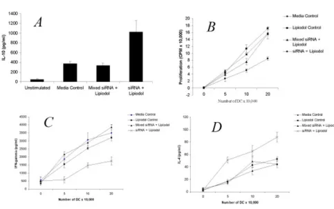
[image:4.612.62.545.92.395.2]Lipiodol transfected DC are immune modulatory Figure 2
Lipiodol transfected DC are immune modulatory. A. Day 7 bone marrow derived DC were cultured alone or transfected with mismatched siRNA, or IL-12p35-specific siRNA using 3 µl/culture lipiodol. Following a 24 hour incubation cells were plated at 1 × 10⁶ in 6 well culture dishes and activated for 24 hours with 10 ng/ml LPS and 10 ng/ml TNF-α. Supernatant was harvested
and analyzed by ELISA for IL-10 production. B. C57/BL6 DC were transfected with mismatched siRNA, IL-12p35-specific siRNA or lipiodol alone, irradiated (3,000 rad) and seeded in triplicate at various concentrations in a flat-bottom 96-well plate. Splenic T cells from BALB/c mice were added as responders (5 × 10⁵ cells/well). The mixed lymphocytes were cultured for 72
IL-10 is mediates immune modulation by IL-12 silenced DC Figure 3
Inhibition of recall response by lipiodol/siRNA vaccine Figure 4
shown). Overall, these data support the notion that lipi-odol is an easy to use method of transfecting bone mar-row derived DC in vitro DC.
Immune modulation by lipiodol/siRNA
We have previously demonstrated that DC silenced for the 12p35 subunit possess an increased production of IL-10 and are poor stimulators of MLR [19]. Using the 3 ul concentration of lipiodol that we found most effective at inhibiting IL-12 production in Fig 1, we verified whether the lipiodol/siRNA treated DC possessed the same immu-nomodulatory properties as previously reported by us. Indeed, we observed that siRNA transfected by lipiodol induced a specific increase in IL-10 production by LPS/ TNF stimulated DC as seen in Fig 2a. Since the specific function of DC in vivo is stimulation of T cell responses, we sought to determine whether the siRNA/lipiodol mix-ture had effects on inhibition of mixed lymphocyte reac-tion (MLR). Indeed, the siRNA specific for IL-12 p35 and not the mismatched (mixed) control inhibited prolifera-tion of responding T cells in a 3 day MLR with naïve BALB/ c splenocytes. Furthermore, as seen in Fig 2b the lipiodol alone did not suppress allostimulatory ability of the DC, indicating that the siRNA itself was specifically inducing inhibition. When supernatants of the MLR were assayed for the prototypic Th1 and Th2 cytokines IFN-γ and IL-4, respectively, an inhibition of IFN-γ Fig 2c, and upregula-tion of IL-4 Fig 2d was observed. This data is in agreement with our previous work in which we demonstrated Th1 to Th2 immune modulation by silencing of the IL-12 p35 subunit [19]. Furthermore, the current data supports the
use of lipiodol as a transfection reagent for induction of immune modulation by siRNA.
Th1 > Th2 shift is associated with IL-10 production
In our previous studies we have observed that silencing IL-12 is associated with an upregulation in IL-10 production [19]. While IL-10 has previously been described to be a Th2 cytokine [29], it has recently been implicated in gen-eration of CD4+ CD25+ T regulatory cells [30] and TR1 cells [31]. We wanted to assess whether siRNA silencing mediated immune modulation is associated with auto-crine IL-10 production by the DC. Indeed, it is known that immature and tolerogenic DC produce this cytokine in an autocrine fashion [32] and that gene-silencing of IL-10 on DC stimulates Th1 immunity and DC maturation [18]. With this in mind we added blocking anti-IL-10 antibod-ies to siRNA treated DC during stimulation of allogeneic BALB/c T cells in MLR. A dose-dependent inhibition in ability to stimulate Th1 responses was seen with addition of anti-IL-10. Specifically, the IL-12 silenced DC stimu-lated the responding T cells to decrease production of
IFN-γ and increase production of IL-4 as depicted in Fig 3a and 3b respectively. Noteworthy is the observation that com-plete inhibition of Th1 stimulation was not achieved by anti-IL-10, implying that stimulation of IFN-γ and inhibi-tion of IL-4 was not completely dependent stimulainhibi-tion of IL-10 and was due to other factors such as suppression of IL-12. Addition of isotype control antibody did not influ-ence cytokine production or proliferation (data not shown).
Table 1
Surface molecules (MFI)
CD80 CD86 CD40
Unactivated DC
Media Control 571.7 (± 23) 1194 (± 125) 29.5 (± 1.5) Lipiodol Control 567.3 (± 21) 1251 (± 173) 28.6 (± 1.3) Mixed siRNA 572.5 (± 10) 1365 (± 157) 29.7 (± 1.3) siRNA alone 559.6 (± 33) 1201 (± 193) 30.1 (± 0.9) siRNA + Lipiodol 577.3 (± 15) 1195 (± 103) 30.4 (± 1.3)
Post-MLR
Lipiodol Control 1962.9 (± 136) 2707.1 (± 576) 140.5 (± 35.4) Mixed siRNA 1933.2 (± 324) 3031.6 (± 489) 170.3 (± 26.9) siRNA alone 1905.7 (± 351) 2759.9 (± 392) 167.2 (± 46.9) siRNA + Lipiodol 2050.8 (± 119) 3051.1 (± 335) 153.3 (± 36.7)
Post-MLR + Anti-IL10
Lipiodol Control 3529 (± 302) 4862 (± 583) 236.0 (± 47.3) Mixed siRNA 3442 (± 247) 4777 (± 773) 242.8 (± 58.9) siRNA alone 3723 (± 453) 4863 (± 669) 237.4 (± 43.5) siRNA + Lipiodol 3985 (± 339) 4957 (± 501) 254.3 (± 49.3)
While we previously demonstrated that IL-12 silenced DC possessed similar levels of costimulatory molecules as wild-type DC [19], we did not assess whether differences were present after stimulation of DC during the MLR proc-ess. This would be anticipated since activated T cells pro-duce a variety of membrane bound (CD40L, CD134L), and soluble factors (IFN-γ, TRANCE) that stimulate DC maturation [33-37]. Extracting DC using magnetic acti-vated cell sorting (MACS) after the MLR demonstrated sig-nificant increases in the mean fluorescent intensity of CD40, CD80 and CD86, however no difference was observed between IL-12 silenced or non-silenced DC. As seen in Table 1, it does not appear that IL-12 silencing leads to Th1>Th2 due to alternation in costimulatory molecule expression. This is particularly interesting since it is known that IL-10 suppresses CD40, CD80, and CD86 expression on DC [38]. Indeed, addition of anti-IL-10 antibody to the MLR significantly increased expression of all 3 costimulatory molecules Table 1. This is in
agree-ment with higher expression of CD40, CD80, and CD86 in IL-10 knockout DC during the process of MLR stimula-tion as compared with wild-type DC [39].
Lipiodol/siRNA induces Th1>Th2 shift in vivo
Having demonstrated above that lipiodol can serve as a transfection reagent for uptake of siRNA in DC, combined with the fact that lipiodol is commonly used for a variety of clinical applications [40,41], we assessed whether lipi-odol/siRNA can be used to modulate immune responses in vivo. Accordingly, we used the KLH and OVA recall responses as an indicator. C57BL/6 mice were immunized with the IL-12 siRNA/lipiodol mixture combined with CFA and either KLH or OVA. 14 days following immuni-zation, recall response experiments were performed using isolated CD4 T cells from draining lymph nodes as previ-ously performed by us [19]. Proliferative responses for both antigens were significantly inhibited in the lipiodol/ siRNA treated animals at restimulation concentrations of
[image:8.612.60.547.91.388.2]In vivo immune modulation lipiodol/siRNA vaccine Figure 5
5 and 10 ug/ml (Fig 4a and 4b). When supernatants were harvested and analyzed by ELISA for cytokine response, a Th1>Th2 shift was observed for both KLH and ovalbumin as demonstrated by higher production of IFN-γ (Fig 5a and 5c) and IL-4 (Fig 5b and 5d), respectively. In order to assess for antigen specificity, in some experiments, mice were immunized with siRNA/lipiodol with KLH and con-currently injected with mixed siRNA/lipiodol and OVA. The recall response to KLH was suppressed in terms of proliferation and possessed a Th2 cytokine profile, whereas the response to OVA was not immune modulated (data not shown). Other experiments not shown included the immunization in absence of lipiodol which did not result in any immunomodulatory effect of siRNA. Overall, these experiments strongly suggest that siRNA can be administered both in vitro and in vivo for immune mod-ulatory purposes using lipiodol as a carrier.
Discussion
Lipiodol (trade name Ultra-Fluide™) is an iodized poppy seed oil fatty acid ethylester commonly used as contrast media for radiography [42]. Commonly, lipiodol alone or with chemotherapy is administered to patients with hepatic cell carcinoma for embolization of tumor-feeding arteries, as well as localized delivery of chemotherapy [43]. The finding that lipiodol selectively is uptaken by endothelial cells through pinocytosis [28], combined with the fact that it has previously been used for siRNA delivery in vivo [26] stimulated us to question whether lipiodol can be used as a carrier for siRNA. We observed that although lipiodol was not as effective as GenePorter for in vitro transfection and gene silencing, the inhibitory effects where several fold more potent than the adminis-tration of naked siRNA.
We therefore continued to seek whether lipiodol transfer of siRNA can still mediate the immune modulatory effects previously reported by us in terms of switching Th1 to Th2 responses [24]. Although this was the case, we also observed that the switch was dependent on IL-10 produc-tion. This is somewhat interesting since although IL-10 is a Th2 cytokine, secretion of IL-10 has also been demon-strated to stimulate CD8 proliferation [44], and even to suppress atopy [45], a manifestation of classical Th2 responses. In this study our data supports a prototypic role of IL-10 secreted by DC in stimulation of IL-4 and IFN-γ production. We are however aware that assessment of other Th1/Th2 associated factors are needed to conclu-sively demonstrate a Th2 switch was occurring. Addition-ally, although in our hands, similar culture situations stimulate IL-10 production primarily by DC, we can not rule out the production of IL-10 by T cells. Future studies involve looking at T cell transcription factors such as T-bet and GATA-3, associated with Th1 and Th2 cells, respec-tively [46].
While we were able to induce in vivo immune modula-tion through administramodula-tion of the lipiodol/siRNA mix-ture, we did not demonstrate that in vivo the DC were indeed the culprits for immune modulation. As previ-ously stated, lipiodol in vivo is pinocytosed by endothe-lial cells [28], and most likely by other cells such as monocytes and macrophages. Indeed both endothelial cells, and macrophages have been demonstrated to pos-sess immune modulatory activity. For example, antigen presentation by unmanipulated endothelium is known to mediate tolerance in various situations [47,48]. Indeed we conceived that the lipiodol itself may be inducing immune modulation due to specific targeting of antigen to endothelial presentation, however this was not the case since lipiodol together with antigen, or together with anti-gen and mixed siRNA did not mediate immune modula-tion. However, the fact that the endothelial cells themselves are protolerogenic may allow for synergy with the tolerogenic siRNA administered. The involvement of macrophages in the immune modulation witnessed is another possibility. Indeed, macrophages have been reported to acquire an M1/M2 phenotype similar to Th1/ Th2 [49]. Preliminary experiments have indicated refrac-toriness to IL-12 production after LPS/TNF stimulation of CD11c DC derived from draining lymph nodes 2–3 days after administration of lipiodol/siRNA, however this will be the subject of an upcoming paper.
Regardless of the cellular mechanisms involved, the abil-ity to immune modulate through administration of a "vaccine composition" with siRNA opens a whole world of novel possibilities both therapeutic and experiments. Through alleviating the need for ex vivo cellular process-ing, simple siRNA based vaccines can be developed for a variety of disease indications where a Th switch is desired. In terms of clinical applicability, the addition of siRNA for embolization of tumors would allow for induction of immune stimulation at the site of tissue necrosis. This would permit induction of immune responses to antigens released during the embolization procedure. Indeed simi-lar approach used by Adema et al has demonstrated suc-cessful induction of immunity through tissue damage combined with immune stimulation [50].
In conclusion, we demonstrate that siRNA can be admin-istered through a clinically useful method, thus opening the possibilities of genetic immune modulation without the use of viruses or ex vivo cellular manipulation.
References
1. Mosmann TR, Cherwinski H, Bond MW, Giedlin MA, Coffman RL:
Two types of murine helper T cell clone. I. Definition accord-ing to profiles of lymphokine activities and secreted proteins.
J Immunol 1986, 136:2348-2357.
3. Singh VK, Mehrotra S, Agarwal SS: The paradigm of Th1 and Th2 cytokines: its relevance to autoimmunity and allergy. Immu-nol Res 1999, 20:147-161.
4. Sospedra M, Martin R: Immunology of Multiple Sclerosis. Annu Rev Immunol 2004.
5. Miossec P, van den Berg W: Th1/Th2 cytokine balance in arthri-tis. Arthritis Rheum 1997, 40:2105-2115.
6. Dabbagh K, Lewis DB: Toll-like receptors and T-helper-1/T-helper-2 responses. Curr Opin Infect Dis 2003, 16:199-204. 7. Steinman RM: The control of immunity and tolerance by
den-dritic cell. Pathol Biol (Paris) 2003, 51:59-60.
8. Agrawal S, Agrawal A, Doughty B, Gerwitz A, Blenis J, Van Dyke T, Pulendran B: Cutting edge: different Toll-like receptor ago-nists instruct dendritic cells to induce distinct Th responses via differential modulation of extracellular signal-regulated kinase-mitogen-activated protein kinase and c-Fos. J Immunol 2003, 171:4984-4989.
9. Redecke V, Hacker H, Datta SK, Fermin A, Pitha PM, Broide DH, Raz E: Cutting edge: activation of Toll-like receptor 2 induces a Th2 immune response and promotes experimental asthma.
J Immunol 2004, 172:2739-2743.
10. Mazzoni A, Segal DM: Controlling the Toll road to dendritic cell polarization. J Leukoc Biol 2004, 75:721-730.
11. Papaccio G, Nicoletti F, Pisanti FA, Bendtzen K, Galdieri M: Preven-tion of spontaneous autoimmune diabetes in NOD mice by transferring in vitro antigen-pulsed syngeneic dendritic cells.
Endocrinology 2000, 141:1500-1505.
12. Feili-Hariri M, Dong X, Alber SM, Watkins SC, Salter RD, Morel PA:
Immunotherapy of NOD mice with bone marrow-derived dendritic cells. Diabetes 1999, 48:2300-2308.
13. Morita Y, Yang J, Gupta R, Shimizu K, Shelden EA, Endres J, Mule JJ, McDonagh KT, Fox DA: Dendritic cells genetically engineered to express IL-4 inhibit murine collagen-induced arthritis. J Clin Invest 2001, 107:1275-1284.
14. Link H, Huang YM, Masterman T, Xiao BG: Vaccination with autologous dendritic cells: from experimental autoimmune encephalomyelitis to multiple sclerosis. J Neuroimmunol 2001,
114:1-7.
15. Yang JS, Xu LY, Huang YM, Van Der Meide PH, Link H, Xiao BG:
Adherent dendritic cells expressing high levels of inter-leukin-10 and low levels of interleukin-12 induce antigen-spe-cific tolerance to experimental autoimmune encephalomyelitis. Immunology 2000, 101:397-403.
16. Tang G: siRNA and miRNA: an insight into RISCs. Trends Bio-chem Sci 2005, 30:106-114.
17. Elbashir SM, Harborth J, Lendeckel W, Yalcin A, Weber K, Tuschl T:
Duplexes of 21-nucleotide RNAs mediate RNA interference in cultured mammalian cells. Nature 2001, 411:494-498. 18. Liu G, Ng H, Akasaki Y, Yuan X, Ehtesham M, Yin D, Black KL, Yu JS:
Small interference RNA modulation of IL-10 in human monocyte-derived dendritic cells enhances the Th1 response. Eur J Immunol 2004, 34:1680-1687.
19. Hill JA, Ichim TE, Kusznieruk KP, Li M, Huang X, Yan X, Zhong R, Cairns E, Bell DA, Min WP: Immune modulation by silencing IL-12 production in dendritic cells using small interfering RNA.
J Immunol 2003, 171:691-696.
20. Flynn MA, Casey DG, Todryk SM, Mahon BP: Efficient delivery of small interfering RNA for inhibition of IL-12p40 expression in vivo. J Inflamm (Lond) 2004, 1:4.
21. Lovett-Racke AE, Rocchini AE, Choy J, Northrop SC, Hussain RZ, Ratts RB, Sikder D, Racke MK: Silencing T-bet defines a critical role in the differentiation of autoreactive T lymphocytes.
Immunity 2004, 21:719-731.
22. Kobayashi T, Yoshimura A: Keeping DCs awake by putting SOCS1 to sleep. Trends Immunol 2005, 26:177-179.
23. Li M, Qian H, Ichim TE, Ge WW, Popov IA, Rycerz K, Neu J, White D, Zhong R, Min WP: Induction of RNA interference in den-dritic cells. Immunol Res 2004, 30:215-230.
24. Ichim TE, Li M, Qian H, Popov IA, Rycerz K, Zheng X, White D, Zhong R, Min WP: RNA interference: a potent tool for gene-specific therapeutics. Am J Transplant 2004, 4:1227-1236. 25. Klein C, Bock CT, Wedemeyer H, Wustefeld T, Locarnini S, Dienes
HP, Kubicka S, Manns MP, Trautwein C: Inhibition of hepatitis B virus replication in vivo by nucleoside analogues and siRNA.
Gastroenterology 2003, 125:9-18.
26. Contreras JL, Vilatoba M, Eckstein C, Bilbao G, Anthony Thompson J, Eckhoff DE: Caspase-8 and caspase-3 small interfering RNA decreases ischemia/reperfusion injury to the liver in mice.
Surgery 2004, 136:390-400.
27. Ichim TE, Zhong R, Min WP: Prevention of allograft rejection by in vitro generated tolerogenic dendritic cells. Transpl Immunol 2003, 11:295-306.
28. Bhattacharya S, Dhillon AP, Winslet MC, Davidson BR, Shukla N, Gupta SD, Al-Mufti R, Hobbs KE: Human liver cancer cells and endothelial cells incorporate iodised oil. Br J Cancer 1996,
73:877-881.
29. Romagnani S: Th1/Th2 cells. Inflamm Bowel Dis 1999, 5:285-294. 30. Horwitz DA, Zheng SG, Gray JD: The role of the combination of
IL-2 and TGF-beta or IL-10 in the generation and function of CD4+ CD25+ and CD8+ regulatory T cell subsets. J Leukoc Biol 2003, 74:471-478.
31. Levings MK, Gregori S, Tresoldi E, Cazzaniga S, Bonini C, Roncarolo MG: Differentiation of Tr1 cells by immature dendritic cells requires IL-10 but not CD25+CD4+ Tr cells. Blood 2005,
105:1162-1169.
32. Corinti S, Albanesi C, la Sala A, Pastore S, Girolomoni G: Regulatory activity of autocrine IL-10 on dendritic cell functions. J Immu-nol 2001, 166:4312-4318.
33. Serra P, Amrani A, Yamanouchi J, Han B, Thiessen S, Utsugi T, Verd-aguer J, Santamaria P: CD40 ligation releases immature den-dritic cells from the control of regulatory CD4+CD25+ T cells. Immunity 2003, 19:877-889.
34. Ohshima Y, Tanaka Y, Tozawa H, Takahashi Y, Maliszewski C, Delespesse G: Expression and function of OX40 ligand on human dendritic cells. J Immunol 1997, 159:3838-3848. 35. Nakahara T, Urabe K, Fukagawa S, Uchi H, Inaba K, Furue M, Moroi
Y: Engagement of human monocyte-derived dendritic cells into interleukin (IL)-12 producers by IL-1beta + interferon (IFN)-gamma. Clin Exp Immunol 2005, 139:476-482.
36. Walsh MC, Choi Y: Biology of the TRANCE axis. Cytokine Growth Factor Rev 2003, 14:251-263.
37. Caux C, Massacrier C, Vanbervliet B, Dubois B, Van Kooten C, Durand I, Banchereau J: Activation of human dendritic cells through CD40 cross-linking. J Exp Med 1994, 180:1263-1272. 38. De Smedt T, Van Mechelen M, De Becker G, Urbain J, Leo O, Moser
M: Effect of interleukin-10 on dendritic cell maturation and function. Eur J Immunol 1997, 27:1229-1235.
39. He Q, Moore TT, Eko FO, Lyn D, Ananaba GA, Martin A, Singh S, James L, Stiles J, Black CM, Igietseme JU: Molecular basis for the potency of IL-10-deficient dendritic cells as a highly efficient APC system for activating Th1 response. J Immunol 2005,
174:4860-4869.
40. Vogl TJ, Wetter A, Lindemayr S, Zangos S: Treatment of unresect-able lung metastases with transpulmonary chemoemboliza-tion: preliminary experience. Radiology 2005, 234:917-922. 41. Di Stefano DR, de Baere T, Denys A, Hakime A, Gorin G, Gillet M,
Saric J, Trillaud H, Petit P, Bartoli JM, Elias D, Delpero JR: Preoper-ative percutaneous portal vein embolization: evaluation of adverse events in 188 patients. Radiology 2005, 234:625-630. 42. Hamm B, Wolf KJ: Contrast material for computed
tomogra-phy and magnetic resonance imaging of the gastrointestinal tract. Curr Opin Radiol 1991, 3:474-482.
43. Bhattacharya S, Novell JR, Winslet MC, Hobbs KE: Iodized oil in the treatment of hepatocellular carcinoma. Br J Surg 1994,
81:1563-1571.
44. Rowbottom AW, Lepper MA, Garland RJ, Cox CV, Corley EG:
Interleukin-10-induced CD8 cell proliferation. Immunology 1999, 98:80-89.
45. Akdis CA, Blaser K: Role of IL-10 in allergen-specific immuno-therapy and normal response to allergens. Microbes Infect 2001, 3:891-898.
46. Hwang ES, Szabo SJ, Schwartzberg PL, Glimcher LH: T helper cell fate specified by kinase-mediated interaction of T-bet with GATA-3. Science 2005, 307:430-433.
47. Rothermel AL, Wang Y, Schechner J, Mook-Kanamori B, Aird WC, Pober JS, Tellides G, Johnson DR: Endothelial cells present anti-gens in vivo. BMC Immunol 2004, 5:5.
Publish with BioMed Central and every scientist can read your work free of charge "BioMed Central will be the most significant development for disseminating the results of biomedical researc h in our lifetime."
Sir Paul Nurse, Cancer Research UK
Your research papers will be:
available free of charge to the entire biomedical community
peer reviewed and published immediately upon acceptance
cited in PubMed and archived on PubMed Central
yours — you keep the copyright
Submit your manuscript here:
http://www.biomedcentral.com/info/publishing_adv.asp
BioMedcentral 49. Modolell M, Corraliza IM, Link F, Soler G, Eichmann K: Reciprocal
regulation of the nitric oxide synthase/arginase balance in mouse bone marrow-derived macrophages by TH1 and TH2 cytokines. Eur J Immunol 1995, 25:1101-1104.