Functional Differences between the Two Progeny Phenotypes of a
Baculovirus
Dianhai Hou,aLeike Zhang,bFei Deng,aWei Fang,aRanran Wang,a* Xijia Liu,aLin Guo,b,cSimon Rayner,aXinwen Chen,a Hualin Wang,aZhihong Hua
State Key Laboratory of Virology, Wuhan Institute of Virology, Chinese Academy of Sciences, Wuhan, Chinaa; State Key Laboratory of Virology, College of Life Sciences,b
and Key Laboratory of Analytical Chemistry for Biology and Medicine (Ministry of Education),cWuhan University, Wuhan, China
The replication of lepidopteran baculoviruses is characterized by the production of two progeny phenotypes: the occlusion-de-rived virus (ODV), which establishes infection in midgut cells, and the budded virus (BV), which disseminates infection to dif-ferent tissues within a susceptible host. To understand the structural, and hence functional, differences between BV and ODV, we employed multiple proteomic methods to reveal the protein compositions and posttranslational modifications of the two phenotypes ofHelicoverpa armigeranucleopolyhedrovirus. In addition, Western blotting and quantitative mass spectrometry were used to identify the localization of proteins in the envelope or nucleocapsid fractions. Comparative protein portfolios of BV and ODV showing the distribution of 54 proteins, encompassing the 21 proteins shared by BV and ODV, the 12 BV-specific pro-teins, and the 21 ODV-specific propro-teins, were obtained. Among the 11 ODV-specific envelope propro-teins, 8 either are essential for or contribute to oral infection. Twenty-three phosphorylated and 6N-glycosylated viral proteins were also identified. While the proteins that are shared by the two phenotypes appear to be important for nucleocapsid assembly and trafficking, the structural and functional differences between the two phenotypes are evidently characterized by the envelope proteins and posttransla-tional modifications. This comparative proteomics study provides new insight into how BV and ODV are formed and why they function differently.
B
aculoviruses are insect-specific pathogens containing largecircular double-stranded DNA genomes. Over millions of years of interdependence between viruses and their natural insect hosts, both have undergone a coevolution, such that lepidopteran baculoviruses have developed a unique biphasic replication cycle that generates two progeny phenotypes, the budded virus (BV) and the occlusion-derived virus (ODV). ODVs are embedded in occlusion bodies (OBs) that offer the virions a certain amount of protection against environmental degradation. Once ingested by a susceptible insect, ODVs are released from OBs within the larval midgut and initiate oral infection. After infecting midgut epithe-lial cells, BVs are synthesized and released to disseminate systemic infection of different tissues within the larval host. The two phe-notypes have been used in a wide range of applications. Due to their expandable genome and the presence of very strong promot-ers, BVs have been established as successful vectors for the expres-sion of thousands of proteins and have also been studied as
poten-tial vectors for gene therapy (1). The OBs of certain baculoviruses
have been widely used in agriculture and forestry as viable
alter-natives to chemical insecticides in insect pest control (2).
The broad applications of baculoviruses provide a strong ra-tionale for identifying the proteins associated with both pheno-types and for understanding their roles in baculovirus infection. While previous proteomic studies have elucidated the protein
compositions of a few baculoviruses (3–8), a comprehensive
in-vestigation into the portfolio and landscape of viral proteins, as well as their associated protein modifications, has not been carried
out. The familyBaculoviridaecontains four genera:
Alphabaculo-virus,Betabaculovirus,Gammabaculovirus, andDeltabaculovirus.
The members ofAlphabaculovirusare subdivided into two groups,
I and II, based on phylogeny. TheHelicoverpa armigera
nucleo-polyhedrovirus (HearNPV), which has a genome size of 131 kb and
contains 135 open reading frames (ORFs), is a group II
Alphabaculo-virusand has been used successfully as a viral pesticide against cotton
bollworms (9). In this study, we systematically investigated the
HearNPV proteome using multiple proteomic techniques to reveal the protein compositions and posttranslational modifications of BV and ODV. Additionally, Western blotting and isobaric tags for rela-tive and absolute quantitation (iTRAQ) were employed to analyze the locations of the structural proteins of BV and ODV. Our results show that the proteins that are shared by BV and ODV appear to be impor-tant for nucleocapsid formation and trafficking, but the two pheno-types differ markedly in their membrane protein compositions. The posttranslational protein modifications and the host proteins associ-ated with BV and ODV are also significantly different. The detailed proteomics analyses exploring all the structural proteins of BV and ODV presented here shed light on the infection mechanisms used by the two distinct phenotypes.
Received28 August 2012 Accepted25 October 2012
Published ahead of print31 October 2012
Address correspondence to Lin Guo, [email protected], or Zhihong Hu, [email protected].
* Present address: Ranran Wang, Department of Microbiology, Perelman School of Medicine, University of Pennsylvania, Philadelphia, Pennsylvania, USA.
D.H., L.Z., and F.D. contributed equally to this work.
Supplemental material for this article may be found athttp://dx.doi.org/10.1128
/JVI.02329-12.
Copyright © 2013, American Society for Microbiology. All Rights Reserved.
doi:10.1128/JVI.02329-12
on November 7, 2019 by guest
http://jvi.asm.org/
MATERIALS AND METHODS
Cell cultures, virus infection, virion purification, and fractionation.A Helicoverpa zeacell line, HzAM1 (10), was maintained at 27°C in Grace’s insect medium supplemented with 10% fetal bovine serum (FBS). The HearNPV G4 strain (9) was used in this study. The BVs were collected from the supernatant of the infected HzAM1 cells and purified as de-scribed previously (4). Polyhedra were isolated and purified from infected H.armigeralarvae (11), and the ODVs were released from polyhedra by alkaline treatment (pH 10.9) for 15 min and purified on continuous (25% to 65%) sucrose gradients. Purified BV and ODV were further separated into envelope (E) and nucleocapsid (NC) fractions (12). The NC and E fractions were precipitated with 3 volumes of 50% acetone-50% metha-nol-0.1% acetic acid and used for further SDS-PAGE and multiple pro-teomic analyses.
SDS-PAGE and in-gel digestion.Purified BV, ODV, and their NC and E proteins were separated on a 12% SDS-PAGE gel. The protein bands or regions were excised from the BV lane, destained, reduced, alkylated, and subjected to in-gel trypsin digestion (11). The peptide mixtures obtained were further desalted by ZipTipC18 (Millipore) and dried by SpeedVac (11).
In-solution digestion and HILIC fractionation of peptides for shot-gun proteomic analysis.Shotgun proteomics is also known as multidi-mensional protein identification technology (MudPIT) (13). Precipitated E and NC proteins were resuspended in lysis buffer (8 M urea, 4 mM CaCl2, 0.2 M Tris-HCl [pH 8.0]), reduced, alkylated, and subjected to
in-solution trypsin digestion as described previously (14). Peptides di-gested from 50g HearNPV virion (BV or ODV) proteins were further fractionated using hydrophilic interaction chromatography (HILIC) as described previously (14). Seven fractions for BV and 10 fractions for ODV were collected.
iTRAQ labeling and SCX fractionation of peptides for quantitative proteomic analyses.Isobaric tags for relative and absolute quantitation (iTRAQ) labeling processing was conducted according to the iTRAQ pro-tocol (Applied Biosystems). Peptides digested in solution from equivalent amounts (50g) of NC and E proteins were used in this study, and two replicate experiments were performed as follows. In one replicate, pep-tides digested from NC and E proteins were labeled with iTRAQ reagents 116 and 117, respectively. In the second replicate, peptides digested from NC and E proteins were labeled with iTRAQ reagents 117 and 116, respec-tively. The labeled peptides were mixed together and fractionated by strong cation exchange (SCX) (15), and seven fractions were collected.
Phosphopeptide enrichment using the IMAC-Fe3ⴙmethod.
In-so-lution-digested peptides from 1.5 mg of virion (BV or ODV) proteins were dissolved in 1% acetic acid and loaded onto immobilized metal af-finity chromatography (IMAC)-Fe3⫹resins to enrich phosphopeptides
(14). The experiment was repeated three times.
Enrichment ofN-linked glycopeptides by the solid-phase extraction method using hydrazide chemistry.Peptides from 1 mg of in-solution-digested virion (BV or ODV) proteins were used for solid-phase extrac-tion ofN-linked glycopeptides (SPEG) as described previously (16) with-out isotope labeling. Two replicate experiments were performed.
Mass spectrometry and database search for protein identification and posttranslational modification assignments.Peptides derived from in-gel digestion were subjected to liquid chromatography-tandem mass spectrometry (LC-MS/MS) analyses using 4000 QTRAP. The mobile phase consisted of two components: (A) 2% acetonitrile with 0.1% formic acid and (B) 98% acetonitrile with 0.1% formic acid. Peptides were loaded onto the precolumn (0.5 mm by 2 mm; Michrom Bioresources, Inc.) at a flow rate of 5l/min and then were eluted into the analytic column at a flow rate of 300 nl/min with a gradient starting from 5% B held for 5 min, programmed to 60% B for 75 min, and held for another 5 min. The precursor ion’s range was chosen fromm/z400 tom/z1,600, and the product ion’s range was chosen fromm/z50 tom/z1,600. For the peptides derived from digestion in solution, LC-MS/MS analyses were conducted using QSTAR Elite as described previously (14).
Analyses of the raw MS spectra generated by LC-MS/MS analyses were performed with the ProteinPilot 4.0 software program (Applied Biosys-tems) using the Paragon algorithm (17) against the ORF database of HearNPV for viral protein identification and posttranslational modification assignments. A lepidopteran protein database (derived from GenBank) was utilized to identify host proteins. For the assignment of phosphorylation sites, the Scaffold PTM (posttranslational modification) (18) was used in combi-nation with ProteinPilot software (Applied Biosystems). Only the phosphor-ylation sites identified by ProteinPilot and by Scaffold PTM (with a probabil-ity ofⱖ0.75) were considered significant.
Putative signal peptide (SP) and transmembrane domains (TMs) were predicted, respectively, by the SignalP 4.0 (http://www.cbs.dtu.dk/services /SignalP/) and TopPred (http://mobyle.pasteur.fr/cgi-bin/MobylePortal /portal.py?form⫽toppred) software packages.
Statistical analyses of iTRAQ peptide data for protein localization. The natural logarithm of the relative peptide ion ratios of the envelope versus the nucleocapsid, i.e., [log ratio⫽ln(E/NC)] obtained with Pro-teinPilot software, was used to infer protein localization. Any proteins with 3 or more identified peptides were evaluated by statistical analyses. A peptide ratio of either 0.000 or 9,999 indicates that only one peak was found, and these data were not considered further.
According to the ProteinPilot software manual, the average protein abundance (APA) ratio is estimated using the formula APA ratio⫽ eweighted average of log ratio. The point estimate of the weighted average of
log ratio,WLR, was calculated using the following equation:
WLR⫽
兺
i⫽1n
wixi
兺
i⫽1
n
wi
wherexiis log(peptide iTRAQ ratioi),n⫽the number of peptide ratios contributing to the estimate of a protein’s average ratio, andwiis equal to 1/(% error)i, the weight forxi.
The 95% confidence intervals of the weighted average of the log ratio were evaluated using the nonparametric bootstrap bias-corrected and ac-celerated (BCa) method (19), implemented in the boot.ci function in the boot R package (20).
Western blot analyses.The polyclonal antibodies against polyhedrin (POLH), HA9(49K), E18, HA66, GP41, P33, E25, VP39, 38K, P6.9, ODV-E66, C42, VP80, and the polyhedron envelope protein (PEP) of HearNPV were generated as described by Deng et al. (4). Previously generated poly-clonal antibodies against HA44, HA100 andper osinfectivity factor 5 (PIF5) (4), P74, PIF1, PIF2 and PIF3 (21), PIF4 (22), P78/83 (23), HA51 (24), FP25K (25), EC43 (26), the fibroblast growth factor (FGF) (27), and fusion protein (F protein) (28) were also used. Western blot analyses were conducted as described previously (4).
RESULTS
Proteins associated with BV and ODV.In this study, we em-ployed 5 proteomic methods to reveal the protein compositions and posttranslational modifications of HearNPV BV and ODV. These included (i) iTRAQ, which involves isobaric tags and iden-tifies proteins with relative and absolute quantifications, (ii) shot-gun proteomics, which involves microcapillary multidimensional chromatography and identifies peptides from protein mixtures, (iii) IMAC, which involves peptide enrichment by immobilized metal ion affinity chromatography and identifies phosphopep-tides, (iv) SDS-PAGE and LC-MS/MS, which involve in-gel tryp-sin digestion and identify the proteins of SDS-PAGE bands, and (v) SPEG proteomics, which involves the solid-phase extraction of
N-linked glycopeptides and identifiesN-glycosylated proteins.
Purified BV, ODV, and their NC and E proteins were separated on a 12% SDS-PAGE gel as shown in Fig. S1 in the supplemental material. The details of the proteomic results are shown in Data
on November 7, 2019 by guest
http://jvi.asm.org/
Sets S1 and S2 in the supplemental material. A summary of the
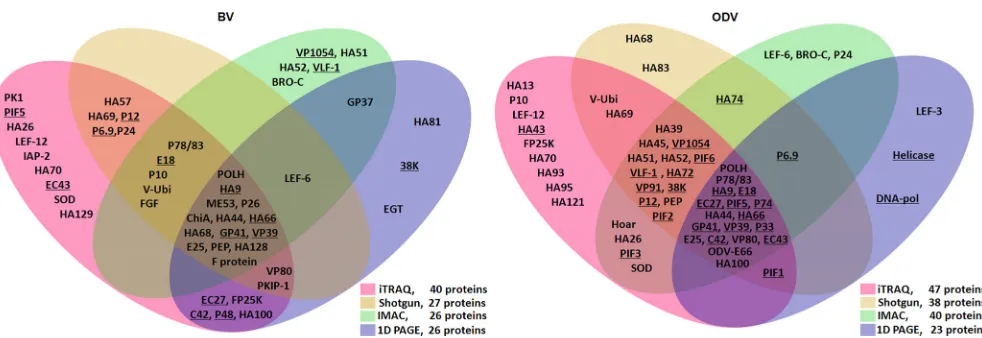
first four proteomic methods is presented inFig. 1, while the SPEG
results are shown inTable 1. In BV, 40 proteins were identified by
iTRAQ, 27 proteins by shotgun proteomics, 26 proteins by IMAC,
and 26 proteins by SDS-PAGE and LC-MS/MS (Fig. 1, left; see also
Data Set S1 in the supplemental material). In addition, SPEG de-tected 6 proteins and further identified viral cathepsin (V-CATH)
as a BV-associated protein (Table 1; see also Data Set S1). When
the data from the various techniques were combined, a total of 51 proteins were identified from BV, of which 33 were identified by at least two proteomics methods.
Similarly, a total of 57 ODV-associated proteins were identi-fied by different proteomics techniques, including 47 proteins by iTRAQ, 38 proteins by shotgun proteomics, 40 proteins by IMAC,
and 23 proteins by SDS-PAGE and LC-MS/MS (Fig. 1, right; see
also Data Set S2 in the supplemental material). Among these pro-teins, 40 were detected by at least two methods.
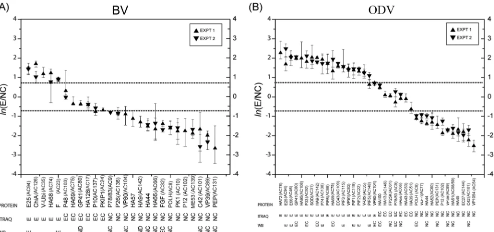
Localization of virion-associated proteins by iTRAQ.In or-der to identify protein locations, BV and ODV were separated into E and NC fractions and analyzed by quantitative mass
spectrom-etry using iTRAQ labeling. Based on the relative protein abun-dances of E/NC in terms of the estimated protein abundance ratio for the envelope versus the nucleocapsid, BV and ODV proteins with 3 or more identified peptides were grouped into one of three
categories: (i) envelope (E)-associated proteins (E/NCⱖ2 [ln(E/
NC) ⱖ0.69]), (ii) envelope-and-nucleocapsid (EC)-associated
proteins (0.5⬍E/NC⬍2 [⫺0.69⬍ln(E/NC)⬍0.69]), and (iii)
nucleocapsid (NC)-associated proteins (E/NCⱕ0.5 [ln(E/NC)ⱕ
⫺0.69]) (see Table S1 in the supplemental material). In this way, a
total of 26 BV proteins comprising 5 envelope-associated pro-teins, 6 envelope-and-nucleocapsid-associated propro-teins, and 15
nucleocapsid-associated proteins were identified (Fig. 2A; see also
Table S1A in the supplemental material). For ODV, a total of 38 proteins consisting of 18 envelope-associated proteins, 8 enve-lope-and-associated proteins, and 12
nucleocapsid-associated proteins were identified (Fig. 2B; see also Table S1B in
the supplemental material).
Identification and localization of proteins by Western blot-ting.To further confirm the locations of the structural proteins, Western blotting was also performed. Polyclonal antibodies
[image:3.585.47.538.62.231.2]FIG 1Summary of protein composition results for HearNPV BV (left) and ODV (right) compiled from multiple proteomic approaches. The proteins that were conserved in all baculoviruses are underlined. The proteins identified by iTRAQ-, shotgun-, IMAC-, and SDS-PAGE and LC-MS/MS (one-dimensional [1D] PAGE)-based proteomics are indicated in their respective regions. The 1D PAGE data of ODV were derived from Deng et al. (4).
TABLE 1Results ofN-glycoproteomic analyses of HearNPV BV using the SPEG method
Protein
HearNPV ORF
AcMNPV
ORF No. of AAsa
Molecular mass (kDa)
TMs and SPsb
DetectedN-glycosylated peptide
Peptide sequencec
Start (AA)
Stop
(AA) N-Glycosylated sites
E18 10 143 81 8.8 TM, SP GADTNAFAFQNPLN#ATMR 51 68 N64
P26 22 136 267 30.5 TM, SP TVSINVIGHQSN#DSDTLDR 46 64 N57
ChiA 41 126 570 65.5 TM, SP FATFDYN#TSGR 98 108 N104
AMLDQVQIQTN#R 346 357 N356
V-CATH 56 127 365 42 TM HFLQQYN#K 57 64 N63
NNN#DSLSTSAQFGVNK 99 114 N101
FGF 113 32 301 34.4 TM, SP N#GTVWGITN#STDSHSVFYR 49 64 N49, N57
NVLVN#SSGVHR 138 148 N142
HSNVN#ATEGVN#QTNFY 207 222 N211, N217
F protein 133 23 677 78.2 TM NKN#LTSCENSETIFH 102 114 N104
NNLLITEYVDMSSTFN#FSR 511 529 N526
EINN#NTIFK 568 576 N571
aAA, amino acid. b
TMs denote putative transmembrane regions with a score ofⱖ0.95; SPs denote predicted signal peptides with a score ofⱖ0.8.
cThe # symbol indicates glycosylation (N).
on November 7, 2019 by guest
http://jvi.asm.org/
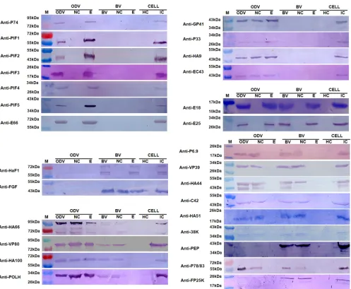
[image:3.585.39.550.535.698.2]against 28 HearNPV proteins were used in Western blot analyses to identify and confirm the iTRAQ-based protein localization data (Fig. 3). The protein localization results are outlined in Table S1 in the supplemental material, and they show the proteins that were identified by at least two methods to total 35 for BV and 41 for ODV. The Western blot protein localization results are
summa-rized at the bottom of the iTRAQ plots inFig. 2.
The Western blot analyses detected 17 BV-associated proteins (Fig. 3), which corroborated the iTRAQ results for the localization of 10 proteins, including 2 envelope-associated proteins (E25 and membrane fusion protein F) and 8 nucleocapsid-associated pro-teins (P78/83, VP80, HA44, HA66, POLH, C42, VP39, and PEP) (Fig. 2A). The Western blots designated FGF as an envelope-and-nucleocapsid-associated protein, while iTRAQ showed it to be predominantly in the nucleocapsid, since the signal from the nu-cleocapsid fraction was significantly higher than that from the envelope fraction. We therefore conclude that FGF is present in both envelope and nucleocapsid fractions, but it is proportion-ately richer in the nucleocapsid. In addition, Western blot analyses detected E18 in the envelope fraction of BV and P6.9, HA51, 38K,
HA100, and FP25K in the nucleocapsid fraction of BV (Fig. 3).
However, either no peptides or fewer than three peptides were identified by iTRAQ for these proteins and, therefore, could not be evaluated by this method. Although iTRAQ detected the pres-ence of GP41 in both the envelope and nucleocapsid fractions and HA9 in the nucleocapsid fraction, neither was observed by West-ern blotting; this is probably because the amounts in BV were too small to be detected under our Western blot conditions.
Twenty-six proteins were detected to be associated with ODV
by Western blotting (Fig. 3). Seven proteins (E25, E66, P74, PIF3,
PIF1, PIF2, and PIF5) were located in the envelope, 3 proteins (VP80, HA100, and HA66) were located in the envelope and nu-cleocapsid fractions, and 5 proteins (HA44, PEP, VP39, HA51,
and C42) were located in the nucleocapsid fraction. These data are
in agreement with the iTRAQ results (Fig. 2B). Of the remaining
proteins, 4 (GP41, P33, HA9, and EC43) were detected in both the envelope and nucleocapsid fractions by Western blotting but were identified as envelope-associated proteins by iTRAQ. Following our interpretation of the BV results, we conclude that these 4 proteins are envelope-and-nucleocapsid-associated proteins, but proportionately higher quantities are present in the envelope rel-ative to the nucleocapsid. For FP25K and P78/83, Western blot-ting detected them in the nucleocapsid fraction, while iTRAQ lo-cated them in both the envelope and nucleocapsid fractions. We therefore conclude that these proteins are located in both the nu-cleocapsid and the envelope, but the amounts in the latter com-ponent were so small that they could not be detected by Western blotting. Finally, our Western blot analyses identified E18 as an envelope protein, P6.9 and 38K as nucleocapsid proteins, and
PIF4 as an envelope protein in ODVs (Fig. 3). No peptides or
fewer than three peptides were identified by iTRAQ in these pro-teins, so they could not be evaluated.
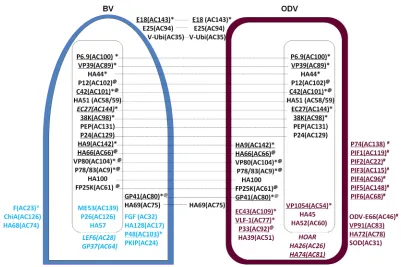
Comparative model of protein compositions and localiza-tions of BV and ODV.By combining all the proteomics (iTRAQ, shotgun MS/MS, IMAC, SDS-PAGE and LC-MS/MS, SPEG, and Western blotting) and protein localization (Western blotting and iTRAQ) data, the comparative protein portfolios of BV and ODV
are presented inFig. 4. AsAutographa californicamultiple
nucle-opolyhedrovirus (AcMNPV) is the type species of the baculovirus family, the AcMNPV homologues of the HearNPV ORFs are also
indicated inFig. 4for general information. Only the proteins
iden-tified by at least two independent methods are included inFig. 4,
except for PIF4, which was detected only by Western blotting. We
also removed POLH and P10 fromFig. 4, because these are the two
most heavily expressed proteins at the late stages of baculovirus infection, but they are not related to BV or ODV formation.
FIG 2Summary of the protein localization results from iTRAQ and Western blotting (WB) for BV (A) and ODV (B). The proteins are ordered along thexaxis according to their estimatedln(E/NC) ratios, which are found along theyaxis. The protein names are shown on the first row below the plot, protein classifications based on iTRAQ data are shown on the second row, and Western blot results are shown on the third row below the plot. Ninety-five percent confidence intervals are shown as vertical lines on the plot. The ORF numbers of the AcMNPV homologues for all the HearNPV ORFs are indicated in brackets.
on November 7, 2019 by guest
http://jvi.asm.org/
[image:4.585.48.541.66.298.2]Of the 54 proteins presented inFig. 4, 21 proteins are shared by BV and ODV (shown in black), 12 proteins are specific to BV-(shown in blue), and 21 proteins are specific to ODV BV-(shown in maroon).
Among the proteins shared by BV and ODV, 3 proteins were located in the envelope of both BV and ODV (E18, E25, and V-ubiquitin [V-Ubi]), and 8 proteins were located in the nucleocap-sid of both BV and ODV (P6.9, VP39, HA44, P12, C42, HA51 [AC59], 38K, and PEP). EC27 was located in the nucleocapsid of ODV, but its location in HearNPV BV was not identified in this study. Six proteins were located in the nucleocapsid fraction of BV (HA9 [AC142], HA66 [AC66], VP80, P78/83, HA100, and FP25K) but appeared to be EC proteins in ODV. One protein was located in both the nucleocapsid and envelope fractions of BV and ODV (GP41). One protein was an EC protein in BV but an enve-lope component of ODV (HA69 [AC75]).
Of the 13 BV-specific proteins, 3 proteins were in the envelope (F protein, chitinase [ChiA], and HA68 [AC74]), 3 proteins were
in the nucleocapsid (ME53, P26, and HA57), 4 proteins were EC proteins (FGF, HA128 [AC17], P48, and protein kinase-interact-ing protein [PKIP-1]), and 3 proteins had no localization data from iTRAQ or Western blots (P24, late expression factor 6 [LEF6], and GP37).
Of the 21 ODV-specific proteins, 11 were in the envelope, in-cluding 8 proteins (P74, PIF1, PIF2, PIF3, PIF4, PIF5, PIF6, and ODV-E66) involved in oral infection (denoted by the # symbol in
Fig. 4) (21,22,29–33). Recently, the truncated ODV-E66 protein was identified as a chondroitin lyase that can efficiently digest
chondroitin and chondroitin 6-sulfate (34). The remaining
enve-lope proteins were VP91, HA72 (AC78), and superoxide dismu-tase (SOD). In addition, 3 proteins were located in the nucleocap-sid (VP1054, HA45, and HA52 [AC60]), 4 proteins were located in both the envelope and the nucleocapsid fractions (EC43, very late expression factor 1 [VLF-1], P33, and HA39 [AC51]), and 3 pro-teins were of unknown location (HOAR, HA26 [AC26], and HA74 [AC81]).
FIG 3Western blot analyses of protein localization in the nucleocapsid and envelope fractions of HearNPV BV and ODV. Purified BV and ODV, as well as their nucleocapsid (NC) and envelope (E) fractions, were loaded for Western blot analyses. Healthy cells (HC) and virus-infected cells (IC) were used as negative and positive controls, respectively. The antibodies used for the Western blots are indicated to the left of each blot diagram. M indicates molecular mass markers.
on November 7, 2019 by guest
http://jvi.asm.org/
[image:5.585.45.542.62.471.2]Posttranslational modifications of virion proteins. Post-translation modifications play important roles in protein
func-tion, and in this study,N-glycosylation, phosphorylation, and
N-terminal acetylation of BV and ODV proteins were analyzed.
N-glycosylation of virion proteins was detected by SPEG
pro-teomics. Although there are many predictedN-glycosylation sites,
a total of 14 were detected in 12 different glycopeptides from 6 BV-associated proteins (E18, P26, ChiA, V-CATH, FGF, and F
protein) (Table 1). No ODV proteins were found to beN
-glyco-sylated.
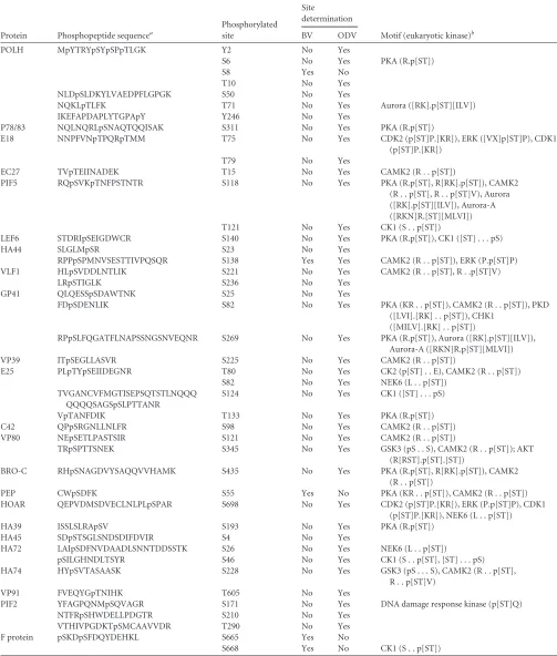
A total of 43 phosphorylation sites in 36 phosphopeptides from
23 viral proteins were identified (Table 2), yielding a Ser/Thr/Tyr
phosphorylation ratio of 31:10:2 (72.1%/22.3%/4.6%). Among all the phosphorylation sites, 38 sites were in ODV-associated pro-teins, while 4 sites were in BV-associated proteins; only 1 site was
identified in both BV and ODV (Table 2), indicating that BV and
ODV have different phosphorylation profiles. Twenty-nine phos-phorylation sites and their surrounding motifs were matched with consensus substrate sequences (motifs) for specific eukaryotic protein kinases by PHOSIDA (Phosphorylation Site Database)
analysis (35) (Table 2).
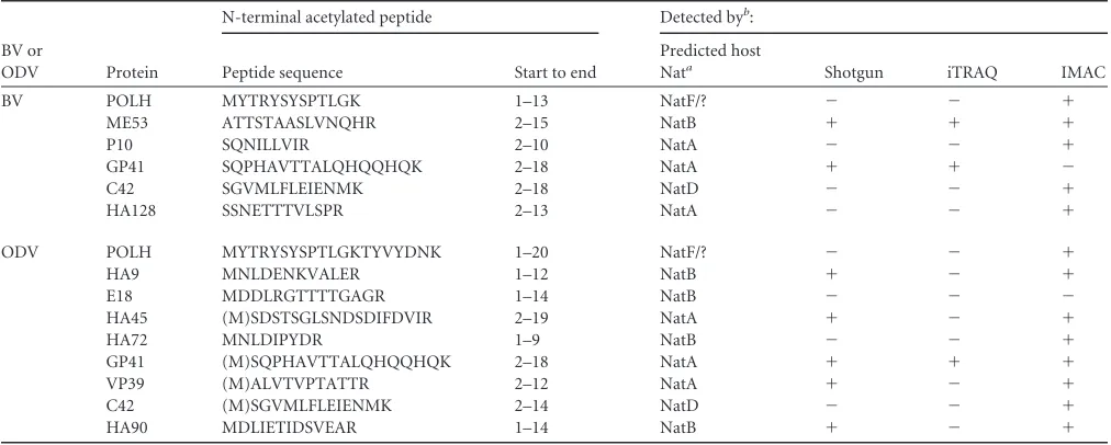
The MS/MS data from multiple proteomic analyses supplied additional information on N-terminal acetylation of HearNPV proteins. We discovered that 12 proteins contained acetylated N
termini (Table 3). Seven proteins (ME53, P10, GP41, C42, HA128,
HA45, and VP39) were found to be acetylated at the second amino acid, indicating that their N-terminal methionine was removed after translation. Eukaryotes contain at least six different
N-termi-nal acetyltransferases (Nats), NatA to NatF, each with a different
substrate specificity (36, 37). The Nat predictions are listed in
Table 3, and the results indicate that NatA, NatB, NatD, and NatF participate in baculovirus N-terminal acetylation.
Virion-associated host proteins.Some viruses contain spe-cific host proteins in their virions to facilitate infection. We iden-tified 101 BV-associated and 21 ODV-associated host proteins that were classified based on their cellular functions, including cytoskeleton assembly, vesicle trafficking, signaling, metabolism, and posttranslational modification (see Table S2 in the supple-mental material). Of all the HearNPV-associated host proteins, 52 have been detected previously in several other envelope viruses (see Table S2 in the supplemental material), implying that envel-oped viruses may use the same or similar cellular pathways during their infection processes. The roles of these virion-associated host proteins in baculovirus replication are still unknown.
DISCUSSION
We have applied multiple proteomic approaches to investigate protein composition and localization in BV and ODV, and we provide detailed comparative schematics of the two
pheno-types (Fig. 4). Our results should help clarify functional
differ-ences between these distinct phenotypes and shed light on how the virus switches from BV to ODV production during its life cycle.
BV and ODV share common proteins important for nucleo-capsid assembly and trafficking.Our study identified 21 proteins shared by BV and ODV, 6 of which are essential for nucleocapsid
FIG 4Comparative schematics showing protein compositions and localizations of HearNPV BV and ODV based on experimental results. The envelope proteins are shown in the figure on the surfaces of BV and ODV, the nucleocapsid proteins are shown inside each rectangle, and the EC proteins are located in the space between the envelope and the nucleocapsid. The proteins shared by BV and ODV are in black, and the proteins located in both BV and ODV are linked by dashed lines; BV-specific proteins are shown in blue, and the ODV-specific proteins are shown in maroon. The underlined proteins are conserved baculovirus core genes, an asterisk denotes proteins essential for BV production, @ denotes proteins involved in nucleocapsid and/or protein traffic, and # denotes proteins involved in oral infection. The BV and ODV proteins with unknown locations are shown in italics. The ORF numbers of the AcMNPV homologues for all the HearNPV ORFs are indicated in brackets.
on November 7, 2019 by guest
http://jvi.asm.org/
[image:6.585.91.492.63.330.2]TABLE 2Summary of protein phosphorylation in HearNPV BV and ODV identified by IMAC
Protein Phosphopeptide sequencea
Phosphorylated site
Site
determination
Motif (eukaryotic kinase)b
BV ODV
POLH MpYTRYpSYpSPpTLGK Y2 No Yes
S6 No Yes PKA (R.p[ST])
S8 Yes No
T10 No Yes
NLDpSLDKYLVAEDPFLGPGK S50 No Yes
NQKLpTLFK T71 No Yes Aurora ([RK].p[ST][ILV])
IKEFAPDAPLYTGPApY Y246 No Yes
P78/83 NQLNQRLpSNAQTQQISAK S311 No Yes PKA (R.p[ST])
E18 NNPFVNpTPQRpTMM T75 No Yes CDK2 (p[ST]P.[KR]), ERK ([VX]p[ST]P), CDK1
(p[ST]P.[KR])
T79 No Yes
EC27 TVpTEIINADEK T15 No Yes CAMK2 (R . . p[ST])
PIF5 RQpSVKpTNFPSTNTR S118 No Yes PKA (R.p[ST], R[RK].p[ST]), CAMK2
(R . . p[ST], R . . p[ST]V), Aurora ([RK].p[ST][ILV]), Aurora-A ([RKN]R.[ST][MLVI])
T121 No Yes CK1 (S . . p[ST])
LEF6 STDRIpSEIGDWCR S140 No Yes PKA (R.p[ST]), CK1 ([ST] . . . pS)
HA44 SLGLMpSR S23 No Yes
RPPpSPMNVSESTTIVPQSQR S138 Yes Yes CAMK2 (R . . p[ST]), ERK (P.p[ST]P)
VLF1 HLpSVDDLNTLIK S221 No Yes CAMK2 (R . . p[ST], R . .p[ST]V)
LRpSTIGLK S236 No Yes
GP41 QLQESSpSDAWTNK S25 No Yes
FDpSDENLIK S82 No Yes PKA (KR . . p[ST]), CAMK2 (R . . p[ST]), PKD
([LVI].[RK] . . p[ST]), CHK1 ([MILV].[RK] . . p[ST])
RPpSLFQGATFLNAPSSNGSNVEQNR S269 No Yes PKA (R.p[ST]), Aurora ([RK].p[ST][ILV]),
Aurora-A ([RKN]R.p[ST][MLVI])
VP39 ITpSEGLLASVR S225 No Yes CAMK2 (R . . p[ST])
E25 PLpTYpSEIIDEGNR T80 No Yes CK2 (p[ST] . . E), CAMK2 (R . . p[ST])
S82 No Yes NEK6 (L . . p[ST])
TVGANCVFMGTISEPSQTSTLNQQQ QQQQSAGSpSLPTTANR
S124 No Yes CK1 ([ST] . . . pS)
VpTANFDIK T133 No Yes PKA (R.p[ST])
C42 QPpSRGNLLNLFR S98 No Yes CAMK2 (R . . p[ST])
VP80 NEpSETLPASTSIR S121 No Yes CAMK2 (R . . p[ST])
TRpSPTTSNEK S345 No Yes GSK3 (pS . . S), CAMK2 (R . . p[ST]); AKT
(R[RST].p[ST].[ST])
BRO-C RHpSNAGDVYSAQQVVHAMK S435 No Yes PKA (R.p[ST], R[RK].p[ST]), CAMK2
(R . . p[ST])
PEP CWpSDFK S55 Yes No PKA (KR . . p[ST]), CAMK2 (R . . p[ST])
HOAR QEPVDMSDVECLNLPLpSPAR S698 No Yes CDK2 (p[ST]P.[KR]), ERK (P.p[ST]P), CDK1
(p[ST]P.[KR]), NEK6 (L . . p[ST])
HA39 ISSLSLRApSV S193 No Yes PKA (R.p[ST])
HA45 SDpSTSGLSNDSDIFDVIR S4 No Yes
HA72 LAIpSDFNVDAADLSNNTDDSSTK S26 No Yes NEK6 (L . . p[ST])
pSILGHNDLTSYR S46 No Yes CK1 (S . . p[ST], [ST] . . . pS)
HA74 HYpSVTASAASK S228 No Yes GSK3 (pS . . . S), CAMK2 (R . . p[ST],
R . . p[ST]V)
VP91 FVEQYGpTNIHK T605 No Yes
PIF2 YFAGPQNMpSQVAGR S171 No Yes DNA damage response kinase (p[ST]Q)
NTFRpSHWDELLPDGTR S210 No Yes
VTHIVPGDKTpSMCAAVVDR T290 No Yes
F protein pSKDpSFDQYDEHKL S665 Yes No
S668 Yes No CK1 (S . . p[ST])
aND, not detectable. b
PKA, protein kinase A; CDK2, cyclin-dependent kinase 2; ERK, extracellular signal-related kinase; CDK1, cyclin-dependent kinase 1; CAMK2, Ca2⫹/calmodulin-dependent protein kinase; CK1, casein kinase 1; PKD, protein kinase; CHK1, checkpoint kinase protein 1; NEK6, NIMA-related kinase 6; GSK3, glycogen synthase kinase 3; AKT, protein kinase B.
on November 7, 2019 by guest
http://jvi.asm.org/
formation: P6.9, VP39, C42, AC142, EC27, and 38K. During bac-ulovirus infection, nucleocapsids assemble in the virogenic stroma (VS) in the nucleus, where DNA replication takes place. The detailed mechanisms of nucleocapsid assembly are largely unknown, but the generally accepted model is that the viral ge-nome is prepackaged with the basic DNA binding protein P6.9 (core protein), and the nucleoprotein complex is then inserted
into preformed tube-like capsid sheaths composed of VP39 (38).
This process is somehow interrupted when P6.9 (39), AC142, C42,
EC27 (40), or 38K (41) is deleted.
In AcMNPV, very late factor 1 (VLF-1), VP1054, and AC109 (EC43) were also found to be components of BV and ODV and are
essential for nucleocapsid assembly (42–44). In our study, VLF-1,
VP1054, and EC43 were identified as ODV components, but their presence was also detected in BV by one of the proteomics meth-ods (see Data Set 1 in the supplemental material). The demarca-tion from the AcMNPV data could be due to (i) the proteins being shared by both BV and ODV, while our requirement of being detected by at least two methods might have been too stringent, or (ii) these proteins being required during nucleocapsid assembly while their integration into the nucleocapsid may not be neces-sary.
Cellular actin cytoskeleton is used during different stages of baculovirus infection, and direct interactions of actin with nucleo-capsid proteins are involved in (i) the transportation of the nu-cleocapsid from the cytoplasm to the nucleus at the early stages of
infection (45), (ii) nucleocapsid assembly at VS (46), (iii) the
transportation of a newly formed nucleocapsid from VS to the ring zone region (RZ) periphery of the inner nuclear membrane
(47), and (iv) egress of the nucleocapsid from the nucleus to the
cytoplasm and budding through the cytoplasmic membrane (45).
Therefore, it is not surprising that we found that many proteins involved in actin interaction are shared by BV and ODV. As ex-amples, P12 is involved in the nuclear localization of globular
actin (G-actin) (48), P78/83 and C42 are involved in actin
poly-merization and nucleocapsid trafficking (45, 49,50), and VP80
interacts with filamentous actin (F-actin) and is involved in the
transportation of a nucleocapsid from VS to RZ (47). AC66, which
contains an actin-binding domain, is involved in nucleocapsid egress from the nucleus to the cytoplasm and in ODV occlusion
(51). The deletion of AC66 resulted in the accumulation of
nu-cleocapsid in VS (51). Therefore, it is likely that AC66 is also
in-volved in nucleocapsid transportation from the VS to the RZ. After being transported to the RZ, either the nucleocapsids egress from the nucleus to form BV in the early stage of infection or they become enveloped to form ODV in the late stage of infec-tion. During ODV formation, abundantly induced intranuclear microvesicles are derived from the inner nuclear membrane, which act as either direct precursors or assembly foci for the ODV envelope. How nucleocapsids egress from the nucleus and what regulates the nucleocapsid to switch from producing BV to form-ing ODV are not clear, but several BV-ODV shared proteins (FP25K, GP41, E18, and E25) may be involved in these processes. FP25K assists in trafficking ODV membrane proteins to the inner
nuclear membrane and microvesicles (52). When FP25K is
de-leted, BV production is increased and ODV synthesis is decreased
(53). GP41 is a tegument protein of AcMNPV ODV that is
mod-ified withO-linkedN-acetylglucosamine (O-GlcNAc) (54). GP41
is essential for BV production, as nucleocapsids failed to egress
from the nucleus whengp41was mutated (55). E18 and E25 are
BV-ODV envelope proteins in AcMNPV (56,57); both are
asso-ciated with virus-induced intranuclear microvesicles (56,57). E18
is essential for BV production (58), and the deletion of E25
re-sulted in a lack of ODV production and a significant reduction in
BV synthesis (59).
Taking these data together, it seems clear that the majority of the shared BV and ODV proteins are involved in nucleocapsid assembly and trafficking.
The membrane proteins distinguish BV and ODV and high-light their different functions.The results of this study emphasize the differences between BV and ODV in their envelope compo-nents. There are 3 BV-specific envelope proteins and 11
ODV-TABLE 3Summary of N-terminal acetylation detected in HearNPV BV and ODV by multiple proteomic methods
BV or
ODV Protein
N-terminal acetylated peptide Detected byb:
Peptide sequence Start to end
Predicted host
Nata Shotgun iTRAQ IMAC
BV POLH MYTRYSYSPTLGK 1–13 NatF/? ⫺ ⫺ ⫹
ME53 ATTSTAASLVNQHR 2–15 NatB ⫹ ⫹ ⫹
P10 SQNILLVIR 2–10 NatA ⫺ ⫺ ⫹
GP41 SQPHAVTTALQHQQHQK 2–18 NatA ⫹ ⫹ ⫺
C42 SGVMLFLEIENMK 2–18 NatD ⫺ ⫺ ⫹
HA128 SSNETTTVLSPR 2–13 NatA ⫺ ⫺ ⫹
ODV POLH MYTRYSYSPTLGKTYVYDNK 1–20 NatF/? ⫺ ⫺ ⫹
HA9 MNLDENKVALER 1–12 NatB ⫹ ⫺ ⫹
E18 MDDLRGTTTTGAGR 1–14 NatB ⫺ ⫺ ⫺
HA45 (M)SDSTSGLSNDSDIFDVIR 2–19 NatA ⫹ ⫺ ⫹
HA72 MNLDIPYDR 1–9 NatB ⫺ ⫺ ⫹
GP41 (M)SQPHAVTTALQHQQHQK 2–18 NatA ⫹ ⫹ ⫹
VP39 (M)ALVTVPTATTR 2–12 NatA ⫹ ⫺ ⫹
C42 (M)SGVMLFLEIENMK 2–14 NatD ⫺ ⫺ ⫹
HA90 MDLIETIDSVEAR 1–14 NatB ⫹ ⫺ ⫹
a
The N-terminal acetyltransferase targeting the peptide sequence was predicted according to the references36and37. NatF/? means the N-terminal methionine may be acetylated by NatF or by unknown N-terminal acetyltransferases (Nats).
b⫹
or⫺indicates that the N-terminal acetylated peptide was detected or not detected in the proteomic analyses, respectively.
on November 7, 2019 by guest
http://jvi.asm.org/
[image:8.585.40.546.77.280.2]specific envelope proteins. The membrane fusion protein F is the most abundant BV membrane protein (see Table S1 in the sup-plemental material), and it is crucial for BV entry into host cells. It binds to an unknown cellular receptor and mediates
low-pH-in-duced fusion after endocytosis (60). The homologue of HA68,
another BV-specific membrane protein, was found to play a role
in BV production (61). Among 11 ODV-specific envelope
pro-teins, 7 proteins are essential for oral infectivity (P74, PIF1, PIF2, PIF3, PIF4, PIF5, and PIF6), and 1 protein plays an important role
in infection (ODV-E66) (21,22,30–34). In addition to the
pro-teins discussed above, VP91 was also found to be a component of
the PIF complex (62). Therefore, the majority of the identified
ODV-specific envelope proteins are involved in and responsible for oral infectivity, whereas the major BV membrane protein is essential for BV infection.
Effects of gene knockout on the BV- and ODV-associated proteins. Gene knockouts assist with the interpretation of whether a protein is a structural protein. A summary of the re-ported gene knockout effects of the homologues of HearNPV BV-and ODV-associated proteins is given in Table S3 in the supple-mental material. The gene knockout effects are denoted by the letters a (lethal with malformed or absent nucleocapsids), b (le-thal), c (inactivation of ODV), d (some effects), and e (no effects). Based on the gene knockout effects, the BV- and ODV-associated proteins are further interpreted (i) to be structural proteins, (ii) to very likely be structural proteins, (iii) to maybe be either structural or trapped proteins, or (iv) to maybe be trapped proteins (see Table S3 in the supplemental material). Since many of the gene
knockout experiments have been donein vitro, the functions of
the BV- and ODV-associated proteinsin vivostill need to be
ex-amined.
N-Glycosylation is found only in BV-associated proteins.
Strikingly, SPEG analysis showed that all the detectedN
-glycosy-lated proteins belonged to BV, and noN-linked glycoproteins
were identified in HearNPV ODVs. Since BVs are formed rela-tively early in the infection process in comparison to ODVs, it is
possible that the hostN-glycosylation system may not be
func-tional at the late stages of infection. However, as all the identified
N-glycosylated proteins contained signal peptides (Table 1), it is
probable that only proteins with signal peptides could enter the
N-glycosylation pathway. This should be important when the
bac-ulovirus system is used to expressN-glycosylated proteins. As
NPVs assemble in the nucleus whileN-glycosylation occurs in the
endoplasmic reticulum (ER), it is not surprising that nucleocapsid
proteins are notN-glycosylated. However, there is evidence that
many ODV envelope proteins containing the inner nuclear mem-brane sorting motif (INM-SM) are transported from the ER to the
INM (52). We propose that those proteins may be transported
from the cytoplasmic face of the ER membrane to the nucleus
without entering the ER lumen forN-glycosylation. This would
explain the lack ofN-glycosylated proteins in ODVs.
Phosphorylation of viral proteins appears to be a common phenomenon during baculovirus infection.Phosphorylation is a common posttranslational protein modification that plays a reg-ulatory role in almost every aspect of the life of a cell. Phosphor-ylation or dephosphorPhosphor-ylation is often associated with protein ac-tivation or inacac-tivation, respectively. Baculovirus proteins
undergo phosphorylation in infected cells (63,64), and multiple
phosphoproteins were detected in BV and ODV (12,63,64). A
total of 23 baculoviral proteins were identified as phosphoproteins
in our study (Table 3). With the exception of P78/83 (65), PEP
(66), and POLH (67), the remaining proteins have not been
re-ported to be phosphoproteins. The number of identified proteins indicates that phosphorylation plays important roles in the infec-tion process. Exploring the regulainfec-tion of the essential phospho-proteins is of interest to us for future studies.
The host proteins associated with baculovirus virions imply that common pathways are shared by many enveloped viruses.
In this study, 21 and 101 host proteins were identified to be asso-ciated with ODV and BV, respectively (see Table S2 in the supple-mental material). The lower number of ODV-associated host pro-teins may be due to the stringent selection of propro-teins for ODV
assembly and the specific incorporation of ODVs into OBs (68). In
contrast, BV apparently was able to trap nonvirion proteins
ran-domly (69), and efficient incorporation of foreign proteins has
been shown by baculovirus surface display systems (70). Although
we cannot rule out the possibility of contamination by host pro-teins during the virion purification process, the identified host proteins may have roles in baculovirus infection. Among the iden-tified host proteins, 52 proteins have also been ideniden-tified in other enveloped viruses (see Table S2 in the supplemental material), and some have been shown to be incorporated specifically into the virion. For example, cyclophilin A has been shown to be recruited
into the virion for the infectivity of HIV-1 (71). It would be
inter-esting to use techniques, such as RNA interference (RNAi), to investigate whether the identified host proteins are involved in
baculovirus infection, as has been shown for other viruses (72).
The methodologies for identifying envelope and nucleocap-sid proteins are applicable to other envelope viruses. In our study, protein localization was initially identified by iTRAQ and subsequently validated by Western blotting. The former is a con-venient technology for determining the localization of multiple proteins at one time. However, one of the challenges with iTRAQ is how to determine an appropriate cutoff ratio for the localization of a protein. In this study, we set a ratio of 2 as the cutoff point for assigning a protein to either the envelope or the nucleocapsid component. A protein was assigned to the envelope when the
E/NC ratio wasⱖ2 [orln(E/NC) wasⱖ0.69] and to the
nucleo-capsid when the E/NC ratio wasⱕ0.5 [orln(E/NC) wasⱕ⫺0.69].
Although the classification appears to be arbitrary, it was based on
the location of the apparent turning points of the curves inFig. 2
and was corroborated by Western blots. We propose that the iTRAQ method and the E/NC cutoff value could find widespread use when investigating the protein localizations of other envelope viruses. It is worth mentioning that when a protein exists in both envelope and nucleocapsid fractions but predominantly in one fraction, the calculated E/NC ratio of iTRAQ sometimes places the protein in that fraction while Western blots may show that it ac-tually existed in both fractions. Thus, Western blotting is recom-mended for amending the results of iTRAQ.
In summary, the data reported here represent the most com-prehensive study of the protein compositions and modifications of BV and ODV to date, and they expand our understanding of how baculovirus structure relates to the distinct functions of both phenotypes. The data will also impact the application of baculo-viruses. For example, as we know now that many ODV-specific genes are not essential for BV synthesis, with the help of synthetic biology, it will be possible to manufacture a mini-BV genome which is more efficient in protein expression and will likely pro-vide a better baculovirus system for vaccine production. On the
on November 7, 2019 by guest
http://jvi.asm.org/
other hand, the ODV-specific proteins responsible for oral infec-tion can be replaced with homologue proteins to produce biocon-trol agents that are deleterious to a variety of target insect pests in agriculture and forestry.
ACKNOWLEDGMENTS
We thank Manli Wang for analyzing host proteins and Cunye Qiao and Yan Bai for their valuable assistance with the analysis of the MS data. We also thank Basil Arif and James Adams for the scientific editing of the manuscript.
This work was supported in part by the Ministry of Science and Tech-nology of China (grant 2009CB118903 to Z.H.), the National Science Foundation of China (grants 30060002 and 31130058 to Z.H. and 30921001 to L.G.), the 111 Project of China (grant B06018 to Wuhan University), and a PSA project from the Ministry of Science and Technol-ogy of China and the Royal Academy of Sciences of the Netherlands (grant 2008DFB30220 to Z.H.).
REFERENCES
1.Hu YC.2006. Baculovirus vectors for gene therapy. Adv. Virus Res.68: 287–320.
2.Bonning BC, Nusawardani T.2007. Introduction to the use of baculovi-ruses as biological insecticides. Methods Mol. Biol.388:359 –366. 3.Braunagel SC, Russell WK, Rosas-Acosta G, Russell DH, Summers MD.
2003. Determination of the protein composition of the occlusion-derived virus ofAutographa californicanucleopolyhedrovirus. Proc. Natl. Acad. Sci. U. S. A.100:9797–9802.
4.Deng F, Wang R, Fang M, Jiang Y, Xu X, Wang H, Chen X, Arif BM, Guo L, Hu Z.2007. Proteomics analysis ofHelicoverpa armigerasingle nucleocapsid nucleopolyhedrovirus identified two new occlusion-derived virus-associated proteins, HA44 and HA100. J. Virol.81:9377–9385. 5.Perera O, Green TB, Stevens SM, Jr, White S, Becnel JJ.2007. Proteins
associated withCulex nigripalpusnucleopolyhedrovirus occluded virions. J. Virol.81:4585– 4590.
6.Wang R, Deng F, Hou D, Zhao Y, Guo L, Wang H, Hu Z. 2010. Proteomics of theAutographa californicanucleopolyhedrovirus budded virions. J. Virol.84:7233–7242.
7.Wang XF, Zhang BQ, Xu HJ, Cui YJ, Xu YP, Zhang MJ, Han YS, Lee YS, Bao YY, Zhang CX.2011. ODV-associated proteins of thePieris rapae granulovirus. J. Proteome Res.10:2817–2827.
8.Xu F, Ince IA, Boeren S, Vlak JM, van Oers MM. 2011. Protein composition of the occlusion derived virus ofChrysodeixis chalcitesnucle-opolyhedrovirus. Virus Res.158:1–7.
9.Chen X, Ijekl WF, Tarchini R, Sun X, Sandbrink H, Wang H, Peters S, Zuidema D, Lankhorst RK, Vlak JM, Hu Z.2001. The sequence of the Helicoverpa armigerasingle nucleocapsid nucleopolyhedrovirus genome. J. Gen. Virol.82:241–257.
10. Mcintosh AH, Ignoffo CM.1983. Characterization of five cell lines es-tablished from species ofHeliothis.Appl. Entomol. Zool.18:262–269. 11. Yu JL, Guo L.2011. Quantitative proteomic analysis ofSalmonella
en-tericaserovar Typhimurium under PhoP/PhoQ activation conditions. J. Proteome Res.10:2992–3002.
12. Braunagel SC, Summers MD.1994.Autographa californicanuclear poly-hedrosis virus, PDV, and ECV viral envelopes and nucleocapsids: struc-tural proteins, antigens, lipid and fatty acid profiles. Virology202:315– 328.
13. Yates JR, Ruse CI, Nakorchevsky A.2009. Proteomics by mass spectrom-etry: approaches, advances, and applications. Annu. Rev. Biomed. Eng. 11:49 –79.
14. Yao Q, Li H, Liu BQ, Huang XY, Guo L.2011. SUMOylation-regulated protein phosphorylation, evidence from quantitative phosphoproteomics analyses. J. Biol. Chem.286:27342–27349.
15. Chen X, Wu D, Zhao Y, Wong BH, Guo L.2011. Increasing phospho-proteome coverage and identification of phosphorylation motifs through combination of different HPLC fractionation methods. J. Chromatogr. B Analyt. Technol. Biomed. Life. Sci.879:25–34.
16. Tian Y, Zhou Y, Elliott S, Aebersold R, Zhang H.2007. Solid-phase extraction ofN-linked glycopeptides. Nat. Protoc.2:334 –339.
17. Shilov IV, Seymour SL, Patel AA, Loboda A, Tang WH, Keating SP, Hunter CL, Nuwaysir LM, Schaeffer DA.2007. The Paragon Algorithm,
a next generation search engine that uses sequence temperature values and feature probabilities to identify peptides from tandem mass spectra. Mol. Cell. Proteomics6:1638 –1655.
18. Vincent-Maloney N, Searle B, Turner M.2011. Probabilistically assign-ing sites of protein modification with scaffold PTM. J. Biomol. Tech.22: S36.
19. Canty A, Ripley B.2008. boot: Bootstrap R (S-Plus) functions. R package version 1.2-34.http://www.r-project.org.
20. Davison AC, Hinkley DV.1997. Bootstrap methods and their applica-tions. Cambridge University Press, Cambridge, United Kingdom. 21. Song J, Wang R, Deng F, Wang H, Hu Z.2008. Functional studies of per
os infectivity factors ofHelicoverpa armigerasingle nucleocapsid nucle-opolyhedrovirus. J. Gen. Virol.89:2331–2338.
22. Huang H, Wang M, Deng F, Wang H, Hu Z.2012. ORF85 of HearNPV encodes the per os infectivity factor 4 (PIF4) and is essential for the for-mation of the PIF complex. Virology427:217–223.
23. Nie Y, Wang Q, Liang C, Fang M, Yu Z, Chen X.2006. Characterization of ORF2 and its encoded protein of theHelicoverpa armigera nucleopoly-hedrovirus. Virus Res.116:129 –135.
24. Zheng F, Huang Y, Long G, Sun X, Wang H.2011.Helicoverpa armigera single nucleocapsid nucleopolyhedrovirus ORF51 is a ChaB homologous gene involved in budded virus production and DNA replication. Virus Res.155:203–212.
25. Wu D, Deng F, Hu ZH, Yuan L, Sun XL.2004. Molecular expression of the fp25k gene of HearNPV inE. coliand preparation of its antiserum. Virol. Sin.19:380 –384.
26. Fang M, Wang Ha, Wang Hu, Yuan L, Chen X, Vlak JM, Hu Z.2003. Open reading frame 94 ofHelicoverpa armigerasingle nucleocapsid nucleo-polyhedrovirus encodes a novel conserved occlusion-derived virion pro-tein, ODV-EC43. J. Gen. Virol.84:3021–3027.
27. Li X, Liang C, Song J, Chen X. 2008. The ORF 113 of Helicoverpa armigerasingle nucleopolyhedrovirus encodes a functional fibroblast growth factor. Virol. Sin.23:321–329.
28. Long G, Westenberg M, Wang H, Vlak JM, Hu Z. 2006. Function, oligomerization andN-linked glycosylation of theHelicoverpa armigera single nucleopolyhedrovirus envelope fusion protein. J. Gen. Virol.87: 839 – 846.
29. Haas-Stapleton EJ, Washburn JO, Volkman LE. 2004. P74 mediates specific binding ofAutographa californicaM nucleopolyhedrovirus occlu-sion-derived virus to primary cellular targets in the midgut epithelia of Heliothis virescenslarvae. J. Virol.78:6786 – 6791.
30. Nie Y, Fang M, Erlandson MA, Theilmann DA.2012. Analysis of the Autographa californicamultiple nucleopolyhedrovirus overlapping gene pair lef3 and ac68 reveals that AC68 is a per os infectivity factor and that LEF3 is critical, but not essential, for virus replication. J. Virol.86:3985– 3994.
31. Ohkawa T, Washburn JO, Sitapara R, Sid E, Volkman LE.2005. Specific binding ofAutographa californicaM nucleopolyhedrovirus occlusion-derived virus to midgut cells ofHeliothis virescenslarvae is mediated by products ofpifgenes Ac119 and Ac022 but not by Ac115. J. Virol.79: 15258 –15264.
32. Sparks WO, Harrison RL, Bonning BC. 2011.Autographa californica multiple nucleopolyhedrovirus ODV-E56 is a per os infectivity factor, but is not essential for binding and fusion of occlusion-derived virus to the host midgut. Virology409:69 –76.
33. Xiang X, Chen L, Hu X, Yu S, Yang R, Wu X. 2011. Autographa californicamultiple nucleopolyhedrovirus odv-e66 is an essential gene re-quired for oral infectivity. Virus Res.158:72–78.
34. Sugiura N, Setoyama Y, Chiba M, Kimata K, Watanabe H. 2011. Baculovirus envelope protein ODV-E66 is a novel chondroitinase with distinct substrate specificity. J. Biol. Chem.286:29026 –29034.
35. Gnad F, Gunawardena J, Mann M.2011. PHOSIDA 2011: the posttrans-lational modification database. Nucleic. Acids Res.39:D253–D260. 36. Arnesen T.2011. Towards a functional understanding of protein
N-terminal acetylation. PLoS Biol.9:e1001074. doi:10.1371/journal.pbio .1001074.
37. Polevoda B, Arnesen T, Sherman F.2009. A synopsis of eukaryotic Nalpha-terminal acetyltransferases: nomenclature, subunits and sub-strates. BMC Proc.3(Suppl 6):S2.
38. Williams GV, Faulkner P.1997. Cytological changes and viral morpho-genesis during baculovirus infection, p 61–107.InMiller LK (ed), The baculoviruses. Plenum, New York, NY.
39. Wang M, Tuladhar E, Shen S, Wang H, van Oers MM, Vlak JM,
on November 7, 2019 by guest
http://jvi.asm.org/
Westenberg M.2010. Specificity of baculovirus P6.9 basic DNA-binding proteins and critical role of the C terminus in virion formation. J. Virol. 84:8821– 8828.
40. Vanarsdall AL, Pearson MN, Rohrmann GF.2007. Characterization of baculovirus constructs lacking either the Ac 101, Ac 142, or the Ac 144 open reading frame. Virology367:187–195.
41. Wu W, Lin T, Pan L, Yu M, Li Z, Pang Y, Yang K.2006.Autographa californicamultiple nucleopolyhedrovirus nucleocapsid assembly is inter-rupted upon deletion of the 38K gene. J. Virol.80:11475–11485. 42. Vanarsdall AL, Okano K, Rohrmann GF.2006. Characterization of the
role of very late expression factor 1 in baculovirus capsid structure and DNA processing. J. Virol.80:1724 –1733.
43. Olszewski J, Miller LK.1997. Identification and characterization of a baculovirus structural protein, VP1054, required for nucleocapsid forma-tion. J. Virol.71:5040 –5050.
44. Lin L, Wang J, Deng R, Ke J, Wu H, Wang X.2009. ac109 is required for the nucleocapsid assembly ofAutographa californicamultiple nucleopoly-hedrovirus. Virus Res.144:130 –135.
45. Ohkawa T, Volkman LE, Welch MD.2010. Actin-based motility drives baculovirus transit to the nucleus and cell surface. J. Cell Biol.190:187– 195.
46. Volkman LE.1988.Autographa californicaMNPV nucleocapsid assembly: inhibition by cytochalasin D. Virology163:547–553.
47. Marek M, Merten OW, Galibert L, Vlak JM, van Oers MM. 2011. Baculovirus VP80 protein and the F-actin cytoskeleton interact and con-nect the viral replication factory with the nuclear periphery. J. Virol.85: 5350 –5362.
48. Gandhi KM, Ohkawa T, Welch MD, Volkman LE.2012. Nuclear local-ization of actin requires AC102 inAutographa californicamultiple nucleo-polyhedrovirus-infected cells. J. Gen. Virol.93:1795–1803.
49. Goley ED, Ohkawa T, Mancuso J, Woodruff JB, D’Alessio JA, Cande WZ, Volkman LE, Welch MD.2006. Dynamic nuclear actin assembly by Arp2/3 complex and a baculovirus WASP-like protein. Science314:464 – 467.
50. Li K, Wang Y, Bai H, Wang Q, Song J, Zhou Y, Wu C, Chen X.2010. The putative pocket protein binding site ofAutographa californica nucleo-polyhedrovirus BV/ODV-C42 is required for virus-induced nuclear actin polymerization. J. Virol.84:7857–7868.
51. Ke J, Wang J, Deng R, Wang X.2008.Autographa californicamultiple nucleopolyhedrovirus ac66 is required for the efficient egress of nucleo-capsids from the nucleus, general synthesis of preoccluded virions and occlusion body formation. Virology374:421– 431.
52. Braunagel SC, Cox V, Summers MD.2009. Baculovirus data suggest a common but multifaceted pathway for sorting proteins to the inner nu-clear membrane. J. Virol.83:1280 –1288.
53. Wu D, Deng F, Sun X, Wang H, Yuan L, Vlak JM, Hu Z. 2005. Functional analysis of FP25K ofHelicoverpa armigerasingle nucleocapsid nucleopolyhedrovirus. J. Gen. Virol.86:2439 –2444.
54. Whitford M, Faulkner P.1992. A structural polypeptide of the baculovi-rusAutographa californicanuclear polyhedrosis virus containsO-linked N-acetylglucosamine. J. Virol.66:3324 –3329.
55. Olszewski J, Miller LK.1997. A role for baculovirus GP41 in budded virus production. Virology233:292–301.
56. Braunagel SC, He H, Ramamurthy P, Summers MD.1996.
Transcrip-tion, translaTranscrip-tion, and cellular localization of threeAutographa californica nuclear polyhedrosis virus structural proteins: ODV-E18, ODV-E35, and ODV-EC27. Virology222:100 –114.
57. Russell RL, Rohrmann GF.1993. A 25-kDa protein is associated with the envelopes of occluded baculovirus virions. Virology195:532–540. 58. McCarthy CB, Theilmann DA.2008. AcMNPV ac143 (odv-e18) is
essen-tial for mediating budded virus production and is the 30th baculovirus core gene. Virology375:277–291.
59. Chen L, Hu X, Xiang X, Yu S, Yang R, Wu X. 2012. Autographa californicamultiple nucleopolyhedrovirus odv-e25 (Ac94) is required for budded virus infectivity and occlusion-derived virus formation. Arch. Vi-rol.157:617– 625.
60. Pearson MN, Russell RL, Rohrmann GF.2001. Characterization of a baculovirus-encoded protein that is associated with infected-cell mem-branes and budded virions. Virology291:22–31.
61. Guo ZJ, Qiu LH, An SH, Yao Q, Park EY, Chen KP, Zhang CX.2010. Open reading frame 60 of theBombyx morinucleopolyhedrovirus plays a role in budded virus production. Virus Res.151:185–191.
62. Peng K, van Lent JW, Boeren S, Fang M, Theilmann DA, Erlandson MA, Vlak JM, van Oers MM.2012. Characterization of novel compo-nents of the baculovirus per os infectivity factor complex. J. Virol.86: 4981– 4988.
63. Kelly DC, Lescott T.1984. Baculovirus replication: phosphorylation of polypeptides synthesized inTrichoplusia ninuclear polyhedrosis virus-infected cells. J. Gen. Virol.65:1183–1191.
64. Maruniak JE, Summers MD.1981.Autographa californicanuclear poly-hedrosis virus phosphoproteins and synthesis of intracellular proteins af-ter virus infection. Virology109:25–34.
65. Vialard JE, Richardson CD.1993. The 1,629-nucleotide open reading frame located downstream of theAutographa californicanuclear polyhe-drosis virus polyhedrin gene encodes a nucleocapsid-associated phospho-protein. J. Virol.67:5859 –5866.
66. Whitt MA, Manning JS.1988. A phosphorylated 34-kDa protein and a subpopulation of polyhedrin are thiol linked to the carbohydrate layer surrounding a baculovirus occlusion body. Virology163:33– 42. 67. Summers MD, Smith GE.1975.Trichoplusia nigranulosis virus granulin:
a phenol-soluble, phosphorylated protein. J. Virol.16:1108 –1116. 68. Ji X, Sutton G, Evans G, Axford D, Owen R, Stuart DI.2010. How
baculovirus polyhedra fit square pegs into round holes to robustly package viruses. EMBO J.29:505–514.
69. Carbonell LF, Miller LK.1987. Baculovirus interaction with nontarget organisms: a virus-borne reporter gene is not expressed in two mamma-lian cell lines. Appl. Environ. Microbiol.53:1412–1417.
70. Makela AR, Oker-Blom C.2006. Baculovirus display: a multifunctional technology for gene delivery and eukaryotic library development. Adv. Virus Res.68:91–112.
71. Franke EK, Yuan HE, Luban J.1994. Specific incorporation of cyclophi-lin A into HIV-1 virions. Nature372:359 –362.
72. Spurgers KB, Alefantis T, Peyser BD, Ruthel GT, Bergeron AA, Costan-tino JA, Enterlein S, Kota KP, Boltz RC, Aman MJ, Delvecchio VG, Bavari S.2010. Identification of essential filovirion-associated host factors by serial proteomic analysis and RNAi screen. Mol. Cell. Proteomics 9:2690 –2703.