Productive Replication and Evolution of HIV-1 in Ferret Cells
Hind J. Fadel,a,cDyana T. Saenz,a,bRebekah Guevara,aVeronika von Messling,dMary Peretz,aand Eric M. Poeschlaa,b,c
Department of Molecular Medicine,aDepartment of Immunology,band Division of Infectious Diseases,cMayo Clinic College of Medicine, Rochester, Minnesota, USA, and
INRS-Institut Armand-Frappier, University of Quebec, Laval, Quebec, Canadad
A rodent or other small animal model for HIV-1 has not been forthcoming, with the principal obstacles being species-specific
restriction mechanisms and deficits in HIV-1 dependency factors. Some Carnivorans may harbor comparatively fewer
impedi-ments. For example, in contrast to mice, the domestic cat genome encodes essential nonreceptor HIV-1 dependency factors. All
Feliformia species and at least one Caniformia species also lack a major lentiviral restriction mechanism (TRIM5
␣
/TRIMCyp
proteins). Here we investigated cells from two species in another carnivore family, the Mustelidae, for permissiveness to the
HIV-1 life cycle.
Mustela putorius furo
(domesticated ferret) primary cells and cell lines did not restrict HIV-1, feline
immuno-deficiency virus (FIV), equine infectious anemia virus (EIAV), or N-tropic murine leukemia virus (MLV) postentry and
sup-ported late HIV-1 life cycle steps comparably to human cells. The ferret TRIM5
␣
gene exon 8, which encodes the B30.2 domain,
was found to be pseudogenized. Strikingly, ferret (but not mink) cells engineered to express human HIV-1 entry receptors
sup-ported productive spreading replication, amplification, and serial passage of wild-type HIV-1. Nevertheless, produced virions
had relatively reduced infectivity and the virus accrued G¡A hypermutations, consistent with APOBEC3 protein pressure.
Fer-ret cell-passaged HIV-1 also evolved amino acid changes in the capsid cyclophilin A binding loop. We conclude that the genome
of this carnivore can provide essential nonreceptor HIV-1 dependency factors and that ferret APOBEC3 proteins with activity
against HIV-1 are likely. Even so, unlike in cat cells, HIV-1 can replicate in ferret cells without
vif
substitution. The virus evolves
in this novel nonprimate cell adaptive landscape. We suggest that further characterization of HIV-1 adaptation in ferret cells and
delineation of Mustelidae restriction factor repertoires are warranted, with a view to the potential for an HIV-1 animal model.
E
xogenous lentiviruses infect species in four mammalian
or-ders: Primates, Perissodactyla, Artiodactyla, and Carnivora.
Endogenous and now apparently extinct lentiviruses have been
identified in several Lagomorpha and lemur genomes (22, 33, 34).
Extant lentiviruses exhibit narrow tropisms with no cross-order
and highly limited cross-species infection. HIV-1, for example,
cannot replicate in a sustained fashion or cause disease in any
species besides
Homo sapiens
(3). These impediments have been
central considerations for animal model development, and they
reflect two complementary issues: viral requirements for specific
cellular cofactors and the antiviral activities of species-specific
re-striction factors such as APOBEC3 proteins, TRIM5 proteins, and
tetherin (52, 59, 60, 63). Lentiviruses have evolved
counterde-fenses to restriction. Impressively, it is now believed that the
pri-mate lentiviral accessory genes (
vif
,
vpu
,
vpr
,
vpx
, and
nef
) are
largely devoted to this role (43). Central plus-strand initiation
provides an additional defense against APOBEC3G editing of the
unduplexed minus strand (28).
Recently, HIV-1 clones that contain only SIVmac
vif
or
vif
and
capsid
sequences were shown to evade macaque TRIM5-alpha and
APOBEC3 restrictions (24, 26, 29, 32), and a
vif
-only chimera
replicated for up to 6 months in pig-tailed macaques (24).
Chronic replication and disease have not yet been observed, but
this approach is promising for achieving an HIV-1 animal model,
and it highlights the centrality of the known restrictions. In
con-trast, progress toward transgenic rodent and other common small
laboratory animal models for HIV-1 has been confounded not
only by multiple specific restrictions but also by complex viral life
cycle blocks, particularly to proper particle assembly (5, 9, 17, 45,
64). Such a model would be valuable and informative whether or
not macaque HIV-1 models become more fully realized, because
of practical limitations intrinsic to research on these nonhuman
primates and because of insights that could be gained from
ob-serving how the host responds and how HIV-1 evolves as it
tran-sitions into a different mammalian order.
Carnivorans comprise over 260 species of placental mammals.
They group phylogenetically into two suborders, the Feliformia
(Felidae, Hyaenidae, Herpestidae, and others) and the Caniformia
(Canidae, Ursidae, Pinnepedia, Mustelidae, etc.). Variants of
fe-line immunodeficiency virus (FIV) currently infect approximately
half of Felidae, and FIV-ancestral lentiviruses have been endemic
in the
Panthera
(lion) lineage since at least the late Pleistocene and
perhaps earlier (4, 53, 68). AIDS very similar to the human
syn-drome results in one feline species, the domestic cat, in which the
virus is pandemic and acquisition occurred relatively recently.
Differences in the respective host-lentiviral equilibria of Primates
and Carnivora are also informative and potentially exploitable.
For example, considering restriction factors, Feliformia lack
func-tioning antiviral Trim5␣
or TRIMCyp genes (48), as does at least
one Caniformia species, the dog (57). The domestic cat does have
an effective APOBEC3 repertoire that restricts HIV-1 (19, 49, 50,
62). However, when FIV Vif was stably expressed in
trans
in a
feline cell line (CrFK) that also expressed HIV-1 entry receptors,
productive spreading replication was enabled (62).
vif
-chimeric
HIV-1 clones that encode FIV Vif in
cis
replicated in such cells, too
(62, 73). The most important implication of these results is that,
Received16 August 2011 Accepted25 November 2011 Published ahead of print14 December 2011
Address correspondence to Eric M. Poeschla, [email protected].
Copyright © 2012, American Society for Microbiology. All Rights Reserved.
doi:10.1128/JVI.06035-11
on November 7, 2019 by guest
http://jvi.asm.org/
except for entry receptors, the domestic cat genome can supply the
dependency factors needed for HIV replication, which is a
funda-mental difference from the mouse (9). SIVmac Vif was also
effec-tive in mediating feline APOBEC3 evasion, showing for the first
time that a Vif could function effectively in a different mammalian
order (62). Since corroborated for SIVmac Vif and extended to
visna virus Vif as well (38), this is an exception to the general
theme of narrow species specificity in evolved retroviral evasions.
Based on these results, we here examined cells of a different
carnivore family, Mustelidae (suborder Caniformia). There are
good precedents for effective Mustelidae models of human viral
diseases. One species, the domesticated ferret, is a favored
exper-imental host for studies of important human RNA virus
patho-gens (influenza virus, severe acute respiratory syndrome [SARS]
coronavirus, and Nipah virus). No Mustelidae antiretroviral
re-striction factors have been cloned or characterized.
MATERIALS AND METHODS
HIV-1 entry receptor-expressing stable Mustelidae cell lines.Adherent cell lines and T cell lines were maintained in Dulbecco modified Eagle medium (DMEM) and RPMI 1640 medium, respectively, with 10% heat-inactivated fetal calf serum (FCS), penicillin-streptomycin, and
L-glutamine. Mpf, a ferret (Mustela putorius furo) brain-derived cell line, and Mv.1.Lu, an American mink (Neovison vison, formerlyMustela vison) fetal lung-derived cell line, were obtained from ATCC. TheM. putorius furolung cell line, FtAEpC, was recently derived as described previously (37). Mpf.CD4.X41cells were derived in two selection steps, with FIV-based lentiviral vectors (55) used consecutively as described previously (62). One vector encoded hCD4 plusneo(G418 resistance), and the sec-ond encoded hCXCR4 pluspac(puromycin resistance), with each recep-tor and resistance gene linked by an intervening internal ribosome entry site (IRES). Cells were selected and maintained in 1 mg/ml G418 and 1 g/ml puromycin. To establish Mpf.CD4.X42, FtAEpC.CD4.X4, and Mv.1.Lu.CD4.X4 cell lines, a single HIV-1-based lentiviral vector derived from TSINcherry (40) was used; the transfer vector has the following elements in series: hCD4-porcine teschovirus 2A (P2A) peptide-hCXCR4-IRES-pac. Cell surface expression was verified by flow cytom-etry using mouse anti-hCXCR4 (RD Systems) and anti-hCD4 (BD Bio-sciences Pharmingen; phycoerythrin and fluorescein isothiocyanate [FITC] conjugated, respectively). Competence for gp120-mediated entry was assayed by infecting with an HIV-1 LAI luciferase reporter virus kindly provided by M. Emerman (54). Luciferase activities were deter-mined by lysing cells with cold phosphate-buffered saline (PBS; 1%) and Tween 20 followed by assay with SteadyGlo or BrightGlo (Promega) in a TopCount NXT microplate scintillation and luminescence counter (Perkin-Elmer). Activities were normalized for total protein (Bio-Rad) or for cell number counted at the time of cell lysate collection. Mean lucifer-ase activity⫾standard deviation (SD) from duplicate measurements was calculated.
Primary mononuclear cells.Spleen, bone marrow, and lymph nodes from 3 different ferret donors were used. Organs were finely minced in PBS supplemented with 2% FCS to allow extravasation of cells. After passage through a strainer, mononuclear cells were purified by Ficoll cen-trifugation and maintained in RPMI supplemented with 10% FCS, 0.05 mM-mercaptoethanol, phytohemagglutinin E (PHA-E; 2g/ml), and human interleukin-2 (IL-2) (in conditioned medium from murine L2.23 feeder cells, a gift of T. Miyazawa). PHA-E was discontinued 48 h after isolation. Human peripheral blood mononuclear cells (PBMC) were pu-rified by Ficoll centrifugation of cells eluted from Mayo Clinic Blood Bank apheresis machine leukoreduction system chambers. Transduction with challenge vector HIV-1luc⫹was performed with six serial 1:3 dilutions in a 24-well plate (200,000 cells/well). At day 5 after transduction, cells were counted and lysed to assay for luciferase activity as described above. Five million cells were then plated in a 10-cm tissue dish and transduced with
equal amounts of HIV-1luc⫹. At day 5, cells were collected, centrifuged, and lysed for immunoblotting as described below. Supernatants were col-lected and concentrated by ultracentrifugation over a sucrose cushion in an SW32Ti swinging bucket rotor at 25,000 rpm for 2 h and then resus-pended in 300l of PBS, a portion of which was set aside for p24 mea-surements and the rest of which was directly lysed in Laemmli buffer with -mercaptoethanol and then boiled for 10 min before being loaded into polyacrylamide gels for immunoblotting. Control supernatants from un-infected cells were collected and processed the same way.
Vectors and viruses.HIV-1luc⫺and HIV-1luc⫹, the vesicular stoma-titis virus G protein (VSV-G)-pseudotyped NL4-3R⫺E⫺⌬426 and NL4-3R⫹E⫺⌬426 luciferase reporter viruses, have been described previously (40). Replication-competent NL4-3 clones that express the Vif proteins of HIV-1 NL4-3 or SIVmac239 (HIV-1VHand HIV-1VS) from avifframe engineered to not overlap integrase are those of Stern et al. (62). TRIP-luc was constructed by exchanging firefly luciferase forgfpin TRIP-green fluorescent protein (GFP), a gift of Pierre Charneau. Replication-competent viruses were produced by transfection of 293T cells with 10g plasmid DNA in 75-cm2flasks. Particle normalization utilized reverse transcriptase (RT) activity or p24 antigen. RT activity was determined using a32P-based RT assay as described previously (40). p24 antigen was measured using the Zeptometrix enzyme-linked immunosorbent assay (ELISA) kit. Mean p24⫾SD from duplicate measurements for each sam-ple was calculated. Infectivity per ng of p24 was determined by titration on GHOST cells according to the NIH AIDS Research and Reference Reagent Program protocol. For infections with p24-normalized viruses, 3⫻105 entry receptor-complemented cells were infected in six-well plates. The cells were washed 24 to 36 h later with DMEM five times to remove input virus, and a time zero p24 sample was collected. Cultures were maintained by splitting them 1:5 or 1:10 when confluent, and supernatants were sam-pled every 2 to 4 days for p24 measurements. Supernatants were filtered (0.45m) before passage to uninfected cells.
Hypermutation analysis.Virus particles were pelleted by ultracentrif-ugation over a sucrose cushion for 2 h at 25,000 rpm. Viral RNA was isolated (RNeasy; Qiagen), and reverse transcribed with a Transcriptor first-strand cDNA synthesis kit (Roche). Genomic segments spanning
gag-vprand the 5=and 3=long terminal repeat (LTR) and leader were amplified with Phusion Hot Start DNA polymerase. Products were gel purified and cloned (StrataClone Ultra Blunt PCR cloning kit; Strat-agene). Eight to 10 independent clones for each virus were sequenced.
Cloning of an Mpf cell cyclophilin A (CypA) cDNA and ferret TRIM5␣exon 8 sequences.Degenerate primers were designed from the canine and feline sequences. The forward primer, which contained a hem-agglutinin (HA) epitope tag, was FelHuFerCypA (5=-ATATGGATCCAC CATGTACCCATACGACGTCCCAGACTACGCTATGGTCAACCCCA YCRTGTT-3=), and the reverse primer was KpnIFelFerCypA (5=-ATATG GTACCTTAGATYTGTCCACAGTCAGCAATGG-3=). Mpf, FtAEpC, and Mv.1.Lu TRIM5␣exon 8 sequences were isolated using the primers described by McEwan et al. (47, 48), i.e., gex8 feT5f, ATCCCTYTYACAG KGTCACA, and gex8 feT5r, MATGAARAGAAYKTATAGATGAGAA ACC, where M⫽A/C, K⫽G/T, R⫽G/A, and Y⫽C/T.
Capsid mutants H87Q and A92T.The H87Q and A92T capsid mu-tants were constructed in the HIV-1–GFP or NL4-3 backbone by site-directed mutagenesis using the QuikChange Lightning site-site-directed mu-tagenesis kit (Agilent). For virus-like particle (VLP) saturation assays, a fixed dose of GFP-encoding vector was coinfected with 4-fold serial dilu-tions of VSV-G-pseudotyped HIV-1 vector encodingpac. Cyclosporine (CsA; Paddock Laboratories) was obtained from the Mayo Clinic phar-macy and used at 5M. GFP-positive cells were counted by fluorescence-activated cell sorting (FACS) 48 after transduction.
Immunoblotting.Cells were lysed in RIPA buffer (150 mM NaCl, 0.5% deoxycholate, 0.1% sodium dodecyl sulfate, 1% NP-40, 150 mM Tris-HCl, pH 8.0) with protease inhibitors (Complete Mini; Boehringer). Protein was quantified with the Bradford assay. Twenty micrograms of lysate was boiled in Laemmli buffer with-mercaptoethanol for 10 min
on November 7, 2019 by guest
http://jvi.asm.org/
and then electrophoresed in 12% Tris-HCl gels (Bio-Rad) and transferred over 1 h to Immobilon P membranes (Millipore). The blocked mem-branes were incubated for 2 h with the primary antibody (Ab) anti-CypA (Santa Cruz rabbit polyclonal 133494) at 1:250 and then washed with Tris-buffered saline–Tween 20 (TBST) three times for 7 min each. After-ward, membranes were incubated for 1 h at room temperature with the secondary Ab, goat anti-rabbit– horseradish peroxidase (HRP) (Calbi-ochem), at 1:4,000. After being washed with TBST 3 times for 10 min each, membranes were incubated in SuperSignal West Pico chemiluminescent substrate (Pierce) for 1 to 2 min and exposed to film. Human and ferret primary mononuclear cell lysates and supernatants were electrophoresed in 10% Tris-HCl gels (Bio-Rad) and transferred over 1 h to Immobilon P membranes (Millipore). Blocked membranes were incubated overnight with primary anti-p24 (mouse monoclonal, Abcam 9071) at 1:2,000 and then washed with TBST three times for 7 min each. Afterward,
mem-branes were incubated for 2 h at room temperature with the secondary Ab, goat anti-mouse–HRP (Calbiochem). After being washed with TBST 3 times for 10 min each, membranes were incubated in Lumi-lightplus West-ern blot substrate (Roche) for 1 to 2 min and exposed to film.
Nucleotide sequence accession number.The sequence of exon 8 of the ferret TRIM5␣gene from ferret (Mpf and FtAEpC) cells was deposited in GenBank under accession no.JQ048543.
RESULTS
Pseudotyped luciferase (
luc
) reporter viruses and vectors were
used initially to compare human, rodent, and carnivore cell lines
(Fig. 1). These included three lines from two Mustelidae species: a
recently established ferret (
Mustela putorius furo
) lung cell line
(FtAEpC cells [37]), an
M. putorius furo
brain cell line (Mpf cells
FIG 1Assessment of gammaretroviral and HIV-1 life cycle stages in Mustelidae and other mammalian cell lines. (A) Lack of restriction to N-tropic MLV luciferase vectors in Mustelidae cell lines. HT1080 cells were used as the positive-control line. The same viral preparation was used for all lines. (B to E) Early and late HIV-1 viral gene expression in Mustelidae and other mammalian cell lines. (B and C) Indicated cells were transduced with increasing doses of VSV-G-pseudotyped reporter virus HIV-1luc. Curves for the three Mustelidae are colored gray. Error bars represent standard deviations of duplicate measurements. (D)
Cells were transduced with an HIV-1 vector in which transfer vector luciferase expression is driven by an internal CMV promoter. Cell lysates from equal numbers of cells were assayed for luciferase activity 5 days later. (E) p24 antigen, measured at day 5 postransduction, in supernatants of the respective cells transduced in panel C.
Fadel et al.
on November 7, 2019 by guest
http://jvi.asm.org/
[image:3.585.43.544.64.476.2][67]), and an American mink (
Neovison vison
, formerly
Mustela
vison
) fetal lung cell line (Mv.1.Lu cells [27]). In agreement with a
previous study that included the latter two (65), we found that all
three Mustelidae cell lines as well as dog and cat cells supported
equivalent N- and NB-murine leukemia virus (MLV) infection,
whereas N-MLV-restricting human HT1080 cells were much less
efficiently infected with N-MLV (Fig. 1A). The Mustelidae and
two other carnivore lines were also readily susceptible to HIV-1luc
reporter virus infection, yielding luciferase activities that equaled
or exceeded those of human cells infected with the same inputs
(Fig. 1B and C). Since
luc
is expressed from the
nef
open reading
frame in HIV-1luc
(40), this virus demonstrates competence for
the following postentry stages: reverse transcription, integration,
and Tat/U3-promoted early (Rev-independent) viral gene
expres-sion (40). Similar results were observed when primary human
PBMC and primary ferret mononuclear cells obtained from
spleen, bone marrow, and lymph nodes were compared (Fig. 2A).
Furthermore, similarly equivalent transduction was observed in
ferret cell lines with a genome-minimized
trans
-packaged HIV-1
vector in which an internal human cytomegalovirus (CMV)
pro-moter drives expression (Fig. 1D) and with analogously organized
single-cycle FIV and equine infectious anemia virus (EIAV)
vec-tors (Fig. 3A and B).
Using primers validated by McEwan et al. to amplify TRIM5␣
exon 8 from mink, dog, and various feline species genomic DNA
(47, 48), we amplified and sequenced exon 8 of the ferret TRIM5␣
gene from ferret (Mpf and FtAEpC) cells (see above) and found
that the Feliformia-specific premature stop codon (48) is lacking,
as was previously reported for the dog and mink exons (48, 57).
However, multiple other stop codons were present in all reading
frames, indicating pseudogenization as in the dog (57). No ferret
TRIMCyp transcript could be identified by PCR using primers
anchored in the ferret CypA sequence (determined in the present
study; see below) and sets of degenerate primers homologous to
Carnivora and human exon 2 (data not shown). Late HIV-1 life
cycle events were assessed initially by measuring HIV-1 p24
pro-duction. Mouse (3T3) cells displayed the previously
well-established (9, 17, 46, 64) assembly block to HIV-1, whereas
Mus-telidae cell lines and primary cells yielded robust HIV-1 p24
production similar to that of human cells (Fig. 1E and Fig. 2B and
FIG 2Early and late HIV-1 viral gene expression in primary human and ferret cells. Primary mononuclear cells from human peripheral blood and ferret spleen, bone marrow, and lymph nodes were transduced with increasing doses of VSV-G pseudotyped reporter virus HIV-1luc⫹. (A) Luciferase activity was measured at 5 days postransduction in cell lysates. (B) Immunoblotting for HIV-1 capsid protein. (C) p24 antigen measured at day 5 postransduction, in supernatants of the respective cells transduced in panel B.
on November 7, 2019 by guest
http://jvi.asm.org/
[image:4.585.87.499.67.441.2]C). Taken together, these data indicate that major pre- and
postin-tegration portions of the HIV-1 life cycle are grossly unimpaired
in each of the three Mustelidae cell lines tested as well as in primary
ferret mononuclear cells derived from spleen, bone marrow, and
lymph nodes. There is an absence of
TRIM5␣/TRIMCyp/Fv1-type postentry blocks to lentiviral and gammaretroviral life cycles,
substantial Tat transactivation function, and substantial
Rev-mediated protein production, assembly, and particle release in
these cells.
Based on these experiments, we proceeded to test directly
whether mink or ferret cells could support productive, spreading
replication of HIV-1. We derived stable cell lines that express
hu-man CD4 and CXCR4 and verified cell surface expression of these
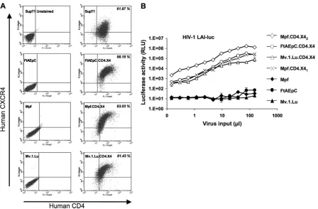
proteins by flow cytometry (Fig. 4A). Entry receptor competence
was verified by infecting the receptor-complemented cells with
HIV-1 LAI-luc (69) (Fig. 4B). The Mustelidae cells were then
chal-lenged with HIV-1 NL4-3 (2) and two variants of NL4-3, HIV-1
VSand HIV-1
VH(62). HIV-1
VSutilizes a separation of the normally
overlapping integrase and
vif
reading frames to encode the Vif
protein of SIVmac (62), and HIV-1
VHis a matched control virus
that encodes HIV-1 Vif in the same manner. HIV-1
VSbut not
HIV-1
VHor wild type HIV-1 NL4-3 replicates in feline
CrFK.CD4.X4 cells (62).
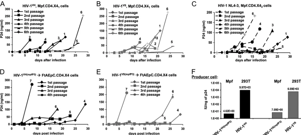
In the present experiments in Mustelidae cells, the pattern was
different. Productive, spreading replication of wild-type HIV-1
NL4-3, HIV-1
VH, and HIV-1
VSwas observed in ferret but not
mink cells (Fig. 5A to C). This was the case in the FtAEpC.CD4.X4
cell line and in two independently derived stable Mpf.CD4.X4 cell
lines (Mpf.CD4.X41
and Mpf.CD4.X42, which were made with
different receptor-transducing systems). The viruses could be
multiply passaged and amplified (Fig. 5D and E). In contrast, the
receptor-complemented mink cell line Mv.1.Lu.CD4.X4, though
clearly enabled for efficient viral entry (Fig. 4B), did not support
productive HIV-1 replication with any of the above viruses,
in-cluding HIV-1 first passaged through the ferret cell lines (data not
shown).
These results showed that wild-type HIV-1 will replicate
pro-ductively and spread in ferret cells if entry receptors are provided.
Nevertheless, we found that virions produced in these cells were
still less infectious per unit of p24 antigen than were virions
pro-duced in human cell lines. Figure 4F shows GHOST cell titrations
of third-passage viruses from the ferret cell line Mpf.CD4.X41,
which we refer to as HIV-1
VH(mpfP3)and HIV-1
VS(mpfP3).
Compar-ison is made to virus produced in maximally permissive human
293T cells. This producer cell-dependent loss of infectivity in
fer-ret cells suggested that APOBEC3 proteins may be targeting
HIV-1. Therefore, we amplified and sequenced long terminal
re-peat (LTR) and
gag/pol-vpr
segments of HIV-1
VH(mpfP3)and
HIV-1
VS(mpfP3)genomes as well as from later FtAEpC cell passages (Fig.
6A). A signature of APOBEC3 protein activity, G
¡
A
hypermuta-tion, was observed. This genome editing is nevertheless manifestly
not lethal since HIV-1 was still amplified exponentially and
pas-saged robustly in ferret cell lines. Another finding was a premature
stop codon in
vpr
, a frequent occurrence when HIV-1 is passaged
in any cultured cells (51). In HIV-1
VH(mpfP3), a
vif
reading
frame-preserving closure of the artificial integrase
-vif
separation arose
through deletion of a 62-nucleotide (nt) segment (Fig. 6B and its
legend). This change, effectively a reversion to wild type, was
found at the 5
=
end of
vif
in 8/10 clones and was likely consequent
to the duplication of 5
=
-GGAAAACAG-3
=
(56, 62) at the 5
=
end of
vif
in the input virus, which facilitated recombination during
re-verse transcription (72) to produce the virus illustrated in Fig. 6B.
While G-to-A changes predominated, other kinds of mutations
were also seen (Fig. 6A). For example, as previously observed to
happen with rat and rabbit APOBEC1 (10, 30), a significant
num-ber of plus-strand C-to-T mutations were observed (45, versus
161 G-to-A changes). This outcome might reflect RNA
deamina-tion as well (10, 30). The dinucleotide contexts observed for
cyt-idine deamination can vary substantially with different APOBEC
proteins (6). Here in ferret cells, it was different from the pattern
typically seen with human A3G, where CC and TC (edited
minus-strand cytidine underlined) contexts predominate (23, 44).
In-stead, a broader dinucleotide context pattern was observed in the
161 G-to-A mutations that occurred with passage in ferret cells:
CC, 46 (29%); TC, 32 (20%); GC, 27 (17%); and AC, 56 (35%).
This is reminiscent of more diverse dinucleotide patterns
ob-served previously for different feline A3 proteins (50).
Vif did not accrue consistent amino acid changes in any of the
FIG 3FIV and EIAV infection. The indicated cells were transduced with increasing doses of luciferase-encoding single-cycle vectors derived from FIV (A) or EIAV (B). Cell lysates from equal numbers of cells were assayed for activity 72 h later. Error bars represent standard deviations of duplicate measurements.
Fadel et al.
on November 7, 2019 by guest
http://jvi.asm.org/
[image:5.585.111.477.65.243.2]viruses. However, capsid did. Two coding changes arose in the
cyclophilin A (CypA) binding loop, which is complexly involved
in the viral life cycle, including in TRIM5 protein restriction (41).
For HIV-1
VH(mpfP3), a T
¡
A change at nt 261 in capsid and, for
HIV-1
VS(mpfP3), a G
¡
A mutation at nt 274 produced,
respec-tively, H87Q and A92T mutations (Fig. 6C). As the selection of
two different mutations in this functionally significant region of
capsid after passage in ferret cells was intriguing, we introduced
them prospectively, alone and in combination, into HIV-1 NL4-3
reporter viruses and full-length clones. Examined in this
geneti-cally defined context, the HIV-1 capsid mutants were found to
have moderately increased infectivity in ferret cells compared to
wild type (WT) (Fig. 7A).
In the case of replicating virus, capsid mutants replicated to
higher peak levels than did wild-type virus in ferret cells (Fig. 7B).
We then tested effects of cyclosporine (Fig. 8A through C). In
these experiments, the increase in infectivity conferred by the
CypA binding loop mutations was again observed in ferret cells
and also in owl monkey kidney (OMK) cells (Fig. 8A through C).
CsA, which disrupts TRIMCyp restriction (59), produced the
well-known dramatic augmenting effect in OMK cells for
wild-type NL4-3 and each of the mutants (Fig. 8A). The moderately
increased infectivity of H87Q in the absence of CsA in these
ex-periments (Fig. 8A) is consistent with that observed previously in
OMK cells (31). In clear contrast, CsA did not boost infectivity in
ferret cells for any of the viruses (Fig. 8B and C); rather, a slight
inhibitory effect was discernible, particularly for A92T. Since
lev-els of CypA have been reported to play a role in determining
HIV-1 infectivity in certain contexts (70), we performed
immu-noblotting for this protein. CypA was clearly present in the ferret
cells, and its levels were also similar to those in human cells (Fig.
8D). A ferret CypA cDNA was isolated by reverse transcriptase
PCR (RT-PCR) (Fig. 8E); sequencing and determination of the
predicted amino acid sequence did not reveal significant
differ-ences in the known HIV-1 capsid-interacting regions (13, 20, 71).
To complete the analysis with respect to the capsid mutants,
VLP saturation experiments were performed. A dose-dependent,
clear VLP saturation effect was observed in rhesus FrHK4 cells as
anticipated (8, 16), but no such effects occurred in ferret cells (Fig.
9A to D).
DISCUSSION
The results of this study show that, unlike the mouse genome (9),
the ferret genome can supply the nonreceptor dependency factors
needed for productive, spreading HIV-1 replication. Moreover, in
contrast to cells of another carnivore that share this property (62,
FIG 4Mustelidae cell lines with functional HIV-1 entry receptors. FIV vectors that coencode theneoorpacresistance markers (62) were used along with G418 and puromycin selection, respectively, to introduce the two receptors serially into Mpf cells, yielding the Mpf.CD4.X41cell line. An HIV-1 TSIN series (40)
lentiviral vector was also constructed to encode the receptor and coreceptor transgenes linked by a porcine teschovirus 2A (P2A) peptide, withpaccoencoded by a downstream IRES. Using this vector, a second Mpf line (Mpf.CD4.X42) as well as FtAEpC.CD4.X4 and Mv.1.Lu.CD4.X4 cell lines was derived and maintained
with puromycin selection. (A) Flow cytometry was performed to determine the expression of human CD4 and CxCR4 on the surface of the derived cells. All cells were labeled with the antibodies except for the SupT1 cells in the top left panel. The parental Mustelidae species cells not complemented with receptors were used as negative controls (left, bottom three panels), and SupT1 was used as a positive control (top right panel). (B) gp120-mediated entry. Cells with or without receptors were infected with increasing doses of native enveloped HIV-1 LAI-luc reporter virus (54). Cell lysates from equal numbers of cells were assayed for luciferase activity 72 h later.
on November 7, 2019 by guest
http://jvi.asm.org/
[image:6.585.70.518.64.358.2]73),
vif
gene substitution was not needed and wild-type HIV-1 was
capable of replication and serial passage. As in feline cells, G
¡
A
hypermutation and producer cell-dependent infectivity
reduc-tions were observed, but in both ferret cell lines they did not
pre-vent productive viral replication. The selection of capsid
muta-tions is also strong corroborative evidence that continuous viral
replication occurred. Whether the HIV-1 or SIVmac Vif protein
produces partial APOBEC3 mitigation in ferret cells, or might be
evolved by repeated passage to acquire it, deserves further specific
analysis. The absence of postentry capsid-targeting defenses
against N-MLV and lentiviruses is consistent with the apparent
lack of an intact Trim5
␣
or TRIMCyp gene in this species. We
observed similarly robust completion of early and late events in
primary ferret cells (Fig. 2). We were not able to complement
primary ferret mononuclear cells with HIV-1 receptors, and
lymphoid-lineage cell lines are not yet available. Therefore, the
extent to which specific relevant primary cell types (CD4
⫹T cells)
in ferrets
in vivo
express all needed nonreceptor dependency
fac-tors and/or might manifest additional restrictions will be a worthy
subject for further study.
The capability to repeatedly passage a primate lentivirus
through the novel adaptive environment of a nonprimate cell
al-lows experimental selection for continued viral evolution. So far,
after three passages, we have observed selection of two capsid
CypA binding loop mutations, H87Q and A92T. We found that
these confer moderately increased infectivity in ferret cells and
FIG 5Productive HIV-1 replication in ferret cells. (A and B) Mpf.CD4.X41cells were infected with HIV-1VHand HIV-1VS, and virus produced was serially
passaged 5 additional times. Input inocula were 1 ng p24. (C) Mpf.CD4.X42cells were infected with 10 ng p24 of HIV-1 NL4-3, and virus produced was serially
passaged 5 additional times. (D and E) Third-passage HIV-1 HIV-1VH(mpfP3)and HIV-1VS(mpfP3)viruses from Mpf.CD4.X4
1replication experiments were
serially passaged 4 times on FtAEpC.CD4.X4 cells. The passage numbers above the curves reflect the total passages on both ferret lines, while the numbers in the symbol keys refer to the number of passages on FtAEpC.CD4.X4 cells. (F) Infectivity determined by titration on GHOST cells.
FIG 6Sequencing of HIV-1VH(mpfP3)and HIV-1VS(mpfP3)viruses isolated from passage 3 of HIV-1VHand HIV-1VSon the Mpf.CD4.X4
1cell line. (A) Sequencing
of 290,607 nt (8 to 10 clones per segment) was performed, revealing 161 G¡A changes. (B) Recombinant HIV-1VHfound in 8/10 clones. Capital letters indicate
original mutations used to separate the integrase andvifframes. (C) Capsid mutations selected in the unstructured CypA binding loop of HIV-1 CA. H87Q and A92T were present in 10 of 10 and 9 of 9 sequenced clones, respectively.
Fadel et al.
on November 7, 2019 by guest
http://jvi.asm.org/
[image:7.585.43.540.66.290.2] [image:7.585.85.504.528.681.2]that disruption of CypA interaction with CsA only slightly affects
this (Fig. 7 and 8). The reasons that these mutations arose are not
clear because of the ambiguities that persist about the roles that
CypA plays in retroviral life cycles, but they may represent optimal
fitness of the CypA binding loop in the presence of CypA but in the
absence of any functionally antiviral TRIM5 protein. CypA, which
is highly conserved between mammals, is a peptidyl-prolyl
isomerase that binds lentiviral capsids. It catalyzes the
cis/trans
isomerization of the G89-P90 peptide bond in the HIV-1 capsid
protein (11). CypA has distinctive and at times opposite
context-dependent effects (18, 20, 42). The protein promotes infectivity in
human cells (25, 61, 66) but is in contrast necessary for Trim5
␣
restriction in rhesus macaque and African green monkey cells (7).
While multiple possibilities have been proposed for the role of
CypA in the viral life cycle, a unifying mechanistic explanation
remains elusive. It has been hypothesized that this peptidyl-prolyl
isomerase protects HIV-1 from human cell restriction by
compet-ing with Trim5
␣
binding (31) or that it shields HIV-1 from an
unknown antiviral factor in human cells since the stimulatory
effect of CypA on HIV-1 infectivity is independent of human
Trim5
␣
(58, 59). Some CypA binding loop mutations alleviate
restriction in Old and New World monkey cells; similar effects are
seen with CsA (15, 31). Of interest, H87Q decreases the binding
affinity of HIV-1 CA for CypA by 4.8-fold (71) and was reported
to confer a replication advantage to HIV-1 in CypA-rich human
cells (21). The mutation is present in 19.7% of natural isolates in
the Los Alamos database (15), and in human cells it can confer
HIV-1 resistance to the effects of CsA (i.e., CypA independence)
(15, 31). H87Q has also been reported to mediate escape from
cytotoxic T lymphocyte responses, emerging in the late phase of
infection in 13.7% of patients infected with HIV-1 (36, 39).
A92T has not been previously reported, but a charge-adding
mutation at this residue, A92E, is known to arise when HIV-1 is
passaged in HeLa cells in the presence of CsA, thus conferring CsA
resistance and in some cell lines actually conferring CsA
depen-dence on the virus (1, 12); this phenomenon was later shown to
reflect high levels of CypA in HeLa cells (70). Our experiments
make it clear that CypA is present at substantial levels in the
Mus-FIG 7Infection of ferret cells with WT, H87Q, A92T, and H87Q/A92T NL4-3 clones. (A) HIV-1GFPreporter viruses. The number of GFP-positive cells was
determined by FACS at 48 h. The upper and lower graphs show the results of two independent experiments with different vector preparations. Student’s 2-tailed
ttest was used to determinePvalues for comparisons of the titers of WT virus with each of the capsid mutants. All calculatedPvalues were⬍0.05 except for the comparison of WT and H87Q/A92T mutants in FtAEpC cells (asterisk, upper right plot;P⫽0.07). (B) Full-length WT and mutant viral clone challenges of Mpf.CD4.X41cells.
on November 7, 2019 by guest
http://jvi.asm.org/
[image:8.585.80.505.70.440.2]telidae cell lines that we used (Fig. 8D) and also that its amino acid
sequence is conserved versus human CypA in the hydrophobic
regions known to form the retroviral capsid-interacting domain
(13) (Fig. 8E). Thus, these mutations that developed in ferret cells
may represent HIV-1 adaptation to a situation where CypA is
present but there is no Trim5 protein pressure.
In contrast to ferret cells, we did not observe spreading HIV-1
replication in the mink (Mv.1.Lu.CD4.X4) cell line. This result,
FIG 8Cyclosporine experiments in ferret cells and CypA alignments. (A to C) Infectious titers of HIV-1 GFP vector were determined in the presence (black columns) and absence (gray columns) of 5M CsA for owl monkey kidney (A), Mpf (B), and FtAEpC (C) cells. (D) Immunoblotting for CypA in human and Mustelidae cell lines. Jurkatppia⫺/⫺cells (14) were used as a negative control. (E) Alignment of the ferret and human CypA amino acid sequences reveals high conservation overall and complete conservation in the central hydrophobic pocket involved in HIV-1 capsid binding. The amino acids involved in HIV-1 capsid binding, which are known from several mutational and high-resolution structural studies (13, 20, 71), are highlighted with boxes. Comparison is made between the human protein reference sequence (top line, accession no. NP_066953) and the predicted amino acid sequence determined from the ferret CypA cDNA isolated from Mpf cell mRNA in the present study (middle line) and a sequence predicted from anM. putorius furowhole-genome shotgun (WGS) sequence contig (bottom line, accession no.AEYP01032892). The dashed arrows at each end indicate the span of the degenerate primers used to obtain the Mpf cell cDNA sequence. Stringent selection pressure for amino acid level conservation is apparent. For example, at the nucleotide sequence level (not shown), there were 49 differences between Mpf cell CypA and human CypA in the coding region of the mRNA (10.1% nonidentity), but virtually all were synonymous. Only three amino acid differences were present, and these were located at both termini (arrowheads indicating the fifth and the final two C-terminal amino acids); the T5I difference (black arrowhead) is uncertain since both threonine and isoleucine were encoded by the degenerate primer, the domestic cat sequence has an isoleucine at this position, and the ferret WGS contig predicts a threonine. Polymporphism is evident in the two ferret sequences; they differed at 10 nt (not shown), but besides T5I there were only two other predicted amino acid differences, A26S and R37H.
FIG 9VLP saturation experiments in ferret cells. FRhK4, Mpf, and FtAEpC cells were infected with a fixed dose of wild-type HIV-1 GFP (A) and capsid mutants H87Q (B), A92T (C), and H87Q/A92T (D), in the presence of increasing amounts of HIV-1 VLPs. While the VLPs clearly released Lv1 restriction in the rhesus macaque cells as anticipated, they had no effect in ferret cells.
Fadel et al.
on November 7, 2019 by guest
http://jvi.asm.org/
[image:9.585.62.524.63.376.2] [image:9.585.53.529.590.691.2]which was verified in repeated experiments, is at variance with a
prior report (35). As was discussed previously (62), it may be
possible to reconcile these differences by considering that a single
round of provirus generation and p24 production occurred in the
experiments of Koito et al. (35).
Our results add to emerging evidence that cells of carnivore
species appear in general to harbor relatively few restrictions to
HIV-1 replication, and prominent among these are
APOBEC3-mediated restrictions. Reference 19 provides a recent review of
carnivore cell restrictions. We also conclude that, like the
domes-tic cat and unlike the mouse, the ferret genome encodes the major
nonreceptor dependency factors for this primate lentivirus. There
are robust precedents for modeling human RNA pathogens in the
ferret. A ferret genome sequencing project is nearing
comple-tion (
http://www.broadinstitute.org/scientific-community/science
/projects/mammals-models/ferret-genome-project
). We suggest
that further characterization of HIV-1 adaptation in ferret cells
and delineation of Mustelidae restriction factor gene repertoires
are warranted, with a view to several possible benefits. These
in-clude potentials for developing an eventual HIV-1 animal model,
for gaining basic insights into mechanisms of species-specific
ret-roviral restriction, and for exploring the extent to which HIV-1
will evolve when confronted with the novel adaptive landscape of
a nonprimate cell.
ACKNOWLEDGMENTS
We thank M. Stern for assistance with initial experiments in Mpf cells, J. Luban for Jurkat CypA-knockout cells, and M. Emerman, J. Olsen, and P. Charneau for plasmids.
We are grateful for funding from NIH AI47536 and AI77344 and the Tietze Foundation.
REFERENCES
1.Aberham C, Weber S, Phares W.1996. Spontaneous mutations in the human immunodeficiency virus type 1 gag gene that affect viral replica-tion in the presence of cyclosporins. J. Virol.70:3536 –3544.
2. Adachi A, et al. 1986. Production of acquired immunodeficiency syndrome-associated retrovirus in human and nonhuman cells trans-fected with an infectious molecular clone. J. Virol.59:284 –291. 3.Ambrose Z, KewalRamani VN, Bieniasz PD, Hatziioannou T. 2007.
HIV/AIDS: in search of an animal model. Trends Biotechnol.25:333–337. 4.Antunes A, et al.2008. The evolutionary dynamics of the lion Panthera leo revealed by host and viral population genomics. PLoS Genet. 4:e1000251.
5.Baumann JG, et al.2004. Murine T cells potently restrict human immu-nodeficiency virus infection. J. Virol.78:12537–12547.
6.Beale RC, et al.2004. Comparison of the differential context-dependence of DNA deamination by APOBEC enzymes: correlation with mutation spectra in vivo. J. Mol. Biol.337:585–596.
7.Berthoux L, Sebastian S, Sokolskaja E, Luban J.2005. Cyclophilin A is required for TRIM5alpha-mediated resistance to HIV-1 in Old World monkey cells. Proc. Natl. Acad. Sci. U. S. A.102:14849 –14853. 8.Besnier C, Takeuchi Y, Towers G. 2002. Restriction of lentivirus in
monkeys. Proc. Natl. Acad. Sci. U. S. A.99:11920 –11925.
9.Bieniasz PD, Cullen BR.2000. Multiple blocks to human immunodefi-ciency virus type 1 replication in rodent cells. J. Virol.74:9868 –9877. 10. Bishop KN, Holmes RK, Sheehy AM, Malim MH. 2004.
APOBEC-mediated editing of viral RNA. Science305:645.
11. Bosco DA, Eisenmesser EZ, Pochapsky S, Sundquist WI, Kern D.2002. Catalysis of cis/trans isomerization in native HIV-1 capsid by human cy-clophilin A. Proc. Natl. Acad. Sci. U. S. A.99:5247–5252.
12. Braaten D, et al.1996. Cyclosporine A-resistant human immunodefi-ciency virus type 1 mutants demonstrate that Gag encodes the functional target of cyclophilin A. J. Virol.70:5170 –5176.
13. Braaten D, Ansari H, Luban J.1997. The hydrophobic pocket of cyclo-philin is the binding site for the human immunodeficiency virus type 1 Gag polyprotein. J. Virol.71:2107–2113.
14. Braaten D, Luban J.2001. Cyclophilin A regulates HIV-1 infectivity, as demonstrated by gene targeting in human T cells. EMBO J.20:1300 –1309. 15. Chatterji U, et al.2005. Naturally occurring capsid substitutions render HIV-1 cyclophilin A independent in human cells and TRIM-cyclophilin-resistant in owl monkey cells. J. Biol. Chem.280:40293– 40300. 16. Cowan S, et al.2002. Cellular inhibitors with Fv1-like activity restrict
human and simian immunodeficiency virus tropism. Proc. Natl. Acad. Sci. U. S. A.99:11914 –11919.
17. Diaz-Griffero F, Taube R, Muehlbauer SM, Brojatsch J.2008. Efficient production of HIV-1 viral-like particles in mouse cells. Biochem. Biophys. Res. Commun.368:463– 469.
18. Dorfman T, Weimann A, Borsetti A, Walsh CT, Gottlinger HG.1997. Active-site residues of cyclophilin A are crucial for its incorporation into human immunodeficiency virus type 1 virions. J. Virol.71:7110 –7113. 19. Fadel H, Poeschla E.2011. Retroviral restriction and dependency factors
in primates and carnivores. Vet. Immunol. Immunopathol.143:179 –189. 20. Gamble TR, et al.1996. Crystal structure of human cyclophilin A bound
to the amino-terminal domain of HIV-1 capsid. Cell87:1285–1294. 21. Gatanaga H, et al.2006. Altered HIV-1 Gag protein interactions with
cyclophilin A (CypA) on the acquisition of H219Q and H219P substitu-tions in the CypA binding loop. J. Biol. Chem.281:1241–1250. 22. Gifford RJ, et al.2008. A transitional endogenous lentivirus from the
genome of a basal primate and implications for lentivirus evolution. Proc. Natl. Acad. Sci. U. S. A.105:20362–20367.
23. Harris RS, et al.2003. DNA deamination mediates innate immunity to retroviral infection. Cell113:803– 809.
24. Hatziioannou T, et al.2009. A macaque model of HIV-1 infection. Proc. Natl. Acad. Sci. U. S. A.106:4425– 4429.
25. Hatziioannou T, Perez-Caballero D, Cowan S, Bieniasz PD. 2005. Cyclophilin interactions with incoming human immunodeficiency virus type 1 capsids with opposing effects on infectivity in human cells. J. Virol. 79:176 –183.
26. Hatziioannou T, et al.2006. Generation of simian-tropic HIV-1 by re-striction factor evasion. Science314:95.
27. Henderson IC, Lieber MM, Todaro GJ.1974. Mink cell line Mv 1 Lu (CCL 64). Focus formation and the generation of “nonproducer” trans-formed cell lines with murine and feline sarcoma viruses. Virology60: 282–287.
28. Hu C, et al.2010. The HIV-1 central polypurine tract functions as a second line of defense against APOBEC3G/F. J. Virol.84:11981–11993. 29. Igarashi T, et al.2007. Human immunodeficiency virus type 1 derivative
with 7% simian immunodeficiency virus genetic content is able to estab-lish infections in pig-tailed macaques. J. Virol.81:11549 –11552. 30. Ikeda T, et al.2008. The antiretroviral potency of APOBEC1 deaminase
from small animal species. Nucleic Acids Res.36:6859 – 6871.
31. Ikeda Y, Ylinen LM, Kahar-Bador M, Towers GJ.2004. Influence of gag on human immunodeficiency virus type 1 species-specific tropism. J. Vi-rol.78:11816 –11822.
32. Kamada K, et al.2006. Generation of HIV-1 derivatives that productively infect macaque monkey lymphoid cells. Proc. Natl. Acad. Sci. U. S. A. 103:16959 –16964.
33. Katzourakis A, Tristem M, Pybus OG, Gifford RJ.2007. Discovery and analysis of the first endogenous lentivirus. Proc. Natl. Acad. Sci. U. S. A. 104:6261– 6265.
34. Keckesova Z, Ylinen LM, Towers GJ, Gifford RJ, Katzourakis A.2009. Identification of a RELIK orthologue in the European hare (Lepus euro-paeus) reveals a minimum age of 12 million years for the lagomorph lentiviruses. Virology384:7–11.
35. Koito A, Kameyama Y, Cheng-Mayer C, Matsushita S.2003. Suscepti-bility of mink (Mustera vision)-derived cells to replication by human im-munodeficiency virus type 1. J. Virol.77:5109 –5117.
36. Kootstra NA, Navis M, Beugeling C, van Dort KA, Schuitemaker H. 2007. The presence of the Trim5alpha escape mutation H87Q in the cap-sid of late stage HIV-1 variants is preceded by a prolonged asymptomatic infection phase. AIDS21:2015–2023.
37. Kugel D, et al.2009. Intranasal administration of alpha interferon re-duces seasonal influenza A virus morbidity in ferrets. J. Virol.83:3843– 3851.
38. LaRue RS, Lengyel J, Jonsson SR, Andresdottir V, Harris RS. 2010. Lentiviral Vif degrades the APOBEC3Z3/APOBEC3H protein of its mam-malian host and is capable of cross-species activity. J. Virol.84:8193– 8201. 39. Leslie AJ, et al.2004. HIV evolution: CTL escape mutation and reversion
after transmission. Nat. Med.10:282–289.
on November 7, 2019 by guest
http://jvi.asm.org/
40. Llano M, et al.2006. An essential role for LEDGF/p75 in HIV integration. Science314:461– 464.
41. Luban J.2007. Cyclophilin A, TRIM5, and resistance to human immu-nodeficiency virus type 1 infection. J. Virol.81:1054 –1061.
42. Luban J, Bossolt KL, Franke EK, Kalpana GV, Goff SP.1993. Human immunodeficiency virus type 1 Gag protein binds to cyclophilins A and B. Cell73:1067–1078.
43. Malim MH, Emerman M.2008. HIV-1 accessory proteins– ensuring viral survival in a hostile environment. Cell Host Microbe3:388 –398. 44. Mangeat B, et al. 2003. Broad antiretroviral defence by human
APOBEC3G through lethal editing of nascent reverse transcripts. Nature 424:99 –103.
45. Mariani R, et al.2001. Mouse-human heterokaryons support efficient human immunodeficiency virus type 1 assembly. J. Virol.75:3141–3151. 46. Mariani R, et al.2000. A block to human immunodeficiency virus type 1
assembly in murine cells. J. Virol.74:3859 –3870.
47. McEwan WA.2010. Factors affecting replication and cross-species trans-mission of feline immunodeficiency virus. Ph.D. thesis. University of Glasgow, Glasgow, United Kingdom.http://theses.gla.ac.uk/1388/. 48. McEwan WA, et al.2009. Truncation of TRIM5 in Feliformia explains the
absence of retroviral restriction in cells of the domestic cat. J. Virol.16: 8270 – 8275.
49. Münk C, et al.2008. Functions, structure, and read-through alternative splicing of feline APOBEC3 genes. Genome Biol.9:R48.
50. Münk C, et al.2007. Multiple restrictions of human immunodeficiency virus type 1 in feline cells. J. Virol.81:7048 –7060.
51. Nakaya T, et al.1996. Serial passage of human immunodeficiency virus type 1 generates misalignment deletions in non-essential accessory genes. Virus Res.46:139 –147.
52. Neil SJ, Zang T, Bieniasz PD.2008. Tetherin inhibits retrovirus release and is antagonized by HIV-1 Vpu. Nature451:425– 430.
53. Pecon-Slattery J, Troyer JL, Johnson WE, O’Brien SJ.2008. Evolution of feline immunodeficiency virus in Felidae: implications for human health and wildlife ecology. Vet. Immunol. Immunopathol.123:32– 44. 54. Peden K, Emerman M, Montagnier L. 1991. Changes in growth
properties on passage in tissue culture of viruses derived from infec-tious molecular clones of HIV-1LAI, HIV-1MAL, and HIV-1ELI. Vi-rology185:661– 672.
55. Poeschla E, Wong-Staal F, Looney D.1998. Efficient transduction of nondividing cells by feline immunodeficiency virus lentiviral vectors. Nat. Med.4:354 –357.
56. Sakurai A, et al.2004. Functional analysis of HIV-1 vif genes derived from Japanese long-term nonprogressors and progressors for AIDS. Microbes Infect.6:799 – 805.
57. Sawyer SL, Emerman M, Malik HS.2007. Discordant evolution of the adjacent antiretroviral genes TRIM22 and TRIM5 in mammals. PLoS Pat-hog.3:e197.
58. Sayah DM, Luban J.2004. Selection for loss of Ref1 activity in human cells releases human immunodeficiency virus type 1 from cyclophilin A dependence during infection. J. Virol.78:12066 –12070.
59. Sayah DM, Sokolskaja E, Berthoux L, Luban J.2004. Cyclophilin A retrotransposition into TRIM5 explains owl monkey resistance to HIV-1. Nature430:569 –573.
60. Sheehy AM, Gaddis NC, Malim MH.2003. The antiretroviral enzyme APOBEC3G is degraded by the proteasome in response to HIV-1 Vif. Nat. Med.9:1404 –1407.
61. Sokolskaja E, Sayah DM, Luban J.2004. Target cell cyclophilin A mod-ulates human immunodeficiency virus type 1 infectivity. J. Virol.78: 12800 –12808.
62. Stern MA, et al.2010. Productive replication of Vif-chimeric HIV-1 in feline cells. J. Virol.84:7378 –7395.
63. Stremlau M, et al.2004. The cytoplasmic body component TRIM5alpha restricts HIV-1 infection in Old World monkeys. Nature427:848 – 853. 64. Swanson CM, Puffer BA, Ahmad KM, Doms RW, Malim MH.2004.
Retroviral mRNA nuclear export elements regulate protein function and virion assembly. EMBO J.23:2632–2640.
65. Towers G, et al.2000. A conserved mechanism of retrovirus restriction in mammals. Proc. Natl. Acad. Sci. U. S. A.97:12295–12299.
66. Towers GJ, et al.2003. Cyclophilin A modulates the sensitivity of HIV-1 to host restriction factors. Nat. Med.9:1138 –1143.
67. Trowbridge RS, Lehmann J, Brophy P.1982. Establishment and char-acterization of ferret cells in culture. In Vitro18:952–960.
68. Troyer JL, et al.2008. FIV cross-species transmission: an evolutionary prospective. Vet. Immunol. Immunopathol.123:159 –166.
69. Yamashita M, Emerman M.2004. Capsid is a dominant determinant of retrovirus infectivity in nondividing cells. J. Virol.78:5670 –5678. 70. Ylinen LM, et al.2009. Cyclophilin A levels dictate infection efficiency of
human immunodeficiency virus type 1 capsid escape mutants A92E and G94D. J. Virol.83:2044 –2047.
71. Yoo S, et al.1997. Molecular recognition in the HIV-1 capsid/cyclophilin A complex. J. Mol. Biol.269:780 –795.
72. Zhang J, Temin HM.1994. Retrovirus recombination depends on the length of sequence identity and is not error prone. J. Virol.68:2409 –2414. 73. Zielonka J, et al.2010. Vif of feline immunodeficiency virus from domes-tic cats protects against APOBEC3 restriction factors from many felids. J. Virol.84:7312–7324.
Fadel et al.
on November 7, 2019 by guest
http://jvi.asm.org/