Genotypic and Phenotypic characterization of Methicillin Resistant
Staphylococcus aureus from ocular isolates and its clinical correlation
DISSERTATION SUBMITTED FOR
MS (Branch III) Ophthalmology
THE TAMILNADU DR. M.G.R. MEDICAL UNIVERSITY
CHENNAI
CERTIFICATE
Certified that this dissertation entitled “GENOTYPIC AND
PHENOTYPIC
CHARACTERIZATION
OF
METHICILLIN
RESISTANT Staphylococcus aureus FROM OCULAR ISOLATES AND
ITS CLINICAL CORRELATION” submitted for MS (Branch III)
Ophthalmology, April 2014, is the bonafide work done by DR.PRIYA.S ,
under our supervision and guidance in the Ocular Microbiology Services of
Aravind Eye Hospital and Post Graduate Institute of Ophthalmology,
Madurai, during her residency period from May 2011 to April 2014.
Dr.LALITHA PRAJNA Dr. M.SRINIVASAN
Chief,
Ocular microbiology
Services DirectorAravind Eye Hospital, Aravind Eye Hospital,
Madurai Madurai
ACKNOWLEDGEMENT
I take this opportunity to pay my respect and homage to
Dr.G.Venkatasamy, our founder and visionary, whose dynamism had led Aravind
against all odds to its epitope.
It is a proud privilege and pleasure to express my thanks and indebtedness
towards my revered mentor and guide DR. Lalitha Prajna, Chief ocular
microbiology services, Aravind eye hospital Madurai, for being a constant source
of motivation and encouragement, which ultimately structured my thesis .
I am grateful to Dr. N.V.Prajna, Director of academics, Aravind Eye Care
System, who offered his excellent guidance and support throughout my residency
programme.
I am very grateful to Dr.R.D.Ravindran, Chairman of Aravind Eye Care
System for having created and environment enriched with all the facilities for
learning and gaining knowledge . I am privileged to have on my side Dr. P.
Namperumalsamy,Chairman emeritus director of research, Dr.G.Natchiar Director
emeritus (human resource department), Dr.M.srinivasan, director emeritus and
other scholars of ophthalmology at Aravind Eye Care System .
My sincere thanks to all paramedical staff for their excellent co-
operation during the study. Last but not the least; I thank my patients who made
Index
Part I
Page No
1. Introduction
1
2. Literature review- Staphylococcus aureus
A. Morphology and classification
4
B. Pathogenesis and virulence factors
7
C. Emergence of Methicillin-Resistant
15
Staphylococcus aureus (MRSA)
Community-Acquired & Hospital-Acquired MRSA
19
D. Global prevalence of MRSA
23
E.
Indian scenario
26
F.
Systemic and Ocular manifestations 31
G. Diagnosis – Phenotypic & Genotypic identification
41
Part II
Page No
1.
Aim and Objectives
50
2.
Material and Methods
a)
Examination, sample and data collection
51
b)
Microbiological work up
52
c)
Genotypic studies
53
3
Results
a)
Demographic and clinical spectrum of MRSA patients 54
b)
Results of phenotypic and molecular studies
60
c)
Antibiotic sensitivity pattern
67
d)
Response to treatment
70
5 . Discussion
72
6) Conclusion
80
INTRODUCTION
Staphylococcus aureus is a bacterium that belongs to the family of
Staphylococcaceae. The bacteria form part of the normal flora of the skin,
intestine, upper respiratory tract and vagina. It can proliferate when there is
appropriate pH, temperature and nutrition and can become pathogenic. The
pathogenicity of S. aureus is determined by the production of several toxins. It
has been documented that there is probably no other bacterium that produces as
many cellular components, enzymes, extra cellular toxins and hemolysins as
this organism
1. It is known for its intrinsic virulence and multidrug resistance
which poses a great challenge to the clinician.
In early 1960s, treatment of S. aureus infections included
semi-synthetic penicillin drugs, such as methicillin. However, soon
methicillin-resistant S. aureus (MRSA) strains started appearing. In the early 1980’s they
became the major cause of nosocomial infections. The possibility of
transmission of health-care associated MRSA - HA-MRSA) to the patient
population was unavoidable. Since 1987, MRSA was also found in the
community where there is no exposure to any known risk factors such as
hospital admission. (community associated- methicillin-resistant S. aureus -
CA-MRSA) Patients presented with severe skin and soft tissue infections and
necrotizing pneumonia. HA-MRSA strains were genetically and phenotypically
different than CA-MRSA strains. CA-MRSA were associated with a smaller
composition, a higher incidence of virulence, and a lack of multidrug resistance.
REVIEW OF LITERATURE
Staphylococcus aureus is a Gram positive, facultative anaerobic
bacterium . They are non-motile, non- sporing and catalase positive
organism. The cocci commonly form irregular clusters with a grape like
appearance under the microscope (Figure 1) The individual coccus size is
approximately 0.5 to 1.5 μm in diameter 5. Although more than 200 species
of Staphylococcus are described, only Staphylococcus aureus and
Staphylococcus epidermidis are significant in their interactions with
humans.Summary of the classification of Staphylococcus aureus is given in
table
1.
Classification of Staphylococcus aureus:
A) Based on coagulase production:
1. Coagulase positive: Eg- S. aureus
2. Coagulase negative: Eg- S. epidermidis, S. saprophyticus
B) Based on pathogenicity:
1. Common pathogen: Eg- S. aureus
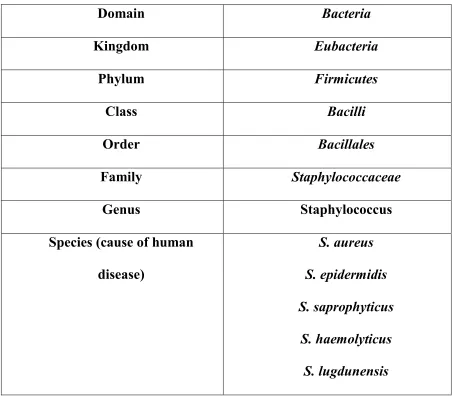
Table 1: Summary of the classification of Staphylococcus aureus
Domain
Bacteria
Kingdom
Eubacteria
Phylum
Firmicutes
Class
Bacilli
Order
Bacillales
Family
Staphylococcaceae
Genus
Staphylococcus
Species (cause of human
disease)
S. aureus
S. epidermidis
S. saprophyticus
S. haemolyticus
Culture Characteristics:
They can be grown in several media given below.
1.
Non selective media:
Nutrient agar
Blood agar
Chocolate agar
2.
Selective media:
Salt-milk agar
Ludlam’s medium
MacConkey’s agar.
Figure 1: Staphylococcus aureus - Gram positive cocci in clusters
Figure 2: Staphylococcus aureus colony morphology in Blood agar plate
[image:12.612.164.466.392.588.2]Important phenotypic characteristics of Staphylococcus aureus :
Gram-positive, cluster-forming coccus (Figure 1)
Nonmotile, nonsporeforming facultative anaerobe
Fermentation of glucose produces mainly lactic acid
Ferments mannitol (distinguishes from S. Epidermidis)
Catalase positive (Figure 3)
Coagulase positive(Figure 4)
Reduces nitrate to nitrite.
Urea hydrolysis test- Positive.
Gelatin liquefaction test- Positive.
Produces Lipase.
Produces Phosphatase.
Figure on left shows MRSA with formation of air bubbles confirms
positive catalase reaction
bubble confirming a negative catal
Figure on left shows MRSA Clot formation
reaction
[image:14.612.205.460.95.235.2]Figure on right shows negative Coag
absence of Clot Formation
Figure 3:Catalase test
[image:14.612.226.438.403.631.2]Figure on left shows MRSA with formation of air bubbles confirms
se reaction Figure on right St.pneumoniae with no air
bubble confirming a negative catalase eaction
Figure 4. Coagulase test
Figure on left shows MRSA Clot formation- a positive Coagulase
Figure on right shows negative Coagulase reaction of S.epidermidis
absence of Clot Formation
Figure on left shows MRSA with formation of air bubbles confirms
Figure on right St.pneumoniae with no air
Source of infection:
A) Exogenous: patients or carriers
B) Endogenous: From colonized site
Mode of transmission:
A) Contact: direct or indirect( through fomites)
B) Inhalation of air borne droplets
C) Hematogenous
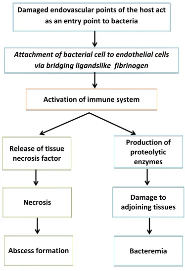
Pathogenisis:
(i) S. aureus cells enter the body through damaged endovascular points of the
host where platelet-fibrin-thrombi complex have formed and attach via
microbial surface components that recognize adhesive matrix molecules
(MSCRAMM) mediated mechanisms. (Figure 5)
(ii) Bacterial cells may attach to endothelial cells via adhesion receptor
interactions or by bridging ligands, including serum components such as
fibrinogen. Upon entry into the host tissue, immune cells phagocytose S.
aureus cells, which promotes the production of proteolytic enzymes and
Figure 5 : Pathogenesis:
Damaged endovascular points of the host act
as an entry point to bacteria
Attachment of bacterial cell to endothelial cells
via bridging ligandslike fibrinogen
Activation of immune system
Release of tissue
necrosis factor
Production of
proteolytic
enzymes
Necrosis
Damage to
adjoining tissues
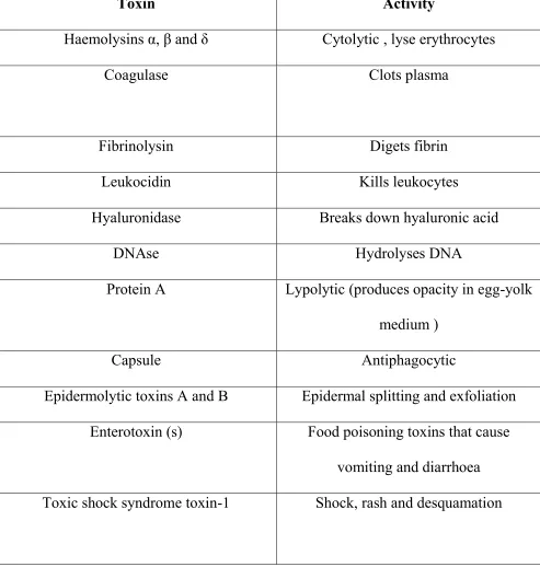
Table 2. Toxins and toxic components produced by Staphylococcus
aureus (Timbury et al., 002)
`
Toxin
Activity
Haemolysins α, β and δ
Cytolytic , lyse erythrocytes
Coagulase
Clots plasma
Fibrinolysin
Digets fibrin
Leukocidin
Kills leukocytes
Hyaluronidase
Breaks down hyaluronic acid
DNAse
Hydrolyses DNA
Protein A
Lypolytic (produces opacity in egg-yolk
medium )
Capsule
Antiphagocytic
Epidermolytic toxins A and B
Epidermal splitting and exfoliation
Enterotoxin (s)
Food poisoning toxins that cause
Virulence factors:
Different strains of S. aureus produce different virulence factor which
result in their ability to multiply and spread across adjacent tissue
1. The cell
wall of S. aureus is composed of a thick peptidoglycan layer, which
contributes to the virulence of the bacterium
6. The peptidoglycan stimulates
the production of cytokines by macrophages resulting in complement system
activation and platelet aggregation
6.
Antimicrobial resistance of S. aureus strains
Staphylococcus aureus causes the tissue destruction as a pathogen
because of the intrinsic virulence and its ability to rapidly adjust to different
environmental conditions
6. The trend of multidrug resistance in S. aureus is
particularly alarming because of the severity and diversity of diseases caused
by this pathogen
9. Despite the availability of novel drugs as an approach to
staphylococcal therapy, the bacteria seem to be able to rapidly develop
resistance to these drugs
10. Perhaps the most commonly known resistance of
S. aureus, is methicillin resistance, which has caused alarming reports with
regard to the spread of S. aureus in hospitals and the community
11-14Penicillin Resistance:
Methicillin resistance in S. aureus strains:
Figure 6: Mechanism of S.aureus resistance to methicillin
Emergence of MRSA
In the 1940s, medical treatment for S. aureus infections became
routine and successful with the discovery of antibiotics, such as penicillin.
However, use of antibiotics has aided natural bacterial evolution by helping
the microbes become resistant to the drugs designed to fight them. In the late
1940s and throughout the 1950s, S aureus developed resistance to penicillin.
1.
Methicillin, a form of penicillin, was introduced to counter the increasing
problem of penicillin-resistant S. aureus. Methicillin was one of most
common types of antibiotics used to treat S. aureus infections; but, in 1961,
British scientists identified the first strains of S. aureus bacteria that resisted
methicillin. This was the so-called birth of MRSA.
The first reported human case of MRSA in the United States came in
1968. Subsequently, new strains of bacteria have developed that can now
resist previously effective drugs, such as methicillin and most related
antibiotics. MRSA is actually resistant to an entire class of penicillin-like
antibiotics called beta-lactams. This class of antibiotics includes penicillin,
amoxicillin, oxacillin, methicillin, and others.
1the first S. aureus strains resistant to vancomycin, which had been one of a
handful of antibiotics of last resort for use against S. aureus. Though it is
feared that this could quickly become a major issue in antibiotic resistance,
vancomycin-resistant strains are still rare.
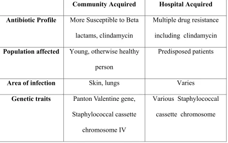
1Community acquired and Hospital acquired MRSA:
MRSA has been circulating in hospitals since the early 1960s but
reports of infection in the community were relatively rare. However, in the
early 1990s strains of highly virulent CA-MRSA were reported in Western
Australia
18. In recent years the incidence of CA-MRSA has been increasing
with outbreaks occurring in various parts of the world
19,20.
While one group believes that CA-MRSA isolates evolved from
HA-MRSA
21another introduces the differences in SCCmec complexes as
evidence that CA-MRSA and HA-MRSA are in fact not related. HA-MRSA
consists of SCCmec types I-III, while CA-MRSA consists of type IV and V
22,23both types are given in table 4 . The guidelines in diagnosing both
Community acquired and Hospital acquired MRSA and the differences are
given in Table 5.
Table 3 : Major Difference Between Community – and Hospital-acquired
Methicillin-resistant Staphylococcus Aureus
Community Acquired
Hospital Acquired
Antibiotic Profile
More Susceptible to Beta
lactams, clindamycin
Multiple drug resistance
including clindamycin
Population affected
Young, otherwise healthy
person
Predisposed patients
Area of infection
Skin, lungs
Varies
Genetic traits
Panton Valentine gene,
Staphylococcal cassette
chromosome IV
Table 4 SCCmec typing in community and health-care -associated
methicillin-resistant Staphylococcus aureus
Strain
SCCmec
type
Antibiotic
resistance
Toxins
PVL genes
Infection
spectrum
HA-MRSA
Types I, II
and III
Multi-drug
resistant
Few
Rare
Bloodstream,
respiratory
tract
and urinary
tract
infections
CA-MRSA Type IV
and V
Resistance is
typically limited
to betalactam
betalactam
and
erythromycin
although
multidrug
resistance
can occur
usually
PVL
presence
Common
Skin and
softtissue
infections and
necrotising
pneumonia
CA-MRSA
community-associated
methicillin-resistant
Staphylococcus
aureus
Table-5 Community -Associated Methicillin – Resistant Staphylococcus
aureus(CA-MSRA) Criteria
Diagnosis of CA MRSA made in the outpatient setting of within 48 hours of
hospital admission.
o
No medical history of MRSA infection or colonization.
o
In the past year, no medical history of the following:
Hospitalization
Admission to a nursing home, skilled nursing facility, or
hospice
Dialysis
Surgery
o
No Permanent indwelling catheters or Medical devices that pass
through the skin into the body
Health Care -Associated Methicillin – Resistant Staphylococcus
aureus(HA-MSRA) Criteria - defined by the CDC
MRSA infection occurring in individuals who have been
-
Hospitalized for more than 48 hours or
-Received surgery within the last year, or
-
Have a permanent indwelling medical device, or
-Reside in a long-term care facility, or
Global prevalence of Health Care associated MRSA :
Isolates of MRSA were initially recovered in hospitals; the first isolate
was detected at a hospital in the United Kingdom. Within a few years, MRSA
was found in other European countries, Japan, and Australia, and the first
isolate in the United States was discovered at Boston Hospital.
25By the late 1980s, MRSA had become endemic in many hospitals,
according to results from large surveillance studies such as the National
Nosocomial Infections Surveillance conducted by the Centers for Disease
Control and Prevention (CDC). In the United States hospitals, the proportion
of S aureus isolated that was resistant to methicillin rose from 2.4% in 1975
to 29% in 1991 ; the proportion of MRSA continued to increase during the
next decade and rose by approximately 3% per year in intensive care units
between 1992 and 2003.
MRSA was also high, but variable, in ICUs in other industrialized countries,
ranging from 21% in Ger many to 59% in Italy , but only 20% in Canada.
26Global prevalence of Community-acquired MRSA :
The most recent and alarming epidemiologic change in the community
since late 1990s is the rapid emergence of MRSA and its increasing
prevalence.Previous ly reported drug resistant organisms were first detected
in the hospital prior to the community.In contrast, the strains causing
community-acquired MRSA (CA-MRSA) infection seems to have arisen
from non-healthcare sources, and show distinct characteristics that
differentiate them from healthcare-associated (HA-MRSA) infections.
CA-MRSA strains have a pathogenic advantages due to their ability to
produce a host of various virulence factors. Even though CA-MRSA
infections were initially limited to selected populations as discussed earlier,
the present scenario has changed.
The carrier state of S. aureus among healthy individuals ranges between
15% to 35%. The risk of these individuals developing infection is 38%. A
further risk of 3% is reported when colonised with methicillin-susceptible S.
aureus (MSSA)
24,25Certain groups of individuals have increased
susceptibility to S. aureus colonisation compared to others including
health-care personnels, nursing home inmates, prison inhabitants, military personnel
and children
26.A review study was conducted by the University of the Witwatersrand
and the University Hospital of Geneva in 2007 which showed that health-care
workers accounted for 93% of personnel to patient transmission of infection
26prevalence was attributed to overcrowding ,poor sanitation and poor
hand-hygiene facilities in the hospitals.
27Figure 7:Global prevalence of MRSA (Grundmann et al., 2006)
MRSA in India: Prevalence & susceptibility pattern:
This study included a total of 26000 isolates . The prevalence of methicillin
resistance during the study period was 41 per cent. Isolation rates for MRSA from
ICU ,ward inpatients and outpatients were 42, 43 and 28 per cent, in 2008 and 49 ,
47 and 28 per cent in 2009 respectively. A study from Chennai reported the
prevalence of MRSA as 40-50 per cent. 29. Majority of the isolates were obtained
from skin and soft tissue infections.This was followed by blood stream and
respiratory infections. As demonstrated by this study,the prevalence of MRSA varies
between regions and between hospitals . Sensitivity to ciprofloxacin was low in both
MSSA (53%) and MRSA (21%). MSSA isolates showed a higher sensitivity to
gentamicin, co-trimoxazole, erythromycin and clindamycin in comparison to MRSA
isolates. None of the isolates were found resistant to vancomycin or linezolid.
CA-MRSA infections are now being increasingly reported from India. In a
study by D’ Souza et al,28he studied 412 confirmed cases of MRSA and found that
54 per cent were proven CA-MRSA.They possessed the SCCmec IV and SCC mec
V genes. These were mainly isolated from SSTIs. These strains also showed variable
resistance to ciprofloxacin, erythromycin, clindamycin and tetracycline. Chatterjee et
al found the overall prevalence of S. aureus nasal colonization was 52.3 per cent and
that of MRSA was 3.89 per cent in the community. 29,30
MRSA is a challenging problem in India. Multidrug resistance is seen more
among MRSA strains as compared with MSSA isolates. Vancomycin and linezolid
Vancomycin-resistance in S. aureus strains
The increased prevalence of MRSA strains in the community resulted in the
increased usage of the glycopeptide, vancomycin . However, the increased usage of
vancomycin to treat MRSA infections lead to the emergence of vancomycin-resistant
staphylococci. The first case of vancomycin resistance among staphylococci was
reported in 1987 and was identified in a Staphylococcus haemolyticus strain . In
1997, the first report of a vancomycin-intermediate resistant S. aureus (VISA) strain
was reported from Japan, with reports subsequently following from other countries
including France ,Scotland and two isolates in South Africa. These VISA isolates
were all MRSA strains7. Complete resistance to vancomycin was reported in
Michigan in the United States in 2002 and subsequently in Pennsylvania two months
later.
Identification of two forms of vancomycin resistance have been demonstrated.
The first form involves the VISA strains with a minimum inhibitory concentration of
8 to 16 μg/ml. The reduced susceptibility to vancomycin by S. aureus is hypothesised
to be a result of changes in peptidoglycan synthesis .
There is a visible irregularly shaped and thickened cell wall in these VISA
strains due to increased amounts of peptidoglycan. Evidently, there is a decrease in
crosslinking of the peptidoglycan strands resulting in the exposure of more
Mechanism of S.aureus Resistance to Vancomycin
Figure 8 : Schematic representation of the mechanisms of S. aureus intermediate
resistance to vancomycin (Lowy, 1998). The vancomycin-intermediate S. aureus
strains synthesise additional quantities of peptidoglycan with increased numbers
of D-Ala-D-Ala residues that bind vancomycin, thus preventing the molecule to
The second form of vancomycin resistance involves vancomycin-resistant S.
aureus (VRSA) with a minimum inhibitory concentration (MIC) of ≥128 μg/ml. The
mechanism is hypothesised to be due to conjugation with vancomycin resistant
Enterococcus faecalis (VRE). The process of conjugation results in the transfer of the
vanA operon of the E. faecalis bacterium to the MRSA strain. The vanA gene
together with its regulator genes, vanSR, from VRE is carried by a transposon,
Tn1546, which is integrated into the plasmid (pLW1043) and conjugatively
transferred into S. aureus. Vancomycin-resistant S. aureus is therefore, an MRSA
with a pLW1043 carrying the vanA gene. The pLW1043 also carries other resistance
mediating genes against gentamycin, penicillin and trimethoprim7.
.
Fluoroquinolone resistance in S. aureus strains
Fluoroquinolones are broad spectrum and bacteriocidal antibiotics. The
fluoroquinolone drugs kill bacteria by inhibiting bacterial DNA synthesis. Important
examples of the fluoroquinolone group include ciprofloxacin, ofloxacin and
norfloxacin. Introduced in the 1980s, fluoroquinolones were initially developed for
the treatment of Gram-negative bacteria, such as Pseudomonas species with limited
activity against Gram-positive bacteria. Over the years, new fluoroquinolones with
increased activity against Gram-positive cocci were developed including
grepafloxacin, levofloxacin, moxifloxacin, sparfloxacin and trovafloxacin. However,
the use of these drugs have been highly regulated because of increased development
Fluoroquinolone resistance of S. aureus emerged rapidly in US hospitals
in 1988 after the introduction of ciprofloxacin with 80% of the infections identified
as MRSA11. Ciprofloxacin was initially developed for the treatment of Gram negative
and Gram-positive bacteria other than S. aureus, thus exposure of S. aureus to
fluoroquinolones was minimal. Staphylococcus aureus resistance to fluoroquinolones
is suggested to be as a result of exposure of the bacteria to fluoroquinolones in the
mucosal and cutaneous surfaces in the nasal cavity. In 2005, MacDougall and
colleagues reported a 38% resistance in 616 S. aureus strains from 17 US hospitals
isolated in 2000 11.Recently, a study reported a 85% fluoroquinolone-resistance in
846 MRSA strains isolated from Kuwaiti hospitals betweenMarch and October 2005.
Diseases caused by S. aureus:
Staphylococcal diseases are usually a result of the production of a toxin or
through the invasion and destruction of tissue. Diseases that arise from exclusively
staphylococcal toxins include staphylococcal scalded skin syndrome (SSSS),
taphylococcal food poisoning and toxic shock syndrome (TSS). Other staphylococcal
diseases include suppurative infections, wound infections and catheter related
Systemic Manifestations:
S. aureus causes a wide variety of suppurative diseases in humans(figure 8a
and 8b). Most infections are minor and superficial.
Minor infections:
1. Impetigo
2.Cellulitis
3.Folliculitis
4.Carbuncles
5.Scalded skin syndrome and
6.Abscesses
7.Serious infections occur in association with a predisposing conditions like
newborns, persons with traumatic or operative wounds, burn victims or other serious
skin lesions, chronic debilitating disorders (diabetes mellitus, cancer, cystic fibrosis)
Major infections:
1. Pneumonia,
2.Meningitis,
3.Osteomyelitis,
4.Endocarditis,
5.Toxic shock syndrome,
6.Bacteremia and
7.Sepsis.
S.aureus is still one of the five most common causes of nosocomial infections and is
often the cause of postsurgical wound infections.
Bacteraemia
Staphylococcus aureus remains a common cause of community onset
bloodstream infections. Staphylococcal bacteraemia mortality rate was
approximately 20% to 50% between 1992 and 1998 in Belgium. The increased risk in
staphylococcal bacteraemia is mostly attributed to catheterisation and patients with a
high nasal carriage (85%) of S. aureus in hospital. It is estimated that more than 50%
of S. aureus associated bacteraemia are acquired in the hospital after surgical
operation or resulting from constant use of contaminated intravascular catheters .
Other risk factors for HA-MRSA bacteremia include immunosuppressive diseases,
of corticosteroids and foreign bodies, which include prosthetic heart valves as well as
central and peripheral venous catheters
Endocarditis
Staphylococcus aureus related endocarditis has accounted for 25% to 35% of
cases worldwide between 1985 to 1993. The infection is abundant in elderly patients,
prosthetic valve patients, intravenous drug users and hospitalized patients. Infective
endocarditis is a complication often arising from S. aureus associated bacteraemia
with a 12% incidence in infants and children in North Carolina,USA, between 1998
and 2001. Echocardiography is one way of exploring the heart valves thus diagnosing
endocarditis. Prognosis of S. aureus related endocarditis is worsened in patients with
HIV infection, as it usually presents as an advanced infective endocarditis.
Toxic shock syndrome
Toxic shock syndrome was first described by Todd and his collaborators
(1978) in Denver, USA, in children aged 8 to 17 years . The disease is characterised
by diarrhoea, erythroderma, high fever, hypotension, mental confusion and renal
failure. Female cases have been associated with caesarean section surgeries,tampoon
use and long-term diaphragm use . Hypovolemic shock develops due to loss of
colloids and fluids . A sunburn-like rash develops within a few hours with the
Food poisoning
Staphylococccus aureus is the leading cause of gastroenteritis resulting
from the consumption of contaminated food. Staphylococcus aureus food poisoning
is due to the release of toxins in the food during its growth, causing symptoms
ranging from abdominal pain to nausea, vomiting and sometimes diarrhoea but never
diarrhoea alone. The onset of S. aureus food poisoning is rapid, ranging from 30 min
to 8 h after ingestion, with spontaneous remission after 24 hrs.
Staphylococcal scalded skin syndrome
Staphylococcal scalded skin syndrome was first described in 1878 by Ritter
von Rittershain as a disease manifested by a bullous exfoliative dermatitis in infants
less than 1 month old. The skin looks and feels as though it had been scalded by hot
water (Figure 9). The disease presents occasionally with an onset of general localised
erythema and spreads to the entire body in less than two days The symptoms are
usually followed by an upper respiratory infection or a purulent conjunctivitis. The
disease has been attributed to the production of an exotoxin known as epidermolytic
Ocular manifestations 31(figures 9-12)
Minor infections:
1.Staphylococcal blepharitis
2. Phlyctenular conjunctivitis
Major infections
1. Preseptal and orbital cellulitis
2.Lid abscess
3.Corneal ulcers
4.Endophthalmitis
5.Blebitis
6.Scleral abscess
MRSA is known to cause a wide spectrum of ocular diseases ranging from
conjunctivitis to panophthalmitis. The commonest manifestation is conjunctivitis31.It
is commonly associated with patients in long term care facilities, particularly in those
with immuno compromised state. Keratitis due to MRSA is usually chronic in onset
and slowly progressive usually not responding to treatment. Patients with obstruction
of nasolacrimal duct are at an increased risk for infection. Scleritis and scleral abscess
due to MRSA can lead to extensive tissue destruction resulting in panophthalmitis.
MRSA causes lid and orbital infections more commonly than methicillin
sensitive strains. Closed space infections like abscesses usually respond well to
surgical drainage. A delay in surgical intervention in such cases can promote
development of resistance. Of special concern to ophthalmic surgeons is the
increasing reports of post operative endophthalmitis. Refractive surgeries have also
not escaped MRSA infections. Cases have been reported following laser insitu
keratomileus and penetrating keratoplasty. Socket infections due to MRSA have also
Fig 9: 2 year old Child infected with MRSA causing Cellulitis
[image:44.612.129.518.392.688.2]Fig 11 : Lower lid coloboma
Diagnosis of MRSA:
Early diagnosis and determination of antimicrobial susceptibility is not only
necessary for the optimal antimicrobial therapy but also to monitor the of the spread
of MRSA strains or resistance genes throughout the hospital and the community 32
Phenotypic identification of MRSA strains:
Upon identifying S. aureus by Gram-staining (Gram-positive cocci),
catalase (positive), fermentation tests (oxidase positive) and tube coagulase (positive)
or DNase (positive), the sample is grown on mannitol salt agar or blood agar at 37oC
for 18 to 24 hrs 33. The colonies appear yellow on mannitol salt agar and creamy
white on blood agar 33. Staphylococcus aureus colonies are subjected to antimicrobial
susceptibility testing by the disk diffusion methods. The Kirby-Bauer disk diffusion
method is the most routinely used detection method for methicillin resistance in S.
aureus in clinical laboratories despite the increasing development of commercial
methods and automated systems 34. The Kirby Bauer disk diffusion method is a
standardised antimicrobial susceptibility test, which is recommended by the Clinical
Antimicrobial susceptibility testing
Staphylococcus aureus colonies grown on Mueller-Hinton agar plates in the
presence of thin disks containing relevant antibiotics at standardized concentrations .
Susceptibility of S. aureus is demonstrated by a clear zone around the disk known as
the zone of Inhibition.(figure 13)
Commercially available susceptibility testing methods are also used in addition to
[image:47.612.157.487.339.614.2]Kirby Bauer disk diffusion method.
Figure 13. Kirby Bauer disk diffusion method
Figure 13 shows Mueller-Hinton agar plate -antibiotic disks A-G. Disks B, E and
G have clear zones indicating susceptibility to these antibiotics. Discs A, C, D
and F show the resistance of the bacterium to these antibiotics
Since conventional identification and antibiotic resistance detection often take
more than 48 hr, molecular based detection techniques, including conventional PCR
and real-time PCR, have been developed for the rapid and accurate identification and
characterization of MRSA isolates 32,36. Molecular techniques are often applied for
the routine diagnostic MRSA detection along with antimicrobial susceptibility testing
methods, partly because susceptibility testing alone is not enough to confirm MRSA
presence due to the sensitivity of the test conditions 37. The identification of MRSA
was simplified by the polymerase chain reaction (PCR) technique 38(figure 14).
Multiplex PCR typing methods of MRSA have been previously described 40.
The M-PCR typing method is based on the characterization of MRSA’s specific ccr
gene complex, which encodes for site-specific recombinases responsible for the
mobility of SCCmec 39. The ccr gene complex together with mec complexes which
are classified into class A, class B, class C and class D 39 can type MRSA isolates
into the different SCCmec types thus enabling researcher to distinguish between
HA-MRSA and CA-HA-MRSA 41.
Recently, another M-PCR assay was developed for the subtyping of the SCCmec
type IV into eight subtypes 42. The “SCCmec IV” M-PCR is important to trace clones
of CA-MRSA characterized by SCCmec type IV to understand the mechanism of
SCCmec assembly and acquisition in these clones 42. The M-PCR assays can be
useful in infection control strategies and be implemented for epidemiological studies
Typing assays of MRSA strains
Following the development of PCR, various techniques became available for
the typing of MRSA and MSSA strains including random amplified polymorphic
DNA (RAPD) and variable number tandem repeat (VNTR) typing techniques. 43.
However, prior to the development of PCR, several molecular techniques were used
for identification and typing of S. aureus and MRSA strains. The section below
discusses the different non-PCR based and PCR based techniques used in the
genotyping of MRSA.
2.10.1 Non-PCR based typing techniques of MRSA strain typing
Before the development of PCR, several efficient typing methods were used for S.
aureus strain typing. These methods including
1)Bacteriophage typing (1952)
2)Capsular typing (1984)
3)PFGE(1984) and
4) Zymotyping
have been applied for discriminating between S. aureus and MRSA strains 44,45.
Amongst these methods, PFGE is the most extensively used method to date for the
typing of MRSA strains as it is the “gold standard”. Most novel MRSA typing studies
couple PFGE as a reference method for MRSA strain typing as it is the most sensitive
PCR-based typing methods for MRSA typing
Following the development of PCR, typing of MRSA strains evolved
detection of polymorphic regions of the MRSA genome
based typing techniques have increased the understanding of MRSA strains by
identifying the different genotypes and related MRSA strains
PCR-based methods include
1)Random amplified polymorphic DNA (RAPD) ,
2) Variable-numbers of tandem repeat
[image:50.612.92.534.429.649.2]techniques including coa ,
Figure 14.PCR for screening
NC - Negative control S1 – S9 - Clinical Isolates
MRSA - Methicillin Resistant Staphylococcus aureus MSSA - Methicillin Susceptible Staphylococcus aureus
NC S1 S2 S3 S4 S5 S6 S7 S8 S9 MRSA
based typing methods for MRSA typing
Following the development of PCR, typing of MRSA strains evolved
polymorphic regions of the MRSA genome. Polymerase chain reaction
typing techniques have increased the understanding of MRSA strains by
different genotypes and related MRSA strains 37.
include
andom amplified polymorphic DNA (RAPD) ,
numbers of tandem repeat (VNTR)- based typing
, spa and hypervariable region typing
PCR for screening mecA gene:
Negative control Clinical Isolates
Methicillin Resistant Staphylococcus aureus Susceptible Staphylococcus aureus
S1 S2 S3 S4 S5 S6 S7 S8 S9 MRSA MSSA M
Following the development of PCR, typing of MRSA strains evolved to the
Polymerase chain reaction
typing techniques have increased the understanding of MRSA strains by
The rate of mutations and genetic rearrangements of strains control the
consistency of the various PCR based typing techniques 43. Using these typing
techniques in combination will provide better results when compared to using one
technique. The sensitivities and specificities can thus be compared when more than
one technique is used. Differentiation between HA-MRSA and CA-MRSA is
primarily based on the harbored SCCmec element 22. Several M-PCR assays have
been proposed to distinguish between these two types of MRSA 40,41
Methicillin-resistant S. aureus classification and sub typing is important for
recognising MRSA outbreaks, determining the source of outbreak and recognizing
virulent strains that might be circulating in the clinical setting 40. The monitoring of
multi-drug resistant MRSA strains (HA-MRSA) and virulent strains (CA-MRSA) is
essential in enforcing the correct and adequate control measures and adjusting
Treatment options
Appropriate surgical drainage is the definitive management of many soft tissue
infections due to MRSA and it acts as an important adjunct to antibiotic therapy in
deep, closed-space infections. Data from the surgical literature suggest that adequate
surgical drainage allows many CA-MRSA infections to resolve regardless of whether
the isolate is susceptible in vitro to the antibiotic chosen 48 .Vancomycin is the
empirical drug of choice for the treatment of MRSA 35 . With MRSA isolates being
widespread, it is imperative that treating physicians de-escalate to β-lactams once the
culture sensitivity results reveal a MSSA isolate. Preservation of glycopeptides and
linezolid for use only in MRSA cases should be encouraged.
Fluoroquinolones, cephalosporins, lincosamide, tetracycline, chloramphenicol
and trimethoprim-sulfamethoxazole can also be used in sensitive cases. But they are
of limited value due to rapid development of resistance during therapy. 7. Adult and
pediatric dosages of agents that may be used for the treatment of CA-MRSA are
Table 6 : Antimicrobial Dosing Recommendations for MRSA
Drug Adult IV Dosage Adult oral Dosage
Peadiatric IV Dosage
Clindamycin 1,200 – 2,700mg/day
divided every six to eight hours↑
300 – 450mg every six hours↑
Age >1 month 20-40mg/kg/day divided every eight hours↑
Doxycycline/ Minocycline
100mg every 12 hours↑
100mg every 12 hours
Age < 8 years: contraindicated
Linezolid 600mg every 12 hours
400mg every 12 hours
600mg every 12 hours
400mg every 12 hours
Age <12 years:10mg/kg every eight to 12 hours:
Rifampin 600mg every 12 hours
600mg every 12 hours
15-20mg/kg/day divided in one to two doses: maximum 600mg/dose↑
Trimethoprim- Sulphamethazo le
15-20mg/kg/day divided every six hours(TMP component)↑
One to two double strength tablets every 12 hours↑
Age > 2 months: 15-20mg/kg/day divided every six hours(TMP component)↑
Vancomycin 15-20mg/kg/dose every 12 hours: then dosage and interval adjusted to trough levels
N/A 15mg/kg/dose every eight hours: then dosage and interval adjusted to trough levels
Treatment of ocular infection
Incision and drainage
based on antibiotic susceptibility pattern .
netilmicin and chloramphenicol to be effective in ocular isolates. (
Eye infections are frequently treated with
chance of antibiotic-resistance development among
Indiscriminate use of antibiotics in the community allows resistant strains to coloni
eyes in the community population
CHART 1
Treatment of ocular infection :
drainage of pus material is very important followed by treatment
based on antibiotic susceptibility pattern . Anna Rita Blanco found vancomycin,
enicol to be effective in ocular isolates. (Chart 1
are frequently treated with multiple topical antibiotics, the
resistance development among microorganisms is high.
Indiscriminate use of antibiotics in the community allows resistant strains to coloni
population.
of pus material is very important followed by treatment
ound vancomycin,
Chart 1).
topical antibiotics, the
microorganisms is high.
AIM
1) To find the prevalent MRSA strains causing ocular infection in South
India.
2) To correlate the ocular manifestations of MRSA with its phenotypic
and genotypic characteristics.
3) To find the antibiotic susceptibility pattern of the prevalent MRSA
strains
Design:- Prospective study.
Participants:-
Patients with culture proven MRSA ocular infection seen between January
2012 to December 2012
Setting :
University affiliated teaching centre attached to a community based eye
hospital offering primary to tertiary care.
Centre :
Aravind Eye Hospital ,Madurai .
Department :
Microbiology, Aravind Eye Hospital ,Madurai
Methodology:- The study was approved by the research committee and the
Institutional Review Board of Aravind eye hospital. A waiver of consent was granted
Sample and data collection:
Clinical isolates were collected by culturing pus, corneal scraping, vitreous fluid,
aqueous humor or conjunctival swab of infected patients. Patient who had a positive
culture for MRSA between January 1, 2012, and December 31, 2012 were included
in this study. Isolates were identified and confirmed as MRSA on the basis of drug
resistance and Polymerized chain reaction(PCR) test the patient was included in the
study. Comprehensive systemic and ophthalmologic histories were obtained from
each patient. A complete ocular examination was performed at every visit, including
best-corrected visual acuity, adnexal examination, slit-lamp biomicroscopy and
fundus examination. Demographic and clinical details were recorded in the proforma
( Appendix).
Data collected included age at time of culture, gender, laterality, clinical
manifestation, pre existing risk factors, diagnosis, treatment and final visual outcome.
Possible risk factors investigated included hospitalization in the past year, recent stay
at a long-term care facility, diabetes, intravenous drug use, immune compromised
state systemic or ocular corticosteroid use, or use of other ocular medications.
Data collected also included the following: antibiotics initially prescribed,
whether antibiotics were begun empirically prior to culture results, if antibiotics were
changed after culture results were known and any procedures performed, including
incision and drainage. Sensitivity (or resistance) of isolates of MRSA to antibiotics
tested was also reviewed. Visual acuity at discharge and subsequent follow ups were
Identification of MRSA:-
Ocular specimens were inoculated on blood agar for 24 – 48 hours at 37° C.
Typical staphylococcal colonies were examined under microscopy by gram staining
and routine biochemical tests like catalase, coagulase, and mannitol fermentation test.
All the confirmed S.aureus strains were subsequently tested for methicillin
resistance based on Kirby- Bauer disk diffusion method using oxacillin(1µg) and
cefoxitin (30µg) discs (Himedia, Mumbai, India).Oxacillin was used instead of
methicillin as it is more stable invitro. The isolates were considered to be methicillin
resistant, if the zone of inhibition was 13mm or less and 17mm or less for oxacillin
and cefoxitin respectively according to CLSI standards (2013).
Confirmation of MRSA:
Molecular methods were used for confirmation of MRSA. It included:
1. Detection of MecA gene by uniplex PCR.
2. Staphylococcal cassette chromosome (SCCmec) typing by multiple
Antibiotic susceptibility pattern:-
The antibiotic susceptibility pattern of various antimicrobial agents such as
levofloxacin,gatifloxacin,moxifloxacin,cefotaxime,gentamycin,tobramycin,ciprofloxa
cin,ofloxacin,chloramphenicol,cefazolin,vancomycin and tetracycline against MRSA
was determined by the modified Kirby- Bauer disc diffusion on Muller Hinton agar
using the criteria of standard zone sizes of inhibition to define sensitivity or
resistance to different antimicrobials (Figure 13) according to CLSI standard.
Clinical correlation:-
Based on the structures involved, ocular infections were grouped into one of
seven diagnoses: conjunctivitis, keratitis, lid disorder, lacrimal system disorder,
wound infection, endophthalmitis and others (e.g., blebitis, buckle or implant
infection and scleritis). If the chart showed more than one diagnosis, the primary
pathology or the more severe diagnosis was chosen.
If the patients had either of the following they were considered to have health
care exposure.
1) A MRSA infection identified after 48 hours of admission to a hospital
2) A history of hospitalization, surgery, dialysis, or residence in a long-term
health care facility within one year of the MRSA culture date
3) A permanent percutaneous medical device present at the time of culture
4) A known positive culture for MRSA prior to the study period.
Patients were followed up for control of infection for 3 to 6 months. Details of
During the study period of
aureus was isolated from 134 patients. Of these, 34(25.3%) were MRSA and 100
[image:59.612.94.519.281.533.2](74.6%) were Methicillin-sensitive S. aureus.
Fig:16 Prevalence of MRSA in ocular samples
Prevalence of MRSA in ocular samples
RESULTS
During the study period of one year, from Jan 2012 to December 2012, S.
aureus was isolated from 134 patients. Of these, 34(25.3%) were MRSA and 100
sensitive S. aureus. (figure 16).
Fig:16 Prevalence of MRSA in ocular samples
Prevalence of MRSA in ocular samples
one year, from Jan 2012 to December 2012, S.
aureus was isolated from 134 patients. Of these, 34(25.3%) were MRSA and 100
MRSA - 34
Of 34 patients who were MRSA positive, nineteen were males and 15 were females
(figure 17) . The mean age was 31.8 years, range, one month to 78 years .
Gender distribution
were MRSA positive, nineteen were males and 15 were females
[image:60.612.94.504.159.422.2](figure 17) . The mean age was 31.8 years, range, one month to 78 years .
Fig:17 Gender distribution
Gender distribution
MALE
FEMALE
were MRSA positive, nineteen were males and 15 were females
(figure 17) . The mean age was 31.8 years, range, one month to 78 years .
MALE - 19
Table 7 : Clinical signs of patients with MRSA infection:
N %
Lids
Lid Edema 22 65%
Blepharitis 1 3%
Trichiasis 2 6%
Lagopthalmas 2 6%
Conjunctiva
Congestion 27 79%
Chemosis 10 29%
Dryeye 1 3%
Discharge 8 24%
Cornea
Edema 8 24%
Cornea ulcer 3 9%
Infiltrate 9 26%
Pannus 2 6%
Vascularization 2 6%
Marginal keratitis 2 6%
Satellite lesion 1 3%
Exposure keratitis 1 3%
Anterior chamber
Evidence iritis 7 21%
Hypopyon 3 9%
Anterior synechiae 1 3%
Lens
Clear lens 24 71%
Cataract 2 6%
Aphakia 4 12%
Posterior synechiae 1 3%
Posterior segment
Vitreous cells 1 3%
Exudates 1 3%
Fundus Normal 20 60%
No view 10 30%
Macular edema 1 3%
Previous treatment taken outside
If yes,
Antibiotic 7 3%
Steroids 4 72%
Native treatment 1 3%
Prior hospitalization 10 31%
Predisposing risk factors local
Trauma 5 17%
Eye surgery 8 25%
Ocular surface disorders 2 7%
Lid anomalies 5 17%
Naso lacrimel duct patency
Free with clear fluid 10 31%
Not free with clear fluid 1 3%
Not free with clear pus 1 3%
Systemic risk factors
Immuno compromised state 2 7%
DM 8 26%
Hemodialysis 1 3%
Table 8: Clinical diagnosis of patients with MRSA infection
Clinical Details No %
1.Orbit
Dermis fat graft infection post evisceration 1 2.9%
Suture Infection post DCR 1 2.9%
Orbital cellulitis and corneal inflitration 1 2.9%
Sling infection 1 2.9%
Socket infection post exentration 1 2.9%
Lacrimal Abscess 4 11.7%
Acute dacryocystitis 1 2.9%
2.Lid
Preseptal Cellulitis 1 2.9%
Lid abscess 9 26.4%
Blepharities 1 2.9%
3.Sclera
Scleral Abscess 1 2.9%
Necrotizing scleritis 1 2.9%
Infectious Nodular Scleritis 1 2.9%
4.Cornea
Corneal inflitration 1 2.9%
Suture infection 1 2.9%
Exposure keratitis due to lagophthalmos 1 2.9%
Neurotropic keratitis 1 2.9%
Graft infection 1 2.9%
Corneal ulcer 2 5.8%
Moorens ulcer 1 2.9%
Corneal graft 1 2.9%
5.Endophthalmitis 1 2.9%
The commonest clinical sign encountered was lid edema (65%) followed by
congestion,chemosis and corneal edema.Majority of the infections were limited to
anterior segment while 2 patients had posterior segment involvement.10(31%) had
prior history of hospitalization.8 out of 10 had undergone prior ocular surgery within
the past year like cataract extraction, exentration, evisceration, lid repair,
keratoplasty, dacryocystorhinostomy and sling surgery. The commonest systemic risk
factor seen in our patients was diabetes(26%).
Lid abscess and lacrimal abscess were the most commonly encountered
diagnosis (38%).This was followed by corneal infections (26%) and sclera
involvement (8.7%).Other infections like cellulitis, blepharitis, conjunctivitis, orbital
cavity infection, dacryocystitis and suture infection were also encountered each
S.N o
Lab
No Infection Sample
Phenotypic Characters Identifi cation of MRSA Confirmatio n and typing Gra ms stain Catal ase& Coag ulase Oxacilli n Cefoxiti n disc diffusio n PC R for Me cA SCC mec Type
1 14519 Sclera – Abscess Pus + + Resistant + V
2 16923 Pre septal cellulitis Pus + + Resistant + V
3 16614
Dermis graft Infection
Pus + + Resistant +
V
4 17816
Suture infection Lacrimal abscess
Pus + + Resistant +
V
5 16538
Orbital cellulitis corneal infilteration
Pus + + Resistant +
III
6 18373 Lid abscess Pus + + Resistant + V
7 20485
Acco corneal infilteration
Conj Swab
+ + Resistant +
V 8 14400 Infected sling Pus + + Resistant + V
9 18314 Lid abscess Pus + + Resistant + V
10 18148
Post excentration cavity infection
Pus + + Resistant +
V
11 14632 Lacrimal abscess Pus + + Resistant + IV
12 16625 Suture Infection
Corneal scraping
+ + Resistant +
IV
13 15115 Lid abscess Pus + + Resistant + IV
14 19983 Neurotrophic ulcer
Corneal scraping
+ + Resistant +
IV
15 12966
Suture infection Lid abscess
Pus + + Resistant + Non
typeab le
16 16825
Lid abscess
Pus + + Resistant + Non
typeab le
[image:65.612.77.569.98.749.2]17 20302 Acute dacryocystitis Pus + + Resistant + IV Table 9 : Phenotypic and genotypic characterisation of MRSA in ocular
Type I to III - Hospital acquired Type IV and V- Community acquired 18 24355 Neurotrophic ulcer
Corneal scraping
+ + Resistant +
III
19 23003 L acrymal abscess Pus + + Resistant + IV
20 23892 Necrotising Scleritis
Conj Swab
+ + Resistant +
IV
21 19881 Lid abscess Pus + + Resistant + IV
22 23052 Graft infection
Corneal scraping
+ + Resistant +
IV
23 21352 Corneal abscess
Corneal scraping
+ + Resistant +
IV
24 13159 Lacrimal abscess Pus + + Resistant + V
25 20242 Blepharitis Pus + + Resistant + V
26 22084 Lid abscess Pus + + Resistant + V
27 13220 Moorens Ulcer
Conj Swab
+ + Resistant +
V
28 16938 graft Infection
Corneal scraping
+ + Resistant +
V
29 17676 Corneal Ulcer
Corneal scraping
+ + Resistant +
V
30 19084 Lacrimal abscess Pus + + Resistant + V
31 21039
Sclera – Abscess
Endophthalmitis Pus
+ + Resistant +
V
32 19148 Scleral Abscess
Conj Swab
+ + Resistant +
V
33 20167
Lid abscess- preseptal
Pus + + Resistant +
V
All isolates were gram positive and showed positive results to catalase and
coagulase tests . All isolates showed resistance to oxacillin and cefoxitin in disc
diffusion method.Confirmation of MRSA was done by PCR which showed all
isolates to be positive for MecA gene.SCCmec typing showed majority of the strains
(88.2%) to be community acquired (type 4 and 5). Two were not typeable and two
isolates were hospital acquired(type 3).Type 1 and 2 were not encountered in our
study. Orbital infections were caused by both types 4 and 5 while corneal and scleral
Systemic co - morbidities and and treatment details are given in table 10. S. N o Lab No Ag e/G end er Comor bidities CA /H A CD C gui del ine Previ ous Diagnosis Treatment hospi talisat ion
Empirical Surgical
1 14519 61/ M
Diabet es Mellitu s
CA Nil Scleral Abscess
Gatifloxacin,in j.amikacin
Incision & Driange
2 16923 1/F Nil CA Nil Preseptal Cellulitis
Ofloxacin,inj.c eftriaxone
Incision & Driange
3 16614 25/ F
Traum a Eye Surger y
HA Yes
Dermis fat graft infection post evisceration Gatifloxacin inj.gentamycin, oral amoxycillin graft removal
4 17816 78/ F
Diabet es Mellitu s
HA YES
Suture Infection post DCR Gatifloxacin,ta b.ofloxacin Incision & Driange
5 16538 1 Mo nth /F
Traum
a HA
Yes IV and oral antibi otic Orbital cellulitis and corneal inflitration Gatifloxacin ,inj.ceftriaxone ,syp.metrogyll Incision & Driange
6 18373 9/
M Nil CA Nil Lid abscess
Moxifloxacin,t ab.cefipime
Incision & Driange
7 20485 68/ M
Diabet es Mellitu s
CA Nil Corneal inflitration
Ofloxacin,chlo ramphenicol
8 14400 9/ M Eye Surger y and lid anomal ies
HA Yes Sling infection
Ofloxacin,cap. amoxycillin
Sling removal
9 18314 28/
M Nil CA Nil Lid abscess
Ofloxacin,tab.c efipime
Incision & Driange
10 18148 41/ M
Eye
surgery HA YES
Socket infection post
exentration
Ofloxacincap.a moxycillin Nil
11 14632 58/ F
Diabet es Mellitu s
CA Lacrimal Abscess
Tobramycin,ta b.amoxyclav
Incision & Driange
12 16625 61/
F Nil HA Yes
Suture
infection Ofloxacin Nil
13 15115 37/
M Nil CA Nil Lid abscess chlorompenicol
Incision & Driange
14 19983 33/ M
Immun io suppres sion
CA Nil
Exposure keratitis due to
lagophthalm os
chlorompenicol Tarsorrhaph y
15 12966 1/F Eye Surger y and lid anomal ies
HA Yes
Suture Infection lid abscess Tobramycin,inj Ceftriaxone Suture removal and Incision & Driange
16 16825 36/
M Nil CA Nil Lid abscess
chlorompenicol ,tab.cefixime
Incision & Driange
17 20302 1 Mo nth /F
Nil CA Nil
18 24355 38/ M Traum a lid anomal y
HA Yes Neurotropic keratitis
Moxifloxacin,c hloramphenicol
Tarsorrhaph y
19 23003 21/ F
Ocular surface disorde r
CA Nil Lacrimal Abscess
gatifloxacin,tab .cefixime
Incision & Driange
20 23892 30/
M Nil CA Nil
Necrotizing scleritis
Moxifloxacin,i nj.amikacin
Nil
21 19881 8/F Eye
surgery CA Nil Lid abscess
Chlorpenicol ,tab.amoxyclav
Nil
22 23052 70/ F
Eye
surgery HA YES
Graft infection Gatifloxacin,fo rtified 10%cefazolin Nil
23 21352 17/ F
Traum a and steriod use
CA Nil Corneal ulcer chlorompenicol ,moxifloxacin, natamycin Therepeutic keratoplasty
24 13159 69/ M
Diabet es Mellitu s
CA Nil Lacrimal Abscess
Ofloxacin,tab.c iprofloxacin
Incision & Driange
25 P2809 831
21/
M Nil CA Nil Blepharities Azithromycin Nil
26 22084 21/ F
Skin furunc ulosis
CA Nil Lid abscess Ofloxacin,chlo ramphenicol
Incision & Driange
27 13220 71/
M Nil CA Nil
Moorens ulcer
Ofloxacin,tab.l evofloxacin
Nil
28 16938 70/ F
Cornea l
sutures
HA YES Corneal graft infection
Gatifloxacin,10 %ceftazidime,n
29 17676 65/
F Nil CA Nil
Corneal
ulcer Ofloxacin
Nil
30 19084 29/ F
Diabet es Mellitu s
CA Nil Lid abscess chlorompenicol ,
Incision & Driange
31 21039 60/ M Eye Surger y and Diabet es Mellitu s
HA Yes Endophthal mitis moxifloxacin,i nj.amikacin,int ravitreal vancomycin,ce ftazidime Scleral debridement , Ac wash
32 19148 53/ M
Diabet es Mellitu s
CA Nil
Infectious Nodular Scleritis Gatifloxacin,in j.amikacin Nil
33 20167 7 mo nth s/F Lid anamol y
CA Nil
Lid abscess and
preseptalcell ulitis
Gatifloxacin Incision & Driange
34 20418 40/
F Nil CA Nil
Lacrimal
Abscess Tobramycin
Antibiotic sensitivity (Bar diagram)
All isolates were 100% sensitive to vancomycin and chloramphenicol , 70% were
sensitive to Cefazolin, 28% to gatifloxacin,ofloxacin and levofloxacin ,6% to
Moxifloxacin, 17% to gentamycin,
sensitive to ciprofloxacin.Antibiotic
0% 10% 20% 30% 40% 50% 60% 70% 80% 90% 100%
Antibiotic sensitivity (Bar diagram) Chart 4
sensitive to vancomycin and chloramphenicol , 70% were
sensitive to Cefazolin, 28% to gatifloxacin,ofloxacin and levofloxacin ,6% to
Moxifloxacin, 17% to gentamycin, 14% to tobramycin , none of the isolates were
sensitive to ciprofloxacin.Antibiotic sensitivity is given in chart 4.
Antibiotic sensitivity
sensitive to vancomycin and chloramphenicol , 70% were
sensitive to Cefazolin, 28% to gatifloxacin,ofloxacin and levofloxacin ,6% to
Table 11: Surgical intervention for patients who had MRSA infection
S.No Surgical Intervention
1
2
3
4 Dacryocystorhinostomy
5 Incision&
6 Scleral debridement
7
8 Infected sling removal
Chart :5 Surgical Intervention
28 out of 34(82.5%) patients needed surgical intervention.The most commonly done
procedure was incision and drainage.
Surgical Intervention
Table 11: Surgical intervention for patients who had MRSA infection
Surgical Intervention n
Suture removal 2
Keratoplasty 2
Tarsorraphy 2
Dacryocystorhinostomy 2
Incision&Drainage 17
Scleral debridement 1
Vitrectomy 1
Infected sling removal 1
Surgical Intervention
28 out of 34(82.5%) patients needed surgical intervention.The most commonly done
procedure was incision and drainage.
Surgical Intervention
Suture removal Keratoplasty Tarsorraphy Dacryocystorhinostomy Incision&Drainage Scleral debridement VitrectomyInfected sling removal
Table 11: Surgical intervention for patients who had MRSA infection
% 6.67 6.67 6.67 6.67 77.27 3.45 3.45 3.45
28 out of 34(82.5%) patients needed surgical intervention.The most commonly done
Suture removal
Dacryocystorhinostomy
Incision&Drainage
Scleral debridement
Inflammatory and infection control was achieved in 30 patients except in
patient no: 15,18,27 and 28 among which patient no 15 was not typeable and patient
no 18 was hospital acquired.
Visual recovery was recorded in 23 patients who completed 6 months follow
up . Of them 10 patients had 6/6 vision and they maintained their initial good vision.
Five patients improved their vision in the study eye. Seven patients did not have
improvemen