ENDOTHELIAL DYSFUNCTION IN SYSTEMIC
LUPUS ERYTHEMATOSUS
DISSERTATION
Submitted in partial fulfillment of the
requirements for the degree of
D.M BRANCH IX -RHEUMATOLOGY
THE TAMILNADU DR. M.G.R MEDICAL UNIVERSITY
CHENNAI – 600032
AUGUST 2010
This is to certify that the dissertation entitled “ENDOTHELIAL DYSFUNCTION IN SYSTEMIC LUPUS ERYTHEMATOSUS” presented here
is the original work done by Dr.S.Rajesh, postgraduate in the Department of
Rheumatology, Madras Medical College & Government General Hospital, Chennai- 600003, in partial fulfillment of the University rules and regulations for the award of
D.M BRANCH IX –RHEUMATOLOGY, under my guidance and supervision
during the academic period from 2007- 2010.
Dr. J. Mohanasundaram MD, Ph D, D N B., Dr. R.Porkodi,MD,DM.,
Dean Professor & Head, Madras Medical College & Dept of Rheumatology,
Government General Hospital, Madras Medical College & Chennai -3 Govt General Hospital, Ch-3
ACKNOWLEDGEMENT
I sincerely thank the Dean, Dr. J. Mohanasundaram, MD, Ph D, D.N.B.,
for having permitted me to carry out this dissertation work at Madras Medical College & Government General Hospital, Chennai.
I gratefully acknowledge and sincerely thank Dr. R.Porkodi, M.D, D.M.,
Professor and Head, Department of Rheumatology, for her valuable suggestions, guidance, constant supervision and moral support without which this study would not have been possible.
I am thankful to Dr. J.Sasikala, M.D., Additional Professor, for her valuable
guidance in doing the biochemical and immunological workup of patients.
I am immensely grateful to Dr. S.Rukmangatharajan M.D, D.M., Reader,
Department of Rheumatology, for the guidance, constant support and valuable suggestions.
I express my gratitude to Dr. S.Rajeswari M.D, D.M., Reader, Department
of Rheumatology, for the valuable guidance, advice and suggestions during the study.
I am extremely thankful to Assistant professors, Dr. R.Ravichandran M.D, Dch, DM., Dr. T.N.Tamilselvam M.D, D.M., and Dr. S. Balameena M.D, Dch,
I express my gratitude to Prof N.Kulashekaran M.D., DMRD, FICR,
Former Professor and Director, Barnard Institute of Radiology, Madras Medical College, Chennai, for permitting me to carry out imaging studies for this work at the institute.
I thank Prof. M.Prabhakaran MD., Professor and Director, Barnard
Institute of Radiology, Madras Medical College, Chennai, and his team of Assistant professors for their help during my study.
I am extremely thankful to the laboratory personnel Mr. R.Sajjad Ahamed, Mr. V. Balasubramanyam, Mrs. C. Radhabai, Mrs. Kumudha Manoharan, Mr.
M.Balasubramani, Mrs. V.Sumathi and Mrs. R Eswari for their invaluable help
in carrying out the immunological investigations without which, this work would not have been possible.
I thank Dr. Kathiravan Mvsc, PhD., Associate professor, Clinical Studies
for statistical analysis and all the paramedical staff members in the Department of Rheumatology, Madras Medical College, Chennai for their full co-operation in conducting the study.
Last but not the least, my sincere thanks to the patients who co-operated for this study, without which the study could not have been completed.
CONTENTS
S.No TOPICS
PAGE
No
1
INTRODUCTION
7
2 REVIEW
OF
LITERATURE
11
3 AIM
38
4 MATERIALS
AND
METHODS
40
5 RESULTS
49
6 DISCUSSION
60
7 CONCLUSION
68
8 BIBLIOGRAPHY
9 APPENDICES
INTRODUCTION
Systemic lupus erythematosus(SLE) is an autoimmune disease, with a wide range of clinical manifestations. In 1976, Urowitz et al. (1) postulated a bimodal
mortality pattern in patients with this disease: in the first part of evolution, mortality is due to severe infections or to disease activity, but after 5 years of SLE course, mortality is caused by the accelerated atherosclerosis and its consequences. With the constantly increasing drugs available in the therapeutic armamentarium, even though the early mortality has been brought under control, the late mortality and morbidity associated with SLE remains at high levels. During the last 3 decades, there have been several studies on atherosclerosis in SLE. It has been proved that atherosclerosis has a high incidence among young women with SLE. These patients have a high prevalence of coronary artery disease and an incidence of myocardial infarction up to 50 times higher than age-matched healthy subjects. This high incidence of atherosclerosis in SLE cannot be attributed only to traditional risk factors (2, 3).
Endothelial function is thought to be an important factor in the pathogenesis of atherosclerosis, hypertension and heart failure. In healthy subjects, endothelium is more than a physical barrier and has several functions, like: a) continuous regulation of vascular tone, b) leucocytes adhesion, c) maintenance of the balance between thrombotic and anticoagulant properties of the blood (4). When these functions of
process. Endothelial dysfunction in SLE is produced by the clustering of traditional risk factors, adverse effects of treatment and SLE itself as an independent risk factor
(5, 6). Systemic inflammation, autoantibodies directed to double stranded DNA
(dsDNA), ribonucleoproteins (nRNP), endothelial cells, phospholipids, circulating immune complexes, activated complement products, lupus nephropathy and dyslipidemia represent some factors related to SLE which contribute to appearance of endothelial dysfunction (7,8). In the 1990s, high-frequency ultrasonographic
imaging of the brachial artery to assess endothelium-dependent flow-mediated vasodilatation (FMD) was developed. The technique provokes the release of nitric oxide, resulting in vasodilatation that can be quantitated as an index of vasomotor function. The noninvasive nature of the technique allows repeated measurements over time to study the effectiveness of various interventions that may affect vascular health (9).
Tremendous interest exists in determining the clinical utility of brachial artery FMD. Investigators have hypothesized that endothelial function may serve as an integrating index of risk factor burden and genetic susceptibility, and that endothelial dysfunction will prove to be a preclinical marker of cardiovascular disease (10). Several studies suggest that the presence of endothelial dysfunction in
the coronary circulation is an independent predictor of cardiovascular disease events
(11, 12). The technique is particularly well suited for study of the earliest stages of
therapy (13). As endothelial dysfunction may represent an early stage in
REVIEW OF LITERATURE
SLE predominantly occurs in women, with a gender ratio of 9:1. Onset is typically in the age group of 20- 30 yrs. Multiple predisposing factors have been identified. The genetic predisposition is complex, likely involving more than 100 genes. HLA-DR and DQ alleles are associated not just with the risk of developing lupus, but with the kinds of autoantibodies produced. Genes that control apoptosis (programmed cell death) are important in murine lupus models and likely in human lupus as well. The proteins to which the lupus patient mounts an autoantibody response are exposed on nuclear blebs during apoptosis. Genes involved in immune complex clearance (Fc-gamma receptor alleles) may predispose patients to lupus nephritis. Gene expression studies have identified an "interferon signature"—a group of genes regulated by interferon-—in the majority of SLE patients. The genetic predisposition to SLE is not overwhelming. Only 10% of patients have a first-degree relative with SLE, and SLE develops in only 2% of children who have an afflicted parent.
contraceptives did not. Pregnancy is associated with SLE flares in some, but not all, studies. Elevation of prolactin may be associated with activity of SLE.
The activity of SLE follows several patterns. The classic pattern, the flare pattern, is characterized by a relapsing-remitting course. However, an equal number of SLE patients have a pattern of continuously active disease. Only a minority of patients have long periods of disease quiescence. The antimalarial drug hydroxychloroquine, which is widely used for cutaneous lupus and lupus arthritis, reduces future flares if patients continue to take it. Over half of SLE patients have acquired permanent damage in one or more organ systems. Although damage, such as renal failure and interstitial pulmonary fibrosis, can occur from SLE itself, immunosuppressive therapy also contributes to certain damage. For example, long-term prednisone therapy may cause osteoporotic fractures, osteonecrosis, cataracts and glaucoma.
hypertension and dyslipidemia. Renal insufficiency can increase serum homocysteine levels.
The description of a bimodal mortality pattern in SLE patients by Urowitz et al. in 1976 was an instrumental step toward identifying their increased risk of
premature atheromatous cardiovascular disease (CVD). Patients who succumbed to lupus early in the disease course were noted to die most often from complications of disease activity (e.g., organ failure) or therapy. Patients who died later often had quiescent disease, and died from CV events. These findings were substantiated by an autopsy series reported by Bulkey et al. in which the majority of a cohort of young
women with a mean age of 35 years had significant obstructive atherosclerotic disease of at least one major coronary artery (14). As survival has improved from better means of disease detection and treatment, atherosclerotic CVD has emerged as a significant cause of morbidity and mortality in SLE. The prevalence of cardiovascular and cerebrovascular events in SLE ranges from 6% to 26% (15, 16, 17, 18, 19, 20, 21, 22, 23 & 24). Ischemic cerebrovascular events (stroke/transient
ischemic attack) have been reported in 10% to 26%, (17, 18, 22, 23 & 24), whereas
MI and angina have been reported in 6% to 11% of SLE patients (15, 16, 19, 20, 21, 22, 23 & 24).
Not surprisingly, autopsy studies as well as the examination of surrogate markers of coronary atherosclerosis in SLE suggest that the prevalence of subclinical atherosclerosis is higher than overt events (14, 25, 26, 27, 28, 29, 30, 31, 32, 33, 34, 35 & 36). Autopsy studies reveal atherosclerotic disease of the coronary
studies demonstrate atherosclerotic vascular disease in 17% to 40% of SLE patients
(27, 28, 29, 30, 31, 32, 33, 34, 35 & 36). In a cohort of SLE women with a mean age
of 44.9 years, 40% were found to have focal carotid plaque as measured by ultrasound (27). Another study of 75 SLE women with a mean age of 38.8 years
demonstrated that 28% had coronary artery calcifications (31). Roman et al. found
carotid plaque in 37% of SLE patients, compared with a prevalence of 15% in age, sex, and race-matched controls (32).
A striking feature of this comorbid condition of SLE is its predilection for premenopausal women. Ward evaluated rates of hospitalization for cardiovascular
events in a cohort of SLE women compared to a control group (37). Younger SLE
women (age 18 to 44) were 2.27 times more likely to be hospitalized with MI and 3.8 times more for congestive heart failure than controls. In the middle-aged women (45 to 64 years), the frequency of hospitalization for heart failure was just 1.39 times higher, and the frequency for MI hospitalization did not differ significantly from that of controls. A study comparing SLE women with age-similar women from the Framingham offspring cohort demonstrated a 50-fold increased risk of MI in the SLE women between 35 and 44 years of age (38). In sharp contrast to women in the
general population, where the risk of atherosclerotic CV events is highest after menopause, the mean age at the first event in SLE women was earlier in their life
Traditional risk factors for CVD occur frequently in SLE, both as a consequence of disease activity and treatment. The presence of subclinical inflammation in the general population has been demonstrated to correlate with the development of a number of traditional risk factors, including insulin resistance, visceral adiposity and hypertension (40, 41 & 42). It is possible that the sustained
systemic inflammation and immune activation in SLE has similar influences on the development of CV risk factors. The Toronto Risk Factor Study compared 250 SLE patients with 250 controls and found that SLE patients had a higher number of CV risk factors per patient as well as a higher prevalence of diabetes, hypertension, and elevated levels of low-density lipoproteins, triglycerides, and homocysteine (43).
Additionally, both Bruce et al. and Costenbader et al. have demonstrated that even
when CV risk factors are identified in SLE patients, they often are not adequately treated (44,45). Although a large part of CVD risk in SLE is likely a result of a high
prevalence of traditional CV risk factors, Esdaile et al. demonstrated that the
presence of CV risk factors alone does not explain the increased incidence of CV events (46).
Glucocorticoid therapy has been implicated in atherosclerosis in both lupus and nonlupus patients, but it is unclear whether this reflects pro-atherogenic effects of the underlying disease process or adverse metabolic effects associated with steroid use (47, 48, 49).
renal-angiotensinogen-aldosterone axis from renal hypoperfusion (50, 51). Although
cohort studies have failed to identify an association between the presence of antiphospholipid antibodies and surrogate markers of coronary atherosclerosis, there is a strong association between surrogate markers of CVD and CV events with hypertension with in SLE (22, 52 and 53). The importance of this association
becomes apparent when more closely evaluated.
Cardiovascular disease and atherosclerosis are a common cause of morbidity and mortality in various SLE cohorts. Autopsy studies from the early 1980s showed severe atherosclerosis in 40% of SLE patients compared with 2% of control subjects, matched for age at the time of death. Analysis of the Swedish Hospital Discharge Register followed by linkage to the Cause of Death Register during the period 1964 to 1995 showed that SLE patients were at increased risk for death as a result of coronary heart disease or stroke (standardized mortality ratio 2.97, 95% confidence interval 2.78 to 3.16). The risk was substantially higher in the younger group of patients (20 to 39 years old; standardized mortality ratio 16, 95% confidence interval 10.4 to 23.6).
to 39), even after adjustment for possible confounding factors, and it correlates with disease activity and damage scores.
Pathogenesis of coronary atherosclerosis in SLE:
Immune Complex or Arteritis
Animal models suggest that this contributes to atherosclerosis. Vascular injury, through immune complexes, followed by exposure to atherosclerotic risk factors, can lead to atherosclerosis in animal models. Immunization of rabbits with heat-shock protein 60/65 leads to aortic intima atherosclerosis. Coronary vasculopathy and myocardial infarctions are found in murine lupus models, often in association with anticardiolipin antibody. Immune complexes from lupus sera accelerated uptake of cholesterol by smooth muscle cells. One small study in human SLE suggested that patients treated with corticosteroids had less intimal proliferation in their coronary vessels, suggesting that suppression of arteritis initially might lead to less atherosclerosis later.
Anti-phospholipid Antibodies
atherosclerosis. In one study, anti-oxLDL was higher in SLE cases with cardiovascular disease. However, several studies have failed to find an association of antioxLDL with arterial thrombosis, arterial disease or atherosclerosis. Finally, one of the plasma protein targets of antiphospholipid antibodies, b2 glycoprotein I, may be an important control against atherosclerosis that is perturbed by anti-phospholipid antibodies. Anti-b2 glycoprotein I accelerates uptake of oxLDL in vitro. There is also interest in lysophosphatidycholine, LPC, a high-affinity ligand for G2A, a lymphocyte expressed protein-coupled receptor. Genetic deletion of the receptor results in autoimmunity. LPC is reduced in SLE and anti-LPC has been detected.
Chronic Infection
In the non-SLE patient, there is great interest in chronic infections, including Chlamydia pneumoniae, as potential causes of atherosclerosis. Simple antibiotic regimens could conceivably eliminate these infections and reduce later coronary artery disease. Whether these chronic infections lead to accelerated atherosclerosis in SLE is currently unknown.
Coronary Artery Disease Risk Factors
9%) and left ventricular hypertrophy (32 vs 5%). On average, SLE patients with coronary artery disease have one less traditional cardiovascular risk factor than a control patient. Although traditional risk factors cannot explain all atherosclerosis in SLE, they contribute significantly to the process.
Several works have found that routine coronary artery disease risk factors are very frequent in SLE patients. In fact, the average SLE patient in a cohort study has 3 or more of these routine CAD risk factors. Some of these risk factors could be due to SLE. Hypertension, for example, is more prevalent in SLE patients with renal disease. Hypertension is associated with coronary artery disease in SLE, including some, but not all, multivariate analyses.
complexes containing b2 glycoprotein I.
The major cause of death in lupus nephritis patients is cardiovascular disease. Traditional cardiovascular risk factors, especially hypertension and hyperlipidemia, are increased. Tubulointerstitial lipid deposits can be found. In juvenile-onset SLE, nephrotic range proteinuria is the strongest risk factor for atherosclerosis. In a longitudinal study, three CAD risk factors, weight, cholesterol, and mean arterial pressure, were worsened by prednisone therapy. In the regression model, a 10-mg increase in prednisone led to a 5.5-lb increase in weight and an 8.9-mg% increase in total cholesterol, adjusting for all other factors known to affect these risk factors. Thus, even if the development of these CAD risk factors is directly due to SLE, prednisone treatment increases their levels.
Based on several studies the risk factor frequency can be approximated as follows:
Risk factor Frequency (%)
Family history 41 Hypertension 48
Hypercholesterolemia 56 Obesity (major) 38 Smoking—ever 56 Smoking—current 35 Sedentary lifestyle 70 Diabetes 7
Homocysteine 15
by increasing the levels of CAD risk factors (hypertension, hypercholesterolemia, hypertriglyceridemia, diabetes mellitus, obesity, and homocysteinemia) or directly, via vascular injury. Accelerated atherosclerosis has occurred in SLE patients without corticosteroid use, but such cases are very rare. In a 1986 review, Nashel made a strong case that the latter occurs. In animals, corticosteroids and ACTH produce vascular injury, alter vascular connective tissue and worsen experimentally induced atherosclerosis. Patients with Cushing’s syndrome, before effective treatment was available, commonly developed accelerated atherosclerosis.
Flow mediated Dilatation:
The capacity of blood vessels to respond to physical and chemical stimuli in the lumen confers the ability to self-regulate tone and to adjust blood flow and distribution in response to changes in the local environment. Many blood vessels respond to an increase in flow, or more precisely shear stress, by dilating. This phenomenon is designated FMD. A principal mediator of FMD is endothelium-derived NO. The precise mechanisms for the acute detection of shear forces and subsequent signal transduction to modulate vasomotor tone are not fully understood. The endothelial cell membrane contains specialized ion channels, such as calcium-activated potassium channels, that open in response to shear stress (54, 55 & 56).
(57, 58). Indeed, endothelial denudation or treatment with a nitric oxide synthase
(NOS) inhibitor abolishes FMD in a variety of arterial vessels. However, it was recently shown that blood vessels from mice genetically engineered to lack the eNOS enzyme (eNOS knockout mice) still respond to shear stress by dilating. In the eNOS knockout mice, FMD seems to be mediated by endothelium-derived prostanoids, as it is blocked by indomethacin (59). Thus, there is some redundancy
in the system, and more than one endothelial mediator is capable of acting as the signal between endothelium and smooth muscle. It is unknown whether other mediators, such as the putative endothelium-derived hyperpolarizing factor, can cause FMD if both NO and prostanoids are deficient.
Several mechanisms may underlie the increase in NO in response to changes in shear stress. Very acute changes may be mediated by the increase in intracellular calcium that occurs when ion channels open (see the previous text). Over slightly longer time periods (minutes), shear-stress-induced phosphorylation of eNOS via a serine/threonine protein kinase, Akt/PKB, increases eNOS activity, even at low calcium concentrations, and this may be important to allow continued output of NO
(60, 61). In addition, other posttranslational modifications of the enzyme
with high resolution ultrasound imaging, under baseline conditions (at rest) and during hyperemia inducedby inflation and deflation of a sphygmomanometer cuff mostly around the forearm distal to the site to be scanned with ultrasound. The induced shear stress caused by the increased blood flowfollowing transient ischemia induces nitric oxide (NO) release,which in turn causes local arterial vasodilatation.
Endothelialfunction, defined as flowmediated dilatation (FMD), is estimatedas the percentage increase in vessel diameter from baselineconditions to maximum vessel diameter during hyperemia. Impaired endothelial function of the brachial artery assessed in thismanner has been reported in asymptomatic children and adultswith elevated cardiovascular risk factors such as smoking (62), hypercholesterolemia (63), hypertension (64), diabetes mellitus (65), and hyperhomocysteinaemia (66).
Althoughthe results of these studies are likely to be internally valid,comparison of the FMD values across studies is troublesome.FMD values vary considerably across populations,ranging from –1.9 to 19.2%.
Masoud El-Magadmi et al (67) studied 62 women with SLE (1997revised
criteria) and 38 healthy women. Demographic and riskfactor data were collected. In patients, disease activity and treatment-related parameters were also assessed. Endothelialfunction was assessed by flow-mediated dilation (FMD) in thebrachial artery in response to reactive hyperemia. Carotid intima-mediathickness (IMT) and the presence of carotid plaques were also assessed in SLE patients. FMD was impaired in SLE patients (median, 3.6%; range, –6.3% to 13.7%; versus median, 6.9%; range, –6.6% to 17.8%, P<0.01). Using multiple regression analysis that
found that systolic blood pressure(P=0.019) and SLE (P=0.017) were significantly
associated with impaired FMD. Within SLE patients, IMT showed a negative correlationwith percent FMD (r=–0.37, P<0.01). In stepwise multipleregression of
SLE patients only, that also included SLE factors and IMT, IMT alone was independently associated with FMD (P=0.037). Two other studies havereported
similar findings. In a study from Sao Paulo, Lima etal noted that the mean± SD
FMD in SLE was 5.0±5.0%compared with 12.0±6.0% in healthy control subjects.In this study, postmenopausal women and subjects with knownCHD risk factors were excluded. Piper et al found in a UKcohort that SLE women had a median FMD of 5.6 (interquartilerange [IQR], 3.1% to 7.2%) compared with 8.0% (IQR, 6.3% to 9.3%) in control subjects.
Valdivielso P et al (68) analyzed endothelial function in systemic lupus
significant association was found in the SLE patients between FMD and LAI (Spearman Rho -0.462, p<0.05). SLE-associated endothelial dysfunction was present in patients who have no prior ischemic events and with the same degree of subclinical arteriosclerosis as controls. Moreover, the endothelial dysfunction was significantly associated with the degree of disease activity. There was difference among these studies with regard to the inclusion of disease duration, comorbities & use of immunosuppressants.
Piper M K et al (70) compared thirty-six female SLE patients with 22
healthy age and sex matched controls. Endothelial dependent vasodilatation (EDD) was assessed at the brachial artery in response to shear stress. SLE patients showed significantly impaired endothelial function (median EDD 5.6%, IQR 3.1-7.2%) compared with healthy controls (median EDD 8.0%, IQR 6.3-9.3%; P = 0.001).
Zahra Seyyedbonakdar et al (69) evaluated the prevalence of vascular
hyperlipidemia, diabetes mellitus, hypothyroidism, history of lupus nephropathy, and history of receiving cyclophosphamide pulses.
Elizabeth Turner et al(71) measured flow-mediated dilation of the brachial
artery using high resolution ultrasound and the presence or absence of coronary calcification by electron beam computed tomography. Twenty patients (17 female) median age (interquartile range) 42.5 (32.0–47.5) years were studied. The median flow-mediated vasodilatation was 3.6% (1.7%–7.7%). In patients with coronary calcification (n = 6), flow-mediated dilation was 2.1% (−0.42%–3.6%) compared with 4.0% (3.5%–8.3%) in those without (p = 0.12). There was no significant relationship between flow-mediated dilation and markers of disease activity, duration of disease, and cardiovascular risk factors. Lower flow-mediated dilation was associated with duration of corticosteroid therapy.
Lai-Shan Tam et al (72) examined whether acute hyperhomocysteinaemia
expression did not change in either group. These results suggest that the prothrombotic tendency after acute hyperhomocysteinaemia is mediated by endothelial dysfunction and platelet activation in patients with SLE and healthy controls.
Parasar Ghosh et al (73) studied Asian Indians with SLE, to find out the
Guidelines for the ultrasound assessment of endothelial-dependent
flow-mediated vasodilation of the brachial artery (9).
Mary C. Corretti, MD et al has led the report submitted by the International
Brachial Artery Reactivity Task Force.
Subject preparation:
Numerous factors affect flow-mediated vascular reactivity, including temperature, food, drugs and sympathetic stimuli, among others. Therefore, subjects should fast for at least 8 to 12 h before the study, and they should be studied in a quiet, temperature-controlled room. All vasoactive medications should be withheld for at least four half-lives, if possible. In addition, subjects should not exercise, should not ingest substances that might affect FMD such as caffeine, high-fat foods and vitamin C or use tobacco for at least 4 to 6 h before the study.
Equipment:
The subject is positioned supine with the arm in a comfortable position for imaging the brachial artery. The brachial artery is imaged above the antecubital fossa. A segment with clear anterior and posterior intimal interfaces between the lumen and vessel wall is selected for continuous 2D grayscale imaging.
Endothelium-dependent FMD: To create a flow stimulus in the brachial
artery, a sphygmomanometric (blood pressure) cuff is first placed either above the antecubital fossa or on the forearm. A baseline rest image is acquired, and blood flow is estimated by time-averaging the pulsed Doppler velocity signal obtained from a midartery sample volume. Thereafter, arterial occlusion is created by cuff inflation to suprasystolic pressure. Typically, the cuff is inflated to at least 50 mm Hg above systolic pressure to occlude arterial inflow for a standardized length of time. This causes ischemia and consequent dilation of downstream resistance vessels via autoregulatory mechanisms. Subsequent cuff deflation induces a brief high-flow state through the brachial artery (reactive hyperemia) to accommodate the dilated resistance vessels. The resulting increase in shear stress causes the brachial artery to dilate. The image of the artery is recorded continuously from 30 s before to 2 min after cuff deflation.
due to a greater flow stimulus resulting from recruitment of more resistance vessels or possibly to direct effects of ischemia on the brachial artery. However, upper-arm occlusion is technically more challenging for accurate data acquisition as the image is distorted by collapse of the brachial artery and shift in soft tissue. The change in brachial artery diameter after cuff release increases as the duration of cuff inflation increases from 30 s to 5 min. The change in diameter is similar after 5 and 10 min of occlusion; therefore, the more easily tolerated 5-min occlusion is typically used. Also, FMD may be studied in the radial, axillary and superficial femoral arteries. Notable caveats are that arteries smaller than 2.5 mm in diameter are difficult to measure and vasodilation is generally less difficult to perceive in vessels larger than 5.0 mm in diameter (78, 79 & 80).
Anatomic landmarks:
a segment of the vessel.
The diameter measurement along a longitudinal segment of vessel is dependent upon the alignment of the image. Skew occurs when the artery is not completely bisected by the plane of the ultrasound beam. With slight skew, the maximal diameter measured is constant, and thus yields a more accurate measurement. Some edge-detection programs can account for skew from transducer angulation (79, 81).
Timing of FMD:
Flow-mediated vasodilation is an endothelium-dependent process that reflects the relaxation of a conduit artery when exposed to increased shear stress. Increased flow, and thereby increased shear stress, through the brachial artery occurs during post occlusive reactive hyperemia. Several studies have suggested that the maximal increase in diameter occur approximately 60 s after release of the occlusive cuff, or 45 to 60 s after peak reactive hyperemic blood flow (80, 82). The increase in
diameter at this time is prevented by the NOS inhibitor NG-monomethyl-L-arginine, indicating that it is an endothelium-dependent process mediated by NO (83, 84).
Other measures of vasodilator response include time to maximum response (85),
duration of the vasodilator response and the area under the dilation curve.
Characterizing FMD:
Evaluating precision of the technique:
Intraobserver and interobserver variability in image acquisition and analysis should be established and periodically reassessed for each condition, including baseline, reactive hyperemia and NTG administration. The highest reproducibility is likely to be shown over a short interval, during which the individual vasodilator response is unlikely to have changed owing to environmental or other influences. This can be accomplished by taking two measurements on the same day within a 10- to 15-min interval, or on separate days in otherwise identical circumstances. Longitudinal studies in which interventions over weeks to months are tested require that reproducibility measurements be performed at longer intervals. The image analysis and measurement of the vasodilator response from repeated studies should be performed by an individual who is blinded as to sequence. Measurement variability is assessed, typically, by a designated core laboratory for multicenter trials, prior to site certification and periodically thereafter to analyze for temporal drifts. Assessment of FMD of the brachial artery in clinical trials has increased because of its seeming ease of use, efficiency and noninvasive nature. Owing to the biological and technical variability of the measurement, several caveats should be considered when planning a clinical trial where FMD is the end point of interest. These include study design, sample size and uniform technique.
Study design:
designs have been successfully employed.
Sample size.
Typically, significant improvement in FMD can be seen with 20 to 30 patients in a crossover design study and 40 to 60 patients in a parallel-group design study. In studies of this size, the minimal statistically significant improvement that can be detected with intervention is an absolute change in FMD of 1.5% to 2%. The sample size depends greatly on the variance of repeated measurement in the control group in a particular vascular laboratory.
With intervention trials, an important parameter to report is the time-dependent reproducibility of FMD. For example, in the placebo group, the pretreatment and post intervention FMD measures are usually reported, and often are very similar. However, if the mean difference between the two measurements for each patient is quite high, it indicates that the variance of the technique might limit interpretation of the study results. An acceptable reproducibility is a mean difference of 2% to 3% in FMD over time (on a baseline vasodilation of about 10%) (93). This
value has not been readily available in published trials.
Methodology:
As discussed above, several techniques have been employed to measure FMD (94, 95). Laboratories should select the method that gives them the most
intervention, FMD might change as a result of the intervention. However, FMD could also be affected by a change in the hyperemic stimulus. Therefore, the flow stimulus should be consistent. Otherwise, any change in FMD of the conduit artery may be related to changes in flow (indirectly mediated by changes in the microcirculation) rather than improvement of endothelial function of the conduit vessel per se.
Ultrasound assessment of brachial artery FMD has yielded important information about vascular function in health and disease, yet several new approaches and technological advances have emerged. Most prior studies examined FMD at a single time point, typically 1 min after cuff release. This practice evolved from the observations that the maximal dilator response occurs at approximately 1 min in healthy subjects (96) and that the necessity for manual acquisition and measurement placed a practical limit on the number of image frames that could be analyzed.
the dilation observed 1 min after cuff release is attributable to NO synthesis (98, 99).
1) To evaluate endothelial function and to assess the extent of dysfunction in newly diagnosed SLE patients by using the measures of flow mediated dilatation of the brachial artery and carotid intima media thickness.
2) To study the correlation of flow mediated dilatation with carotid intima medial thickness.
3) To study the relationship between endothelial dysfunction and clinical characteristics of SLE.
Inclusion Criteria:
1) Newly diagnosed SLE patients by ACR criteria of age 16 yrs and above.
Exclusion Criteria:
1) Patients with co morbidities like hypertension, diabetes mellitus and hyperlipidemia.
2) Patients with history of cardiovascular disease (angina, myocardial infarction, congestive cardiac failure).
3) Patients with renal failure (creatinine >3 mg/dl or creatinine clearance <30
ml/min).
4) Patients who were on long term medications like prednisolone, other immunosuppressants & statins prior to our evaluation.
5) Patients with clinical evidence of upper limb vascular insufficiency in the form of pre gangrene or gangrene.
6) Patients with overlap syndrome.
7) Infections in the previous four weeks.
Subjects:
Patients were recruited from the rheumatology outpatient clinic and wards of Government General Hospital, Chennai, during the period February 2009- February 2010. Fifty eligible patients older than 16 years of age were enrolled. They fulfilled at least four classification criteria for systemic lupus erythematosus by 1997 ACR revised criteria, with no known preexisting cardiovascular disease; and were willing to undergo measurement of flow-mediated dilation.
Healthy control subjects were recruited from the clinically normal secretarial and support staff as well as from friends of patients. All subjects gave written informed consent to take part in this study, which was approved by the ethical committee.
Clinical and Laboratory Assessment:
with the dilute Russell viper venom test & activated partial thromboplastin time; were also assayed.
ELISA results were interpreted as follows:
ANA
< 0.9 – Negative.
0.9- 1.1 - Borderline positive. > 1.1 - Positive.
Anti dsDNA
< 0.9 - Negative
0.91 – 1.09 – Equivocal > 1.1 – Positive.
ACL IgG
< 10 GPL– Negative
10- 15GPL – Borderline positive. 15- 80 GPL– Moderate positive. >80 GPL– High positive.
ACL IgM
< 15 MPL – Negative
21- 80 – Moderate positive > 80 – High positive.
Complement Assay (Nephelometry)
Normal range: C3 - 88 – 201 mg/dl. C4 – 16 – 47 mg/dl.
Endothelial function was assessed with high-resolution B-mode Doppler (Siemens Alterces with a 7.5 MHz linear-array transducer Photo (A)) examination
of the brachial artery using the protocol described as in the guidelines cited above
(9). We measured flow-mediated dilation (FMD)in response to reactive hyperemia.
All subjects were studied between8 and 11 AM after a12-hour overnight fast. They were asked notto smoke on the morning of study and to avoid alcohol for 48hours. The brachial artery was scanned 10 cm above the antecubital fossa (Photo B).
Distance measured was from anterior to posterior M lines (media-adventitia interface), and every measurement was taken by sonologist blinded to cases and controls. Then, ischemia was induced by inflating the pneumatic cuff to a pressure 50 mmHg above systolic one, in order to obliterate the brachial artery. After 5 minutes, the cuff was deflated and the diameter was measured after 60 seconds post-deflation (Df).
A)
(B)
Patients and control subjects also had the intima-mediathickness (IMT) of their carotid artery measured using high-resolution B-mode Doppler (Siemens Alterces with a 3-15 MHz linear-array transducer). The commoncarotid artery was scanned longitudinally, and the IMT measurementwas taken in the proximal part of the common carotid artery, 1 cm proximal to the carotid bulb as the maximum distance betweenthe intima-lumen and adventitia-media interfaces in areas without carotid plaque (100). IMT was determined as the average of 6 measurements,3 each
from the left and right common carotid arteries. This is shown in the Photo E.
We alsonoted the presence or absence of carotid plaques, with plaquebeing defined using the criteria described by Li et al (101). The intraclass correlation
coefficient for IMT measurements, assessedin 15 subjects on 2 separate occasions 2 weeks apart, was 0.92(95% CI, 0.84 to 1.00).
Statistical Analysis
The statistical analysis was performed using the SPSS (version 17.0).
Results are presented as mean ± SD, except for frequencies, which are expressed as percentages.
Unpaired student’s t- test has been used for comparing the FMD, carotid IMT and other features of the study and control group.
p< 0.05 was considered statistically significant.
Pearson’s test has been used for studying the correlation between flow mediated dilatation and variables.
RESULTS
The demographic, clinical and investigation features of the studied groups were as follows:
TABLE 1: Characteristics of the group
Parameter Patient (n=50) Control (n=50) P value
Male/Females 3/47 3/47 1.00
Mean Age (yrs) 23 ± 5.41 23 ± 5.41 1.00
BMI (kg/m2) 19.6 ± 1.77 20 ± 1.71 0.31
Mean duration of symptoms (months)
7.3 0 -
Systolic BP (mmHg) 112 ± 8.03 111 ± 7.7 0.59 Diastolic BP (mmHg) 79.6 ± 5.83 79.2 ± 5.52 0.75 Total Cholesterol
(mg/dl)
169 ± 23.4 175 ± 19.7 0.13
Triglycerides (mg/dl) 114 ± 11.9 113 ± 10.2 0.56
HDL (mg/dl) 43.6 ± 3.5 42.9 ± 2.51 0.20
LDL (mg/dl) 108 ± 10.3 105 ± 8.61 0.09
Age distribution of the population
40%
30% 20%
10%
17-20 (n=20) 21-25 (n=15)
Clinical characteristics of the SLE patients:
The patient group had the following clinical features in the proportion given: mucocutaneous – 88% (n=44); constitutional – 86% (n=43); musculoskeletal – 41% (n=82%); serositis – 22% (n=11); hematological – 6% (n=12%); neuropsychiatric – 8% (n=4); renal – 2% (n=1); Raynaud’s – 0. The mean SLEDAI score was 11.6.The median SLEDAI score was 11 (range, 3 to 29). The median SLICC was 0 (range 0 to 1). Since we had chosen patients at diagnosis, and by excluding the patients who had been on immunosuppressants prior to our evaluation, we had a relatively naïve population who had not been on specific disease modifying drugs.
On evaluation, the patient population had the following immunological features.
TABLE 2: Immunological profile the patient group was as follows:
Parameter Patient group (n=50)
ANA 100% (n=50)
Anti dsDNA 78% (n=39)
Low C3/C4 assay 36% (n=18)
ACL IgG/IgM 16% (n=8)
LAC 10% (n=5)
0
5
10
15
20
25
30
35
40
45
50
Immunological profile
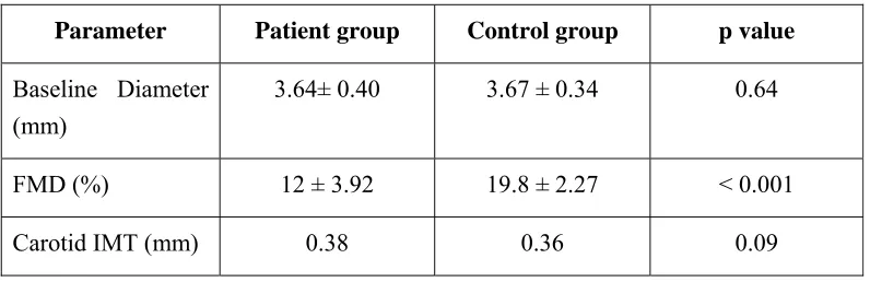
TABLE 3: Study characteristics in the patient and control group
Parameter Patient group Control group p value
Baseline Diameter (mm)
3.64± 0.40 3.67 ± 0.34 0.64
FMD (%) 12 ± 3.92 19.8 ± 2.27 < 0.001
Carotid IMT (mm) 0.38 0.36 0.09
The patients and the controls had a similar degree of baseline diameter of the brachial artery (3.64 ± 0.40 vs. 3.67 ± 0.34 mm, p = 0.64) and carotid IMT (0.38 ±
0.08 vs. 0.36 ± 0.06, p value 0.09). However, the SLE patients had worse
endothelial function than the controls (FMD 12 ± 3.92% vs. 19.8 ± 2.27%, p<0.001).
The endothelial function, assessed by vascular ultrasonography on brachial artery, is shown (Baseline Photo (C) & post dilatation Photo (D). The carotid IMT
0% 5% 10% 15% 20% 25% 30% 35% 40%
F MD C AR O T ID IMT
C)
(E)
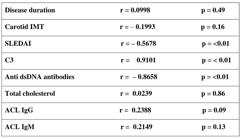
Correlations between FMD and SLE parameters:
[image:57.612.118.503.222.445.2]The correlations between FMD and biological and immunological parameters are shown in Table 4.
TABLE 4: Parameter Correlation coefficient
Disease duration r = 0.0998 p = 0.49
Carotid IMT r = – 0.1993 p = 0.16 SLEDAI r = – 0.5678 p = <0.01
C3 r = 0.9101 p = < 0.01
Anti dsDNA antibodies r = –0.8658 p = <0.01
Total cholesterol r = 0.0239 p = 0.86
ACL IgG r = 0.2388 p = 0.09
ACL IgM r = 0.2149 p = 0.13
Value of r Qualitative Description of the
Strength
–1 perfect negative
(–1, –0.75) strong negative (–0.75, –0.5) moderate negative (–0.5, –0.25) weak negative (–0.25, 0.25) no linear association
(0.25, 0.5) weak positive (0.5, 0.75) moderate positive
(0.75, 1) strong positive
1 perfect positive
The patients with SLE have a high incidence of atherosclerosis with its main consequence: coronary artery disease. Epidemiological studies have shown that SLE women aged 35 – 44 years were over 50 times more likely to develop myocardial infarction than women of similar age from general population (3).
Anatomo-pathological investigations have revealed that the SLE patients were prone to develop a premature atherosclerosis (102). The increased risk of atherosclerosis is
not exclusively related to traditional risk factors alone (103). In the last years, SLE
itself appeared like an independent risk factor for atherosclerosis, acting through autoimmune vascular injury (6). In patients with systemic lupus erythematosus,
atherosclerosis has a long period of subclinical evolution. The first reversible step in the atherogenesis process is represented by the endothelial dysfunction (104).
Endothelial dysfunction appears when the normal functions of the endothelial cells (control of vascular tone and blood pressure, regulation of leucocytes traffic from the blood to the tissues, and platelet adhesion and aggregation, maintenance of the balance between blood coagulation and fibrinolysis, control of growth, development and differentiation of the vessel wall cells) are lost or dysregulated (105). A non-invasive method for the assessment of endothelial
dysfunction is represented by flow mediated vasodilation (endothelium dependent dilation) (106). In our study, FMD in SLE patients was significantly lower than in
control subjects (FMD 12±3.92% vs. 19.8±2.27%, p<0.001). There are several studies with similar results, starting with the first study, performed by Lima et al. (107), who showed that SLE patients presented lower FMD than sex and
age-matched controls (5.0±5.0% vs. 12.0± 6.0%) , even in subjects without traditional cardiovascular risk factors (70). Tani et al. (16) identified the same pattern of FMD
in SLE patients as well as by Valdivielso et al (68) (FMD 12.4+/-4.4% vs.
16.9+/-5.5%, p<0.05). The reduced values of FMD in patients with SLE were found by
Piper and Turner in their studies, too (67, 71). Our study concurred with the FMD
results of the other indian study done by Parasar Ghosh et al (73) which was
impaired in SLE patients compared to controls (9.97% vs. 18.97%, p < 0.00001).
The Carotid IMT values showed no significant difference in our study and control population (0.38 ±0.08 vs. 0.36 ± 0.06, p value 0.09). Similar results were
obtained in the study done by Valdivielso et al (68) (0.58+/-0.08 mm vs.
0.57+/-0.07 mm, NS). Parasar Ghosh et al (73) have reported higher carotid IMT in SLE
which was not the case in our study. Masoud El-Magadmi et al (67) & Zahra Seyyedbonakdar et al (69) also observed a negative correlation with percent FMD
in their studies. The reason for this difference could be that in early stages of the disease to which our patients belonged to, identification of a functional abnormality (FMD) would have preceded the structural changes that occur in the carotid arteries in the form of intima medial thickness that occurs on a later stage. Moreover the other factors which would contribute to subclinical atherosclerosis like comorbities, immunosuppressants were not excluded in their studies as against ours.
We found a moderate negative correlation between FMD and SLEDAI (r = –
0.7321, p < 0.001). This correlation between FMD and the disease activity was
identified in other studies (96, 108 & 109). Valdivielso et al (68) also have reported
significant correlation between FMD and LAI (Lupus Activity Index). AntidsDNA had a strong negative correlation, while C3 complement assay had strong positive correlation with flow mediated dilatation. There was no linear correlation between other factors like disease duration, cholesterol, anticardiolipin antibodies.
When comparing our study with the other indian study done by Parasar Ghosh et al (73) regarding other factors, our study population has less disease
FMD using vascular ultrasonography on brachial artery represents a useful, non-invasive method for the assessment of the endothelial dysfunction. Reactive hyperemia produces a shear stress stimulus that induces the normal endothelium to release nitric oxide (NO), which acts as a vasodilator. Impaired endothelial function is associated with a reduced release of NO and a lower vasodilation (110). The effect
of disease states and/or interventions on the blood flow response to cuff occlusion (reactive hyperemia) is under explored. Current technology limits the utility of spectral Doppler to reproducibly assess changes in flow, which might provide useful information about endothelial function of the microvasculature.
Ongoing studies in several large populations, including the Framingham Heart Study and the Cardiovascular Health Study, shall determine whether endothelial dysfunction in the brachial artery will identify patients at risk for developing coronary artery disease, cerebral vascular disease and/or peripheral vascular disease. The technique is particularly well suited for study of the earliest stages of atherosclerosis in children and young adults, thus providing maximal opportunity for prevention.
useful measure of cardiovascular risk on an individual or group basis. To that end, the methodology will need to mature, with formal opportunities for training, certification and continuing medical education, as currently exist for other cardiovascular testing modalities.
FUTURE DIRECTIONS
Ultrasound assessment of brachial artery FMD has yielded important information about vascular function in health and disease, yet several new approaches and technological advances have emerged. Most prior studies examined FMD at a single time point, typically 1 min after cuff release. This practice evolved from the observations that the maximal dilator response occurs at approximately 1 min in healthy subjects (111) and that the necessity for manual acquisition and
measurement placed a practical limit on the number of image frames that could be analyzed. Commercially available technology now makes it possible to acquire multiple images of the brachial artery automatically using the ECG signal as a trigger and to measure arterial diameter automatically using computer-based edge detection techniques. This approach allows investigators to examine the entire time course of brachial dilation in response to reactive hyperemia , the true peak response, the time to peak and the overall duration of FMD as discussed in the previous text. The time course and extent of brachial expansion within a single cardiac cycle, possibly reflecting vessel compliance, can be examined. In the carotid artery, compliance has been shown to correlate with cardio- vascular risk (112).
About 70% of the dilation observed 1 min after cuff release is attributable to NO synthesis.
1) Endothelial dysfunction is present in SLE patients even in the absence of traditional cardiovascular risk factors.
2) FMD using vascular ultrasonography on brachial artery represents a non-invasive, reproducible and cost effective tool for the assessment of endothelial dysfunction.
3) Impaired flow mediated dilatation of brachial artery may be an early physiological feature of endothelial dysfunction and may precede the anatomical increase in carotid intimal thickness.
4) FMD of brachial artery has significant negative correlation with disease activity and antidsDNA antibody.
5) Future prospects of FMD technique include
a) early screening module for endothelial dysfunction and to start on drugs with pleotrophic effect like statins, ACE inhibitors and Aspirin.
1. Urowitz, M.B., A.A. Bookman, B.E. Koehler et al., The bimodal mortality
pattern of systemic lupus erythematosus, Am. J. Med., 1976, 60,221–225.
2. Bruce I.N., D.D. Gladman, M.B. Urowitz. Detection and modification of risk factors for coronary artery disease in patients with SLE: a quality improvement study, Clin. Exp. Rheumatol., 1998, 16,435–440.
3. Manzi S, E.N. Meilahn, J.E. Rairie. Age-specific incidence rates of myocardial infarction and angina in women with systemic lupus erythematosus: comparison with the Framingham study, Am. J. Epidemiol., 1997, 145, 408–415.
4. Pearson J.D., Normal endothelial cell function, Lupus, 2000, 9, 183–188.
5. Ahmad Y, J. Shelmerdine, H. Bodill et al. Subclinical atherosclerosis in systemic lupus erythematosus(SLE): the relative contribution of classic risk factors and the lupus phenotype, Rheumatology, 2007, 46(6), 983–988.
6. Roman M.J, B.A. Shanker, A. Davis. Prevalence and correlates of accelerated atherosclerosis in systemic lupus erythematosus, N. Engl. J. Med., 2003, 349,2399–2406.
7. Hahn B.H. Systemic lupus erythematosus and accelerated atherosclerosis, N. Engl. J. Med, 2003, 349, 2379–80.
8. Yazici Z.A, E. Raschi, A. Patel et al, Human monoclonal anti-endothelial cell IgG-derived from a systemic lupus erythematosus patient binds and activates human endothelium in vitro, Int. Immunol, 2001, 13,349–357.
9. Mary C. Corretti, MD, Todd J. Anderson, MD, Emelia J. Benjamin, MD,
Report. Guidelines for the ultrasound assessment of endothelial-dependent flow-mediated vasodilation of the brachial artery. Journal of American College of Cardiology, 2002; 39:257-265.
10. Vogel RA, Corretti MC. Estrogens, progestins, and heart disease: can endothelial function divine the benefit? Circulation 1998; 97:1223–6.
11. Suwaida JA, Hamasaki S, Higano ST, Nishimura RA, Holmes DR, Lerman A. Long-term follow-up of patients with mild coronary artery disease and endothelial dysfunction. Circulation 2000; 101:948–54.
12. Schachinger V, Britten MB, Zeiher AM. Prognostic impact of coronary vasodilator dysfunction on adverse long-term outcome of coronary heart disease. Circulation 2000; 101:1899–906.
13. Raza K, Thambyrajah J, Townend JN, et al. Suppression of inflammation in primary systemic vasculitis restores vascular endothelial function: lessons for atherosclerotic disease? Circulation. 2000; 102: 1470–1472.
14. Bulkley BH, Roberts WC. The heart in systemic lupus erythematosus and the changes induced in it by corticosteroid therapy. A study of 36 necropsy patients. Am J Med 1975; 58:243-264.
15. Sturfelt G, Eskilsson F, Nived O, et al. Cardiovascular disease in systemic lupus erythematosus. A study of 75 patients from a defined population. Medicine (Baltimore) 1992; 71:216-223.
16. Jouhikainen T, Pohjola-Sintonen S, Stephansson E. Lupus anticoagulant and cardiac manifestations in systemic lupus erythematosus. Lupus 1994; 3:167-172.
abnormalities and risk of ischemic cerebrovascular accidents in patients with systemic lupus erythematosus. Lupus 2003;12:805-812.
19. Urowitz M, Bookman A, Koehler B, et al. The bimodal mortality pattern of systemic lupus erythematosus. Am J Med 1976; 60:221-225.
20. Petri M, Perez-Gutthann S, Spence D, et al. Risk factors for coronary artery disease in patients with systemic lupus erythematosus. Am J Med 1992; 93:513-519. P.676
21. Hearth-Holmes M, Baethge BA, Broadwell L, et al. Dietary treatment of hyperlipidemia in patients with systemic lupus erythematosus. J Rheumatol 1995; 22:450-454.
22. Rahman P, Aguero S, Gladman DD, et al. Vascular events in hypertensive patients with systemic lupus erythematosus. Lupus 2000; 9: 672-675.
23. Bessant R, Hignorani A, Patel L, et al. Risk of coronary heart disease and stroke in a large British cohort of patients with systemic lupus erythematosus. Rheumatology 2004; 43:924-929.
24. Toloza SMA, Uribe AG, McGwin G, et al. Systemic lupus erythematosus in a multiethnic US cohort (LUMINA). Arthritis Rheum 2004; 50:3947-3957.
25. Haider Y, Roberts W. Coronary artery disease in systemic lupus erythematosus; quantification of degrees of narrowing in 22 necropsy patients (21 women) aged 16-37 years. Am J Med 1981; 70:775-781.
27. Manzi S, Selzer F, Sutton-Tyrrell K, et al. Prevalence and risk factors of carotid plaque in women with systemic lupus erythematosus. Arthritis Rheum 1999; 42:51-60.
28. Bruce IN, Burns RJ, Gladman DD, et al. Single photon emission computed tomography dual isotope myocardial perfusion imaging in women with systemic lupus erythematosus. I. Prevalence and distribution of abnormalities. J Rheumatol 2000; 27:2372-2377.
29. Asanuma Y, Oeser A, Shintani A, et al. Premature coronary artery atherosclerosis in systemic lupus erythematosus. N Engl J Med 2003; 349:2407-2415.
30. Doria A, Shoenfeld R, Wu R, et al. Risk factors for subclinical atherosclerosis in a prospective cohort of patients with systemic lupus erythematosus. Ann Rheum Dis 2003; 62:1071-1077.
31. Manger K, Kusus M, Forster C, et al. Factors associated with coronary artery calcification in young female patients with SLE. Ann Rheum Dis 2003; 62:846-850.
32. Roman M, Shanker B, Davis A, et al. Prevalence and correlates of accelerated atherosclerosis in systemic lupus erythematosus. N Engl J Med 2003; 349:2399-2406.
33. Sella EMC, Sato EI, Leite WA, et al. Myocardial perfusion scintigraphy and coronary disease risk factors in systemic lupus erythematosus. Ann Rheum Dis 2003; 62:1066-1070.
femoral arteries of patients with systemic lupus erythematosus: a controlled study. J Rheumatol 2004; 31:909-914.
36. Jimenez S, Garcia-Criado M, Tassies D, et al. Preclinical vascular disease in systemic lupus erythematosus and primary antiphospholipid syndrome. Rheumatology 2005; 44:756-761.
37. Ward M. Premature morbidity from cardiovascular and cerebrovascular diseases in women with systemic lupus erythematosus. Arthritis Rheum 1999; 42:338-346.
38. Manzi S, Meilahn EN, Rairie JE, et al. Age-specific incidence rates of myocardial infarction and angina in women with systemic lupus erythematosus: comparison with the Framingham Study. Am J Epidemiol 1997; 145:408-415.
39. Bruce I, Gladman D, Urowitz M. Premature atherosclerosis in systemic lupus erythematosus. Rheum Dis Clin North Am 2000; 26:257-278.
40. Han TS, Sattar M, Williams K, et al. Prospective study of C-reactive protein in relation to the development of diabetes and the metabolic syndrome in the Mexico City diabetes study. Diabetes Care 2002; 25:2016-2021.
41. Saijo Y, Kiyota N, Kawasaki Y, et al. Relationship between C-reactive protein and visceral adipose tissue in healthy Japanese subjects. Diabetes Obes Metab 2004; 6:249-258.
42. Sung KC, Suh JY, Kim BS, et al. High sensitivity C-reactive protein as an independent risk factor for essential hypertension. AJH 2003; 16:429-433.
44. Bruce IN, Gladman DD, Urowitz MB. Detection and modification of risk factors for coronary artery disease in patients with systemic lupus erythematosus: a quality improvement study. Clin Exp Rheum 1998; 16:435-440.
45. Costenbader K, Wright E, Matthew L, et al. Cardiac risk factor awareness and management in patients with systemic lupus erythematosus. Arthritis Rheum 2004; 51:983-988.
46. Esdaile J, Abrahamowicz M, Grodzicky T, et al. Traditional Framingham risk factors fail to fully account for accelerated atherosclerosis in systemic lupus erythematosus. Arthritis Rheum 2001; 44:2331-2337.
47. Wei L, MacDonald T, Walker B. Taking glucocorticoids by prescription is associated with subsequent cardiovascular disease. Ann Intern Med 2004; 141:764-770.
48. Zonana-Nacach A, Barr S, Magder L, et al. Damage in systemic lupus erythematosus and its association with corticosteroids. Arthritis Rheum 2000; 43:1801-1808.
49. Qi D, Pulinilkunnil T, An D, et al. Single-dose dexamethasone induces whole-body insulin resistance and alters both cardiac fatty acid and carbohydrate metabolism. Diabetes 2004; 53: 1790-1797.
50. Rollino C, Boero R, Elia F, et al. Antiphospholipid antibodies and hypertension. Lupus 2004; 13:769-772.
51. Godfrey T, Khamashta M, Hughes G. Antiphospholipid syndrome and renal artery stenosis. Q J Med 2000; 93:127-129.
systemic lupus erythematosus. Am J Kidney Dis 1993; 21(suppl):58-60.
54. Cooke JP, Rossitch E, Jr, Andon NA, Loscalzo J, Dzau VJ. Flow activates an endothelial potassium channel to release an endogenous nitrovasodilator. J Clin Invest 1991; 88:1663–71.
55. Miura H, Wachtel RE, Liu Y, et al. Flow-induced dilation of human coronary arterioles: important role of Ca-activated K channels. Circulation 2001; 103:1992–8.
56. Olesen SP, Clapham DE, Davies PF. Haemodynamic shear stress activates a K_ current in endothelial cells. Nature 1988; 331:168–70.
57. Pohl U, Holtz J, Busse R, Bassenge E. Crucial role of the endothelium in the vasodilator response to flow in vivo. Hypertension 1985; 8:37-44.
58. Joannides R, Haefeli WE, Linder L, et al. Nitric oxide is responsible for flow-dependent dilatation of human peripheral conduit arteries in vivo. Circulation 1995; 91:1314–9.
59. Sun D, Huang A, Smith CJ, et al. Enhanced release of prostaglandins contributes to flow-induced arteriolar dilatation in eNOS knockout mice. Circ Res 1999; 85:288–93.
60. Corson MA, James NL, Latta SSE, Nerem RM, Berk BC, Harrison DG. Phosphorylation of endothelial nitric oxide synthase in response to fluid shear stress. Circ Res 1996; 79:984–91.
62. Corretti MC, Plotnick GD, Vogel RA. Smoking correlates with flow-mediated brachial artery vasoactivity but not cold pressor vasoactivity in men with coronary artery disease. Int J Card Imaging 1998; 14:11–1.
63. Vogel RA, Corretti MC, Plotnick GD. Changes in flow-mediated brachial artery vasoactivity with lowering of desirable cholesterol levels in healthy middle-aged men. Am J Cardiol 1996; 77:37- 40.
64. Li J, Zhao SP, Li XP, Zhuo QC, Gao M, Lu SK. Non-invasive detection of endothelial dysfunction in patients with essential hypertension. Int J Cardiol
1997; 61:165–169.
65. Lambert J, Aarsen M, Donker AJ, Stehouwer CD. Endothelium-dependent and independent vasodilation of large arteries in normoalbuminuric insulin-dependent diabetes mellitus. Arterioscler Thromb Vasc Biol 1996; 16:705–
711.
66. Lambert J, van den BM, Steyn M, Rauwerda JA, Donker AJ, Stehouwer CD. Familial hyperhomocysteinaemia and endothelium-dependent vasodilatation and arterial distensibility of large arteries. Cardiovasc Res 1999; 42:743–751
67. Masoud El-Magadmi, MB; Helena Bodill, MSc; Yasmeen Ahmad, MB, MRCP; Paul N. Durrington, MD et al.Independent Risk Factor for Endothelial Dysfunction in Women..Circulation. 2004; 110:399-404.
68. Valdivielso P, Gómez-Doblas JJ, Macias M, Haro-Liger M, Fernández-Nebro A, Sánchez-Chaparro MA, González-Santos P. Clin Exp Rheumatol. 2008 Sep-Oct; 26(5):827-33. Lupus-associated endothelial dysfunction, disease activity and arteriosclerosis.
Medwin J, Hiller L, Martin U, Townend J, Bacon P A, Gordon C.Impaired endothelial function in systemic lupus erythematosus.Lupus 2007;16(2): 84-8.
71. Elizabeth Turner, Victor Dishy, Cecilia P Chung, Paul Harris, Rosanna Pierce, Yu Asanuma, Annette Oeser, Tebeb Gebretsadik, Ayumi Shintani, Paolo Raggi, and C Michael Stein. Endothelial Function in Systemic Lupus Erythematosus: Relationship to Disease Activity, Cardiovascular Risk Factors, Corticosteroid Therapy, and Coronary Calcification. Vasc Health Risk Manag. 2005 December; 1(4): 357–360.
72. Lai-Shan Tam, Boli Fan, Edmund K Li, G Neil Thomas, So F Yim, Christopher J Haines, Brian Tomlinson. Patients with systemic lupus erythematosus show increased platele activation and endothelial dysfunction induced by acute hyperhomocysteinemia. The Journal of Rheumatology,July 1, 2003vol. 30(7) 1479-1484.
73. Ghosh Parasar, Kumar Amresh, Kumar Sudeep, Aggarwal Amita, Sinha Nakul, Misra Ramnath . Subclinical atherosclerosis and endothelial dysfunction in young South-Asian patients with systemic lupus erythematosus. Clinical Rheumatology, 2009, vol. 28 (11)1259-1265.
74. Hashimoto M, Eto M, Akishita M et al (1999) Correlation between flow-mediated vasodilatation of the brachial artery and intima–media thickness in the carotid artery in men. Arterioscler Thromb Vasc Biol 19:2795–2800.
76. Uehata A, Lieberman EH, Gerhard MD, et al. Noninvasive assessment of endothelium-dependent flow-mediated dilation of the brachial artery. Vasc Med 1997; 2:87–92.
77. Vogel RA, Corretti MC, Plotnick GD. A comparison of the assessment of flow-mediated brachial artery vasodilation using upper versus lower arm arterial occlusion in subjects with and without coronary risk factors. Clin Cardiol 2000;23:571–5.
78. Sorensen KE, Celermajer DS, Spiegelhalter DJ, et al. Non-invasive measurement of human endothelium dependent arterial responses: accuracy and reproducibility. Br Heart J 1995; 74:247–53.
79. Stadler RW, Karl WC, Lees RS. New methods for arterial diameter measurement from B-mode images. Ultrasound Med Biol 1996; 22: 25–34.
80. Corretti MC, Plotnick GD, Vogel RA. Technical aspects of evaluating brachial artery vasodilatation using high-frequency ultrasound. Am J Physiol 1995; 268:H1397–H1404.
81. Stadler RW, Taylor JA, Lees RS. Comparison of B-mode, M-mode and echo-tracking methods for measurement of the arterial distension waveform. Ultrasound Med Biol 1997; 23:879–87.
86. Celermajer DS, Sorensen KE, Gooch VM, et al. Non-invasive detection of endothelial dysfunction in children and adults at risk of atherosclerosis. Lancet 1992; 340:1111–5.
87. Levine GN, Frei B, Koulouris SN, Gerhard MD, Keaney JF, Jr., Vita JA. Ascorbic acid reverses endothelial vasomotor dysfunction in patients with coronary artery disease. Circulation 1996; 93:1107–13.
transient impairment of endothelium-dependent brachial artery vasoactivity following a single high-fat meal. JAMA 1997; 278:1682–6.
90. Gerhard M, Walsh BW, Tawakol A, et al. Estradiol therapy combined with progesterone and endothelium-dependent vasodilation in postmenopausal women. Circulation 1998; 98:1158–63.
91. Koh KK, Cardillo C, Bui MN, et al. Vascular effects of estrogen and cholesterol-lowering therapies in hypercholesterolemic postmenopausal women. Circulation 1999; 99:354–60.
92. Wilmink HW, Banga JD, Hijmering M, Erkelens WD, Stroes ES, Rabelink TJ. Effect of angiotensin-converting enzyme inhibition and angiotensin II type 1 receptor antagonism on postprandial endothelial function. J Am Coll Cardiol 1999; 34:140–5.
94. Bellamy MF, McDowell IF, Ramsey MW, et al. Hyperhomocysteinemia after an oral methionine load acutely impairs endothelial function in healthy adults. Circulation 1998; 98:1848–52.
95. Hornig B, Arakawa N, Haussmann D, Drexler H. Differential effects of quinaprilat and enalaprilat on endothelial function of conduit arteries in patients with chronic heart failure. Circulation 1998; 98: 2842–8.
96. Corretti MC, Plotnick GD, Vogel RA. Technical aspects of evaluating brachial artery vasodilatation using high-frequency ultrasound. Am J Physiol 1995; 268:H1397–H1404.