0022-538X/96/$04.0010
Copyrightq1996, American Society for Microbiology
cis-Acting Elements in Human Immunodeficiency Virus Type 1
RNAs Direct Viral Transcripts to Distinct
Intranuclear Locations
EVELYN BERTHOLD
ANDFRANK MALDARELLI*
Laboratory of Molecular Microbiology, National Institute of Allergy and Infectious Diseases, Bethesda, Maryland 20892
Two distinct intranuclear locations were identified for alternatively spliced RNA transcripts expressed from
the pNL4-3 infectious molecular clone of human immunodeficiency virus (HIV) type 1. Multiply spliced HIV
RNA encoding
tat
was detected within the nucleus in large clusters; immunostaining and colocalization studies
using laser-scanning confocal microscopy revealed that these structures contained the non-small nuclear
ribonucleoprotein RNA processing factor, SC35. In contrast, unspliced
gag
RNA was detected in much smaller
granules distributed throughout the nucleus, with little or no association with SC35-containing granules.
Analyses of nuclear RNA expressed from recombinant plasmids encoding
gag
(pCMVgag-2) alone or
tat
(pCMVtat-2) alone revealed distributions corresponding to those obtained with pNL4-3, indicating that
expression within the context of the HIV provirus was not required for the distinct RNA locations detected for
these transcripts. The presence of unspliced
gag
RNA in small granules was confirmed in infections of H9
T-lymphocytic cells, indicating that
gag
localization was not restricted to transient expression systems. The
intranuclear distribution of
gag
RNA was dependent on specific RNA sequences. Deletion of a portion of the
gag
gene of pCMVgag-2, containing a
cis
-repressing inhibitory region, resulted in redirection of unspliced
gag
RNA from small granules into large SC35-containing clusters. The addition of the Rev-responsive element,
RRE, to the deleted pCMVgag-2 construct resulted in RNA transcripts which were no longer associated with
SC35. We also identified a cellular intron, rabbit
b
-globin-intervening sequence 2 (IVS-2) which, when
introduced into pCMVgag-2, redirected unspliced
gag
RNA into SC35-containing granules and permitted
rev
-independent Gag expression. These findings suggest that redirecting intranuclear RNA localization may
influence gene expression. Color micrographs from this article are available for view at http//128.231.216.2/
lmmhome.htm.
Replication of human immunodeficiency virus type 1
(HIV-1) is regulated by transcriptional and posttranscriptional
events, which require interactions between viral and
host-en-coded factors (48). The entire HIV-1 genome is expressed
from a unique promoter located in the 5
9
long terminal repeat
(LTR) portion of the integrated provirus, and a 9.2-kb
ge-nome-length transcript is alternatively spliced, yielding
numer-ous transcripts encoding HIV-1 gene products (4, 24, 53, 58,
63, 66). Regulation of differential HIV RNA processing is
complex; expression of unspliced and singly spliced RNA
spe-cies requires binding of the HIV-1 regulatory gene product,
Rev, to a highly structured RNA target, the Rev-responsive
element (RRE), while expression of multiply spliced 1.8-kb
RNA occurs in the presence or absence of rev (5, 11, 16, 17, 25,
26, 28, 29, 44–47). Additional cis-acting sequences within the
HIV-1 gag/pol and env genes which inhibit their own expression
are reported to participate in regulation of RNA expression as
well. These sequences, denoted instability sequences (INS [52,
64, 65]), cis-repressor sequences (CRS [12, 15, 61]), or
inhib-itory regions (IR [42]), all share the requirement for the RRE
sequence in cis and rev in trans for full expression in the
cytoplasm. The nature of the inhibitory block conferred by
repressor sequences and the mechanisms by which rev relieves
cis inhibition remain unknown. Recently, rev-independent gag
expression has been achieved by a series of conservative base
substitutions which interrupt AT-rich regions (64), and
rev-independent env expression was observed when the env gene
was positioned downstream of an efficiently spliced
b
-globin
intron (IVS-2) and when cryptic splice sites within env were
removed, redefining the env gene as an exon (27).
In previous RNA fractionation experiments, we reported
that HIV inhibitory sequences were localized predominantly to
the nucleus (42). In the present communication, we
investi-gated the intranuclear location of HIV RNA sequences by
using in situ hybridization and confocal microscopic
tech-niques. HIV RNA species were detected in two distinct
in-tranuclear locations; a multiply spliced RNA species encoding
the tat gene was identified in nuclear speckles and colocalized
with the RNA processing factor SC35. In contrast, unspliced
RNA was identified in small granules which did not colocalize
with SC35. Further analysis revealed that the presence of the
RRE or inhibitory gag sequences in cis was capable of directing
RNA localization within the nucleus. In addition, we identified
that the
b
-globin (IVS-2), which has been shown to enable
rev-independent env expression (27), redirected HIV gag RNA
into SC35-containing granules and allowed rev-independent
gag expression. These findings indicate that cis-acting
se-quences can direct the intranuclear location of RNA
tran-scripts and suggest that physical compartmentalization within
the nucleus may influence HIV RNA processing and gene
expression.
MATERIALS AND METHODS
Cells and transfection.HeLa cells were maintained in DMEM supplemented with 10% fetal bovine serum, glutamine, and antibiotics (maintenance medium) (70). Uninfected H9 cells and H9 cells chronically infected with HIV IIIB (H9IIIB) or with HIV pNL4-3 (H9NL43) were maintained in RPMI 1640 sup-plemented with 10% fetal bovine serum, glutamine, and antibiotics (43). HIV
* Corresponding author. Mailing address: Laboratory of Molecular Microbiology, NIAID, NIH, 9000 Rockville Pike, Bethesda, MD 20892. Phone: (301) 402-4072. Fax: (301) 402-0226. Electronic mail address: [email protected].
4667
on November 9, 2019 by guest
http://jvi.asm.org/
infections were initiated by cell-cell transmission as previously described (43, 62) and examined 21 h after cocultivation. HeLa cells were transfected with 20mg of plasmid DNA, as previously described (70), except that the cells did not undergo glycerol shock. For in situ hybridization and/or immunofluorescence studies, transfected cells were washed, scraped, and reseeded into 12-well plates 4 to 5 h after DNA addition. T-lymphocytic cells were attached to glass slides by cyto-centrifugation or by coating slides with poly-lysine. For RNA analyses, cells were washed and refed with maintenance medium 4 to 5 h after DNA addition and harvested 24 to 48 h later. In experiments comparing Gag protein expression, plasmids were cotransfected with pCMV-CAT as an internal control; lysates prepared from these transfections were normalized for transfection efficiency by measuring chloramphenicol acetyltransferase enzyme activity (42), and then p24 determinations were performed.
Plasmids.The pNL4-3 infectious molecular clone of HIV-1 (1), tat-expressing pCMV-tat-2 (22), rev-expressing pCMV-rev (2), pCMV-CAT (2), and rabbit
b-globin-expressing pAL4-SV (9) have been previously described. A rev-defec-tive derivarev-defec-tive of pNL4-3, pNL4-3rev2, was constructed by restricting it with
BamHI, extending the recessed strand with the Klenow fragment of Pol I, and
religating the blunt ends with DNA ligase. Recombinant plasmids expressing portions of the HIV-1 gag/pol gene from the cytomegalovirus immediate-early promoter were constructed by a multistep strategy. The BglII restriction frag-ment (nucleotide [nt] 679 to 2096, numbered according to the pNL4-3 entry in reference 51) from pNL4-3 containing the HIV-1 gag p17, p24, and a portion of p9 was ligated to BamHI-restricted, calf intestinal phosphatase-treated pCMV-CAT vector, yielding pCMVgag-0. To provide the HIV LTR polyadenylation signal, the EcoRV-EcoRI fragment containing the HIV LTR from the pNLA-1 plasmid (69) was ligated to pCMVgag-0 restricted with SmaI-EcoRI, generating pCMVgag-3. Sequences 39to the LTR were removed by restricting pCMVgag-3 with EcoRI and NarI, extending the recessed strand with Klenow Pol I, and religating the blunt ends, yielding pCMVgag-2 (see Fig. 6). An RRE-containing derivative of pCMVgag-2 was constructed by inserting the RRE into a unique
BglII site of pCMVgag-2. A DNA fragment (nt 7721 to 7993) including the RRE
was obtained by PCR, amplifying pNL4-3 with oligonucleotides containing the
BglII recognition sequence at the 59end: 59GAAGATCTTCGAAGAGTGGT GCAGA 39and 59CCTAGTTGTCGAGGAGGTCTAGAAG 39; the amplified DNA fragment was restricted with BglII and ligated to the BglII-restricted, calf intestinal phosphatase-treated pCMVgag-2 (see Fig. 6). Deletion mutants of pCMVgag-2 and pCMVgag-2RRE were obtained by restricting each plasmid with AccI and religating, generating pCMVgag-2ACC and pCMVgag-2AC-CRRE, respectively (see Fig. 6). A derivative of pCMVgag-2 containing the second intervening sequence of rabbitb-globin upstream of the major splice donor was constructed by obtaining the BamHI-EcoRI fragment from rabbit
b-globin plasmid pAL4-SV, extending the recessed strands with Klenow, and blunt-end ligating to BssHII-restricted, blunt-ended, phosphatase-treated pCMVgag-2. The structures of the recombinant clones were analyzed by restric-tion digests, and the sequences of the relevant porrestric-tions of the constructs were confirmed by dideoxy sequencing.
In situ hybridization.DNA restriction fragments, whole plasmids, or PCR-amplified DNAs were used to specifically detect RNA by hybridization as de-scribed by Johnson et al. (32). HeLa cells on glass coverslips were harvested 24 to 48 h after transfection; coverslips were washed two to three times with Dulbecco’s phosphate-buffered saline (PBS) and fixed at room temperature (RT) for 10 min in 4% paraformaldehyde–PBS buffered to pH 7.4. Coverslips were washed two times with PBS and stored in 70% ethanol at 48C. For hybrid-ization, the probe was redissolved in 120ml of 83% formamide containing 0.83 mg of tRNA per ml and 0.83mg of sheared salmon sperm DNA per ml, heated to 708C for 10 min, and then immediately chilled. The probe was mixed 1:1 with hybridization buffer (43SSC [13SSC is 0.15 M NaCl plus 0.015 M sodium citrate], 0.4% bovine serum albumin [BSA], 0.4 mM vanadyl riboside complexes, 20% dextran sulfate), and 20ml of this mixture was applied to each coverslip. The coverslips were covered with parafilm and incubated at 378C for 3 h overnight in a humidified chamber. After hybridization, cells were washed sequentially in 50% formamide in 23SSC at 378C for 20 min, 23SSC at 378C for 20 min, 13
SSC at RT for 20 min, and 43SSC at RT for 10 min. The hybridized probes were detected by incubation with Texas red-conjugated streptavidin (Molecular Probes, Eugene Ore.; 2mg/ml in 43SSC–1% BSA) for 30 min to 1 h at 378C, washed three times with 43SSC for 10 min at RT, and mounted onto glass slides in Fluormount (Virotech International, Rockville, Md.). For colocalization ex-periments, coverslips were incubated in PBS–1% BSA at 378C for 30 min with specific mouse monoclonal antiserum SC35 (gift of T. Maniatis [21]), human serum from an HIV-seropositive individual, mouse monoclonal anticoilin (gift of E. Chan), rabbit anti-Rev (gift of G. Pavlakis), mouse monoclonal anti-p24 (New England Nuclear), or fluorescein isothiocyanate-conjugated mouse monoclonal anti-p24 (Seramun). The sera were visualized by incubating with the correspond-ing fluorescein-conjugated goat anti-mouse, goat anti-human, or goat anti-rabbit serum.
The cells were examined by laser-scanning confocal microscopy with a Zeiss LSM 4 confocal microscope system (Carl Zeiss, Thornwood, N.Y.). A 488-nm incident beam from an Ar-Kr laser and a 515-565 band-pass filter were used to detect fluorescein, and a 568-nm incident beam and 590-nm long-pass filter were used to identify the Texas red signal; the 568-nm incident beam and Nomarski optics were employed to obtain a light microscopic image. The fluorescein, Texas
red, and Nomarski images were stored in separate channels as black and white images; to determine colocalization, the RNA image (pseudocolored red) and the SC35 immunofluorescence image (pseudocolored green) were electronically recombined; the colocalized image is pseudocolored yellow-orange; colocaliza-tion was confirmed in all cases by using the colocalizacolocaliza-tion funccolocaliza-tion of the LSM-4 software package. Where no colocalization was detected, the entire cell was optically sectioned, either manually or automatically, to ensure that no significant areas of colocalization were present. At least four independent transfections were performed to compare the constructs, and several hundred total cells were analyzed for each transfected plasmid. No colocalization studies were possible using in situ hybridization and anti-Rev sera, because Rev reactivity was lost after the in situ hybridization procedure. Color micrographs from this article are available for view at http//128.231.216.2/lmmhome.htm.
RNA analyses.Whole-cell RNA preparations were obtained by guanidinium extractions as described previously (42), or polyadenylated RNA was isolated from transfected HeLa cells using an Invitrogen fast track mRNA purification kit (Invitrogen, San Diego, Calif.). RNA was electrophoresed in 1% agarose-form-aldehyde gels, transferred to nitrocellulose, and hybridized to a32
P-end-labeled oligonucleotide complementary to the major splice donor region of pNL4-3 (59 CCCATCTCTCTCCTTCTAGCCTCCGCTAGTCAAAATTTTTGGCGTACT CACCAGTCGCCGCCCCTCGCCTCTTGCCGTGCGC 39), as previously de-scribed (42).
Probe preparation.DNA probes were prepared from restriction fragments (probe ACC, AccI position 959-AccI position 1678) from PCR fragments (probe D, position 736 to 959 using oligonucleotide primers 59GGCGACTGGTGAG TACGCCAAAAATT 39and 59CAGCCTTCTGATGTCTCTAAAAGGACC 39; probe E, position 769 to 2011 using oligonucleotide primers 59GCGGAG GCTAGAAGGAGAG 39 and 59 GGGCCCTGCAATTTTTGGC 39; probe RRE, position 7721 to 7993 using oligonucleotide primers used above to amplify the RRE for cloning), or from entire plasmids (pCMVTAT-2 and pNL4-3) as noted by Johnson et al. (32). Probes were labeled by incorporation of biotin 14-dATP in nick translation reactions using the BIONICK labeling system (GIBCO Life Sciences, Gaithersburg, Md.) under conditions stipulated by the supplier. Each probe preparation was mixed with 10mg of sheared salmon sperm DNA–1/10 volume 3M sodium acetate and then precipitated with 3 volumes of 95% ethanol and centrifuged, and the pellet was washed in 70% ethanol. To detect polyadenylated RNA, a poly(T) (64-mer) oligonucleotide was synthesized containing a biotinylated dT at every fourth residue.
p24 determination.Quantitative p24 antigen levels in extracts of transfected cells were measured with the Coulter HIV-1 p24 antigen capture assay kit (Coulter Corp., Hialeah, Fla.).
RESULTS
Two distinct patterns of HIV-1 RNA intranuclear
distribu-tion.
HIV RNA processing results in three distinct size classes
of viral RNA, 9.2, 4.5, and 1.8 kb in size. We investigated the
intracellular distribution of the different HIV RNA species by
fluorescent in situ hybridization (FISH) and confocal
micros-copy. HIV RNA transiently expressed in HeLa cells
trans-fected with the infectious molecular clone pNL4-3 was
hybrid-ized in situ to complementary biotinylated DNA probes (Fig.
1). Hybridized probes were detected by staining with Texas red
streptavidin, and the cells were examined by laser-scanning
confocal microscopy. As shown in Fig. 2, hybridization to gag
probe ACC (see Fig. 1 for position) revealed that unspliced
HIV RNA was present in the cytoplasm and was distributed
within the nucleus in small granules finely stippling the
nucleo-plasm but was excluded from the nucleolus (Fig. 2c).
RNA-expressing cells were actively producing HIV protein in the
cytoplasm; immunostaining with anti-p24 antisera (Fig. 2a)
revealed that all cells expressing HIV RNA were producing
p24
Gagprotein. Hybridization with biotinylated probes was
specific for HIV RNA; after RNase digestion, p24-expressing
cells (Fig. 2b) no longer contained any hybridization signal
(Fig. 2d). The distribution of HIV unspliced RNA in small
granules within the nucleus was also demonstrated with a
sec-ond probe, D (Fig. 1) (data not shown). FISH using a probe,
RRE (Fig. 1), complementary to the Rev-responsive element
contained within the env gene, also revealed a finely stippled
pattern of RNA in the nucleus (Fig. 3b), similar to that
de-tected with a probe ACC complementary to unspliced HIV
RNA (Fig. 3a). Since both unspliced gag and singly spliced vif
and vpu/env RNA transcripts contain the RRE sequence, it is
4668 BERTHOLD AND MALDARELLI J. VIROL.
on November 9, 2019 by guest
http://jvi.asm.org/
not possible to determine whether the RRE hybridization
sig-nal corresponds to unspliced or singly spliced transcripts.
How-ever, because the hybridization signal obtained with the RRE
probe was present in the nucleus as a uniformly fine stippling,
we infer that both unspliced and singly spliced HIV RNA
transcripts were present in small granules.
In contrast to the distribution of unspliced and singly spliced
transcripts, the majority of transcripts encoding the multiply
spliced tat gene accumulated in fewer, larger clusters of
ap-proximately 2
m
m in size distributed in the nucleus exclusive of
the nucleolus (Fig. 3c). A small amount of the RNA
hybridiz-ing to the tat probe was present in small granules as well (Fig.
3c); it is likely that some of this stippled signal includes
un-spliced, full-length RNA identified by the tat probe. These
findings suggest that the sites in the nucleus where HIV
un-spliced and multiply un-spliced RNAs accumulate are distinct.
To characterize the intranuclear locations of HIV RNA
spe-cies, we immunostained cells after hybridization with antisera
elicited against the non-small nuclear ribonucleoprotein
splic-ing factor, SC35, which is organized into distinct units
(“speck-les”) (67) within the nucleoplasm (Fig. 3d, e, and f). RNA
hybridization and the SC35 immunostaining images of a field
of cells were obtained by laser-scanning confocal microscopy
and were recombined to determine whether HIV RNA and
SC35 colocalized in the same nuclear compartment.
Combin-ing the in situ hybridization signal (Fig. 3a) with the
corre-sponding immunostaining image (Fig. 3d) revealed that little
colocalization of HIV unspliced gag RNA and SC35 was
pres-ent in HeLa cells transfected with pNL4-3, indicating that the
vast majority of steady-state gag RNA did not associate with
SC35-containing clusters (Fig. 3g; RNA signal is
pseudocol-ored red, SC35 signal is pseudocolpseudocol-ored green, and colocalizing
signal is yellow). Optical sectioning through entire transfected
cells confirmed the presence of gag RNA exclusively in small
granules; the small amount of gag signal which did colocalize
with SC35 (Fig. 3g, yellow) did not conform to the contour of
SC35-containing speckles but appeared to be a completely
random association. Similarly, combining the hybridization
sig-nal obtained with probe RRE (Fig. 3b) with the SC35
immu-nostaining image (Fig. 3e) revealed little specific association of
HIV RNA and SC35-containing speckles (Fig. 3h; RNA is
pseudocolored red, SC35 is pseudocolored green, and
colocal-FIG. 1. Schematic of probes used for HIV in situ hybridization. (A) The pNL4-3 infectious molecular clone of HIV is shown, coded to illustrate genes expressed from unspliced (black), singly spliced (white), and multiply spliced (gray) mRNA transcripts. Biotinylated probes were prepared from an AccI restriction fragment (ACC), BssHII-EcoRI1EcoRI-BamHI restriction fragments (B), or DNA fragments of the gag (D and E) and env (RRE) gene amplified by PCR, as described in
Materials and Methods. (B) pCMVtat-2 is a cDNA clone of HIV-1 tat under the transcriptional control of the cytomegalovirus immediate-early promoter (22). A biotinylated probe was prepared from the plasmid as described by Johnson and coworkers (32).
on November 9, 2019 by guest
http://jvi.asm.org/
ization is yellow). Again, optical sectioning revealed no specific
correlation between the RNA and the protein signal. These
data suggest that neither unspliced nor singly spliced HIV
RNAs accumulated in SC35-containing nuclear speckles.
In contrast to the findings with unspliced or singly spliced
HIV RNA, combining the hybridization signal obtained with
[image:4.612.132.484.69.593.2]the tat probe (Fig. 3c) with the corresponding SC35
immuno-staining image (Fig. 3f) revealed a high degree of
colocaliza-tion (Fig. 3i; RNA is pseudocolored red, SC35 signal is
pseudo-colored green, and colocalizing signal is yellow). Although tat
RNA was highly concentrated in some SC35-containing
gran-ules, other SC35-containing granules contained little tat RNA,
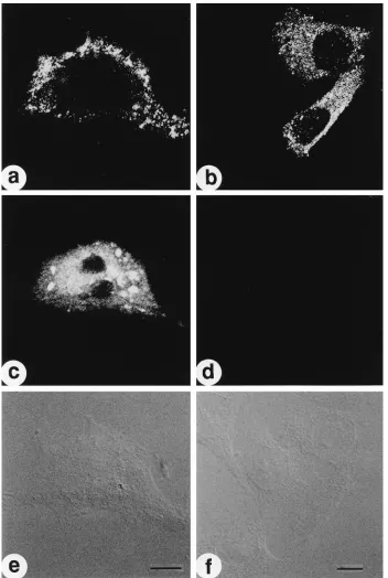
FIG. 2. In situ detection of HIV unspliced HIV mRNA and p24Gag
protein. HeLa cells transfected with the infectious molecular clone pNL4-3 were fixed, treated in the absence (a, c, and e) or presence (b, d, and f) of RNase as described in Materials and Methods, hybridized to biotinylated DNA probe ACC (see Fig. 1 for position), and immunostained with antibody to p24. The hybridization (c and d), immunofluorescence (a and b), and Nomarski (e and f) signals of a field were imaged separately by laser-scanning confocal microscopy and individually stored as black and white images as described in Materials and Methods. In cells prepared in the absence of RNase treatment, a representative example demonstrates that p24 expression (a) and gag RNA expression (b) occur in the same cell (Nomarski, c). RNA was present in the nucleus and cytoplasm but was excluded from the nucleolus. After RNase digestion, p24-expressing cells (b) no longer contained gag RNA (d); the Nomarski image identifies several nontransfected cells with no detectable p24 immunostaining. Bar510mm.
4670 BERTHOLD AND MALDARELLI J. VIROL.
on November 9, 2019 by guest
http://jvi.asm.org/
FIG. 3. Comparison of intranuclear distributions of unspliced and spliced HIV RNA. HeLa cells were transfected with pNL4-3, fixed, hybridized to biotinylated DNA probes complementary to unspliced (left column), singly spliced (middle column), and multiply spliced (right column) HIV RNA species, and immunostained with anti-SC35. Images of in situ hybridization (a to c), SC35 (d to f), and Nomarski imaging (j to l) were obtained. The in situ and immunostaining images were recombined (g to i) to determine colocalization; RNA is pseudocolored red, SC35 is pseudocolored green, and colocalization is yellow-orange. A representative cell showing hybridization with gag probe ACC (a) and SC35 staining (d) was recombined (g), revealing little colocalization between the unspliced gag RNA and SC35. Similarly, recombining the hybridization signal obtained using probe RRE complementary to env RRE sequence (b) with the corresponding SC35 immunostaining image (e) revealed little specific colocalization (h). In contrast, the distribution of multiply spliced RNA complementary to the tat probe (c) was similar to the distribution of SC35 immunostaining (f) and a high degree of colocalization (i, yellow-orange) was observed. Bar510mm.
on November 9, 2019 by guest
http://jvi.asm.org/
suggesting that these granules may not all be equivalent in
accumulating tat RNA. Colocalization of multiply spliced HIV
RNA species with SC35 in large clusters did not require
ex-pression within the context of the HIV provirus; colocalization
of tat RNA and SC35-containing speckles was also observed in
HeLa cells transiently expressing tat RNA from the cDNA
expression plasmid pCMVtat-2 or from subviral expression
constructs under the direction of the HIV promoter and was
detected using probes made from HIV- or CMV-tat expression
constructs (data not shown). Complete or partial SC35-RNA
association has been described for the majority of RNA signals
or tracks and for specific endogenous RNAs as well (reviewed
in reference 67). Consistent with such observations, we
de-tected colocalization of SC35 with the majority, but not all, of
the cellular polyadenylated RNA signal obtained using an
oli-go(dT) probe (data not shown).
These studies suggest that, in steady-state conditions,
alter-natively spliced products of a single 9.2-kb HIV RNA
tran-script accumulate in distinct nuclear locations. Multiply spliced
RNAs associate with large SC35-containing clusters, typical of
endogenous RNA, whereas unspliced and singly spliced HIV
RNA transcripts rarely colocalize with SC35-containing
nu-clear speckles. The presence of unspliced RNA nu-clearly distinct
from SC35 suggests a less common RNA distribution, similar
to that described for a minority of polyadenylated cellular RNA
species (8). We confirmed the uncommon RNA nuclear
local-ization for HIV unspliced RNA in T-lymphocytic cells infected
with HIV. FISH was used to investigate the distribution of
un-spliced HIV RNA in cultures of H9 cells chronically infected
with HIV IIIB (H9IIIB) or HIV pNL4-3 (H9NL43). FISH of
H9IIIB revealed unspliced HIV RNA present in the nucleus in
small granules (Fig. 4, H9IIIB), similar in size that detected for
unspliced HIV RNA in transient expression experiments in HeLa
cells (compare Fig. 4, H9IIIB, with Fig. 3a). In contrast,
unin-fected H9 cells contained only a background fluorescence
sig-nal (Fig. 4, H9). Since coculture of uninfected H9 cells with H9
cells chronically infected with HIV initiates rapid cell-cell
fu-sion and a well-defined cycle of HIV transmisfu-sion, replication,
and apoptosis (43), we used the cocultivation technique to
obtain large numbers of newly infected cells and syncytia at a
time during replication (21 h of coculture) when virus RNA
production is high and before apoptotic nuclear destruction is
extensive. As shown in Fig. 4, FISH of unspliced HIV RNA in
nuclei of syncytia was present in numerous small granules (Fig.
4a); costaining with SC35 (Fig. 4b) and overlay of the in situ
signal (Fig. 4c, red) and immunofluorescent signal (Fig. 4c, green)
revealed only occasional colocalization between HIV unspliced
RNA and SC35. These findings reveal that unspliced HIV RNA
expressed after infection was present in areas of the nucleoplasm
independent of SC35-containing granules, in a distribution
sim-ilar to that observed in HeLa cells transiently expressing HIV.
To determine whether the intranuclear distribution of HIV
gag RNA was dependent on expression of the rev gene product,
we compared HIV RNA expression in HeLa cells transfected
with rev
1
and rev-defective pNL4-3. As shown in Fig. 5,
un-spliced gag RNA expressed from rev
2
pNL4-3 was present in
the nuclei but not in the cytoplasm of transfected cells (Fig.
5b), whereas unspliced gag RNA expressed from rev
1
pNL4-3
was present in the nucleus and cytoplasm of transfected cells
(Fig. 5a). Unspliced intranuclear gag RNA was present in small
granules in both rev
1
and rev
2
pNL4-3 transfected cells,
although the number of granules containing HIV RNA was
often greater in the absence of Rev (compare Fig. 5a and b).
Little colocalization of gag RNA expressed from rev-defective
pNL4-3 with SC35-containing speckles was detected in the
composite (Fig. 5f) obtained by combining the in situ
hybrid-ization image (Fig. 5b) with the corresponding SC35
immuno-staining image (Fig. 5d); as expected, a similar absence of
colo-calization was observed in the composite (Fig. 5e) obtained by
combining the in situ hybridization image (Fig. 5a) with the
SC35-immunostaining image (Fig. 5c) for the rev
1
pNL4-3.
These results suggest that rev expression does not appreciably
alter the intranuclear appearance of unspliced HIV RNA and
suggests that localization may be an intrinsic property of the
RNA transcripts themselves.
In addition to a nuclear distribution, unspliced HIV RNA
was also identified in the cytoplasm of many transfected cells
(compare cytoplasmic RNA in Fig. 2c, 3a and b, and 5a), often
in large collections of 3- to 5-
m
m size exhibiting intense
fluo-rescence. Cytoplasmic staining was specific; RNA
accumula-tions were abolished after RNase treatment (Fig. 2d) and were
not present in cells transfected with the rev
2
pNL4-3 clone
(Fig. 5b). Large cytoplasmic accumulations of RNA are not
restricted to virus-encoded genes but may be characteristic of
transient expression, since we also detected large cytoplasmic
accumulations of globin RNA in cells tranfected with the
b
-globin-expressing pAL4-SV plasmid (data not shown).
Nuclear RNA localization in a
gag
reporter construct.
To
investigate potential cis determinants of the intranuclear
local-ization of HIV gag RNA, we constructed a subgenomic
gag-expressing plasmid, pCMVgag-2 (Fig. 6), which exclusively
ex-presses the HIV-1 gag gene independently of the HIV LTR.
Analysis of pCMVgag-2 RNA expression by Northern blotting
revealed two distinct RNA species, a 1,900-nt species
cor-responding to full-length transcript and a more abundant
shorter transcript of approximately 900 nt (data not shown).
The shorter transcript is likely to be the product of splicing to
cryptic splice acceptors within the 3
9
portion of the gag gene
(5a). In situ hybridization using probe ACC in HeLa cells
transfected with pCMVgag-2 revealed the presence of
un-spliced gag RNA in small granules randomly distributed
throughout the nucleoplasm exclusive of the nucleolar region
(Fig. 7a). Using a probe generated from the entire pCMVgag-2
plasmid to detect spliced and unspliced RNA, we identified a
strong hybridization signal in the cytoplasm of
pCMVgag-2-transfected cells, corresponding to the exported spliced RNA
(Fig. 7b). The fact that spliced RNA is present in the nucleus
and cytoplasm while unspliced RNA is localized exclusively to
the nucleus suggests that nuclear sequestration of unspliced
gag RNA was a specific consequence of intragenic sequences.
The intranuclear location of unspliced gag RNA was further
investigated by using FISH-SC35 immunostaining
colocaliza-tion studies. As shown in Fig. 8, unspliced RNA expressed
from pCMVgag-2 was present in small granules (Fig. 8a), a
pattern which was distinct from the distribution of
SC35-con-taining speckles (Fig. 8d); colocalization studies (Fig. 8g)
re-vealed no colocalization of unspliced gag RNA expressed from
pCMVgag-2 and nuclear speckles, similar to the distribution of
unspliced gag RNA expressed from pNL4-3 (compare Fig. 8a
with Fig. 2c, 3a, and 5a). In addition, no colocalization of
unspliced HIV RNA from pCMVgag-2 was detected by using
antisera to another nuclear protein, coilin, which is a
constit-uent of the RNA-containing coiled bodies (data not shown).
These data indicate that the intranuclear distribution of
un-spliced gag RNA appeared similar whether the RNA was
ex-pressed within the context of the HIV provirus or as a simple
expression plasmid under transcriptional control of a
heterol-ogous promoter. Because the pCMVgag-2 plasmid contains
inhibitory sequences but does not contain the RRE sequence
in cis and is not coexpressed with Rev, no unspliced gag RNA
was detected in the cytoplasm of transfected cells using a probe
complementary to the unspliced gag RNA (Fig. 8a); similarly,
4672 BERTHOLD AND MALDARELLI J. VIROL.
on November 9, 2019 by guest
http://jvi.asm.org/
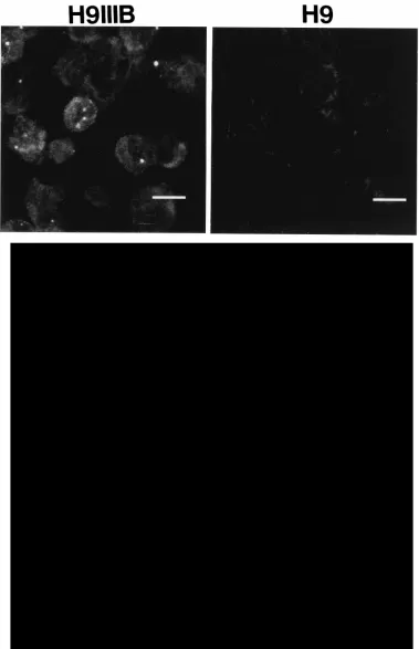
FIG. 4. Detection of HIV unspliced RNA in HIV-infected H9 cells. (Top) H9 cells chronically infected with HIV IIIB or control uninfected H9 cells were cytospun onto microscope slides, fixed, and hybridized in situ to HIV probe B (Fig. 1). Representative cells demonstrating HIV-specific signals were detected in chronically infected but not in uninfected cells. Bar510mm. (Bottom) Detection of unspliced RNA after HIV cell-cell transmission. H9 cells chronically infected with HIV pNL4-3 were mixed with uninfected H9 cells (one infected cell to four uninfected cells) to establish rapid HIV cell-cell transmission. At 21 h after cocultivation, cells were harvested and hybridized in situ (a) using probe B (Fig. 1) and immunostained for SC35 (b). The acquired images were recombined and pseudocolored (RNA is red, SC35 is green) to investigate colocalization (c); the corresponding Nomarski image (d) is included to identify cell-cell fusion. Bar510mm.
on November 9, 2019 by guest
http://jvi.asm.org/
quantitative assay of Gag protein production revealed only
trace amounts of p24 protein (10 pg per 10
6cells) present in
HeLa cells transfected with this construct.
To determine whether the presence of the RRE sequence
altered HIV RNA intranuclear localization, a derivative of
pCMVgag-2 was constructed containing the RRE sequence 3
9
to the gag coding sequence (Fig. 6, pCMVgag-2RRE), and
FISH-immunofluorescence was performed after transient
ex-pression in HeLa cells. As shown in Fig. 8b, addition of RRE
to pCMVgag-2 plasmid did not result in marked changes in the
distribution of unspliced gag RNA; as shown in the composite
image in Fig. 8h, no colocalization of the gag RNA in situ
hybridization signal with the SC35 immunostaining signal was
detected (Fig. 8b, RNA; Fig. 8e, SC35; Fig. 8h, overlay).
Ad-dition of the RRE alone did not allow gag expression in the
cytoplasm; no RNA hybridization signal was identified in the
cytoplasm of transfected cells (Fig. 8b), and only trace p24
protein production was detected by antigen capture assay (
,
10
pg of p24 per 10
6cells). However, gag expression from
pCM-Vgag-2RRE was rev responsive; cotransfection of
rev-express-ing plasmid with pCMVgag-2RRE resulted in marked
cyto-plasmic accumulation of unspliced gag RNA (Fig. 8c), and a
10-fold increase in p24 protein production was detected (100
pg of p24 per 10
6cells) compared with that measured in pCMV
gag-2-transfected cells. The distribution of unspliced gag RNA
in the nucleus expressed from pCMVgag-2RRE in the
pres-ence of Rev (Fig. 8c) was similar to that of pCMVgag-2RRE
alone (Fig. 8b); a composite image (Fig. 8i) of the in situ
hybridization signal of unspliced gag RNA expressed from
pCMVgag-2RRE plus Rev (Fig. 8c) and SC35 immunostaining
(Fig. 8f) revealed no colocalization of gag RNA with
SC35-containing speckles.
To investigate the distribution of the rev gene product, we
performed additional immunofluorescence studies to identify
Rev intranuclearly. Rev immunostaining revealed that the
ma-jority of Rev was present in or surrounding the nucleolus (Fig.
9a); Rev signal in the nucleoplasm was generally distributed in
a finely granular pattern (Fig. 9a). Costaining with anti-SC35
(Fig. 9b) and overlay of the Rev protein (red) and SC35
pro-tein (green) signal revealed that the majority of Rev propro-tein
did not accumulate with SC35 (Fig. 9c), although some
asso-ciation with SC35-containing granules was present, usually
near the periphery of granules rather than a precise
colocal-ization (Fig. 9c). In these transient expression studies, the
distribution of Rev protein in the nucleoplasm was similar to
the distribution of unspliced gag RNA. These findings suggest
the possibility that a portion of the rev gene product is present
in the same nuclear compartment as unspliced gag RNA.
Features present in the
gag
coding sequence and the RRE
direct intranuclear localization.
To determine whether
cis-acting gag repressor sequences participated in intranuclear
dis-tribution of gag RNA, a 719-nt deletion in the gag gene was
constructed which removes an intragenic cis-inhibitory
se-quence and RNA expression from the resulting plasmid,
pCMVgag-2ACC (Fig. 6), was investigated by transient
expres-sion in HeLa cells. In contrast to the RNA distribution
ex-pressed from pCMVgag-2 (Fig. 10a), unspliced RNA from the
gag deletion construct was consistently present in the nucleus
in large structures (Fig. 10b). The distribution of unspliced
RNA expressed from pCMVgag-2ACC (Fig. 10b) was similar
to the distribution of SC35-containing nuclear speckles (Fig.
10e). Combining the in situ hybridization image (Fig. 10b) and
the SC35 immunostaining image (Fig. 10e) revealed that the
large intranuclear collections of gag RNA colocalized with
SC35-containing nuclear speckles (Fig. 10h, colocalization in
yellow). Thus, these data indicate that the removal of cis
se-FIG. 5. Comparison of intranuclear distributions of unspliced HIV mRNAs in the presence and absence of rev expression. HeLa cells were transfected with
rev1(a, c, e, and g) or rev2(b, d, f, and h) pNL4-3, and cells were hybridized with ACC probe (a and b) and immunostained for SC35 (c and d). The images acquired were recombined and pseudocolored (RNA is red, SC35 is green) to investigate colocalization (e and f); the corresponding Nomarski image (g and h) is included. rev expression allowed RNA expression in the nucleus and cytoplasm (a); overlay of the SC35 immunostaining image (c) onto the in situ hybridization image (a) revealed no significant colocalization (e). In the absence of rev, un-spliced RNA (b) was confined to the nucleus (compare with the nuclear outline evident in the Nomarski image in panel h). Recombining the RNA image (b) with the corresponding SC35 immunostaining (d) revealed no significant colo-calization (f; RNA is red, SC35 is green, colocolo-calization is yellow). Bar510mm.
4674 BERTHOLD AND MALDARELLI J. VIROL.
on November 9, 2019 by guest
http://jvi.asm.org/
quences altered the intranuclear accumulation of gag RNA. To
investigate whether the presence of the RRE sequence
af-fected the intranuclear localization of the deleted gag RNA, a
plasmid, pCMVgag-2ACCRRE, was constructed containing
the intragenic gag deletion and the RRE sequence 3
9
to the
remaining gag coding sequence (Fig. 6). In marked contrast to
the findings with pCMVgag-2ACC, none of the unspliced
RNA expressed from pCMVgag-2ACCRRE was associated
with SC35-containing clumps (Fig. 10c). Combining the in situ
hybridization image of RNA expressed from pCMVgag-2AC
CRRE (Fig. 10c) with the SC35 immunostaining image (Fig.
10f) revealed only random colocalization (Fig. 10i).
Coexpres-sion of Rev with pCMVgag-2ACCRRE did not alter gag
in-tranuclear RNA localization, and the gag RNA appeared
sim-ilar to unspliced gag RNA from pCMVgag-2ACCRRE alone
(data not shown). These observations indicate that, like the
cis-acting intragenic gag sequence, the RRE directed
intranu-clear localization of unspliced gag RNA.
Redirection of intranuclear RNA localization correlates
with increased Gag expression.
The finding that specific HIV
gag RNA domains redirected intranuclear localization did not
permit us to examine the effect of nuclear localization on Gag
protein expression because the removal of the cis-acting
se-quence disrupted the gag reading frames. As an alternative, we
investigated a cellular cis-acting sequence which, when placed
upstream of the gag coding region, redirected gag RNA and
which permitted rev-independent Gag expression. Previously,
Hammarskjo
¨ld and coworkers (27) determined that the
pres-ence in cis of the second intervening sequpres-ence from rabbit
b
-globin (IVS-2), permitted rev-independent expression of
HIV env. We therefore investigated whether the presence of
the IVS-2 in cis redirected gag RNA localization and conferred
rev-independent Gag expression. We constructed recombinant
[image:9.612.60.295.73.427.2]plasmids derived from pCMVgag-2 which contained
b
-globin
IVS-2 in the sense orientation (pCMVgag-2BG) or, as a
con-trol, in the inverted, reverse complement orientation
(pCMV-gag-2BGi, Fig. 6) and then analyzed the intranuclear
distribu-tion of unspliced HIV gag RNA and the producdistribu-tion of Gag
protein after transient expression in HeLa cells. Introduction
of the
b
-globin IVS-2 into pCMVgag-2 in the sense orientation
(pCMVgag-2BG) resulted in the distribution of RNA into
large granules (Fig. 11b). Immunostaining with SC35 antibody
(Fig. 11e) and overlay of the RNA hybridization and SC35
immunofluorescence signals (Fig. 11h) revealed marked
colo-calization (Fig. 11h; gag RNA is red, SC35 is green). In
con-trast, gag RNA expressed from pCMVgag-2 (Fig. 11a) or from
the control plasmid pCMVgag-2BGi, containing the
b
-globin
IVS-2 in the inverted orientation (Fig. 11c), was present in
small granules. Costaining of 2 and
pCMVgag-2BGi with SC35 (Fig. 11d and f, respectively) and overlay of
the SC35 immunofluorescence and RNA hybridization signals
revealed no colocalization between SC35 and gag RNA
ex-pressed from pCMVgag-2 (Fig. 11g; RNA is red, SC35 is
green) or pCMVgag-2BGi (Fig. 11i; RNA is red, SC35 is
green). These findings revealed that the presence of the
b
-glo-bin IVS-2 in cis resulted in a marked intranuclear
redistribu-tion of unspliced HIV gag RNA into SC35-containing granules.
To investigate whether redirecting gag RNA into
SC35-con-taining granules was associated with an increase in Gag protein
expression, we performed FISH-immunostaining experiments
to detect the expression of gag RNA and Gag protein (Fig. 12).
Gag protein expression was undetectable in cells transfected
with pCMVgag-2 (Fig. 12a, gag RNA; Fig. 12d, Gag protein
immunostaining). In contrast, expression from pCMVgag-2BG
containing the
b
-globin IVS-2 in the sense orientation resulted
FIG. 6. Schematic of HIV gag expression plasmids. gag expression plasmids were constructed containing the cytomegalovirus immediate-early promoter di-recting the HIV gag gene and the HIV LTR as a polyadenylation signal (pCMVgag-2) as described in Materials and Methods; a derivative was cloned containing the HIV RRE cloned into a BglII site 39to the gag coding sequence (pCMVgag-2RRE). Removal of cis-inhibitory sequences by AccI deletion of 2 and 2RRE yielded 2ACC and pCMVgag-2ACCRRE, respectively. pCMVgag-2BG contains theb-globin IVS-2 intron cloned in the sense orientation 59to the major splice donor in the BssHII site; pCMVgag-2BGi contains the IVS-2 intron cloned in the inverted orientation.
FIG. 7. Identification of unspliced and spliced mRNA expressed from pCMVgag-2. HeLa cells were transfected with pCMVgag-2, and mRNA was detected in situ with biotinylated DNA probe ACC (a) to detect unspliced gag RNA or with biotinylated pCMVgag-2 plasmid probe (b) to detect spliced and unspliced RNA. Bar510mm.
on November 9, 2019 by guest
http://jvi.asm.org/
[image:9.612.318.552.568.684.2]4676
on November 9, 2019 by guest
http://jvi.asm.org/
in Gag protein expression (Fig. 12b, unspliced gag RNA; Fig.
12e, Gag protein immunostaining). Quantitative p24 antigen
determinations revealed a sevenfold increase in p24
produc-tion in pCMVgag-2BG-transfected cells (70 pg of p24 per 10
6cells) compared with that expressed in pCMVgag-2 (10 pg of
p24 per 10
6cells). Gag protein expression was undetectable
from cells expressing the control plasmid pCMVgag-2BGi
(Fig. 12c, unspliced gag RNA; Fig. 12f, Gag protein
immuno-staining). Quantitative p24 determinations confirmed that only
trace amounts (10 pg of p24 per 10
6cells) of Gag protein were
expressed in pCMVgag-2BGi-transfected cells. The finding
that the presence of the
b
-globin IVS-2 permitted
rev-indepen-dent Gag protein expression and redistributed gag RNA
sim-ilar to that obtained for rev-independent tat transcripts
(com-pare Fig. 11b and 12b with Fig. 3c) suggests that redirecting
nuclear RNA localization may affect gene expression.
DISCUSSION
HIV-1 expression proceeds by a regulated program of
alter-native splicing of full-length 9.2-kb primary transcripts into
three different size classes of mRNA. Unspliced 9.2-kb mRNA
and singly spliced 4.0-kb mRNA species require the binding of
the HIV gene product Rev to the RRE for cytoplasmic
expres-sion, whereas multiply spliced 1.8-kb transcripts are expressed
in the presence or absence of rev. The in situ hybridization
findings presented here reveal that dependent and
rev-independent RNA transcripts accumulate in distinct
intranu-clear sites, demonstrating that the alternatively spliced
prod-ucts of HIV are in turn alternatively localized within the
nucleus. cis sequences were responsible for intranuclear
local-ization, and the portion of the gag gene we investigated
con-tains inhibitory regions which are capable of repressing their
own expression and which are rescued by the RRE in cis and
by the rev gene product in trans (12, 42, 65). Inhibitory
se-quences have been suggested to function in regulating HIV
RNA processing by several potential mechanisms, including
reducing the half-life of RNA, retaining RNA in the nucleus,
and preventing translation of RNA in the cytoplasm (12, 42,
64, 65). In the experiments reported here, expression of gag
RNA from rev-defective pNL4-3 or from the pCMVgag-2
plas-mid yielded nuclear RNA containing inhibitory sequences
present exclusively in the nucleus, consistent with previous
biochemical fractionation studies of transient expression of
recombinant chloramphenicol acetyltransferase reporter
plas-mids containing gag/pol inhibitory sequences (42). Our finding
that the inhibitory regions may direct gag RNA localization
suggests that cis repression may be accomplished through
nu-clear compartmentalization. Sequence-dependent RNA
local-ization has been proposed (38) as a mechanism by which
cel-lular gene expression may be regulated in the nucleus and
compartmentalized in the cytoplasm (34, 41, 60). Our findings
suggest that, in addition to defining intron/exon boundaries
and polyadenyation and methylation sites, HIV pre-mRNA
transcripts contain signals which determine its nuclear
accu-mulation site and may affect its processing and expression.
[image:11.612.60.554.533.697.2]The rev gene product is an RNA binding protein capable of
shuttling between the nucleus and the cytoplasm (33, 49, 59);
the reported models for the mechanism of Rev function
in-clude: (i) improving the efficiency of nucleocytoplasmic
trans-port (19, 25, 26, 44); (ii) influencing splice site utilization (39);
(iii) facilitating dissociation from splicing complexes (9, 35,
36); (iv) stabilizing unspliced RNA (20, 66); and (v) activating
HIV mRNA translation in certain cell types (5, 14, 59). Rev
protein has been detected predominantly in the nucleolus
(13, 57) and in the nucleoplasm (33, 40), associated, in part,
with SC35-containing granules. In our experiments, Rev was
present predominantly in the nucleoli and in small
SC35-inde-pendent granules in the nucleoplasm (Fig. 9). Thus, a portion
FIG. 9. Independent distributions of Rev protein and SC35. HeLa cells transfected with pCMVgag-2RRE and pCMVRev were immunostained for Rev (a) and SC35 (b); overlay of the corresponding images (c; Rev is red, SC35 is green, colocalization is yellow) revealed distinct distributions, although some Rev protein associated with the periphery of SC35-containing granules. Bar510mm.
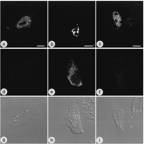
FIG. 8. Identification of HIV gag RNA from recombinant expression plasmids. HeLa cells transfected with pCMVgag-2 (left column), pCMVgag-2RRE (middle column), or pCMVgag-2RRE and pCMV-Rev (right column) were fixed and hybridized to gag probe ACC (a to c) and immunostained with anti-SC35 (d to f). The presence of colocalization was determined by overlaying the RNA and SC35 images (g to h; RNA is red, SC35 is green, colocalization is yellow). The corresponding Nomarski image (j to l) was acquired for each field. Unspliced RNA from pCMVgag-2 (a) contained exclusively nuclear RNA. Combining the in situ hybridization image (a) and the corresponding SC35 image (d) revealed little colocalization (g). The addition of the RRE did not change the pattern of nuclear RNA (b); overlay of the SC35 image (e) yielded no significant colocalization (h). Cotransfection of pCMVgag-2RRE with rev-expressing pCMV-Rev resulted in expression of unspliced RNA in the nucleus and cytoplasm (c). Combining the SC35 immunostaining image (f) with the RNA hybridization image (c) revealed little colocalization (i; RNA is red, SC35 is green). Bar510mm.
on November 9, 2019 by guest
http://jvi.asm.org/
FIG. 10. cis-acting sequences direct RNA localization. HeLa cells transfected with 2 (left column), 2ACC (middle column), or pCMVgag-2ACCRRE (right column) were hybridized to probe D, and confocal images of RNA hybridization (a to c) and SC35 immunostaining (d to f) were acquired and recombined (g to h) to determine colocalization. Nomarski images (j to l) are included for comparison. Cells transfected with pCMVgag-2 expressed RNA exclusively in the nucleus (a); overlay of SC35-immunostaining image (d) revealed no colocalization (g; RNA is red, SC35 is green). Cells transfected with pCMVgag-2ACC, containing a deletion of cis-inhibitory sequences, contained RNA in large accumulations (b); combining the RNA image with the corresponding SC35 immunostaining image (e) revealed marked colocalization (h; RNA is red, SC35 is green, colocalization is yellow). Expression of unspliced RNA from pCMVgag-2ACCRRE, containing the deletion in the gag inhibitory sequence and the addition of the RRE (Fig. 6), resulted in RNA present in fine granules (c); overlay with the corresponding SC35 image (f) revealed that these transcripts no longer colocalized with SC35 (i; RNA is red, SC35 is green). Bar510mm.
4678 BERTHOLD AND MALDARELLI J. VIROL.
on November 9, 2019 by guest
http://jvi.asm.org/
of Rev is present in the same compartment in which we
de-tected unspliced and singly spliced, rev-dependent RNA
tran-scripts. It is possible that cis-acting sequences direct RNA into
the nucleoplasm where Rev is available to bind the RRE and
shuttle the RNA to the cytoplasm.
In our experiments, both gag inhibitory sequences and the
RRE contained RNA localization activity (Fig. 10a, b, and c);
since the RRE sequence itself may contain an inhibitory
[image:13.612.60.555.72.557.2]se-quence (6), it is possible that we detected the effect of an
RRE-associated inhibitory sequence on the localization of
pCMVgag-2ACCRRE RNA (Fig. 10c). We have not yet
in-vestigated the nuclear localization activity of other
cis-repres-sor sequences in env to determine whether they all contain a
similar localization activity. Interestingly, Lawrence and
co-workers (37) identified unspliced HIV RNA, containing tat
exons and a portion of the env sequence, including the RRE
FIG. 11. Redirection of gag RNA byb-globin intron in cis. HeLa cells were transfected with pCMVgag-2 (a, d, and g), pCMVgag-2BG (b, e, and h) containing the
b-globin intron upstream of the major splice donor in the sense orientation, or pCMVgag-2BGi containing IVS-2 in the inverse complement orientation (Fig. 6). In situ hybridization using probe ACC (a, b, and c) to detect unspliced RNA revealed that the presence of theb-globin intron redirected HIV unspliced RNA into large granules (b) compared with the distribution of unspliced RNA in small granules expressed from pCMVgag-2 (a) or pCMVgag-2BGi (c). Immunostaining for SC35 (d, e, and f) and combining the corresponding in situ and immunofluorescence images (g, h, and i; RNA is red, SC35 is green) revealed that gag RNA expressed from pCMVgag-2BG associated with SC35-containing granules (h), whereas no colocalization was detected between SC35 and unspliced RNA expressed from pCMVgag-2 (g) or pCMVgag-2BGi (i). Bar510mm.
on November 9, 2019 by guest
http://jvi.asm.org/
and repressor sequences, in both a stippled pattern and a large
patchy distribution in nuclei of COS-7 cells, and these monkey
cells contained unspliced HIV RNA in the cytoplasm in the
absence of rev. It is likely that the differences in
nuclear/cyto-plasmic distribution of RNA in COS and HeLa cells detected
by in situ and biochemical techniques represent cell-type
and/or species-specific differences, but it will be of interest to
determine whether transport in COS cells may take place by
both a rev-dependent and a rev-independent pathway.
Ultrastructural and biochemical studies of gene expression
suggest a dynamic relationship between the physical
organiza-tion of the nucleus and the funcorganiza-tional events of pre-mRNA
processing. Recent visualization of RNA splicing occurring
along transcript “tracks” extending from transcription sites to
the nuclear periphery suggests that RNA processing is directly
coupled to RNA synthesis (8, 30, 72–74). Although tracks may
occur close to, or coincident with, SC35-containing nuclear
speckles, many tracks are clearly distinct from any large
gran-FIG. 12. Rev-independent expression of gag RNA containingb-globin IVS-2 in cis. HeLa cells were transfected with pCMVgag-2 (a, d, and g), pCMVgag-2BG (b, e, and h), or pCMVgag-2BGi (c, f, and i); unspliced RNA was detected by in situ hybridization using probe E (a, b, and c), and p24 was detected using fluorescein isothiocyanate-conjugated anti-p24 monoclonal antibody (d, e, and f). Cells transfected with pCMVgag-2 (a) or pCMVgag-2BGi (c) contained unspliced gag RNA present in small granules (a), but no Gag protein was detected by p24 immunostaining (pCMVgag-2 and pCMVgag-2BGi, panels d and f, respectively). In contrast, cells transfected with pCMVgag-2BG contained unspliced gag RNA in large granules (b) and p24 protein expressed in the cytoplasm (e). Corresponding Nomarski images are presented (g, h, and i). Bar510mm.
4680 BERTHOLD AND MALDARELLI J. VIROL.
on November 9, 2019 by guest
http://jvi.asm.org/
[image:14.612.62.556.69.559.2]ules and the role of nuclear speckles is uncertain. Since splicing
factors have been reported to accumulate within speckles (7,
67, 68), several investigators have suggested that nuclear
speckles may represent repositories from which splicing factors
are recruited (30, 31, 75). Others have detected marked
reor-ganization of nuclear speckles upon cell differentiation and
dissolution of speckles upon transcriptional arrest, suggesting a
more dynamic function for SC35-containing granules (3, 50,
54, 73). Huang and colleagues (30), noting the substantial
portion of nuclear RNA associated with SC35-containing
structures, have suggested that speckle-associated RNA may
play a role in RNA processing itself. It is also possible that
SC35-containing granules have functions in addition to storing
splicing factors; for example, our study revealed that tat RNA
was concentrated in some SC35-containing granules, whereas
other SC35-containing structures in the same nucleus had little
or no tat RNA. Recently, Luznik and coworkers (40) described
changes in the appearance of SC35-containing granules after
HIV infection. It will be of interest to determine whether such
changes occur in all SC35-containing granules or in those
con-taining tat RNA. In addition, we are investigating whether
association of tat RNA with nuclear speckles requires specific
sequences within multiply spliced HIV RNAs or occurs
be-cause of the absence of inhibitory-type signals.
The functional consequences of redirecting RNA to distinct
intranuclear locations remain uncertain. Our experiments
identified that a cellular sequence, IVS-2 from rabbit
b
-globin,
may also direct RNA, indicating that RNA localization
se-quences are not restricted to HIV and that redirecting
intranu-clear localization is associated with differences in gene
expres-sion. In our studies, the presence of the
b
-globin intron
re-sulted in rev-independent expression, similar to the previously
reported effect of the
b
-globin intron on env expression. The
intranuclear distribution of rev-independent gag RNA
ex-pressed from pCMVgag-2BG was similar to the distribution of
rev-independent tat RNA. We speculate that the model
pro-posed by Hammarskjo
¨ld and coworkers (27) to explain
rev-independent env expression may apply to rev-rev-independent gag
expression as well and that it includes redirecting RNA into a
distinct pathway, reflected in our experiments as a marked change
in intranuclear accumulation into SC35-containing granules.
The mechanism of how intragenic sequences affect nuclear
RNA distribution presumably involves interactions with
nu-clear factors. Several novel nunu-clear proteins binding the RRE
have been identified (56, 71) whose precise functions in HIV
RNA processing remain to be elucidated. A specific
interac-tion between a cis-repressor sequence in pol and an abundant
nuclear factor, heterogeneous nuclear ribonucleoprotein C
(hnRNP-C), has been reported (55). hnRNP-C binds
prefer-entially to U-rich sequences along RNA transcripts and may
influence 3
9
splice site selection (10, 18, 23). HIV gag/pol
se-quences contain AU-rich regions, which may account for
hnRNP-C binding; interestingly, Schwartz and coworkers (64)
demonstrated that interrupting multiple U-rich sequences
re-sults in loss of the inhibitory effect of gag sequences and
ren-ders them rev independent. It is conceivable that binding of
nuclear factors such as hnRNP-C to intragenic sequences may
be required for cis repression, but little is known about the
interactions of nuclear factors and RNA processing in the
context of the intact nucleus. By combining in situ localization
and RNA binding studies to characterize functional and spatial
relationships between inhibitory sequences and nuclear
fac-tors, we hope to identify the events in RNA processing critical
to HIV replication, which may serve as potential sites for
therapeutic intervention.
ACKNOWLEDGMENTS
We thank G. Pavlakis, E. Chan, and T. Maniatis for gifts of antisera, Alicia Buckler-White for sequencing and oligonucleotide synthesis, and D. F. J. Purcell for helpful discussions. We are indebted to Klaus Strebel and Malcolm A. Martin for advice and support of the project and for critical reviews of the manuscript.
REFERENCES
1. Adachi, A., H. E. Gendelman, S. Koenig, T. Folks, R. Willey, A. Rabson, and
M. A. Martin.1986. Production of acquired immunodeficiency syndrome-associated retrovirus in human and nonhuman cells transfected with an infectious molecular clone. J. Virol. 59:284–291.
2. Ahmad, N., and S. Venkatesan. 1988. nef protein of HIV-1 is a transcrip-tional repressor of HIV-1 LTR. Science 241:1481–1485.
3. Antoniou M., M. Carmo-Fonseca, J. Ferreira, and A. I. Lamond. 1994. Nuclear organization of splicing snRNPs during differentiation of murine erythroleukemia cells in vitro. J. Cell. Biol. 123:1055–1068.
4. Arrigo, S., S. Weitsman, J. A. Zack, and I. S. Y. Chen. 1990. Characterization and expression of novel singly spliced RNA species of human immunodefi-ciency virus type 1. J. Virol. 64:4585–4588.
5. Arrigo, S. J., and I. S. Y. Chen. 1991. Rev is necessary for translation but not cytoplasmic accumulation of HIV-1 vif, vpr, and env/vpu 2 RNAs. Genes Dev. 5:808–819.
5a.Berthold, E., and F. Maldarelli. Unpublished observations.
6. Brighty, D. W., and M. Rosenberg. 1994. A cis-acting sequence that overlaps the rev-responsive element of human immunodeficiency virus type 1 regu-lates nuclear retention of env mRNAs independently of known splice signals. Proc. Natl. Acad. Sci. USA 91:8314–8318.
7. Carmo-Fonseca, M. R., B. S. Pepperkok, W. Sproat, M. Ansorge, S.
Swan-son, and A. I. Lamond.1991. In vivo detection of snRNP-rich organelles in the nuclei of mammalian cells. EMBO J. 10:1863–1873.
8. Carter, K. C., D. Bowman, W. Carrington, K. Fogarty, J. A. McNeil, F. S.
Fay, and J. B. Lawrence.1993. A three-dimensional view of precursor messen-ger RNA metabolism within the mammalian nucleus. Science 259:1330–1335. 9. Chang, D. D., and P. A. Sharp. 1989. Regulation by HIV Rev depends upon
recognition of splice sites. Cell 59:789–795.
10. Chou, Z. F., F. Chen, and J. Wilusz. 1994. Sequence and position require-ments for uridylate-rich downstream elerequire-ments of polyadenylation signals. Nucleic Acids Res. 22:2525–2531.
11. Cochrane, A. W., C. A. Chen, and C. A. Rosen. 1990. Specific interaction of the human immunodeficiency virus rev protein with a structured region in the env mRNA. Proc. Natl. Acad. Sci. USA 87:1198–1202.
12. Cochrane, A. W., K. J. Jones, S. Beidas, P. J. Dillon, A. M. Skalka, and C. A.
Rosen. 1991. Identification and characterization of intragenic sequences which repress human immunodeficiency virus structural gene expression. J. Virol. 65:5305–5313.
13. Cullen, B. R., J. Hauber, K. Campbell, J. G. Sodroski, W. A. Haseltine, and
C. A. Rosen.1988. Subcellular localization of the human immunodeficiency virus trans-acting art gene product. J. Virol. 62:2498–2501.
14. D’Agostino, D. M., B. K. Felber, J. E. Harrison, and G. N. Pavlakis. 1992. The Rev protein of human immunodeficiency virus type 1 promotes polyso-mal association and translation of gag/pol and vpu/env mRNAs. Mol. Cell. Biol. 12:1375–1386.
15. Dayton, A. I., E. F. Terwilliger, J. Potz, M. Kowalski, J. G. Sodroski, and
W. A. Haseltine. 1988. Cis-acting sequences responsive to the rev gene product of the human immunodeficiency virus. J. Acquired Immune Defic. Syndr. 1:441–452.
16. Dayton, E. T., D. A. Konings, D. M. Powell, B. A. Shapiro, L. Butini, J. V.
Maizel, and A. I. Dayton.1992. Extensive sequence-specific information throughout the CAR/RRE, the target sequence of the human immunodefi-ciency virus type 1 Rev protein. J. Virol. 66:1139–1151.
17. Dayton, E. T., D. M. Powell, and A. I. Dayton. 1989. Functional analysis of CAR, the target sequence for the Rev protein of HIV-1. Science 246: 1625–1629.
18. Dreyfuss, G., M. J. Matunis, S. Pinol-Roma, and C. H. Burd. 1993. hnRNP proteins and the biogenesis of mRNA. Annu. Rev. Biochem. 62:289–321. 19. Emerman, M., R. Vazeux, and K. Peden. 1989. The rev gene product of the
human immunodeficiency virus affects envelope-specific RNA localization. Cell 57:1155–1165.
20. Felber, B. K., M. Hadzopoulou-Cladaras, C. Cladaras, T. Copeland, and
G. N. Pavlakis.1989. Rev protein of human immunodeficiency virus 1 affects the stability and transport of the viral mRNA. Proc. Natl. Acad. Sci. USA 86: 1495–1499.
21. Fu, X.-D., and T. Maniatis. 1990. Factor required for mammalian spliceo-some regulation is localized to discrete regions in the nucleus. Nature (Lon-don) 343:437–441.
22. Gatignol, A., A. Buckler-White, B. Berkhout, and K.-T. Jeang. 1991. Char-acterization of a human TAR RNA binding protein that activates the HIV-1 LTR. Science 251:1597–1600.
23. Gorlach, M., C. G. Burd, and G. Dreyfuss. 1994. The determinants of RNA-binding specificity of the heterogeneous nuclear ribonucleoprotein C