0022-538X/96/$04.00
1
0
Copyright
q
1996, American Society for Microbiology
Prolonged Gene Expression and Cell Survival after Infection by
a Herpes Simplex Virus Mutant Defective in the
Immediate-Early Genes Encoding ICP4, ICP27, and ICP22
NAXIN WU,
1SIMON C. WATKINS,
2PRISCILLA A. SCHAFFER,
3ANDNEAL A. D
ELUCA
1*
Department of Molecular Genetics and Biochemistry
1and Department of Cell Biology and Physiology,
2University of Pittsburgh
School of Medicine, Pittsburgh, Pennsylvania 15261, and Division of Molecular Genetics, Dana-Farber Cancer Institute, and
Department of Microbiology and Molecular Genetics, Harvard Medical School, Boston, Massachusetts 02115
3Received 19 March 1996/Accepted 21 May 1996
Very early in infection, herpes simplex virus (HSV) expresses four immediate-early (IE) regulatory proteins,
ICP4, ICP0, ICP22, and ICP27. The systematic inactivation of sets of the IE proteins in
cis
, and the subsequent
phenotypic analysis of the resulting mutants, should provide insights into how these proteins function in the
HSV life cycle and also into the specific macromolecular events that are altered or perturbed in cells infected
with virus strains blocked very early in infection. This approach may also provide a rational basis to assess the
efficacy and safety of HSV mutants for use in gene transfer experiments. In this study, we generated and
examined the phenotype of an HSV mutant simultaneously mutated in the ICP4, ICP27, and ICP22 genes of
HSV. Unlike mutants deficient in ICP4 (
d
120), ICP4 and ICP27 (
d
92), and ICP4 and ICP22 (
d
96), mutants
defective in ICP4, ICP27, and ICP22 (
d
95) were visually much less toxic to Vero and human embryonic lung
cells. Cells infected with
d
95 at a multiplicity of infection of 10 PFU per cell retained a relatively normal
morphology and expressed genes from the viral and cellular genomes for at least 3 days postinfection. The
other mutant backgrounds were too toxic to allow examination of gene expression past 1 day postinfection.
However, when cell survival was measured by the capacity of the infected cells to form colonies,
d
95 inhibited
colony formation similarly to
d
92. This apparent paradox was reconciled by the observation that host cell DNA
synthesis was inhibited in cells infected with
d
120,
d
92,
d
96, and
d
95. In addition, all of the mutants exhibited
pronounced and distinctive alterations in nuclear morphology, as determined by electron microscopy. The
appearance of
d
95-infected cells deviated from that of uninfected cells in that large circular structures formed
in the nucleus.
d
95-infected cells abundantly expressed ICP0, which accumulated in fine punctate structures
in the nucleus at early times postinfection and coalesced or grew to the large circular objects that were revealed
by electron microscopy. Therefore, while the abundant accumulation of ICP0 in the absence of ICP4, ICP22,
and ICP27 may allow for prolonged gene expression, cell survival is impaired, in part, as a result of the
inhibition of cellular DNA synthesis.
The more than 75 genes of herpes simplex virus type 1
(HSV-1) (40, 41) are expressed in a regulated and sequential
manner such that three broad categories of genes,
immediate-early (IE), immediate-early (E), and late (L), can be defined (26, 27). The
five genes classically designated IE genes are expressed shortly
after the genome arrives in the nucleus and in the absence of
prior de novo viral protein synthesis (26, 27). Transcription of
the IE genes is activated by the virion protein VP16 (3, 8),
which functions as a complex on IE promoters with cellular
Oct1 and other host cell proteins (19–21, 33, 46, 71). The IE
genes encode the proteins infected cell polypeptide 4 (ICP4),
ICP27, ICP0, ICP22, and ICP47 (10, 26, 50, 74).
ICP4, ICP0, ICP27, and ICP22 are nuclear phosphoproteins
(50, 77) and possess regulatory activities which are thought to
prime the cell for, and participate in, the efficient cascade of
subsequent viral gene expression, DNA replication, and the
production of progeny virions. ICP4 is a large multifunctional
protein. It can act as a transcription factor that either represses
(12, 22, 45, 48, 56) or activates (12, 16, 18, 47, 53) transcription
through contacts with the general transcriptional machinery
(22, 67). ICP4 is largely responsible for the transition from the
IE to E phase of viral gene expression (14, 52, 73). ICP0 will
activate most test promoters in transient assays (16, 18, 47, 53)
and has been found to elevate levels of viral gene expression
and growth in tissue culture and in the trigeminal ganglia of
mice (6, 7, 37). It also facilitates the reactivation of virus from
latency in the mouse model (6, 37). ICP27 also appears to be
multifunctional. Several studies have shown that it can
modu-late the activity of ICP4 and ICP0 (44, 54, 66), as well as the
modification state of ICP4 (44, 54, 70). ICP27 has also been
shown to regulate viral and cellular mRNA processing events
(9, 23, 24, 42, 43, 61–63, 68). The combined activities of ICP27
contribute to efficient DNA replication and late gene
expres-sion (39, 57); however, recent studies have shown that ICP27
also significantly contributes to elevated levels of early gene
expression (59, 72). The contribution of ICP27 to the elevated
levels of some early proteins has provided an explanation for
the requirement for ICP27 for viral DNA replication (72).
ICP22 is not essential for growth in many cell types, including
Vero cells, and acts to promote efficient late gene expression in
a cell-type-dependent manner (65). It has also been shown to
be involved in the production of a novel modified form of RNA
polymerase II (55). How all four of these proteins function
together to orchestrate the regulatory cascade seen in
HSV-infected cells remains to be determined. Additionally, the
ef-fects of these proteins on host cell metabolism are unknown.
In the absence of ICP4, only the remaining IE proteins,
* Corresponding author. Mailing address: E1257 Biomedical
Sci-ence Tower, Department of Molecular Genetics and Biochemistry,
University of Pittsburgh School of Medicine, Pittsburgh, PA 15261.
Phone: (412) 648-9947. Fax: (412) 624-1401.
6358
on November 9, 2019 by guest
http://jvi.asm.org/
ICP6, and the L/STs (OrfP) are efficiently expressed (4, 11, 34,
35, 79). Infection of cells with ICP4 mutants promotes the
rapid destruction of most cells in culture (29), whereas
UV-irradiated virus (29) and mutant viruses that are also deficient
in the activation function of VP16 (30) are significantly less
toxic. ICP4 mutants also cause chromosomal aberrations and
rapid cell death (29). Johnson and colleagues have shown that
either ICP4, ICP0, ICP27, or ICP22 can significantly reduce
the transformation efficiency of cultured cells to G418
resis-tance (30). Therefore, a tenable hypothesis is that the activities
of IE proteins perturb host cell metabolism, reducing cell
via-bility.
We have been constructing viruses that have deletions of
specific IE genes and sets of IE genes in an effort to uncover
possible interactions between IE proteins and to understand
how HSV initially alters host cell metabolism. In addition,
current virus-based strategies for using HSV as a gene transfer
vehicle have not met with success. For this purpose, it is
de-sirable to gain an understanding of how HSV affects host cell
metabolism in the absence of ICP4, as well as construct
mu-tants in the genes that contribute to the observed deleterious
effects. We have previously reported viruses that contain
mu-tations in the essential IE genes, ICP4 and ICP27, and the
complementing cell lines used for their isolation and
propaga-tion (59). This report describes the consequences of the
addi-tional inactivation of the ICP22 gene. In contrast to viruses
deficient in ICP4 alone or ICP4 and ICP27, viruses deficient in
ICP4, ICP27, and ICP22 minimally affect cell structure. Viral
and cellular gene expression continues for at least 3 days.
However, cellular DNA replication and cell division are
inhib-ited. These findings have implications for how ICP0 might
affect host cell metabolism and also indicate the need to
elim-inate ICP0 expression if HSV is to be effectively used as a
replication-defective gene transfer vehicle.
MATERIALS AND METHODS
Cells and viruses.Vero cells and human embryonic lung (HEL) cells were propagated in Dulbecco’s modified Eagle’s medium (DMEM) containing 10% fetal bovine serum as described previously (76). The Vero-derived cell lines which provide HSV-1 IE functions in trans, E26 (ICP4 and ICP27), E5 (ICP4), and E8 (ICP27), were previously reported (11, 13, 59). All viruses are derived from wild-type HSV-1 strain KOS. The ICP27 and ICP22 double mutant, DMP, contains the 5dl1.2 (39) and n199 (55) mutations, respectively. The ICP4 and ICP27 double mutant d92 was previously described (59). d120 (ICP42) and 5dl1.2 (ICP272) were previously described (11, 39). Mutant viruses were prop-agated and titers were determined on the appropriate cell lines complementing the defective essential viral function for productive replication. Stocks of mutant virus were tested for the quantity of viral DNA reaching the nucleus following infection and also for the expression of ICP0 by immunofluorescence as a function of multiplicity of infection (MOI) to ascertain differences in the ratio of infectious virus to PFU. The results of these tests did not indicate a significant difference in this ratio with the different viruses.
DNA preparation and Southern blot analysis.Small-scale viral DNA prepa-rations were obtained from 2 3105 productively infected cells. Cells were harvested when cytopathic effects were generalized. After a cycle of freezing and thawing, the suspension was sonicated and pelleted in a microcentrifuge for 30 min at 48C. The pellet was washed with Tris-buffered saline and lysed in digestion buffer containing 0.8% sodium dodecyl sulfate (SDS) and 400mg of proteinase K per ml for 4 h at 378C. After phenol-chloroform extraction, the DNA was precipitated by ethanol and resuspended in TE (10 mM Tris-HCl [pH 8.0], 1 mM EDTA).
For Southern blot analysis, viral DNA samples were digested with an appro-priate restriction enzyme. Digested DNA samples were subjected to electro-phoresis on a 0.7% agarose gel, stained with ethidium bromide, photographed, and transferred to a nitrocellulose membrane. Prehybridization, hybridization, and washing were performed according to standard protocols (60, 69). The membrane was exposed to Amersham Hyperfilm MP.
RNA preparation and Northern (RNA) blot analysis.RNA samples were prepared by infecting 1.53106cells in 60-mm-diameter culture dishes with viruses at an MOI of 10 PFU per cell. At the appropriate time postinfection, total RNA was isolated by using the Biotecx Ultraspec RNA isolation system (3a) as recommended by the manufacturer. The final RNA pellet was resuspended in
diethylpyrocarbonate-treated water, and its concentration was determined by measuring the optical density at 260 nm.
For Northern blot analysis, 5mg of RNA was denatured in denaturing buffer (65% formamide, 8% formaldehyde, 1 mM EDTA, 20 mM morpholinepropane-sulfonic acid [MOPS], 8 mM sodium acetate) with ethidium bromide (0.5mg/ml) at 688C for 15 min. After being mixed with RNA loading buffer, the samples were subjected to electrophoresis on 1.3% formamide denaturing agarose gels at 35 V overnight with constant buffer circulation (28). Following electrophoresis, the gel was rinsed in water, and equal amounts of RNA samples were checked and recorded by photography on a UV transilluminator. The RNA was transferred to a nitrocellulose membrane in 203SSC (3 M sodium chloride, 0.3 M trisodium citrate), air dried, and baked at 808C for 2 h. Prehybridization and hybridization were performed as described previously (60). To detect HSV-1 ICP0, tk (thymi-dine kinase gene), and cellularb-tubulin mRNAs,32
P-labeled nick-translated plasmid fragments from pW3dHS8 (58) digested with SacI and PstI, pTKSS (59) digested with EcoRI and BamHI, and pRT3beta (5) digested with PstI were used, respectively. Quantitation of Northern blots was performed with the AMBIS 4000 radioanalysis imaging detector system.
Electrophoresis of proteins.Viral and cellular protein expression was analyzed by SDS-polyacrylamide gel electrophoresis (PAGE) of virus-infected cells la-beled with32P
ior [35S]methionine. Vero or HEL cells (53105) were seeded on 35-mm-diameter culture dishes. Viral infection was carried out in 0.1 ml of DMEM at an MOI of 10 PFU per cell at 378C and 5% CO2with occasional rocking for 1 h. For [35S]methionine labeling, 100mCi of [35S]methionine was added to methionine-deficient DMEM for 1 h at the indicated time. For32
Pi labeling, 100mCi of32P
iwas added to phosphate-deficient DMEM at 2 to 6 h postinfection. After labeling, the cell monolayer was washed twice with Tris-buffered saline containing 500 mM N-p-tosyl-L-lysine chloromethyl ketone (TLCK). The infected cells were lysed in protein sample buffer (2% SDS, 50 mM Tris-HCl [pH 7.0], 5%b-mercaptoethanol, 0.005% bromophenol blue, 5% su-crose) and subjected to electrophoresis on an SDS–9% polyacrylamide gel. The gel was dried under vacuum and exposed to Amersham Hyperfilm.
DNA synthesis assay.To analyze DNA synthesis in cells, 1.53106
cells seeded on 60-mm-diameter culture dishes were infected with virus at an MOI of 10 PFU per cell and then incubated for 1 h at 378C and 5% CO2in 0.2 ml of medium. Following adsorption, the monolayers were washed twice and fresh medium was added. At the indicated time points, 100mCi of [3H]thymidine (New England Nuclear) was added for 3 h. After labeling, total DNA was isolated as described above except that the samples were also treated with RNase. Purified DNA was dissolved in TE, and its concentration was determined by measuring the optical density at 260 nm. The amount of3
H incorporated was determined by liquid scintillation spectroscopy.
Colony-forming inhibition assays.Monolayers of 1.53106
Vero cells were infected as described above with the indicated virus at the indicated MOI. An uninfected monolayer was maintained as a control. At 6 h postinfection, the monolayers were washed with Tris-buffered saline and trypsinized to generate single-cell suspensions. The suspensions were serially diluted and plated on 60-mm-diameter petri dishes in quadruplicate. For the efficient development of colonies, the fetal bovine serum concentration was raised to 20% and the me-dium was changed every 4 days. Approximately 2 weeks postplating, the colonies were fixed, stained with crystal violet, and counted. The fraction of surviving cells was represented relative to that obtained with uninfected monolayers.
Electron microscopy.Confluent monolayers of infected and uninfected cells were fixed in 2.5% glutaraldehyde in phosphate-buffered saline (PBS) for 1 h. Following fixation, the monolayers were postfixed with 1% osmium tetroxide containing 0.1% potassium ferricyanide, dehydrated through graded alcohol, and embedded with epoxy resin. Sections (50 to 60 nm) were cut with a Reichert Ultracut E ultramicrotome, mounted on 200-mesh grids, double stained with 2% uranyl acetate (7 min) and 1% lead citrate (3 min), and examined in a JEOL 100 CX electron microscope.
Immunofluorescence.Infected and uninfected cells were prepared on circular coverslips. Following incubation of the cultures, the culture medium was re-moved, and the cells were washed three times (5 min each) in PBS, fixed and permeabilized in2208C methanol for 15 min, air dried, and then rehydrated in PBS. This procedure was followed by three washes (5 min each) in PBS con-taining 0.5% bovine serum albumin (BSA) and 0.15% glycine (BSA buffer). Nonspecific activity was blocked with 5% normal goat serum in BSA buffer. Subsequently, the sections were incubated for 2 h with a mouse monoclonal antibody against ICP0 (Goodwin Institute, Fla.) at a dilution of 1:100. Following incubation, the sections were washed three times in BSA buffer, and the primary antibody was revealed with a specific goat anti-mouse–Cy3.18 immunoconjugate (Jackson Laboratories). To image the DNA, cells were then washed for 2 min with Hoescht 33258 (2mg/ml) and mounted in Gelvatol (Monsanto). Image fields were collected directly at the microscope, using a 603, high-numerical aperture, color-corrected oil immersion objective; a high-sensitivity, integrating three-chip Sony color camera (700 by 600 pixels); and a Corecco frame grabber board in conjunction with a Nikon FXA photomicroscope. For fluorescent images, ap-propriate cubes, in perfect registration, were used to collect the ICP0 and DNA signals. A further differential interference contrast image was collected to assess cellular morphology at high resolution.
V
OL. 70, 1996
HSV-1 MUTANTS DEFECTIVE IN ICP4, ICP27, AND ICP22
6359
on November 9, 2019 by guest
http://jvi.asm.org/
RESULTS
An ICP4-, ICP27-, ICP22-deficient (d95) virus was
gener-ated by coinfecting E26 cells (59), which supply ICP4 and
ICP27, with d92 (59) and the virus DMP. DMP is defective for
ICP27 and ICP22 by virtue of the 5dl1.2 (39) and n199 (55)
alleles, respectively. As previously described, d92 is defective
for ICP4 and ICP27 by virtue of the d120 (11) and 5dl1.2 alleles
(39), respectively. Therefore, both viruses used in this cross
contain the 5dl1.2 allele, ensuring that the progeny will also
contain this allele. The progeny from the coinfection were
plaqued on E26 cells, and individual plaques were isolated and
screened for the ability to grow on ICP4- and ICP27-expressing
E26 cells and not on E8 cells, which supply only ICP27. This
manipulation was performed to restrict the further analysis of
progeny to isolates that were genetically deficient in ICP4.
Isolates that grew only on E26 cells were then screened for the
incorporation of the n199 allele by Southern blot hybridization.
n199 is marked by an HpaI site, which is part of a linker that
specifies the stop codon conferring the ICP22
2phenotype.
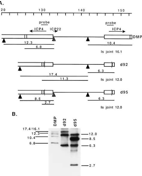
Figure 1A shows the genome of HSV from nucleotide 120 to
the S terminus in the parental orientation, the locations of the
genes for ICP4 and ICP22, and the structures of DMP, d92,
and d95 relative to the relevant HpaI restriction sites (vertical
arrowheads). Also shown are the expected sizes of the HpaI
fragments that span the ICP4 gene. The expected size of the Is
joint fragment is listed for clarity. Figure 1B shows a Southern
blot of the HpaI restriction digests of d92, DMP, and d95
probed with the BamHI Y fragment (Fig. 1A), demonstrating
the incorporation of the n199 insertion into the d95
back-ground. The sizes of the shortened fragments in the digest of
d95 relative to d92 are consistent with the incorporation of the
n199 allele into d95. The sizes of the shortened fragments in
the digest of d95 relative to DMP are consistent with the
incorporation of both of the 4.1-kb deletions of the ICP4
cod-ing sequence in d92 into d95. Therefore, the HpaI pattern of
this region of d95 is consistent with the presence of mutations
in both copies of the ICP4 gene and in the ICP22 gene. The
plaquing behavior of d95 on E26, E5, and E8 cells is consistent
with the presence of mutations in both copies of the ICP4 gene
and the ICP27 gene.
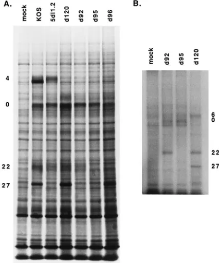
To visualize the IE proteins synthesized in mutant-infected
cells and verify the lack of ICP4, ICP27, and ICP22 synthesis,
cycloheximide-treated Vero cell monolayers were infected with
the indicated viruses at an MOI of 10 PFU per cell and
incu-bated in the presence of cycloheximide for 6 h. The
cyclohex-imide was then removed by washing the monolayer, and
incu-bation was continued in the presence of actinomycin D and
[
35S]methionine. Under these conditions, only the IE proteins
are labeled (10, 26). While ICP4, ICP0, ICP27, and ICP22 were
visible in the profiles of KOS-infected cells, the individual
mutants were missing the bands corresponding to the intended
mutations in the IE genes (Fig. 2A). Thus, d95 does not
syn-thesize ICP4, ICP27, or ICP22. The proteins synsyn-thesized in
cells infected with d120, 5dl1.2, and d92 are consistent with the
previously reported genotypes and phenotypes of these viruses
(11, 39, 59). Also included on this gel is a sample of
d96-infected cells. d96 was generated by a backcross of d95 with
wild-type virus and screening for progeny that grow on E5 cells
and not on Vero cells. d96 does not synthesize ICP4 or ICP22.
To further demonstrate that d95 does not synthesize ICP4,
ICP27, or ICP22, cells infected with d120 (ICP4
2), d92 (ICP4
2ICP27
2), and d95 (ICP4
2ICP27
2ICP22
2) were metabolically
labeled with
32P
i
, and extracts of these cells were analyzed by
SDS-PAGE. ICP27 and ICP22 are readily labeled with
32P,
making this approach a very good one to visualize these
pro-teins. The resulting autoradiogram is shown in Fig. 2B. The
band corresponding to ICP27 was missing in both d95 and d92,
while that corresponding to ICP22 was missing in d95. The lack
of ICP22 in d95 was also evident in the [
35S]methionine profile
in Fig. 4. None of the mutants in Fig. 2 synthesized ICP4.
Prolonged viral and cellular gene expression in
d
95-infected
cells.
Levels of viral and cellular gene expression were
com-pared in d92- and d95-infected Vero cells by SDS-PAGE
anal-ysis and Northern blot analanal-ysis. One effect that became evident
early in the course of this study was that cells infected with
d120, d96, and d92 at an MOI of 10 PFU per cell could be
analyzed only up to 1 day postinfection, whereas cells infected
with d95 retained a morphology more closely resembling, but
not identical to, that of uninfected cells (Fig. 3). While the
d120- and d92-infected cell monolayers were virtually
de-stroyed at 2 days postinfection, the d95 monolayer was intact
(Fig. 3A). It is also interesting that there were fewer
d95-infected than und95-infected cells at this time and that many of the
d95-infected cells consisted of two nuclei in one cytoplasmic
boundary. The same general effects on toxicity and cell number
were observed on HEL cells (Fig. 3B), although it was difficult
to observe multinucleated cells at this level of resolution. As a
FIG. 1. (A) Schematic genome map from nucleotide 120 to the S terminusshowing the locations of ICP4 and ICP22. The mutant virus, DMP, encodes a truncated ICP22 peptide of 199 amino acids by virtue of the insertion of a HpaI linker encoding stop codons in all three reading frames (indicated by vertical arrowheads under the ICP22 gene). The remaining arrowheads mark the natural sites of HpaI cleavage. Boxed regions represent the repeat region of HSV-1 viral genome. The lined region represents the unique short region of the HSV-1 genome. ICP22 and ICP4 (two copies) transcripts are represented by arrows. The
HpaI restriction fragments and their sizes (in kilobases) are represented under
the maps of the viruses. (B) Southern blot analysis. DMP, d92, and d95 viral DNAs were digested by HpaI and subjected to electrophoresis in a 0.7% agarose gel. The fractionated DNA fragments were transferred to a nitrocellulose mem-brane and probed with32
P-labeled 1.84-kb BamHI Y fragment (probe). The sizes (in kilobases) of the bands are marked.
on November 9, 2019 by guest
http://jvi.asm.org/
[image:3.612.60.297.70.364.2]consequence of the cytotoxicity of d120, d92, and d96, it was
only possible to analyze viral and cellular gene expression in
d95-infected cells past 1 day postinfection.
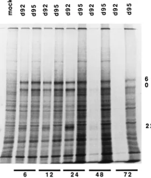
d92- and d95-infected Vero cells were analyzed for viral and
cellular protein synthesis. Monolayers of Vero cells were
in-fected at an MOI of 10 PFU per cell and labeled for 1 h with
[
35S]methionine at the indicated times postinfection, and cell
extracts were subjected to SDS-PAGE analysis. The resulting
SDS-PAGE profile is shown in Fig. 4. Several observations can
be made from these results. (i) The ICP22 band was clearly
evident in the 6-, 12-, and 24-h d92 samples and not in the
corresponding d95 samples. (ii) There was very little d92
sam-ple at 2 and 3 days postinfection. This was due to loss of cells
at this time and is consistent with the results shown in Fig. 3.
(iii) Cellular protein synthesis in d95-infected cells remained
quite high even at 3 days postinfection. This is evident by
comparison with the mock-infected sample. (iv) The viral
pro-teins ICP0 and ICP6 were abundantly expressed even at 3 days
postinfection. d120 and d96 behaved like d92 with respect to
the lack of longevity of protein synthesis (data not shown). This
is presumably due to the toxic effects of these viruses and is
consistent with the results of Johnson and colleagues (29).
To further assess gene expression in d95-infected cells, the
[image:4.612.318.553.70.600.2]abundances of several RNA species were determined. Figure
5A shows levels of ICP0, tk, and cellular
b
-tubulin RNAs in
d120-, d92-, and d95-infected cells and in uninfected cells at 6
and 24 h postinfection. Also shown is an experiment in which
the levels of these transcripts were determined in uninfected
FIG. 2. IE proteins specified by wild-type and mutant viruses. (A)Cyclohex-imide reversal experiment. Vero cell monolayers were pretreated for 1 h by incubation in cycloheximide (100mg/ml)-containing medium, infected with KOS (wild type), 5dl1.2 (ICP272), d120 (ICP42), d92 (ICP42ICP272), d95 (ICP42 ICP272ICP222), and d96 (ICP42ICP222) at an MOI of 10 PFU per cell, and incubated in the presence of cycloheximide for 6 h. The monolayers were then washed twice and further incubated for 3 h in presence of actinomycin D (10 mg/ml) and [35
S]methionine (100mCi per plate). The cells were lysed in SDS sample buffer and subjected to electrophoresis on an SDS–9% polyacrylamide gel. The viral proteins ICP4, ICP0, ICP22, and ICP27 are indicated on the left. (B) Phosphoprotein synthesis in d95-, d120-, and d92-infected cells. Monolayers of Vero cells on 35-mm-diameter petri dishes were infected with d92, d95, and
d120 at an MOI of 10 PFU per cell. At 2 h postinfection, the medium was
replaced with phosphate-deficient medium containing 100mCi of 32 Pi. Cell extracts were lysed and analyzed on an SDS–9% polyacrylamide gel. ICP6, ICP0, ICP22, and ICP27 are marked on the right.
FIG. 3. Apparent cytotoxicity of d95, d120, and d92. (A) Toxicity to Vero cells. Confluent monolayer of 53105Vero cells were infected with d120, d92, and d95 at an MOI of 10 PFU per cell and incubated for 2 days. The culture were photographed through a 403phase-contrast objective. (B) Toxicity to HEL cells. The assay was performed as described above except that 106HEL cells were used per monolayer.
V
OL. 70, 1996
HSV-1 MUTANTS DEFECTIVE IN ICP4, ICP27, AND ICP22
6361
on November 9, 2019 by guest
http://jvi.asm.org/
[image:4.612.70.283.72.328.2]and d95-infected cells at 24, 48, and 72 h postinfection (Fig.
5B). It should be noted that while ICP0 is abundantly
tran-scribed in the absence of ICP4, tk is not. The levels of tk seen
in the absence of ICP4 are approximately 2 to 4% of those seen
in the presence of ICP4 (28). Consistent with previous studies,
ICP0 RNA was slightly increased in size in d92-infected cells
relative to d120-infected cells, and the abundance of tk RNA
was less in d92-infected cells than in d120-infected cells, at 6 h
postinfection (59). Curiously, deletion of ICP22 from the d92
background suppressed these effects. The effect on ICP0 RNA
was less evident at 24 h postinfection, and the effect on tk RNA
was no longer observed. Consistent with the labeling of cellular
proteins in the SDS-PAGE profile in Fig. 4, the abundance of
b
-tubulin RNA was greatest in the d95-infected cells, being
comparable to that in uninfected cells. Therefore, despite the
equal loading of total cellular RNA as determined
spectropho-tometrically and by the ethidium bromide staining patterns of
the rRNA,
b
-tubulin RNA was less abundant in d120- and
d92-infected cells than in d95-infected cells. This finding
im-plies that the stability or the transcription of these messages is
reduced as a consequence of the genes expressed in d120 and
d92 and that the further removal of ICP22 relieved this effect.
The abundances of all three of the messages in d95-infected
cells remained relatively unchanged up to 3 days postinfection
(Fig. 5B). However, after 3 to 4 days at an MOI of 10, the
monolayer lost its integrity; consequently, these times were not
analyzed. The same patterns of expression of ICP0, tk, and
b
-tubulin RNAs seen in Vero cells were also seen in HEL cells
(data not shown).
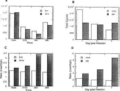
Quantitative analysis of the
b
-tubulin RNA in Fig. 5 is
shown in Fig. 6. At 6 and 24 h postinfection, the levels of
b
-tubulin RNA were reduced in d120-, d92-, and d95-infected
cells, with the least reduction seen in d95-infected cells (Fig.
6A). While the levels of
b
-tubulin RNA declined in uninfected
cells over the course of 3 days,
b
-tubulin RNA levels in
d95-infected cells remained constant over this time period (Fig.
6B), as did the levels of ICP0 and tk RNAs over this time
interval (Fig. 5B). The simplest interpretation of the data is
that HSV proteins expressed from the d95 genome, including
ICP0, allow for transcription to continue at a constant rate
over the 3-day period.
It is clear from Fig. 5 that
b
-tubulin RNA is present in two
species. This has been previously reported and results from the
use of alternative polyadenylation signals (36). Figures 6C and
D show the ratios of the low (1.8-kb)- to high
(2.6-kb)-molec-ular-size species, indicative of the relative usage of the
proxi-mal and distal poly(A) sites. This usage changed as a function
of the viral genetic background by 1 day postinfection.
Utili-zation of the proximal signal increased when ICP27 was
de-leted, as demonstrated by the increase in the low/high ratio in
d92- and d95-infected cells relative to d120- and mock-infected
cells at 24 h postinfection (Fig. 6C). The usage of the proximal
poly(A) site became more pronounced by 2 and 3 days
postin-fection in d95-infected cells relative to uninfected cells (Fig.
6D). Therefore, the HSV proteins expressed from the d95
genome, including ICP0, result in the alteration of 3
9
process-ing relative to uninfected cells in the case of
b
-tubulin RNA. It
is also possible that the d95 background results in an altered
relative stability of the two processed forms of the message.
Apparently, the added expression of ICP27 results in the
al-teration of poly(A) site usage back to a proportion observed in
uninfected cells.
Inhibition of cell division and DNA replication in
d
95-in-fected cells.
While cells infected with d95 do not exhibit rapid
rounding up and detachment from the monolayer and continue
to express viral genes for at least 3 days, they do not increase
in number. This is evident to some degree in Fig. 3 and is
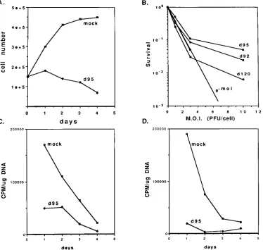
represented quantitatively in Fig. 7A. While uninfected Vero
cells in a monolayer increased in number over 2 days,
d95-infected cells did not. Rather, a marginal decrease in cell
number was evident. Therefore, it appears the growth
poten-tial of d95-infected cells was inhibited.
[image:5.612.103.252.74.249.2]To assess the growth potential of d95-infected cells, two
experiments were performed. The first involved infecting
monolayers of Vero cells with d120, d92, and d95 at several
multiplicities, followed by trypsinization and plating to
mea-sure CFU (Fig. 7B). The second meamea-sured incorporation of
[
3H]thymidine into infected cell DNA (Fig. 7C and D). In the
FIG. 4. Viral and cellular protein synthesis in d92- and d95-infected cells. Vero cells (53105
) seeded on a 35-mm-diameter petri dish were infected with
d92 and d95 at an MOI of 10 PFU per cell. At 6, 12, 24, 48, and 72 h
postin-fection, cells were pulse-labeled for 1 h by incubation in presence of 100mCi of 35
S-labeled methionine per ml. After the labeling period, the cells were solubi-lized in SDS sample buffer and electrophoresed on an SDS–9% polyacrylamide gel. The positions of ICP0, ICP6, and ICP22 are indicated on the right.
FIG. 5. Accumulation of ICP0, tk, andb-tubulin RNAs in d120-, d92-, and
d95-infected Vero cells. Vero cells were mock infected or infected with d120, d92, and d95 at an MOI of 10 PFU per cell. At 6 and 24 h postinfection, total
RNA was isolated and 5mg of each sample was subjected to Northern blot analysis. ICP0, tk, andb-tubulin RNAs were probed with the probes described in Materials and Methods (A). Uninfected and d95-infected Vero cells (MOI of 10) incubated for 1, 2, and 3 days were analyzed in a similar manner (B). Because of alternative polyadenylation site usage, twob-tubulin mRNAs were detected. The 1.8-kb species results from utilization of the proximal poly(A) site, and the 2.6-kb species results from utilization of the distal poly(A) site.
on November 9, 2019 by guest
http://jvi.asm.org/
[image:5.612.63.299.487.642.2]colony-forming assay, d92 inhibited cell viability less than d120.
Interestingly, d95 was only marginally less inhibitory than d92,
despite the dramatically different appearance of d92- and
d95-infected cells shown in Fig. 3. Figure 7B also shows the
prob-ability of the cells not being infected following inoculation at a
given MOI. The survival curves indicate that up to an MOI of
3, a single PFU is very efficient in inhibiting colony formation.
At an MOI of 10, survival is greater than would be expected
from the pattern seen at the lower MOIs. This finding indicates
that the inhibitory effects may be saturable or that there may
be subpopulations of cells that are less susceptible to the
in-hibitory effects of IE proteins. In summary, all of these viruses
had a significant inhibitory effect on colony-forming ability,
indicating that fundamental cellular processes required for
cells to form colonies are perturbed by HSV, even when ICP4,
ICP27, and ICP22 are not expressed.
To study this effect in greater detail, we determined if
cel-lular DNA synthesis was inhibited in d95-infected cells.
Ac-cordingly, Vero and HEL cells were infected with d95 at an
MOI of 10 PFU per cell. At 1, 2, 3, and 4 days postinfection,
d95-infected and uninfected cells were labeled for 3 h with
[
3H]thymidine. Following the labeling period, DNA from the
cells was isolated and the amount of
3H incorporated per
microgram of DNA was determined. As is evident in Fig. 7C
and D, d95 infection significantly inhibited cellular DNA
rep-lication in both Vero and HEL cells, respectively. The
reduc-tion in labeling of uninfected cells at 3 and 4 days postinfecreduc-tion
is consistent with results of Fig. 7A, probably reflecting contact
inhibition. To determine the level of DNA synthesis as a
func-tion of the other IE mutant backgrounds, Vero cells were
infected with d120, d92, d95, and d96 at an MOI of 10 PFU per
cell and labeled with [
3H]thymidine from 21 to 24 h
postinfec-tion. As described above, DNA was isolated and the quantity
of
3H incorporated per microgram of DNA was determined.
The resulting levels of incorporation of [
3H]thymidine relative
to that in uninfected cells were 25% for d120, 12% for d92,
13% for d95, and 25% for d96. These results suggest that the
cellular environment in all of these mutant backgrounds is
incompatible with uninfected levels of cellular DNA synthesis.
Perturbation of nuclear structure by HSV IE gene mutants.
It has been known for some time that ICP4 mutants of HSV
have a deleterious effect on cellular morphology and
chroma-tin structure (29, 49). To determine the contributions of the IE
proteins to morphological changes in the cell, Vero cells were
infected at an MOI of 10 PFU per cell with d120, d92, d95, and
d96 and processed for electron microscopy at 24 h
postinfec-tion. Figure 8 shows that all of the mutants elicit changes or the
formation of novel structures relative to uninfected cells.
Nor-mal cell morphology is shown in Fig. 8A. Infection of cells with
d120 (Fig. 8B) resulted in the accumulation of small dense
intranuclear granules. The nucleus commonly had a highly
convoluted profile, and frequently a series of large
protein-aceous cytoplasmic bodies was seen. d96 (Fig. 8F) had
mor-phologic sequelae similar to those for d120 except that the
small dense intranuclear granules were absent. In d92-infected
cells (Fig. 8C), no large cytoplasmic bodies were seen; rather,
these structures were confined to the nucleus. The d92-infected
cells also showed small nuclear granules (as seen in the
d120-FIG. 6. Quantitation of mRNA accumulation and poly(A) site usage of cellularb-tubulin. (A) Counts of32P hybridizing to the 1.8- and 2.6-kb species in Fig. 5A were determined as described in Materials and Methods. (B) Counts of32P hybridizing to the 1.8- and 2.6-kb species in Fig. 5B were determined as described in Materials and Methods. (C) The ratios of the net counts of the 1.8-kb over 2.6-kb species in Fig. 5A. (D) The ratios of the net counts of the 1.8-kb over 2.6-kb species in Fig. 5B.V
OL. 70, 1996
HSV-1 MUTANTS DEFECTIVE IN ICP4, ICP27, AND ICP22
6363
on November 9, 2019 by guest
http://jvi.asm.org/
[image:6.612.115.503.76.372.2]infected cells); however, they commonly appeared as larger
condensed structures about the large nuclear bodies (Fig. 8C
and D). d95-infected cells appeared to be the least affected;
however, numerous large and regularly shaped nuclear
inclu-sions were evident in these cells (Fig. 8E). These incluinclu-sions
were noticeably absent from uninfected cells and are
reminis-cent of the nuclear bodies seen in d92-infected cells.
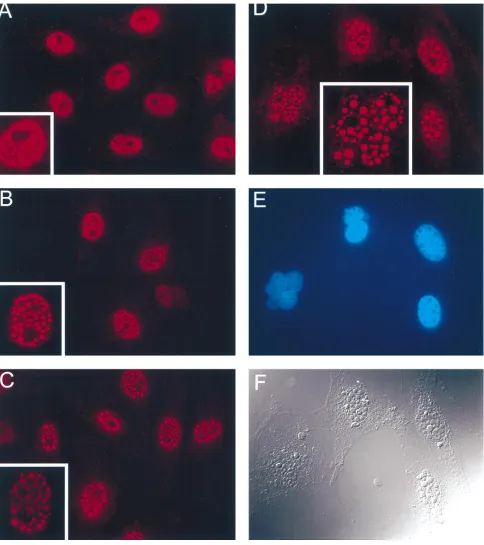
ICP0 is abundantly synthesized in d95-infected cells (Fig. 4).
To address the possibility that ICP0 is present in the dense
nuclear bodies in d95-infected cells, d95-infected cells were
stained with ICP0 antibody and processed for
immunofluores-cence microscopy. Shown in Fig. 9 are fluorescent images of
d95-infected cells (MOI of 10 PFU per cell) stained with a
monoclonal antibody to ICP0 at 6, 12, 24, and 48 h
postinfec-tion. The insets show an enlargement of a nucleus from the
larger field. As previously observed (31, 81, 82), ICP0
accumu-lated in fine punctate structures at early times (6 h)
postinfec-tion (Fig. 8A). Subsequently, the continued accumulapostinfec-tion of
ICP0 in the nucleus resulted in the formation of fewer but
much larger ICP0-containing bodies (Fig. 8B to D). The
per-turbation of nuclear structure and number often seen with IE
mutants is evident in Fig. 8D and E. Figure 8E shows the same
field as in Fig. 8D, but the DNA has been specifically stained
with Hoescht dye. From this micrograph, it is clear that the
ICP0-containing structures do not contain DNA. Figure 8F is
a differential interference image of the same cell as in Fig. 8D
and E. The ICP0-containing structures are easily resolved in
this image. Thus, in the absence of ICP4, ICP27, and ICP22,
ICP0 accumulated to very high levels in the nucleus and
local-ized to dense, spherical bodies. The formation of these
struc-tures represents the only obvious deviation from the
morphol-ogy of uninfected cell nuclei.
DISCUSSION
[image:7.612.124.498.73.428.2]The ability of virus-encoded functions to rapidly usurp host
cell metabolic mechanisms along with the function of viral
systems enables HSV to express its 75 to 80 genes, replicate its
genome, and assemble progeny within 6 h of infection in
sus-ceptible cells. While significant progress has been made toward
FIG. 7. Growth potential of d95-infected cells. (A) Cell number as a function of time postinfection with d95. Monolayers of Vero cells were mock infected or infected with d95 at an MOI of 10 PFU per cell. At 1, 2, 3, and 4 days postinfection, the monolayers were trypsinized and the cells were counted in a hemocytometer. Shown are the cell counts per milliliter from a 3-ml suspension. (B) Colony inhibition assay. Confluent monolayers of Vero cells were mock infected or infected with the indicated viruses and MOIs. The monolayers were trypsinized at 6 h postinfection and plated for CFU as described in Materials and Methods. Shown are the surviving fraction of infected cells relative to uninfected cells. Also shown is the probability of a cell not becoming infected as a function of MOI (e2moi). (C) Incorporation of [3
H]thymidine into Vero cell DNA as a function of time after infection with d95. Monolayers of Vero cells were mock infected or infected with d95 at an MOI of 10 PFU per cell. At 1, 2, 3, and 4 days postinfection, the cultures were labeled for 3 h with [3
H]thymidine, and the cellular DNA was extracted, quantified, and counted for3
H as described in Materials and Methods. (D) Incorporation of [3
H]thymidine into HEL cell DNA as a function of time after infection with d95. This experiment was performed like that in panel C except that HEL cells were used in place of Vero cells.
on November 9, 2019 by guest
http://jvi.asm.org/
FIG. 8. Electron micrographs of Vero cells after infection with different IE mutants. Eighty percent confluent monolayers of Vero cells were uninfected or infected with d120, d92, d96, or d95 at an MOI of 10 PFU/ml. At 24 h postinfection, the monolayers were processed for electron microscopy as described in Materials and Methods. (A) Uninfected cells show a typical cell and nuclear morphology, with large prominent nucleoli and thin heterochromatin (bar55mm). (B) Cells infected with d120. Small intranuclear (arrowheads) and large cytoplasmic (arrows) inclusions are apparent. Magnification as in panel A. (C and D) In cells infected with d92, at low power (C), large (arrowheads) and small intranuclear particles are apparent. At high power (D), the small particles are seen to exist both as small stellate granules and as structures condensed about the larger inclusions (arrows). C, magnification as in panel A; D, bar50.5mm. (E) Cells infected with d95. Large abundant nuclear inclusions are seen (arrowheads). Serial sectioning shows these structures to be spherical (not shown). Magnification as in panel A. (F) Cells infected with d96. The nucleus appears similar to that seen in control cells (A); however, abundant, large cytoplasmic inclusions are apparent (arrowheads). Magnification as in panel A.
V
OL. 70, 1996
HSV-1 MUTANTS DEFECTIVE IN ICP4, ICP27, AND ICP22
6365
on November 9, 2019 by guest
http://jvi.asm.org/
the understanding of viral functions, less light has been shed on
how these functions perturb or alter host cell processes.
It has been known for some time that infection of cells with
viruses deficient in ICP4 results in rapid cell death (29)
[image:9.612.66.550.67.612.2]accom-panied by chromosomal damage (29, 49) and alterations in
nuclear structures. In the absence of ICP4, at least five other
gene products, ICP27, ICP0, ICP22, ICP47, ICP6, and OrfP,
are efficiently synthesized (4, 11, 34, 35, 79). Many of these
FIG. 9. Localization of ICP0 in d95-infected cells. Eighty percent confluent monolayers of Vero cells were infected with d95 at an MOI of 10 PFU/ml and processed for immunofluorescence at 6 (A), 12 (B), 24 (C), and 48 (D to F) h postinfection, using a monoclonal antibody to ICP0 as described in Materials and Methods. At early time points, the ICP0-containing nuclear inclusions appear small and granular, with a high nucleoplasmic staining. With time, the size of the inclusions increases dramatically, while the nucleoplasmic background staining decreases. (E) When the DNA within the nucleus is counterstained with Hoescht dye, it is apparent that the ICP0-staining granules contain no DNA. (F) When the cells are examined by differential interference contrast microscopy, while the cellular cytoplasmic profile appears normal, the multiple nuclear inclusions are apparent. Panels D to F are the same field.on November 9, 2019 by guest
http://jvi.asm.org/
proteins have been shown to have activities that may perturb
host cell functions. Indeed, it has been demonstrated that
ICP27, ICP0, and ICP22 reduce the efficiency with which cells
are transformed with a selectable marker, implying that they
reduce cell viability (30).
In this study, the effects of a series of HSV mutants defective
in ICP4, ICP22, and ICP27 on gene expression, host cell
via-bility, cellular DNA replication, cell division, and nuclear
ul-trastructure were studied. These experiments revealed that
infection of cells with a mutant defective in ICP4, ICP22, and
ICP27 (d95) resulted in prolonged cell viability, as measured
by viral and cellular gene expression (Fig. 4 and 5) and overall
cell morphology relative to d120- and d92-infected cells. The
colony-forming capacity, or the capacity of d95-infected cells to
multiply, was not significantly greater than that of d92-infected
cells (Fig. 7B), despite the large difference observed in the
appearance of d92- and d95-infected monolayers (Fig. 3).
Ad-ditionally, d95 had the least effect on nuclear morphology of all
of the mutants tested (Fig. 8). The only deviation from
unin-fected cell morphology was the accumulation of dense,
spher-ical structures in the nucleus, which were subsequently shown
to contain ICP0 (Fig. 9).
Gene expression in mutant virus-infected cells.
In
HSV-1-infected cells, the transcription of viral and cellular genes is
carried out by host RNA polymerase II (1). ICP4 has a direct
effect on transcription, and ICP0, ICP27, and ICP22 have all
been implicated in modulating transcription. ICP0 is a
promis-cuous transactivator which has been reported to transactivate a
variety of viral and cellular promoters (16, 18, 47, 53). ICP0 is
abundantly expressed in d95-infected Vero cells. The level of
ICP0 transcript and the rate of ICP0 protein synthesis were
relatively constant from days 1 to 3 postinfection. In addition,
the accumulation of a cellular message was maintained at a
high level throughout this time period. It is possible that the
expression of ICP0 in the absence of the deleterious effects of
other IE genes allows for the observed prolonged gene
expres-sion.
The striking difference in cell morphology and survival was
observed when ICP22 was inactivated. Mutants in ICP22 show
reduced transcription and expression of certain viral genes (55,
65). ICP22 also induces a novel phosphorylated form of
cellu-lar RNA polymerase II (55). These observations are all
con-sistent with transcriptional effects, with the presence of ICP22
correlating with more efficient viral transcription. By virtue of
the prolonged accumulation of the products of viral and
cel-lular genes, we apparently see more efficient gene expression in
the absence of ICP22. Previous studies of ICP22, including
some using the same ICP22 allele in d95, have all focused on
the effect of ICP22 in an otherwise wild-type background. It is
possible that the prolonged gene expression that is observed in
this study with d95 compared with d92 is related not to a direct
effect of ICP22 on gene expression but rather to some other
deleterious effect that is relieved by the inactivation of ICP22.
Comparison of the activities of the transcriptional apparatus in
d92- and d95-infected cells should shed some light on this
question.
ICP27 has been shown to affect two aspects of mRNA
pro-cessing: splicing and poly(A) site usage (23, 42, 43, 61–63).
Effects on mRNA structure were observed in two cases in this
study. The first is that the tk and ICP0 messages were slightly
larger and appeared more heterogeneous when synthesized in
the d92 (ICP4
2ICP27
2) background compared with the d120
(ICP4
2) background. This effect was suppressed when ICP22
was mutated (d95). The second is that the deletion of ICP27
from the d120 background resulted in a change in the relative
utilization of the
b
-tubulin poly(A) sites (Fig. 5 and 6) at 24 h
postinfection. The further deletion of ICP22 had little or no
effect on poly(A) site utilization; however, the effect became
more pronounced because of the extended life span of the
infected cells. This observation also demonstrates that in the
d95 background, the poly(A) site usage may be different from
that in uninfected cells. This effect is unexpected since none of
the proteins expressed in this background is known to have an
effect on poly(A) site usage. These observations on viral and
cellular messages, the former in particular, are consistent with
effects of ICP27 on poly(A) site selection (42, 43). The effect
on ICP0 and tk messages also suggests a functional interaction
between ICP27 and ICP22. Deleting ICP27 (in d92) resulted in
altered mRNA structure relative to d120, and the further
in-activation of ICP22 in d95 suppressed this effect.
Cytotoxicity and the inhibition of DNA synthesis.
In the
absence of ICP4, ICP27, and ICP22, the infected cell survives
longer than cells infected with an ICP4
2ICP27
2mutant, as is
clear from microscopic examination of cells and studies on cell
and viral gene expression. Despite this, the ability of
d95-infected cells to proliferate and form colonies was impaired.
This impairment is due at least in part to factors contributing
to the inhibition of cell DNA synthesis. Presumably, the
pro-teins synthesized in the d95 background (ICP6, ICP0, ICP47,
and OrfP) are responsible for this effect. ICP47, a cytoplasmic
protein that has been shown to affect the processing of major
histocompatibility complex class I molecules (80), is probably
not responsible for this effect. ICP6 is also probably not
in-volved since a virus deleted for ICP6, ICP4, and ICP27 still
inhibits cell DNA synthesis (unpublished data). This leaves
OrfP and ICP0 as potential candidates for this effect.
Addi-tionally, ICP0 may induce changes in cellular gene expression
that result in the inhibition of cellular DNA synthesis or cell
cycle arrest. It is also possible that the virion-associated host
cell shutoff function, UL41 (53a), also contributes to the
shut-off of host cell DNA synthesis. However, two observations
suggest that this is at most a minor component of the shutoff of
DNA synthesis: (i) at the MOIs used in this study, d95 did not
efficiently shut off host cell protein synthesis (Fig. 3), and (ii) a
virus lacking ICP4, ICP27, and UL41 still shuts off host cell
DNA synthesis (data not shown).
ICP0 clearly alters the nuclear ultrastructure and potentially
the compartmentalization of nuclear proteins involved in cell
cycle regulation. In cells infected with wild-type virus (31) or an
ICP4
2ICP27
2mutant (d92 [82]), ICP0 accumulates in many
small punctate intranuclear structures. Everett and Maul have
shown that these structures are coincident with
PML-contain-ing structures (17, 38), also known as ND10, PODs, or Kr
bodies (2, 15, 32, 75, 78). The function of these structures is
unknown but is thought to be involved in the proliferative or
differentiation state of the cell (15, 32, 75). In a wild-type virus
background, ICP0 dissociates these structures late in infection
and itself becomes more diffusely localized throughout the cell
(17, 38). In d95-infected cells, ICP0 accumulates over the
course of 3 days (Fig. 3). Early in infection, its localization is
similar to that seen previously, i.e., an abundance of small
punctate structures is evident over a diffuse background.
How-ever, with time, ICP0 accumulates into increasingly large
tures, which are clearly visible by light microscopy. These
struc-tures do not contain significant quantities of DNA. One
possibility is that they represent inclusion bodies which have
nucleated at the ND10 structures. From previous results, it is
likely that ND10 structures are disturbed, and it is also possible
that cellular molecules, which interact with ICP0, are
seques-tered into these inclusion bodies. Further studies on the
com-positions of these structures and the cellular proteins that they
may contain are in progress. A remaining formal possibility is
V
OL. 70, 1996
HSV-1 MUTANTS DEFECTIVE IN ICP4, ICP27, AND ICP22
6367
on November 9, 2019 by guest
http://jvi.asm.org/
that a truncated ICP22 molecule is synthesized as a function of
n199 allele, and this peptide is involved in the phenotype of
d95. However, the n199 allele imparts a growth restriction
similar to that seen with complete deletions of the ICP22 gene
(51). d95 titers are reduced 5- to 10-fold relative to d92 titers
(data not shown). Also, the region of ICP22 that is most
con-served among the herpesviruses (25, 51) is excluded by virtue
of the n199 mutation. An additional unlikely possibility is that
a fortuitous gain-of-function mutation contributes to the
phe-notype of d95.
In the absence of ICP4, ICP27, and ICP22, cell DNA
syn-thesis was inhibited and ICP0 accumulated to very high levels
in discrete structures in the nucleus. The formation of large
nuclear inclusions correlated with the absence of ICP27. This
finding may reflect previous observations that the expression of
ICP27 in the absence of ICP4 results in the cytoplasmic
local-ization of ICP0 (81, 82). Also of note is the observation that the
expression of ICP22 results in the formation of fine granular
structures in the nucleus. In the presence of nuclear ICP0, as in
d92, these appeared to coalesce around the ICP0-containing
bodies (Fig. 8D). This finding raises the possibility that
mole-cules present as a function of ICP22 interact with ICP0.
Fur-ther studies on the identities and compositions of the
ICP22-dependent structures will be necessary to address this question.
The observations described in this study reflect some of the
effects of the IE proteins on host cell metabolism. Presumably
some of these changes occur early in the viral infection and
prime the infected cell for productive viral infection. It is also
possible that some of the observed effects are exaggerated or
are a consequence of IE protein overexpression. In either case,
these results also bear on the use of IE deletion mutants of
HSV for use as gene transfer vehicles. While viral backgrounds
such as d95 may have utility in some cases or in certain cell
types, it is highly likely that all of the regulatory IE genes are
deleterious to host cell survival and that they will all have to be
deleted if HSV is to be generally used as a vector.
ACKNOWLEDGMENTS
This work was supported by NIH grants AI30612, DK44935, and
CA20260.
REFERENCES
1. Alwine, J., W. Stein, and C. Hill. 1974. Transcription of herpes simplex type 1 DNA in nuclei isolated from infected Hep-2 and KB cells. Virology 60: 302–307.
2. Ascoli, C. A., and G. G. Maul. 1991. Identification of a novel nuclear domain. J. Cell Biol. 112:785–795.
3. Batterson, W., and B. Roizman. 1983. Characterization of the herpes simplex virion-associated factor responsible for the induction of alpha genes. J. Virol. 46:371–377.
3a.Biotecx. 1992. Bulletin 27. Biotecx, Houston, Tex.
4. Bohenzky, R. A., A. G. Papavassiliou, I. H. Gelman, and S. Silverstein. 1993. Identification of a promoter mapping within the reiterated sequences that flank the herpes simplex virus type 1 ULregion. J. Virol. 67:632–642. 5. Bond, J. F., G. S. Robinson, and S. R. Farmer. 1984. Differential expression
of two neural cell-specificb-tubulin mRNAs during rat brain development. Mol. Cell. Biol. 4:1313–1319.
6. Cai, W., T. L. Astor, L. M. Liptak, C. Cho, D. M. Coen, and P. A. Schaffer. 1993. The herpes simplex virus type 1 regulatory protein ICP0 enhances virus replication during acute infection and reactivation from latency. J. Virol. 67:7501–7512.
7. Cai, W., and P. A. Schaffer. 1992. Herpes simplex virus type 1 ICP0 regulates expression of immediate-early, early, and late genes in productively infected cells. J. Virol. 66:2904–2915.
8. Campbell, M. E., J. W. Palfreyman, and C. M. Preston. 1984. Identification of herpes simplex virus DNA sequences which encode a trans-acting polypeptide responsible for stimulation of immediate early transcription. J. Mol. Biol. 180:1–19.
9. Chapman, C. J., J. D. Harris, M. A. Hardwicke, R. M. Sandri-Goldin, M. K. Collins, and D. S. Latchman.1992. Promoter independent activation of heterologous virus gene expression by the herpes simplex virus
immediate-early protein ICP27. Virology 186:573–578.
10. Clements, J. B., R. J. Watson, and N. M. Wilkie. 1977. Temporal regulation of herpes simplex virus type 1 transcription: location of transcripts on the viral genome. Cell 12:275–285.
11. DeLuca, N. A., A. M. McCarthy, and P. A. Schaffer. 1985. Isolation and characterization of deletion mutants of herpes simplex virus type 1 in the gene encoding immediate-early regulatory protein ICP4. J. Virol. 56:558– 570.
12. DeLuca, N. A., and P. A. Schaffer. 1985. Activation of immediate-early, early, and late promoters by temperature-sensitive and wild-type forms of herpes simplex virus type 1 protein ICP4. Mol. Cell. Biol. 5:1997–2208.
13. DeLuca, N. A., and P. A. Schaffer. 1987. Activities of herpes simplex virus type 1 (HSV-1) ICP4 genes specifying nonsense peptides. Nucleic Acids Res. 15:4491–4511.
14. Dixon, R. A., and P. A. Schaffer. 1980. Fine-structure mapping and functional analysis of temperature-sensitive mutants in the gene encoding the herpes simplex virus type 1 immediate early protein VP175. J. Virol. 36:189–203. 15. Dyck, J. A., G. G. Maul, W. H. Miller, Jr., J. D. Chen, A. Kakizuka, and R. M.
Evans.1994. A novel macromolecular structure is a target of the promyelo-cyte-retinoic acid receptor oncoprotein. Cell 76:333–343.
16. Everett, R. D. 1984. Trans activation of transcription by herpes virus prod-ucts: requirement for two HSV-1 immediate-early polypeptides for maxi-mum activity. EMBO J. 3:3135–3141.
17. Everett, R. D., and G. G. Maul. 1994. HSV-1 IE protein Vmw110 causes redistribution of PML. EMBO J. 13:5062–5069.
18. Gelman, I. H., and S. Silverstein. 1985. Identification of immediate early genes from herpes simplex virus that transactivate the virus thymidine kinase gene. Proc. Natl. Acad. Sci. USA 82:5265–5269.
19. Gerster, T., and R. G. Roeder. 1988. A herpesvirus trans-activating protein interacts with transcription factor OTF-1 and other cellular proteins. Proc. Natl. Acad. Sci. USA 85:6347–6351.
20. Goding, C. R., and P. O’Hare. 1989. Herpes simplex virus Vmw65-octamer binding protein interaction: a paradigm for combinatorial control of tran-scription. Virology 173:363–367.
21. Greaves, R., and P. O’Hare. 1989. Separation of requirements for protein-DNA complex assembly from those for functional activity in the herpes simplex virus regulatory protein Vmw65. J. Virol. 63:1641–1650. 22. Gu, B., R. Rivera-Gonzalez, C. A. Smith, and N. A. DeLuca. 1993. Herpes
simplex virus infected cell polypeptide 4 preferentially represses Sp1-acti-vated over basal transcription from its own promoter. Proc. Natl. Acad. Sci. USA 90:9528–9532.
23. Hardwicke, M. A., and R. M. Sandri-Goldin. 1994. The herpes simplex virus regulatory protein ICP27 contributes to the decrease in cellular mRNA levels during infection. J. Virol. 68:4797–4810.
24. Hardy, W. R., and R. M. Sandri-Goldin. 1994. Herpes simplex virus inhibits host cell splicing, and regulatory protein ICP27 is required for this effect. J. Virol. 68:7790–7799.
25. Holden, V. R., R. R. Yalamanchili, R. N. Harty, and D. J. O’Callaghan. 1992. ICP22 homolog of equine herpesvirus 1: expression from early and late promoters. J. Virol. 66:664–673.
26. Honess, R. W., and B. Roizman. 1974. Regulation of herpesvirus macromo-lecular synthesis. I. Cascade regulation of the synthesis of three groups of viral proteins. J. Virol. 14:8–19.
27. Honess, R. W., and B. Roizman. 1975. Regulation of herpesvirus macromo-lecular synthesis: sequential transition of polypeptide synthesis requires functional viral polypeptides. Proc. Natl. Acad. Sci. USA 72:1276–1280. 28. Imbalzano, A. N., D. Coen, and N. A. DeLuca. 1991. Herpes simplex virus
transactivator ICP4 operationally substitutes for the cellular transcription factor Sp1 for efficient expression of the viral thymidine kinase gene. J. Virol. 65:565–574.
29. Johnson, P. A., A. Miyanohara, F. Levine, T. Cahill, and T. Friedmann. 1992. Cytotoxicity of a replication-defective mutant of herpes simplex virus type 1. J. Virol. 66:2952–2965.
30. Johnson, P. A., M. J. Wang, and T. Friedmann. 1994. Improved cell survival by the reduction of immediate-early gene expression in replication-defective mutants of herpes simplex virus type 1 but not by mutation of the virion host shutoff function. J. Virol. 68:6347–6362.
31. Knipe, D. M., and J. L. Smith. 1986. A mutant herpesvirus protein leads to a block in nuclear localization of other viral proteins. Mol. Cell. Biol. 6:2371– 2381.
32. Koken, M. H., F. Puvion-Dutilleul, M. C. Guillemin, A. Viron, G. Linares-Cruz, N. Stuurman, L. de Jong, C. Szostecki, F. Calvo, C. Chomienne, L. Degos, E. Puvion, and H. de The.1994. The t (15;17) translocation alters a nuclear body in a retinoic acid-reversible fashion. EMBO J. 13:1073–1083. 33. Kristie, T. M., J. H. LeBowitz, and P. A. Sharp. 1989. The octamer-binding proteins form multi-protein—DNA complexes with the HSV alpha TIF regulatory protein. EMBO J. 8:4229–4238.
34. Lagunoff, M., and B. Roizman. 1994. Expression of a herpes simplex virus 1 open reading frame antisense to theg134.5 gene and transcribed by an RNA 39coterminal with the unspliced latency-associated transcript. J. Virol. 68: 6021–6028.
35. Lagunoff, M., and B. Roizman. 1995. The regulation of synthesis and