Open Access
Research
Rev-dependent lentiviral expression vector
Yuntao Wu
1,2, Margaret H Beddall
1and Jon W Marsh*
1Address: 1Section on Molecular Virology, Laboratory of Cellular and Molecular Regulation, NIMH, Bethesda, MD, 20892-4483, USA and 2National
Center for Biodefense and Infectious Diseases, Department of Molecular and Microbiology, George Mason University, Manassas, VA, 20110, USA
Email: Yuntao Wu - [email protected]; Margaret H Beddall - [email protected]; Jon W Marsh* - [email protected] * Corresponding author
Abstract
Background: HIV-responsive expression vectors are all based on the HIV promoter, the long terminal repeat (LTR). While responsive to an early HIV protein, Tat, the LTR is also responsive to cellular activation states and to the local chromatin activity where the integration has occurred. This can result in high HIV-independent activity, and has restricted the use of LTR-based reporter vectors to cloned cells, where aberrantly high expressing (HIV-negative) cells can be eliminated. Enhancements in specificity would increase opportunities for expression vector use in detection of HIV as well as in experimental gene expression in HIV-infected cells.
Results: We have constructed an expression vector that possesses, in addition to the Tat-responsive LTR, numerous HIV DNA sequences that include the Rev-response element and HIV splicing sites that are efficiently used in human cells. It also contains a reading frame that is removed by cellular splicing activity in the absence of HIV Rev. The vector was incorporated into a lentiviral reporter virus, permitting detection of replicating HIV in living cell populations. The activity of the vector was measured by expression of green fluorescence protein (GFP) reporter and by PCR of reporter transcript following HIV infection. The vector displayed full HIV dependency.
Conclusion: As with the earlier developed Tat-dependent expression vectors, the Rev system described here is an exploitation of an evolved HIV process. The inclusion of Rev-dependency renders the LTR-based expression vector highly dependent on the presence of replicating HIV. The application of this vector as reported here, an HIV-dependent reporter virus, offers a novel alternative approach to existing methods, in situ PCR or HIV antigen staining, to identify HIV-positive cells. The vector permits examination of living cells, can express any gene for basic or clinical experimentation, and as a pseudo-typed lentivirus has access to most cell types and tissues.
Background
All HIV-dependent expression vectors in common use are based on the HIV long terminal repeat promoter (LTR). An early HIV gene product, Tat, increases the level of tran-script that is initiated at the LTR. The placement of reporter genes downstream of the LTR results in a respon-siveness to the synthesis of Tat, a measure of HIV
replica-tion. The earliest indicator lines made use of reporter enzymes, such as luciferase and β-galactosidase [1-4], per-mitting a direct measurement of reporter gene induction. Tat appears to increase HIV transcriptional activity by two mechanisms. The first identified Tat activity is not directed towards the proviral DNA promoter, but rather through direct association with the growing nascent RNA chain.
Published: 7 February 2007
Retrovirology 2007, 4:12 doi:10.1186/1742-4690-4-12
Received: 20 December 2006 Accepted: 7 February 2007
This article is available from: http://www.retrovirology.com/content/4/1/12
© 2007 Wu et al; licensee BioMed Central Ltd.
Tat associates with a 5' RNA loop structure [5-7], the trans-activation response element (TAR), to promote comple-tion of the initiated transcript [8-10], an activity also defined as processivity or anti-termination. More recent work has provided evidence that Tat also stimulates assembly of transcription factors to the DNA promoter [11]; that is, Tat promotes initiation, as well as elongation [12]. However, the LTR as a promoter is inherently leaky. Following integration of the HIV DNA into the host chro-matin, the LTR transcribes the early gene products Tat, Rev, and Nef. That is, there is a required basal level of tran-scription that is Tat-independent. In addition, the site of integration or insertion of the LTR-based expression vec-tor can mediate high levels of transcriptional activity in some cells [13], leading to expression from the reporter LTR in the absence of HIV. For example, in the generation of HIV indicator cells with LTR-based reporters, it has been necessary to remove 25% or more of the stably trans-fected cells [4,14] since they generate reporter transcript in the absence of HIV. While high expression cells can be removed in the generation of reporter clones, this inher-ent leakiness previnher-ents the use of viral vectors to deliver the LTR-based reporter construct to detect the presence of existing HIV-positive cells in a mixed population.
By eliminating this non-specific activity, that is, non-HIV induction of signal, from an HIV expression vector, a wider use of this convenient and efficient tool would be possible. In addition to Tat, HIV transcriptional activity is also affected by an early gene product, Rev. Rev binds to a 3' loop structure, the Rev response element (RRE), present in unspliced and singly-spliced HIV transcripts, to permit nuclear export and translation of these mRNAs [15-17]. This viral specific activity exploits an essential cellular process. The removal of non-coding regions of transcripts (introns) prior to translation is critical to all cells. Introns are operationally defined by the presence of strong splic-ing sites, and HIV exploits this cellular activity by includ-ing multiple splice sites with varyinclud-ing activity [18,19] to generate multiple coding regions within the same stretch of HIV RNA. The existence of these sites results in the gen-eration of fully spliced transcripts in the early phase of HIV infection [20,21]. Once Rev is expressed, RRE-con-taining HIV transcripts can be delivered to the cytosolic translational machinery.
It is of interest that in HIV infection protein expression from singly or non-spliced HIV transcripts appears abso-lutely dependent on Rev expression [15,22,23]. This dependency is lost with seemingly minor modifications to HIV DNAs [24,25], and the earliest reported RRE-contain-ing expression constructs [26,27] can display varied Rev-independent expression. These were designed to elucidate Rev function, and the focus was not to eliminate back-ground signal. In this report, by inclusion of multiple
components of the HIV genome, we have constructed a lentiviral expression vector that displays a full dependency on the presence of HIV. As it is silent in HIV-negative cells, it differs dramatically from LTR-based systems.
Results
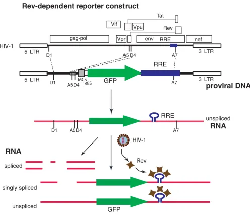
To test the potential of Rev from HIV infection to control reporter gene expression in an HIV-dependent fashion, we constructed an HIV-like vector as shown in Fig. 1. Overall, we incorporated four separate segments of the HIV genome into the expression vector; however, no HIV gene is expressed from this construct. The 5' end of the vector consists of the HIV 5' LTR, the splice donor 1 site, D1, and a portion of the gag open reading frame that includes the packaging signal. The second HIV segment is from the
tat1/rev1 exon that includes splice acceptor site 5, A5, and splice donor site 4, D4. The third segment of HIV DNA is from the env exon and encompasses the RRE, and the splice acceptor site 7, A7. The last segment includes the entire 3' LTR along with a small portion of the nef reading frame 5' to the LTR. In infectious HIV, the joining of splice donor 1 with splice acceptor site 5 (removal of D1/A5 intron) is utilized in transcripts for both Envelope and Nef proteins. In HIV NL4-3 infection of PBMC, the single spliced D1/A5 transcript represents 80% of all Env mes-sage [19]. Nef transcript requires a second splicing event, and the most common is removal of the segment between D4 and A7 [19]. The vector has the capacity to express two genes. A multiple-cloning site (MCS) for one of the expressed genes is immediately down-stream of the A5/ D4 HIV splice sites; however, this cloning site has not been utilized in this report. This is followed by an internal ribosome entry site (IRES), which is upstream to the reporter gene used in this report, green fluorescent protein or GFP. The reporter gene is adjoined to the RRE-containing
env exon segment. Together these segments compose the transfer vector. The full-length transcript generated from the construct possesses efficient HIV splice sites that medi-ate the removal of the reporter open reading frame (see Fig. 1). The reporter gene is only expressed from the RRE-containing unspliced or singly spliced transcripts, and thus requires the presence of Rev, a process that mimics HIV late gene expression. Co-transfection of this plasmid with an Env-coding plasmid and a packaging construct results in production of an infectious non-replicative len-tiviral vector [28] capable of transferring the Rev-depend-ent reporter system into cells.
To permit a quantitative measurement of reporter gene induction from the lentiviral construct in response to HIV infection, we tested the vector in cells infected or not infected with HIV. CEM-SS cells, a human T cell line, were first infected with a modified, non-replicative NL4-3 HIV, where the gene for murine CD24 was inserted into the nef
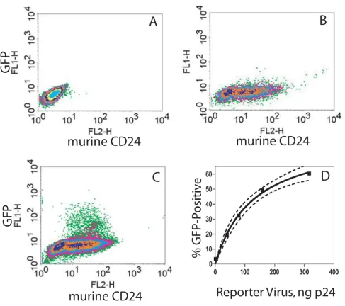
genes, but lacks an env gene and therefore must be pseudo-typed (see Methods). Staining of cells for murine CD24 thus confirms the presence of HIV in a cell popula-tion (compare lack of staining in Fig. 2A to CD24-posi-tive, HIV-infected cells in Fig. 2B). The resultant mixed populations of HIV-infected and non-infected CEM-SS cells were then transduced with a predetermined titer (see Methods) of the VSV-Env pseudo-typed Rev-dependent indicator lentiviral vector (Fig. 2A and 2C). In Fig. 2C, in order to determine whether a single reporter vector was
adequately robust, we added one infectious indicator vec-tor per five cells. If we assume that the infection is a Pois-son process, then only two percent of the cells should contain two or more integrated indicator vectors. After 3 days, GFP was found expressed only in HIV-infected (CD24-positive) cells (Fig. 2C); HIV-free cells transduced with equivalent levels of indicator viral vector displayed no GFP signal (Fig. 2A). Consistent with the viral input, we found that approximately twenty percent of the CD24-positive cells (HIV+) were also GFP-positive (Fig. 2C; also
[image:3.612.56.552.86.513.2]Rev-dependent reporter construct Figure 1
Rev-dependent reporter construct. A. Comparison of the HIV-1 genome and the Rev-dependent vector. The vector contains both LTRs along with the 5' end of the gag gene, splice donors and acceptors, and a portion of the env that includes the Rev response element. No intact HIV genes are present. As used in this report, the lentiviral vector contains an open reading frame encoding green fluorescent protein (GFP). B. In the absence of HIV infection the reporter provirus undergoes basal transcrip-tion generating a single message that is rapidly spliced, and results in the removal of the GFP reading frame. In the presence of HIV Rev, singly and non-spliced transcript are delivered to the cytosol, and the reporter gene is expressed.
RNA
spliced
5 LTR 3 LTR
D1 A7
D4
A5
GFP
RRE
IRES
MCS
unspliced
D1 A5D4 A7
unspliced
singly spliced
GFP
Rev
RRE
RNA
Rev-dependent reporter construct
5 LTR 3 LTR
D1 A5D4 A7
gag-pol env nef
Vif
Rev Tat
Vpr
Vpu
RRE
HIV-1
HIV-1
see legend to Fig. 2). This finding demonstrates that a sin-gle infection of the reporter vector was adequate to detect the presence of HIV in the cell. To examine increased dos-age of reporter virus, we utilized a second preparation of the lentiviral vector that had been concentrated by ultra-centrifugation. As shown in Fig. 2D, the percent of HIV-infected cells (CD24-positive) that expressed GFP increased proportionally with increasing reporter viral vector input. The curve that fits this data is a rectangular hyperbola, defining this as a saturable process, and pre-dicts that a maximum of 80–90% of the HIV-infected cells examined can be labeled by the Rev-dependent reporter virus (see legend Fig. 2 and Discussion).
Lentiviral vectors possess the capacity to infect differenti-ated macrophages, a known target cell for in vivo HIV rep-lication. However, regulation of HIV LTR transcriptional activity in macrophages, compared to T cells, involves a different set of cellular factors [30,31]. We thus examined the capacity of the lentiviral expression vector to respond to HIV infection of macrophages. Human primary mono-cyte-derived macrophages were first infected with the CCR5-utilizing HIV-1AD8 followed 24 hours later by
addi-tion of the reporter lentiviral vector under condiaddi-tions sim-ilar to the above CEM cell experiment. After 5 days the cells were fixed and stained for the presence of intracellu-lar HIV p24 (Fig. 3). HIV-infected macrophages, stained for anti-p24, display cytosolic, perinuclear areas of high intensity due to accumulated viral particles, along with formation of large cells [32,33]. High intensity focal stain-ing with anti-p24 (red fluorescence) was seen in multiple cells (see Fig. 3B, cells 1–9). Staining of cells in non-infected control populations was absent for both anti-p24 and GFP (data not shown), and GFP was expressed only in cells that were p24-positive (compare Fig. 3B and 3C). Of 308 cells examined here, 97 were p24-positive, and 22 of these were also GFP-positive. Under the conditions used in this experiment, and consistent with the viral input, approximately one fifth of the p24-positive macro-phages displayed GFP production. As with the CEM exper-iment (Fig. 2), this finding suggests that a single infecting indicator viral vector in macrophages is capable of report-ing the presence of HIV.
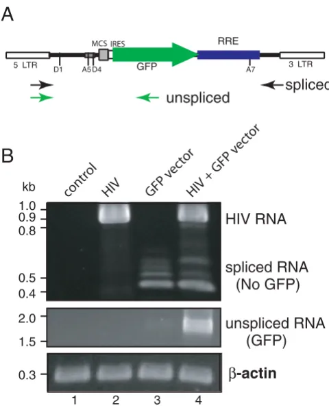
The Rev-dependent system outlined in Fig. 1 operates under the assumption that in the absence of HIV there is elimination, through splicing, of GFP-encoding tran-script. Upon Rev expression, the non-spliced RRE-con-taining GFP transcript would be rescued. To test this hypothesis, we performed RT-PCR on control and HIV-infected CEM-SS cells that were secondarily exposed to the reporter GFP-encoding viral vector at high input levels (see Fig. 4 legend). High levels of reporter vector were used to challenge the cell's capacity to remove GFP-encod-ing non-spliced transcript in the absence of HIV, that is, to
remove false-positive reporter message. We used two sets of primers that amplified the spliced or non-spliced tran-scripts from the lentiviral construct (Fig. 4A), and com-pared spliced to unspliced reporter transcript in four cell populations: uninfected cells, cells infected with HIV only, cells infected with reporter virus only, and cells infected with both virus. Note that the vectors used to detect spliced transcript will also recognize HIV tran-scripts. As shown in Fig. 4B, HIV-specific transcript, con-sistent with the size of fully spliced transcript amplified with these primers (0.94 kb), appeared in the two cell populations infected with HIV (lanes 2 and 4). In cells exposed to the reporter vector but lacking HIV, we observed basal levels of transcription from the LTR pro-moter of the reporter vector (lane 3). This transcriptional activity represents HIV-independent LTR-mediated signal (the leakiness seen with LTR-based systems); however, with this Rev-dependent system the transcripts were all spliced (lane 3; spliced RNA vs unspliced RNA) and thus lacked the capacity to generate the GFP reporter. Follow-ing exposure to the reporter viral vector, the generation of a non-spliced, reporter-encoding message (seen in lane 4) was dependent on the presence of HIV infection. These data confirmed that expression of GFP-encoding tran-script was HIV-dependent, and that the dependency corre-sponded to the prevention of transcript splicing, consistent with Rev activity.
Discussion
Specific expression of reporter by the Rev-dependent lentivirus in HIV infected T cells Figure 2
Specific expression of reporter by the Rev-dependent lentivirus in HIV infected T cells. CEM-SS cells were not infected (A) or were infected (B, C) with HIV-1 NL4-3.HSA.R+E-, an HIV-1 NL4-3 clone with the murine heat stable antigen (HSA; CD24) gene inserted into the nef gene. Both uninfected (A) and infected (C) cells were then transduced with the Rev-dependent reporter virus vNL-GFP-RRE(SA) (5 ng p24/106 cells; VSV-G envelope) or not transduced (B). At 72 hrs post transduction, cells were stained with R-phycoerythrin conjugated rat-anti-mouse CD24 antibody and analyzed by flow cytometer for CD24 and GPF expression. GFP was expressed only in HIV-1 infected (C), but not in HIV-1 uninfected (A) cells. Fifty thousand cells were examined in each run. In this run 61% of total cells were CD24-positive and of the CD24-positive population 17% were GFP-positive. Thresholds for CD24-positive and GFP-positive were set at a fluorescent intensity of 10 on the x- and y-axis respectively. Two independent analyses yielded the same result. D. Percent HIV-infected cells that express GFP versus reporter vector input. A concentrated preparation of reporter particles was used at varied dosages in HIV-infected CEM cells, as above. Flow cytometry studies yielded percent CD24-positive cells that became GFP-positive after 3 days (y-axis) versus reporter virus input (ng p24). Non-linear curve fitting (rectangular hyperbola) yielded R2 = 99.4; maximum GFP-positive = 86.1 ± 6.3% (best-fit values ± std. error); KD = 128.1 ± 21.5 ng p24. Dashed lines are 95% confidence bands.
murine CD24
GFP
G
FP
B
A
C
murine CD24
% GFP-P
ositiv
e
Reporter Virus, ng p24
not become infected (approximately one infectious parti-cle per five cells), less than 2% of the cells should possess more than one reporter provirus. Expression of GFP reporter from the HIV-positive cells closely approximated the input reporter viral vector level (Fig. 2 and 3), suggest-ing that undetected expression of integrated vectors is not prominent. Note, however, that expression of the reporter gene is achieved in cells that are also highly supportive of transcriptional activity from the HIV provirus, that is, LTR activity is supported. Both HIV and the reporter vector use the LTR promoter.
We also examined high doses of reporter viral vector, where a majority of the cells are infected with the Rev-dependent reporter vector. Under these conditions we did not see intact reporter (unspliced) transcript or protein in HIV-negative cells. The nature of the curve that fits the reporter vector detection of HIV-infected cells (Fig. 2D) suggests that the rate-limiting step towards reporter expression follows Michaelis-Menten kinetics. This is characteristic of a single binding-site event, but at this time we are unable to define this rate-limiting process, such as the viral particle binding to the cellular receptor. Additional mechanisms leading to this non-linear func-tion would include the potentially equivalent susceptibil-ity of increasing numbers of infected cells to additional infection, as characterized by a Poisson distribution.
Con-centration of this viral particle by centrifugation permitted this examination of high vector input, but it also dimin-ished the infectivity of the viral particles. This data also predicts that most of the HIV-infected cells will be suscep-tible to detection by the reporter virus. Of course, in mixed cell populations or tissues there will be variable susceptibilities to infection by the reporter virus as pres-ently used. The inability to infect all cells by the reporter virus is perhaps the most limiting aspect of this applica-tion of the Rev-dependent vector.
In this report we have utilized the term Rev-dependence to distinguish this construct from the existing LTR-based Tat-responsive HIV reporter systems; however, with the intact LTR, we would speculate that this Rev-dependent vector should maintain responsiveness to Tat expression as well. This dual dependency is likely to contribute to the higher stringency seen. In this document, we have characterized this Rev-dependent vector in only one application (viral reporter vector), an application that is challenging and not feasible with the LTR-based reporters. At the same time we view this application as supplementary to existing methods to test cells for the presence of HIV replication. These include the staining or probing of HIV proteins and nucleic acids in fixed cells, and may include amplification with PCR [34-36]. We have also incorporated the vector into continuous cell lines (data not shown). As
[image:6.612.57.556.87.255.2]intro-Specific expression of reporter by the Rev-dependent lentiviral vector in HIV-infected primary macrophages Figure 3
duced into a transformed T cell, we find that expression from this Rev-dependent vector is insensitive to cellular activation states, thus maintaining a negative background in the absence of HIV, but has enhanced sensitivity to HIV infection, relative to many of the existing non-T cell indi-cator cell lines. Although this report has been limited to the expression of reporter genes, the construct's incorpo-ration into a lentivirus and the specificity acquired should also permit its use as an experimental agent to express any gene in HIV-infected cells.
Conclusion
The inclusion of Rev-dependent processing to an LTR-based vector has resulted in a highly specific HIV-depend-ent expression system.
Methods
Viruses and cellsViruses and cells were obtained through the NIH AIDS Research and Reference Reagent Program: pNL4-3.HSA.R+E- from Dr. Nathaniel Landau [29]; CEM-SS from Dr. Peter L. Nara [37]; J1.1 cells from Dr. Thomas Folks [38]. HIV-1AD8 is a macrophage-tropic molecular
clone [39]. Macrophages were derived from peripheral blood monocytes of healthy donors from the Department of Transfusion Medicine at the National Institutes of Health. Cells were cultured in RPMI media supplemented with 10% fetal bovine serum (Hyclone) and recombinant human macrophage colony stimulating factor (rH-MCSF) (R & D Systems) at 10 ng/ml. Media was changed every 48 hours for ten days.
DNA constructs
pCMV∆R8.2 and pMD.G have been described previously [28]. pNL-GFP-RRE was constructed by complete deletion of all HIV ORFs of pNL4-3 [40] by replacing the 8.1 kb BssHII-BlpI fragment of the HIV-1 genomes with an insert containing the GFP ORF and the HIV-1 Rev-responsive element (RRE) including the HIV-1 sequence immediately following the BssHII site and the first 336 nucleotides of the gag ORF (the gag reading frame was disrupted by a frame shift mutation at the ClaI site by blunt end liga-tion), the GFP ORF was derived from pIRES-hrGFP-1a (Stratagene) by PCR amplification (5' CTCGAAATTAAC-CCTCACTAAAGG 3'; 5'ATCGTGTACGGCCGAATTGGGT-ACACTTACCTG 3'), and the fragment containing RRE (corresponding to position 7612 to 8469 of the HIV-1NL4-3 genome). pNL-GFP-RRE-(SA) was constructed by insertion of a PCR fragment into the NotI-SmaI site of pNL-GFP-RRE, in front of the GFP ORF. The insert carry-ing the HIV-1 A5 spliccarry-ing acceptor and D4 donor was amplified by primers: 5' ATAAGAATGCGGCCGCATCTC-CTATGGCAGGAAG 3'; 5' AATCACCCGGGTGCTAC-TACTAATGCTACTATTGC 3'. The sequence of pNL-GFP-RRE-(SA) has been deposited in GenBank (accession number EF408805).
Virus generation
Stocks of the HIV-1NL4-3 and HIV-1AD8 were prepared by transfection of HeLa cells with cloned proviral DNA. HIV-1 based lentiviral vectors carrying reporter genes were made by cotransfection of DNA constructs as follows: 2 × 10⁶ 293T cells were cotransfected with 10 ug of
pNL-GFP-RRE-SA, 7.5 ug of pCMV∆R8.2, and 2.5 ug of pMD.G using the calcium phosphate method (Promega). Viral particles were harvested 2 days after cotransfection and
[image:7.612.54.294.87.383.2]fil-Reverse transcriptase PCR analyses of gene expression from lentiviral vector NL-GFP-RRE-(SA) in response to HIV-1 infection
Figure 4
Reverse transcriptase PCR analyses of gene expression from lentiviral vector NL-GFP-RRE-(SA) in response to HIV-1 infection. (A) A diagram of PCR primers used. Two sets of primers were used to detect the spliced (black arrows) and unspliced (green arrows) transcripts by reverse transcriptase PCR (RT-PCR) as described in Methods. The mRNA mole-cules from infected and uninfected cells were extracted at day 3 after lentiviral vector infection and analyzed by RT-PCR for the spliced transcripts and unspliced transcripts as shown in (B). (B) CEM-SS cells were either uninfected (lane 1, con-trol), infected with HIV alone (lane 2, HIV, VSV-G pseudo-typed, 564 ng p24 per million cells), infected with NL-GFP-RRE-(SA) alone (lane 3, 1764 ng p24 virion per million cells), or infected with HIV first, then NL-GPF-RRE(SA) 24 hours later (lane 4). The cellular β-actin transcript was also ampli-fied to ensure that comparable numbers of cells were used for the analyses.
B
5 LTR 3 LTR
D1 A5D4 GFP A7 RRE IRES
MCS
spliced
unspliced
A
spliced RNA
(No GFP)
unspliced RNA
(GFP)
HIV RNA
1 2 3 4
β
-actin
0.32.0
1.5 kb
0.9 0.8 1.0
0.5 0.4
HIV + GFP v ector
GFP v ector
HIV cont
tered through a 0.45 um filter and stored at -80°. One preparation was concentrated by ultracentrifugation [41]. The titer (TCID50) of the lentiviral indicator vNL-GFP-RRE(SA) virus preparation was estimated by serial dilu-tion into activated (TNF-treated) HIV-positive J1.1 Jurkat cells [38] following the method of Reed and Muench [42].
RT-PCR analysis
Total cellular poly(A+) mRNA was purified from cells by
MicroPoly(A)Pure mRNA isolation kit (Ambion) as rec-ommended by the manufacturer. Reverse transcription was accomplished using the RETROscript First-Strand Synthesis Kit (Ambion) with random decamers as the first-strand primers. Following cDNA synthesis, PCR was carried out using primer 5'TAATCGGCCGAACAGGGACTTGAAAGCGAAAG3' and 5'CAGGCACAAGCAGCATTGTTAG 3' to amplify spliced lentiviral transcripts, and primer 5' TAATCGGCCGAACAGGGACTTGAAAGCGAAAG3' and 5'ATCGTGTACGGCCGAATTGGGTACACTTACCTG3' to amplify non-spliced GFP transcripts. The PCR condition was: 1 × PCR buffer, 125 uM dNTPs, 1.5 mM Mg2+, 50
pmol of each primer, 1 U SuperTaq Plus (Ambion) in 50 ul, with 30 cycles of 20 sec at 94°, and 180 sec at 68°. One fifth of the product was analyzed on 2% agarose gel. Cel-lular β-actin transcripts were amplified using Quantum-RNA β-actin Internal Standards (Ambion) with similar conditions as above except using 20 pmol of each actin primer and runs at 20 sec at 94°, 30 sec at 55°, and 40 sec at 68°.
Reporter virus detection of HIV infection
CEM-SS cells, infected with VSV pseudo-typed HIV-1 NL4-3.HSA.R+E- [29], were enriched by biotin conjugated rat-anti-mouse CD24 antibody and streptavidin conjugated magnetic beads and further cultured for two weeks to remove the beads. The enriched population was then infected with the Rev-dependent lentiviral vector, vNL-GFP-RRE(SA). At 72 hrs post transduction, cells were stained with R-phycoerythrin conjugated rat-anti-mouse CD24 antibody and analyzed by flow cytometer for CD24 and GPF expression. Curve fitting and statistical analysis were achieved with GraphPad Prism software.
Monocyte-derived macrophages in six well plates were infected with HIVAD8 (36 to 360 ng p24) in 500 ul of RPMI for 3 hours, then washed and returned to 2 ml RPMI. The following day, 5 ng p24 of pNL-GFP-RRE(SA) was applied to the macrophages. At 5 days post infection, cells were intracellularly stained on ice. Briefly, cells were rinsed once with Hank's buffer followed by treatment with Cytofix/Cytoperm (Becton Dickinson) for 20 min, then 2 ml of cold methanol for 15 min. After washing with Per-meabilization/Wash buffer (Becton Dickinson), cells were stained with anti-p24 HIV antibody 183.H12-5C (1:250)
[43-45] for 90 min, followed by washing and staining with Alexa Fluor-568 goat anti-mouse IgG (Invitrogen) (1:250) for 30 minutes.
Fluorescent microscopy of monocyte-derived macrophages
The GFP-expressing and anti-p24 stained cells were pho-tographed using a Leica DMIRB/E inverted research microscope attached to a Orca DCAM ER camera (Hamamtsu), using an XF102.2 filter (Omega Optical Inc.) for the Alexa Fluor-568 (p24) detection and a GFP filter (Leica) for the GFP expressing cells. P24 expression and GFP expression were captured with similar exposures, typically 1000 milliseconds. Files were saved as Tiffs and color was added on ImageJ software version 1.33u (Ras-band, W., NIH-public domain). Dimensions were deter-mined through a stage micrometer (Electron Microscopy Sciences).
Flow cytometry
Infected or uninfected cells were re-suspended into 100 ul Hank's staining buffer (Hank's buffer plus 0.1% bovine serum albumin, 0.1% sodium azide, and 25 mM HEPES, pH 7.2) for staining with antibodies at concentrations rec-ommended by manufactures. Following staining for 30 min on ice, cells were either washed with Hank's buffer or, in the case of HIV-infected cells, fixed with 500 ul of Cytofix/Cytoperm (Becton Dickenson) for 20 min on ice for flow cytometry analysis on a FACScan analyzer (Bec-ton Dickenson). The murine CD24 antibody was from Southern Biotechnology.
Competing interests
A patent application for the vector has been submitted by NIMH with YW and JWM.
Authors' contributions
YW, MB, and JM performed experiments, performed data analysis, contributed to experimental design, and contrib-uted to writing this paper.
Acknowledgements
We thank Dongyang Yu for technical expertise with the PCR run, and Drs. Michael Emerman, Tomris Mustafa, and Sundararajan Venkatesan for their comments and criticisms concerning this manuscript. This research was supported by the Intramural Research Program of the NIMH/NIH and by the NIH grant 5R21NS51130-2 awarded to YW.
References
1. Felber BK, Pavlakis GN: A Quantitative Bioassay for Hiv-1 Based on Trans-Activation. Science 1988, 239(4836):184-187.
4. Kimpton J, Emerman M: Detection of Replication-Competent and Pseudotyped Human-Immunodeficiency-Virus with a Sensitive Cell-Line on the Basis of Activation of an Integrated Beta-Galac-tosidase Gene. Journal of Virology 1992, 66(4):2232-2239.
5. Muesing MA, Smith DH, Capon DJ: Regulation of Messenger-Rna Accumulation by a Human-Immunodeficiency-Virus Transacti-vator Protein. Cell 1987, 48(4):691-701.
6. Dingwall C, Ernberg I, Gait MJ, Green SM, Heaphy S, Karn J, Lowe AD, Singh M, Skinner MA, Valerio R: Human Immunodeficiency Virus-1 Tat Protein Binds Trans-Activation-Responsive Region (Tar) Rna Invitro. Proceedings of the National Academy of Sciences of the United States of America 1989, 86(18):6925-6929.
7. Berkhout B, Silverman RH, Jeang KT: Tat Trans-Activates the Human Immunodeficiency Virus through a Nascent Rna Target. Cell 1989, 59(2):273-282.
8. Kao SY, Calman AF, Luciw PA, Peterlin BM: Anti-Termination of Tran-scription within the Long Terminal Repeat of Hiv-1 by Tat Gene-Product. Nature 1987, 330(6147):489-493.
9. Laspia MF, Rice AP, Mathews MB: Hiv-1 Tat Protein Increases Tran-scriptional Initiation and Stabilizes Elongation. Cell 1989,
59(2):283-292.
10. Kato H, Sumimoto H, Pognonec P, Chen CH, Rosen CA, Roeder RG: Hiv-1 Tat Acts as a Processivity Factor Invitro in Conjunction with Cellular Elongation-Factors. Genes & Development 1992,
6(4):655-666.
11. Raha T, Cheng SWG, Green MR: HIV-1 Tat stimulates transcription complex assembly through recruitment of TBP in the absence of TAFs. Plos Biology 2005, 3(2):221-230.
12. Brady J, Kashanchi F: Tat gets the "green" light on transcription ini-tiation. Retrovirology 2005, 2:.
13. Jordan A, Defechereux P, Verdin E: The site of HIV-1 integration in the human genome determines basal transcriptional activity and response to Tat transactivation. Embo Journal 2001,
20(7):1726-1738.
14. Gervaix A, West D, Leoni LM, Richman DD, WongStaal F, Corbeil J: A new reporter cell line to monitor HIV infection and drug suscep-tibility in vitro. Proceedings of the National Academy of Sciences of the United States of America 1997, 94(9):4653-4658.
15. Feinberg MB, Jarrett RF, Aldovini A, Gallo RC, Wongstaal F: Htlv-Iii Expression and Production Involve Complex Regulation at the Levels of Splicing and Translation of Viral-Rna. Cell 1986,
46(6):807-817.
16. Malim MH, Hauber J, Fenrick R, Cullen BR: Immunodeficiency Virus Rev Trans-Activator Modulates the Expression of the Viral Reg-ulatory Genes. Nature 1988, 335(6186):181-183.
17. Emerman M, Vazeux R, Peden K: The Rev Gene-Product of the Human Immunodeficiency Virus Affects Envelope-Specific Rna Localization. Cell 1989, 57(7):1155-1165.
18. Robertguroff M, Popovic M, Gartner S, Markham P, Gallo RC, Reitz MS:
Structure and Expression of Tat-Specific, Rev-Specific, and Nef-Specific Transcripts of Human-Immunodeficiency-Virus Type-1 in Infected Lymphocytes and Macrophages. Journal of Virology 1990,
64(7):3391-3398.
19. Purcell DFJ, Martin MA: Alternative Splicing of Human-Immunode-ficiency-Virus Type-1 Messenger-Rna Modulates Viral Protein Expression, Replication, and Infectivity. Journal of Virology 1993,
67(11):6365-6378.
20. Kim SY, Byrn R, Groopman J, Baltimore D: Temporal Aspects of DNA and Rna-Synthesis During Human Immunodeficiency Virus-Infection - Evidence for Differential Gene-Expression. Journal of Virology 1989, 63(9):3708-3713.
21. Klotman ME, Kim S, Buchbinder A, Derossi A, Baltimore D, Wongstaal F:
Kinetics of Expression of Multiply Spliced Rna in Early Human-Immunodeficiency-Virus Type-1 Infection of Lymphocytes and Monocytes. Proceedings of the National Academy of Sciences of the United States of America 1991, 88(11):5011-5015.
22. Sodroski J, Goh WC, Rosen C, Dayton A, Terwilliger E, Haseltine W: A 2nd Posttranscriptional Transactivator Gene Required for Htlv-Iii Replication. Nature 1986, 321(6068):412-417.
23. Knight DM, Flomerfelt FA, Ghrayeb J: Expression of the Art Trs Pro-tein of Hiv and Study of Its Role in Viral Envelope Synthesis. Sci-ence 1987, 236(4803):837-840.
24. Schwartz S, Campbell M, Nasioulas G, Harrison J, Felber BK, Pavlakis GN:
Mutational Inactivation of an Inhibitory Sequence in Human-Immunodeficiency-Virus Type-1 Results in Rev-Independent Gag Expression. Journal of Virology 1992, 66(12):7176-7182.
25. Kotsopoulou E, Kim VN, Kingsman AJ, Kingsman SM, Mitrophanous KA:
A Rev-independent human immunodeficiency virus type 1 (HIV-1)-based vector that exploits a codon-optimized HIV-1 gag-pol gene. Journal of Virology 2000, 74(10):4839-4852.
26. Hope TJ, Huang XJ, McDonald D, Parslow TG: Steroid-Receptor Fusion of the Human-Immunodeficiency-Virus Type-1 Rev Transactivator - Mapping Cryptic Functions of the Arginine-Rich Motif. Proceedings of the National Academy of Sciences of the United States of America 1990, 87(19):7787-7791.
27. Ragheb JA, Bressler P, Daucher M, Chiang L, Chuah MKL, Vandendriess-che T, Morgan RA: Analysis of Transdominant Mutants of the Hiv
Type-1 Rev Protein for Their Ability to Inhibit Rev Function, Hiv Type-1 Replication, and Their Use as Anti-Hiv Gene Therapeu-tics. Aids Research and Human Retroviruses 1995, 11(11):1343-1353. 28. Naldini L, Blomer U, Gallay P, Ory D, Mulligan R, Gage FH, Verma IM,
Trono D: In vivo gene delivery and stable transduction of nondi-viding cells by a lentiviral vector. Science 1996, 272(5259):263-267. 29. He JL, Choe S, Walker R, Dimarzio P, Morgan DO, Landau NR: Human-Immunodeficiency-Virus Type-1 Viral-Protein-R (Vpr) Arrests Cells in the G(2) Phase of the Cell-Cycle by Inhibiting P34(Cdc2) Activity. Journal of Virology 1995, 69(11):6705-6711.
30. Henderson AJ, Calame KL: CCAAT/enhancer binding protein (C/ EBP) sites are required for HIV-1 replication in primary macro-phages but not CD4(+) T cells. Proceedings of the National Academy of Sciences of the United States of America 1997, 94(16):8714-8719. 31. Lee ES, Sarma D, Zhou HY, Henderson AJ: CCAAT/enhancer binding
proteins are not required for HIV-1 entry but regulate proviral transcription by recruiting coactivators to the long-terminal repeat in monocytic cells. Virology 2002, 299(1):20-31.
32. Orenstein JM, Meltzer MS, Phipps T, Gendelman HE: Cytoplasmic Assembly and Accumulation of Human Immunodeficiency Virus Type-1 and Type-2 in Recombinant Human Colony-Stimulating Factor-1-Treated Human-Monocytes - an Ultrastructural-Study.
Journal of Virology 1988, 62(8):2578-2586.
33. Pelchen-Matthews A, Kramer B, Marsh M: Infectious HIV-1 assembles in late endosomes in primary macrophages. Journal of Cell Biology 2003, 162(3):443-455.
34. Bagasra O, Hauptman SP, Lischner HW, Sachs M, Pomerantz RJ: Detec-tion of Human-Immunodeficiency-Virus Type-1 Provirus in Mononuclear-Cells by Insitu Polymerase Chain-Reaction. New England Journal of Medicine 1992, 326(21):1385-1391.
35. Patterson BK, Till M, Otto P, Goolsby C, Furtado MR, McBride LJ, Wolin-sky SM: Detection of Hiv-1 DNA and Messenger-Rna in Individual Cells by Pcr-Driven Insitu Hybridization and Flow-Cytometry.
Science 1993, 260(5110):976-979.
36. Embretson J, Zupancic M, Beneke J, Till M, Wolinsky S, Ribas JL, Burke A, Haase AT: Analysis of Human Immunodeficiency Virus-Infected Tissues by Amplification and Insitu Hybridization Reveals Latent and Permissive Infections at Single-Cell Resolution. Proceedings of the National Academy of Sciences of the United States of America 1993,
90(1):357-361.
37. Nara PL, Hatch WC, Dunlop NM, Robey WG, Arthur LO, Gonda MA, Fischinger PJ: Simple, Rapid, Quantitative, Syncytium-Forming Microassay for the Detection of Human Immunodeficiency Virus Neutralizing Antibody. Aids Research and Human Retroviruses 1987,
3(3):283-302.
38. Perez VL, Rowe T, Justement JS, Butera ST, June CH, Folks TM: An Hiv-1-Infected T-Cell Clone Defective in Il-2 Production and Ca2+ Mobilization after Cd3 Stimulation. Journal of Immunology 1991,
147(9):3145-3148.
39. Theodore TS, Englund G, BucklerWhite A, Buckler CE, Martin MA, Peden KWC: Construction and characterization of a stable full-length macrophage-tropic HIV type 1 molecular clone that directs the production of high titers of progeny virions. Aids Research and Human Retroviruses 1996, 12(3):191-194.
40. Adachi A, Gendelman HE, Koenig S, Folks T, Willey R, Rabson A, Martin MA: Production of Acquired Immunodeficiency Syndrome-Asso-ciated Retrovirus in Human and Nonhuman Cells Transfected with an Infectious Molecular Clone. Journal of Virology 1986,
59(2):284-291.
41. Burns JC, Friedmann T, Driever W, Burrascano M, Yee JK: Vesicular Stomatitis-Virus G Glycoprotein Pseudotyped Retroviral Vec-tors - Concentration to Very High-Titer and Efficient Gene-Transfer into Mammalian and Nonmammalian Cells. Proceedings of the National Academy of Sciences of the United States of America 1993,
90(17):8033-8037.
42. Reed LJ, Muench H: A simple method of estimating fifty percent endpoints. American Journal of Hygiene 1938, 27(3):493-497.
43. Chesebro B, Wehrly K, Nishio J, Perryman S: Macrophage-Tropic Human-Immunodeficiency-Virus Isolates from Different Patients Exhibit Unusual V3 Envelope Sequence Homogeneity in Comparison with T-Cell-Tropic Isolates - Definition of Critical Amino-Acids Involved in Cell Tropism. Journal of Virology 1992,
66(11):6547-6554.
44. Wehrly K, Chesebro B: p24 antigen capture assay for quantification of human immunodeficiency virus using readily available inex-pensive reagents. Methods-a Companion to Methods in Enzymology 1997,
12(4):288-293.
45. Toohey K, Wehrly K, Nishio J, Perryman S, Chesebro B: Human-Immu-nodeficiency-Virus Envelope V1 and V2 Regions Influence Repli-cation Efficiency in Macrophages by Affecting Virus Spread.