FORMULATION AND EVALUATION OF SUSTAINED
RELEASE MUCOADHESIVE CIPROFLOXACIN HCl
MICROPARTICLES
A Dissertation submitted to
THE TAMIL NADU DR. M.G.R. MEDICAL UNIVERSITY, CHENNAI
In partial fulfillment of the requirement for the award of the degree of
MASTER OF PHARMACY
(PHARMACEUTICS)
Submitted by
Reg. No: 26104211
Under the guidance of
Mrs. P.Amsa, M. Pharm.,
Department of Pharmaceutics
MAY 2012
Department of Pharmaceutics,
Nandha College of Pharmacy, Erode-638 052
CERTIFICATE
This is to certify that the work embodied in this thesis entitled, “FORMULATION
AND EVALUATION OF SUSTAINED RELEASE MUCOADHESIVE CIPROFLOXACIN HCl MICROPARTICLES” submitted to The Tamilnadu Dr. M.G.R. Medical University, Chennai, was carried out by ( Reg.No.26104211) in the Department of Pharmaceutics, Nandha College of Pharmacy, Erode-52 for the partial fulfillment for the award of degree of Master of Pharmacy in Pharmaceutics under my supervision.
This work is original and has not been submitted in part or full for another degree or diploma of this or any other university.
Asst.Prof.P.Amsa,
Place : Erode - 52 Project Guide
The work presented in this thesis entitled “FORMULATION AND EVALUATION
OF SUSTAINED RELEASE MUCOADHESIVE CIPROFLOXACIN HCl MICROPARTICLES” was carried out by me in the Department of Pharmaceutics, Nandha College of Pharmacy, Erode-52 under the direct supervision of Assist. Prof
.
P.Amsa,
M.Pharm.,
Nandha College of Pharmacy, Erode-52.This work is original and has not been submitted in part or full for the award of any other degree or diploma of any other University.
Place: Erode Reg. No.
26104211
Date:
First of all, I think the god who is constantly showering his blessing on me, it is my great privilege.
Pride and honour in expressing my humble thanks to my esteemed teacher and guide
Mrs. P.AMSA., M.Pharm, Asst. Professor, Department of Pharamaceutics, Nandha College of Pharmacy, Erode for her valuable guidance, keen interest, inspiration and constant encouragement throughout the course of this investigation. Without her valuable advice and deep-rooted knowledge, this work would not have been a reality.
It is proud to express my sincere thanks to Dr. T.Sivakumar, M.Pharm, Ph.D., principal of Nandha College of Pharmacy, with a deep sense of gratitude for his encouragement co-operation, kind suggestion and providing the best facilities during this work.
I express my profound indulge to Dr. S.Tamilzharsi, M.Pharm, Ph.D, HOD., DEPARTMENT OF PHARMACEUTICS, Nandha College of Pharmacy, Erode -52, for providing much of his effort in the form of suggestion, guidance, encouragement throughout the course of this thesis.
I express my loyal thanks to Thiru.V. Shanmugam, B.Com, Chairman and Mr. Nandha Kumar Pradeep, M.B.A, Secretary, Nandha College of Pharmacy, Erode-52,
for providing all the facilities to make this work a success.
I owe my warm and humble thanks to Mr. M.Jagadeeswaran, M.Pharm., Asst. Prof. Dept. of Pharm. Analysis, Mr. K.Raja, M.Parm (Ph.D)., Asst. Prof. Dept. of Pharmaceutics, Dr.Sengottuvelu., M.Pharm., Ph.D, Departmet of Pharmacology, Dr. R.DuraiSamy, M.Pharm.Ph.D., HOD, Dept. of Pharmacognosy. Dr.P.R. Radhika, M.Pharm., Ph.D., Prof. Dept. of Pharmaceutics, kind suggestion and providing the best facilities during this work.
I express my deepest and special thanks to my batch mates Mr. Ravi, Mr. Praveen, Mr.Sanath, Mr.Rajanish Reddy, Mr.Subhash, Mr.Prakash, Ms.Pavani, Mrs.Reepa Patel for their kind of co-operation help and encouragement throughout my post- graduation.
teaching staff of Nandha College of Pharmacy for their kind co-operation and help throughout the course.
With no words I can hearties and deep gratitude to my dear friends who always believed In me and stood with me in good and bad times, special thanks to them for their friendship adherent love affection , encouragement they always showered on me. I am thanking to my juniors who have contributed directly and indirectly during my dissertation.
To completion of this dissertation is not only fulfillment of my dreams but also the dreams of my parents who have taken lots of pain for me in completion of higher studies.
A word thanks to all those gentle people associate with this work directly or indirectly whose names have been to unable to mention here.
Thank you to one and all…………
Place: Erode – 52. Reg.No: 26104211
Date: M.Pharm, II Year,
µg - Microgram
mg - Milligram
ng - Nanogram
gm - Gram
µm - Micrometer
nm - Nanometer
cm - Centimeter
ml - Mililitre
hrs - Hours
DDS - Drug Delivery System
CR - Controlled Release
SRM - Sustained Release Mucoadhesive
G.I.T. - Gastro Intestinal Tract
CMC - Carboxy Methyl Cellulose
HPMC - Hydroxy Propyl Methyl Cellulose
FN - Formulation Number
I
SR.NO PARTICULARS PAGE NO.
1.0 2.0 3.0 4.0 5.0 6.0 7.0 INTRODUCTION
1.1 Microspheres
1.2 Polymer Used In Microspheres 1.3 Route Of Administration
1.3.1 Oral Delivery 1.3.2 Parenteral Delivery
1.4 General Methods Of Preparation 1.5 Application of Microspheres 1.6 Mucoadhesion
1.7 Fundamentals Of Bioadhesion 1.8 Mechanism Of Bioadhesion
REVIEW OF LITERATURE DRUG PROFILE
POLYMER PROFILE RESEARCH ENVISAGED
5.1 Aim of work 5.2 Plan of work
MATERIALS AND EQUIPMENTS EXPERIMENTAL INVISTIGATION
7.0 Preformulation Study 7.1 Organoleptic Properties 7.2 Standard caliberation curve
7.2.1 Preparation of standard curve of Ciprofloxacin HCl With 0.1 N HCl.
II
8.0
9.0 10.0 11.0
7.3 Drug Excipients interaction study by FTIR Spectrum 7.4 Preparation of Microspheres
7.5 Evaluation of microparticles
7.5.1 Bulk density of the microparticles 7.5.2 Particle Size Analysis
7.5.3 Drug Content
7.5.4 Entrapment efficiency
7.5.5 In-vitro test for mucoadhesion 7.5.6 In-vitro drug release study 7.5.7 Kinetic modeling
7.5.8 Morphology
7.5.9 HPLC Analysis of Ciprofloxacin HCl 7.5.10 Stability studies
RESULTS AND DISCUSSION
SUMMARY
CONCLUSION REFERENCES
48
65
67
SL NO. TABLE PAGE NO.
1 List of chemicals 38
2 List of Equipments 38
3 Description and composition of ciprofloxacin HCl microspheres. 41
4 Mechanism of drug release as per Korsmeyer Eqution / Peppa’s
model
45
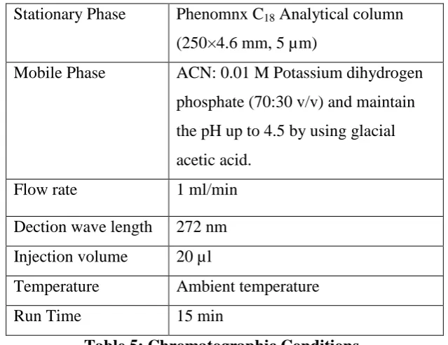
4 HPLC Chromatic conditions 47
5 Calibration curve of Ciprofloxacin. 48
6 FT-IR Studies 49
7 Evaluation parameters 53
8 In-vitro release study parameters 54
9 Correlation coefficients of different mathematical models for formulations F-1
58
Department of Pharmaceutics, Nandha College of Pharmacy, Erode-52. Page 1
1.0. INTRODUCTION
The drug should be delivered to site at the specific target sites at a rate and concentration that permit optimal therapeutic efficacy while reducing side effects to minimum. The other aspect to be considered in drug delivery is patient compliance during the drug therapy.
The concept of the advanced drug delivery systems especially those offering a sustained and controlled action of drug to desired area of effect, attained great appeal for nearly half a century. However, prior to advent of improved alternate methods, drug delivery systems were considered only as a means of getting the drug in to the patient’s body. The actual practice of controlled release began with advent of timed release coating to the pills or solid drug particles in order to mask their unacceptable taste or make them more palatable.
In the mid 1940 - 1960s, the concept of chemical microencapsulation technology began as an alternative means of delivering drugs. In continued quest for the more refined systems, in 1980s polymer membrane technology came to be known at forefront. Further, the process of targeting and site specific delivery with absolute accuracy can be achieved by attaching bioactive molecule to liposomes, bio-erodible polymer, implants, monoclonal antibodies and various particulate carriers (e.g., nanoparticles and microspheres, etc.). The micro-particulate delivery systems are considered and accepted as a reliable means to deliver the drug to the target site with specificity, if modified and to maintain the desired concentration at the site of interest without untoward effect(s).
The term microcapsule, which explains as a spherical particle with size varying from 1µm to (1000 µm) 1 mm, containing a core substance. Microspheres comprises of strict sense, spherical empty particles. However, the terms microcapsules and microspheres are often used synonymously.
1.1. Microspheres
1, 2, 20Department of Pharmaceutics, Nandha College of Pharmacy, Erode-52. Page 2
sustained controlled release fashion. One such approach is using microspheres as carrier for drugs.
Definition of Microspheres:
The Microspheres are characteristically free flowing powders consisting of proteins or synthetic polymers, which are biodegradable in nature, and ideally having a particle size less than 200 µm3.solid bio degradable microspheres incorporating a drug and dispersed or dissolved throughout particle matrix have the potential for the controlled release of drugs.
Microspheres are defined as homogeneous, monolithic particles in the size range of about 1µm-1000 µm and are widely used as drug carriers for controlled release. These systems have significant importance in biomedical applications. Microspheres can be produced for protection of core material, reduction of gastric irritation decrease in volatility, conversion of liquid to pseudo-solid, cell microencapsulation and for designing pulsatile drug delivery systems. Administration of the drug in the form of microspheres usually improves the treatment by providing the localization of the active substances at the site of action and by prolonging release of drugs.
Prerequisites for Ideal Carriers
3, 13 Longer duration of action Control of drug release
Increased of therapeutic efficiency Protection of drugs
Reduction of toxicity Biocompatibity Relative stability
Department of Pharmaceutics, Nandha College of Pharmacy, Erode-52. Page 3
1.2. Polymer used in Microspheres
These materials include the polymer of natural and synthetic origin and also modified natural substances
Synthetic Polymer
a. Non-Biodegradable
Poly methyl methacrylate9 Acrolein11 Glycidyl methacrylate
b. Biodegradable
Lactides and glycolides and their copolymers25 Polyalkyl cyano acrylate
Natural Polymer
a. Proteins
Albumins29,31 Gelatin30 Collagenb. Carbohydrates
Starch24 Agarose Carrageenan Chitosan
c. Chemically modified carbohydrates
Poly (acryl) dextran Poly (acryl) starch
Department of Pharmaceutics, Nandha College of Pharmacy, Erode-52. Page 4
1.3. Route of Administration
Microspheres can be used for the delivery of drugs via different routes. Route of administration is selected depending on the drug properties, disease state being treated and the age and condition of the patient. Desirable properties of the microspheres to be used for the delivery will also change depending on the route of administration.
1.3.1 Oral Delivery:
Oral delivery is the simplest way of drug administration. In oral drug delivery, the microspheres have to pass through frequently changing environments in the GI tract. There’s also patient to patient variation in GI content, stomach emptying time and peristaltic activity. Although constraints of the oral route are numerous, on the whole, it offers less potential danger than the parenteral route. The relatively brief transit time of about 12 hrs through the GI tract limits the duration of action that can be expected via the oral route. Recently, it has been reported that microspheres of <10 µm in size are taken up by the payer’s patches and may increase the retention time in the stomach. Also microspheres made from polymers with mucoadhesive properties get attached the stomach and prolong the residence time in the stomach. Bioavailability of the drugs with limited solubility in the stomach or intestine and small absorption rate constant can be increased by increasing the retention time in the stomach. improved drug delivery was observed compared to the microspheres administered alone.
1.3.2 Parenteral Delivery:
Department of Pharmaceutics, Nandha College of Pharmacy, Erode-52. Page 5
Drug Loading and Drug Release Kinetics
3, 13The active components are loaded over the microspheres principally using two methods, i.e. during the preparation of microspheres or after the formation of the microspheres by incubating them with the drug protein. The active components can be loaded by means of the physical entrapment, chemical linking and surface adsorption. The entrapment largely depends on the method of preparation and nature of the drug or polymer.
Maximum loading can be achieved by incorporating the drug during the time of preparation but it may get affected by many other process variables such as method of preparation, presence of additives (e.g. cross linking agent,surfactant,stabilizers,etc.) heat of polymerization, agitation intensity etc. Release of the active constituents is an important consideration is case of microspheres.
Mechanism of Drug Release
1, 3, 13 Osmotically driven burst mechanism Pore diffusion mechanism Erosion or degradation of the polymer
a)
Osmotically Driven Burst Mechanism:
Water diffuse into the core through biodegradable or non-biodegradable coating, creating sufficient pressure that ruptures the membrane. The burst effect is mainly controlled by three factors the macromolecules/Polymer ratio, particle size of the dispersed macromolecule and the particle size of the microspheres
.
b)
Pore Diffusion Method:
Penetrating water front continue to diffuse towards the core.
c)
Erosion:
Department of Pharmaceutics, Nandha College of Pharmacy, Erode-52. Page 6
1.4. General Methods of Preparation
1, 3The Microsphere can be prepared by using any of the several techniques discussed in the following
Single emulsion technique Double emulsion technique Polymerization technique
a. Normal polymerization Bulk polymerization Suspension polymerization Emulsion polymerization
b. Interfacial polymerization
Phase separation coacervation techniques Spray drying and spray congealing Solvent extraction
Solvent evaporation technique
But the choice of technique mainly depend on the nature of polymer used, the drug, the intended use and the duration of therapy
Some formulation and technology related factors are mentioned below The particle size requirement
The drug or the protein should not be adversely affected by the process Reproducibility of the release profile and method
No stability problem
There should be no toxic products associated with the final product.
Method of Preparation
Preparation of Microsphere should satisfy certain criteria:
The ability to incorporate reasonably high concentration of the drug
Stability of the preparation after synthesis with a clinically acceptable shelf life Controlled particle size and dispersability in aqueous vehicles for injection Release of active drug with a good control over a wide time scale.
Department of Pharmaceutics, Nandha College of Pharmacy, Erode-52. Page 7
Single Emulsion Technique:
The micro particulate carriers of natural polymers i.e. those of proteins and carbohydrates are prepared by single emulsion technique. The natural polymer are dissolved or dispersed in aqueous medium followed by dispersion in non-aqueous medium like liquid paraffin.
Next cross linking of the dispersed globule is carried out. The cross linking is achieved either by means of heat or by using the chemical cross linkers. E.g. the chemical cross linking agents are glutaraldehyde, formaldehyde, Di acid chloride etc.
Disadvantages
Heat denaturation is not suitable for thermolabile substance.
Chemical cross linking suffers the excessive exposure of active ingredient to chemicals if added at the time of preparation and then subjected to centrifugation, washing and separation.
Double Emulsion Technique:
Double emulsion method of involves the formation of the multiple emulsions or the double emulsion of type w/o/w and is best suited for water soluble drugs, peptides, proteins and vaccines.
This method can be used with both the natural as well as synthetic polymers. The aqueous protein solution is dispersed in a lipophilic organic continuous phase. This protein solution may contain the active constituents. The continuous phase generally consisted of the polymer solution that eventually encapsulates of the protein contained in dispersed aqueous phase.
Department of Pharmaceutics, Nandha College of Pharmacy, Erode-52. Page 8
Polymerization Techniques
The polymerization techniques conventionally used for the preparation of the microspheres are mainly classified as:
a. Normal polymerization
b. Interfacial polymerization both are carried out in liquid phase.
a. Normal Polymerization
It is carried out using different technique as bulk, suspension, precipitation, Emulsion and micellar polymerization
.
Bulk Polymerization Technique
Monomer or a mixture of monomer along with the initiate polymerization. Polymer so obtained may be moulded as microspheres. Drug loading may be done during the process of polymerization
.
Suspension Polymerization Technique
Suspension polymerization also referred as bead or pearl polymerization is carried out by heating the monomer or mixture of monomer as droplets dispersion in a continuous aqueous phase. The droplet may also contain an initiator and other additives.
Emulsion Polymerization Technique
It is differs from suspension polymerization as due to the presence initiator in the aqueous phase, which later on diffuses to the surface of micelles
.
b. Interfacial Polymerization
It involves the reaction of various monomers at the interface between the two immiscible liquid phases to from a film of polymer that essentially envelopes the dispersed phase.
Phase Separation Coacervation Technique:
This process is based on the principle of decreasing the solubility of the polymer in organic phase to affect the formation of polymer rich phase called the coacervates.
Department of Pharmaceutics, Nandha College of Pharmacy, Erode-52. Page 9
achieving the coacervates determines the distribution of the polymer film, the particle size and agglomeration of the formed particle.
The agglomeration must be avoided by stirring the suspension using a suitable speed stirrer since as the process of microspheres formation begins the formed polymerize globules start to stick and form the agglomerates. Therefore the process variables are critical as they control the kinetics of the formed particles since there is no defined state of equilibrium attainment.
Spray Drying and Spray Congealing
These methods are based on the drying of the mist of the polymer and drug in the air. Depending upon the removal of the solvent or cooling of the solution, the two processes are named spray drying and spray congealing respectively.
The polymer is first dissolved in a suitable volatile organic solvent such as dichloromethane, acetone, etc. The drug in the solid form is then dispersed in the polymer solution under high speed homogenization. This dispersion is then atomized in a stream of hot air. The atomization leads to the formation of the small droplets or the fine mist from which the solvent evaporates instantaneously leading the formation of the microspheres in a size range 1-100µm.
Microparticles are separated from the hot air by means of the cyclone separator while the trace of solvent is removed by vacuum drying
.
Advantage
Feasibility of operation under aseptic condition. The spray drying process is used to encapsulate various penicillin’s. Thiamine mononitrate of mono and diglycerides of stearic acid and palmitic acid using spray congealing. Very rapid solvent evaporation, however leads to the formation of porous microparticles
.
Solvent Extraction
Solvent extraction method is used for the preparation of microparticles, involves removal of the organic phase by extraction of organic solvents such as isopropanol .organic phase is removed by extraction with water. This process decrease the hardening time for the microspheres.
Department of Pharmaceutics, Nandha College of Pharmacy, Erode-52. Page 10
temperature of water, ratio of emulsion volume to the water and the solubility profile of the polymer.
Solvent Evaporation
1, 4Solvent evaporation is carried out by maintain emulsion at reduced pressure or by stirring the emulsion so that the organic phase evaporation out. In the latter case, the emulsion is added to the large quantity of water into which organic phase diffuse out. The solid microspheres are subsequently obtained by filtration and washing.
Advantage
High encapsulation efficiency of moderately water soluble and water-insoluble compounds
Water soluble drugs such as theophylline, caffeine, and salicylic acid could be loaded efficiently using an o/o emulsion method.
Versatile and easy
It’s possible to achieve various drug release profiles by the regulation of copolymer ratio, molecular weight, and size of the microsphere, drug loading, porosity and other formulation parameter.
1.5. Applications of Microspheres
1, 3, 13:
Microspheres in Vaccine Delivery:
The prerequisite of a vaccine protection against the microorganism or its toxic product. An ideal vaccine must fulfill the requirement of efficacy, safety, convenience in application and cost. The aspect of safety and minimization of adverse reaction is a complex issue. The aspect of safety and the degree of the production of antibody responses are closely related to mode of application. Biodegradable delivery systems for vaccines that are given by parenteral (subcutaneous, intramuscular, intradermal) carrier lies since they offer specific advantages like:
Improved antigenicity by adjuvant action Modulation of antigen release
Department of Pharmaceutics, Nandha College of Pharmacy, Erode-52. Page 11
Targeting Using Micro particulate Carriers
The concept of targeting i.e. site specific drug delivery is a well-established dogma, which is gaining full attention. The therapeutic efficacy of the drug release on its access and specific interaction with its candidate receptors. The ability to leave the pool in reproducible, efficient and specific manner is center to drug action mediated by use of a carrier system. Placement of the particles indiscrete anatomical compartment leads to their retention either because of the physical properties of the environment or biophysical interaction of the particle with the cellular content of the target tissue.
Monoclonal Antibodies Mediated Microspheres Targeting
Monoclonal antibodies targeting microspheres are immuno microspheres. This targeting is a method used to achieve selective targeting to the specific sites. Monoclonal antibodies are extremely specific molecule. This extreme specificity of monoclonal antibodies (Mabs) can be utilized to target microspheres loaded bioactive molecule to selected sites. Mabs can be directly attached to the microspheres by means of covalent coupling. The free aldehyde groups, amino groups or hydroxyl groups on the surface of the microspheres can be linked to the antibodies. The Mabs can be attached to microspheres by any of the following methods
Non specific adsorption Specific adsorption Direct coupling Coupling via reagent
Chemoembolization
Department of Pharmaceutics, Nandha College of Pharmacy, Erode-52. Page 12
Imaging
The microspheres have been extensively studied and used for the targeting purposes. Various cells, cell lines, tissue and organs can be imaged using radio labeled microspheres. The particle size range of Microspheres is an important factor in determining the imaging of particular sites. The particle injected intravenously apart from the portal vein will become entrapped in the capillary bed of the lungs. This phenomenon is ex0ploited for the scintiographic imaging of the tumor masses in lungs using labeled human serum albumin microspheres.
Topical Porous Microspheres
Microsponges are porous microspheres having myriad of interconnected voids of particle size range 5-300 µm. These microsponges having capacity to entrap wide range of active ingredients such as emollients, fragrances, essential oils etc., are used as the topical carriers system further, these porous microspheres with active ingredient can be incorporated into formulation such as creams, lotions and powders. Microsponges consist of non-collapsible structures with porous surface through which active ingredients are released in a controlled manner.
Surface Modified Microspheres
Different approaches have been utilized to change the surface properties of carriers to product them against phagocytic clearance and to alter their body distribution patterns. The adsorptions of the poloxamer on the surface of the polystyrene, polyester or poly methyl methacrylate microspheres renders them more hydrophilic and hence decrease their MPS uptake. Protein microspheres covalently modified by PEG derivatives show decreased immunogenicity and clearance.
The most studied surface modifiers are: Antibodies and their fragments Proteins
Mono, oligo and polysaccharides
Department of Pharmaceutics, Nandha College of Pharmacy, Erode-52. Page 13
Such modification are provided surface of microspheres in order to achieve the targeting to the discrete organs and to avoid rapid clearance from the body.
1.6. Mucoadhesion
3, 4:
Mucoadhesion is a promising approach in the design of the drug delivery systems to prolong the residence time of the dosage form the site of application or absorption and to facilitate intimate contact of the dosage form with the underlying absorption surface to improve and enhance the bioavailability of drugs. Several studies reported mucoadhesive drug delivery systems in the form of tablet, films, patches and gels for oral, buccal, nasal, ocular, and topical routes.
Oral route is the most convenient route for the delivery of most of the drugs because of more flexibility in designing of dosage form and administration of the dosage form. The oral drug delivery systems (DDS) designing depends upon various parameters such as type of delivery system, type of disease treated, length of the therapy, patient, properties of the drug. The main drawback of the conventional dosage forms is frequent administration of dosage form and targeting of the drug is not possible. To overcome this drawback novel drug delivery systems are developed. These novel drug delivery systems are of two types viz. Controlled drug delivery systems (CDDS), targeted drug delivery systems. CDDS will release the drug in a controlled manner in the GIT and they are not specifically delivering the drug to GIT sites. But in case of targeted drug delivery systems the drug will be released to a particular site of the GIT, e.g. colon specific DDS.
Department of Pharmaceutics, Nandha College of Pharmacy, Erode-52. Page 14
1.7. Fundamentals of Bioadhesion
When the bio adhesive system come in contact with the biological membrane it forms an adhesive bond between the polymer and the biological membrane by forming contact between the two surfaces and forming secondary bonds by noncovalent interactions. The bond formation between polymer and membrane depends upon properties of polymer and biological membrane.
Bioadhesive Polymers:
The bio adhesive property of the polymer depends upon various properties of the polymer. The properties involved are, molecular weight of the polymer, chain length and cross-linking density of the polymer, charges and ionization, hydrophilic functional groups and degree of hydration.
Two classes of polymers appear to be of interest for bio adhesion: hydrophilic polymers and hydrogels. The large class of hydrophilic polymers, those containing carboxyl groups exhibits the best bio adhesive properties Bio adhesive polymers include sodium alginate, methylcellulose, carboxy methyl cellulose, hydroxy methyl cellulose, and cationic hydrogels such as chitosan. In general, hydrogels have most often been used for bio adhesive drug delivery because of the belief that polymer-mucin chain entanglement is an essential component in bio adhesive bond formation. However, other factors, such as surface energy, surface texture, electrical charge, and hydrophilic functional groups, may be equally important. It has recently been shown that nonhydrogel polymers that are high in hydrophilic functional groups can also produce intense bio adhesive interactions and can be utilized to improve bioavailability of orally administered compounds.
1.8. Mechanism of Bioadhesion
23The mechanisms responsible in the formation of bio adhesive bonds are not fully known, however most research has described bio adhesive bond formation as a three step process.
Step 1: Wetting and swelling of polymer
Department of Pharmaceutics, Nandha College of Pharmacy, Erode-52. Page 15
Wetting and Swelling of Polymer (Wetting Theory):
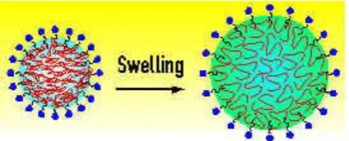
[image:24.595.176.421.279.378.2]The wetting and swelling step occurs when the polymer spreads over the surface of the biological substrate or mucosal membrane in order to develop an intimate contact with the substrate. The ability of the adhesive to spread spontaneously on mucin influences development of intimate contact between the mucoadhesive and mucin and consequently influences the mucoadhesive strength. Bio adhesives are able to adhere to or bond with biological tissues by the help of the surface tension and forces that exist at the site of adsorption or contact. Swelling of polymers occurs because the components within the polymers have an affinity for water.
Fig: Swelling of a polymer
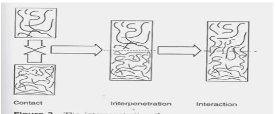
Interpenetration between the Polymer Chains and the Mucosal Membrane (Diffusion Theory):
Department of Pharmaceutics, Nandha College of Pharmacy, Erode-52. Page 16
Fig: The interpenetration of polymer chains
Formation of Chemical Bonds between the Entangled Chains:
This step involves the formation of weak chemical bonds between the entangled polymer chains. The types of bonding formed between the chains include primary bonds such as covalent bonds and weaker secondary interactions such as Vander Waals Interactions and hydrogen bonds. Both primary and secondary bonds are exploited in the manufacture of bio adhesive formulations in which strong adhesions between polymers are formed.
Mechanisms of bio adhesion:
Department of Pharmaceutics, Nandha College of Pharmacy, Erode-52. Page 17
Factors affecting on Mucoadhesion
1. Polymer related factors:
i) Molecular weight
ii) Concentration of active polymer iii) Flexibility of polymer chains iv) Special confirmation
v) Swelling
2. Environment related factors:
i) pH of polymer - substrate interface ii) Applied strength
iii) Initial contact time
3. Physiological factors:
i) Mucin turns over ii) Disease state
Bioadhesion Advantages:
Advantages and applications of oral bio adhesive dosage forms are as follows:
a) Bio adhesive drug delivery systems can prolong GI transit time and improving oral drug absorption should ideally be nontoxic, non-absorbable from the GI tract.
b) They prolong the residence time at the site of action or absorption.
c) Preferably form a strong non-covalent bond with mucin-epithelial cell surfaces thereby they adhere to the tissues and cause localized drug release.
d) Adhere quickly to moist tissue.
e) Allow easy incorporation of drug and offer no hindrance to its release.
Department of Pharmaceutics, Nandha College of Pharmacy, Erode-52. Page 18
possible.
g) Economical:
Most of the routes of administration like ocular, nasal, rectal, buccal, respiratory, vaginal, are coated with the mucus layer, mucoadhesives increase the residence time and provide intimate contact between a dosage form and the absorbing tissue which results in high drug concentration in a local area and high drug flux through the absorbing tissue. The intimate contact may increase the total permeability of the high molecular weight drugs like proteins and peptides.
Microencapsulation:
Department of pharmaceutics, Nandha college of pharmacy, Erode. Page 19
2.0. LITERATURE REVIEW
Alagusunduram. M et al., (2009)2 studied Microspheres are characteristically free flowing powders consisting of proteins or synthetic polymers which are biodegradable in nature and ideally having a particle size less than 200 μm. A well designed controlled drug delivery system can overcome some of the problems of conventional therapy and enhance the therapeutic efficacy of a given drug. There are various approaches in delivering a therapeutic substance to the target site in a sustained controlled release fashion. One such approach is using microspheres as carriers for drugs. It is the reliable means to deliver the drug to the target site with specificity, if modified, and to maintain the desired concentration at the site of interest without untoward effects. Microspheres received much attention not only for prolonged release, but also for targeting of anticancer drugs to the tumor. In future by combining various other strategies, microspheres will find the central place in novel drug delivery, particularly in diseased cell sorting, diagnostics, gene & genetic materials, safe, targeted and effective in-vivo delivery and supplements as miniature versions of diseased organ and tissues in the body.
Meral Yuce et al., (2008)12 studied the Indomethacin loaded microspheres of ethyl cellulose were prepared by the emulsion solvent evaporation techniques. The aim of this work was to investigate the influence of process variation polymer type via viscosity grade of ethyl cellulose. Microspheres exhibited lower burst effect with decreased drug release rate, when the drug was incorporated with ethyl cellulose N100 and higher ratio of each polymer. Therefore Indomethacin release from ethyl cellulose microspheres could not be evaluated by any of the kinetic models.
Department of pharmaceutics, Nandha college of pharmacy, Erode. Page 20
The percent drug entrapment was up to 80% and they could sustain drug release over a period of 8 and half hours
Malay and Das et al., (2006)15 designed diltiazem loaded mucoadhesive micro spheres by emulsification, internal gelatin techniques with a maximum incorporation efficiency of 93.29 ± 0.26% the scanning electron microscopy indicated that the microspheres were spherical in shape and drug remained dispersed in the polymer matrix. The in-vitro drug release mechanism was non-fickian type controlled by swelling and relaxation of polymer. There was no significant change in drug content and cumulative drug release of drug loaded microspheres stored at different storage conditions after 8 weeks of study.
Rajendra Kotadiya et al., (2009)20 studied the theophylline loaded agar microspores. The effect of hydrocolloid on the physicochemical properties of the microspheres was investigated. The similar result of prepared formulation (t50=211.78) and marketed product
(t50=209.29) suggest sustained release of the drug.
Rossoul dinarvand et al., (2005)21, 49 prepared gelatin micro spheres by polymerization techniques using glutaraldehyde as the cross linking agent. They investigated the effect of time of cross linking and amount of cross linking agent on the swelling properties of micro spheres and their release. The micro encapsulation efficiency micro spheres appearance, particle size, swelling ratio and drug release profile were also studied micro spheres with larger amount of cross linking agent showed reduced swelling ratio. In-vitro release pattern of lactic acid from gelatin micro spheres showed lymphatic profile and release rates were reduced upon increasing the amount of cross linking agent.
Department of pharmaceutics, Nandha college of pharmacy, Erode. Page 21
from the formulations was different at different concentrations of polymers and different RPM and temperature. The best cumulative release was achieved after 24 hrs i.e. 91.6%. The Mucoadhesive property of the ethylcellulose microspheres was evaluated by in-vitro wash off test. The microspheres exhibited 75% mucoadhesion and showed good drug entrapment efficiency. By, above results it was concluded that ethyl cellulose microspheres showed reproducible results, with good Mucoadhesive properties and good surface morphology.
Bagherwal A et al., (2010)41 Ciprofloxacin HCl belong to the fluoroquinoline derivatives which is widely used in the long term therapy for treatment of a wide range of infections including anthrax, biliary tract infection, bone and joint infection, gastrointestinal including traveler’s diarrhea and Campylobacter enteritis, Shiegella, meningococcal meningitis prophylaxis, surgical infection prophylaxis, tuberculosis, leprosy and topically in the treatment of eye infections. Hence there is a potential need for floating tablet as sustained release dosage form for this drug. HPMC and carbomer are the polymers, used as suspending agent, viscosity increasing agent and tablet binder coating agents. In the present study, it was aimed to formulate floating tablet of ciprofloxacin HCl with HPMC and carbomer in different proportion (4%, 8% and 12%) by direct compression techniques using polymers lactose, Magnesium Streate, talc with sodium bicarbonate. All the prepared formulation were found to complies with the official tests like precompression parameter like angle of repose and post compression parameters like Shape, tablet dimensions, hardness, friability test, weight variation test, floating test, content uniformity and in-vitro dissolution study. In-vitro release studies were carried out using USP XXII dissolution test apparatus. The mean percentage of ciprofloxacin released at various time intervals was calculated and plotted against time. The mechanism of drug release with all the formulations was dominantly diffusion and followed zero order kinetics. It was observed that the integrity of the drug is not affected by formulation procedure. The results revealed the drug polymer ratio showed greater drug release than other formulations.
Department of pharmaceutics, Nandha college of pharmacy, Erode. Page 22
bone infections. The HAP microspheres were made by chemical precipitation followed by a spray-drying technique. Agglomerated microspheres were prepared by a wet granulation process using a granulator. Implants were prepared by direct compression of the granules on a Carver press. Ciprofloxacin was analyzed by high-performance liquid chromatography. Characterizations of the HAP microspheres include particle size, size distribution, physical state of the drug in the microsphere, and microstructure of the drug delivery system before and after in vitro release. The particle size, porosity, and morphology of the microspheres were dependent on viscosity and concentration of the slurry as well as the atomization pressure used during spray drying. Even at the highest drug load (2% wt/wt), the drug was present in a noncrystalline state. The drug release from the agglomerated microspheres was quick and almost complete within 1 hour. However, Compressing the same amount of agglomerated microspheres into an implant greatly reduced the rate of ciprofloxacin release. Only 12% (wt/wt) of the drug was released from the implant within 1hr.
Jeong YI et al., (2009) created ciprofloxacin HCl (CIP)-encapsulated poly (DL-lactide -co-glycoside) (PLGA) microspheres by the solvent evaporation method. Their antibacterial activity was evaluated with pathogenic microorganisms in vitro and in vivo. Since the half-life of CIP in the blood stream is short, sustained-release properties of microspheres may provide enhanced antibacterial activity. CIP- encapsulated microspheres of PLGA were prepared by the O/O method. CIP-encapsulated PLGA microspheres showed spherical shapes under a scanning electron microscope (SEM) and their particle sizes ranged from 10 to 50 micron. In an in vitro drug release study, CIP was continuously released over 3 weeks from the microspheres, and a burst effect was observed for the first 3 days. In the in vitro antibacterial activity test, CIP-microspheres showed lower antibacterial activity compared to free CIP because of their sustained-release properties, while empty microspheres did not affect the growth of microorganisms. In the in vivo antibacterial activity test, the number of microorganisms following treatment with CIP- encapsulated microspheres was significantly lower than after treatment with free CIP at 5 days post injection. These results suggest that encapsulated CIP keeps its antibacterial activity after microencapsulation.
Department of pharmaceutics, Nandha college of pharmacy, Erode. Page 23
surface methodology. Ciprofloxacin HCl has an absorption window in the stomach and in the upper part of the small intestine. A 32 full factorial design was employed to optimize the formulation wherein hydroxylpropyl methyl cellulose K4M (HPMC K4M) (X1) and Carbopol 934 (X2) were taken as independent variables and amount of drug release after 12 hrs (Y1), t50 (Y2), and t85 (Y3) were taken as the dependent variables. The capsules were prepared by physical blending of drug and the polymers in varying ratios. The release data were evaluated by the model dependent (curve fitting) method using the PCP Disso v2.08 software. Optimization studies were carried out using the Design Expert Software (Version 7.1.6). Formulations were evaluated for in vitro buoyancy and in vitro release studies. The in vitro drug release followed zero order kinetics and the drug release mechanism was found to be anomalous or non-fickian type. It was found that both HPMC and Carbopol and their interaction had significant impact on the release and floating properties of the delivery system. The similarity factor f2 was found to be 62.32 for the developed formulation indicating the release was similar to that of the marketed formulation (Cifran). Thus, a combination of HPMC K4M and Carbopol 934 can be used to increase the gastric residence time and drug release for a period of 12hrs.
Department of pharmaceutics, Nandha college of pharmacy, Erode. Page 24
Faizi Muzaffar et al., (2010)45 Mucoadhesive microspheres include microparticles and microcapsules (having a core of the drug) of 1-1000μm in diameter and consisting either entirely of a mucoadhesive polymer or having an outer coating of it, respectively. Amoxicillin microspheres were formulated by using solvent evaporation technique. Using Eudragit RS 100 as matrix polymer. Evaluation of the prepared mucoadhesive microspheres was done for % yield, particle size analysis, particle size distribution angle of repose, and determination of drug content, shape, surface characterization, drug entrapment and finally cumulative drug release from microspheres by suitable and reliable official methodologies.
Arunachalam.A et al., (2010) in the present study, gelatin microspheres containing oflaxacin were prepared by coacervation phase separation method and characterized by optical microscopy and scanning electron microscopy. The microspheres were analyzed for drug entrapment, bulk density, angle of repose, particle size and In-vitro release pattern. The effect of process variables on microsphere size was studied and based on these preliminary studies, different batches of microspheres were prepared by altering the drug: polymer ratio and cross-linking with glutaraldehyde. The size of microspheres was in range of 42- 45 μm. They were spherical in shape as evidenced by photomicrographs and scanning electron microscopy. The percent drug entrapment was in the range of 78-90 % and they could sustain drug release over a period of 8 hrs.
Department of pharmaceutics, Nandha college of pharmacy, Erode. Page 25
Pham H H et al., (2002) synthesized and characterized hydroxyapatite (HAP) microspheres, agglomerated microspheres, and implants containing ciprofloxacin. This delivery system is to be used as an implantable drug delivery system for the treatment of bone infections. The HAP microspheres were made by chemical precipitation followed by a spray-drying technique. Agglomerated microspheres were prepared by a wet granulation process using a granulator. Implants were prepared by direct compression of the granules on a Carver press. Ciprofloxacin was analyzed by high-performance liquid chromatography. Characterizations of the HAP microspheres include particle size, size distribution, physical state of the drug in the microsphere, and microstructure of the drug delivery system before and after in vitro release. The particle size, porosity, and morphology of the microspheres were dependent on viscosity and concentration of the slurry as well as the atomization pressure used during spray drying. Even at the highest drug load (2% wt /wt), the drug was present in a noncrystalline state. The drug release from the agglomerated microspheres was quick and almost complete within 1 hour. However, compressing the same amount of agglomerated microspheres into an implant greatly reduced the rate of ciprofloxacin release. Only 12% (wt/wt) of the drug was released from the implant within 1 hour. The in vitro release of ciprofloxacin from these implants follows a diffusion-controlled mechanism. This method provides a unique way of producing various shapes and drug loads of HAP microspheres that can be easily manufactured on a commercial scale.
Department of pharmaceutics, Nandha college of pharmacy, Erode. Page 26
Duygu Gurcan et al., (2010) study was to evaluate ciprofloxacin hydrochloride-loaded chitosan microspheres for nasal administration. Microspheres were prepared by spray drying method and evaluated with respect to the particle size, morphological properties, drug-polymer interaction, production yield, drug content, encapsulation efficiency, in-vitro drug release and kinetic assessment and in-vivo bioavailability. The particle size of microspheres prepared ranged from 3.3 to 6.7 μm. The microspheres showed spherical shape and smooth surface. For all formulations, drug loading capacity and microsphere yield were higher than 74% and 38%, respectively. Based on in-vitro evaluation of microspheres, the most suitable formulation has chosen for in vivo nasal application to rats. In-vivo studies showed that, absolute bioavailability of CIPRO formulations (oral solution, nasal solution and nasal microsphere suspension) were found as 8.57%, 15.7% and 32.9%, respectively. According to the obtained data, CIPRO-loaded chitosan microspheres prepared with spray-drying method are able to prove sustained release and could be use via nasal route as an alternative to oral route.
Tamizharasi S et al., (2009)48prepared and evaluated poly (e-caprolactone) microspheres of Repaglinide by using the solvent evaporation techniques. The in-vitro release study showed that Repaglinide release from all formulations was slow and sustained over 12 hrs. Application of the in vitro drug release data to various kinetic equations indicated zero order release from Repaglinide microspheres.
Department of Pharmaceutics, Nandha College of Pharmacy. Page 28
3.0. DRUG AND POLYMER PROFILE
Drug Profile - CIPROFLOXACINE HCl50:
Ciprofloxacin HCl is a synthetic fluroquinone antibiotic.
Systematic (IUPAC) name: 1-cyclopropyl- 6-fluoro- 4-oxo- 7-piperazin- 1-yl- quinolone- 3- Carboxylic acid.
Chemical data:
Molecular formula : C17H18FN3O3. HCl
Molecular weight : 331.346
Structure:
Fig: Structure of Ciprofloxacin Physico-chemical properties:
Description : Yellowish to light yellowish crystalline powder.
Standards : Ciprofloxacin contains not less than 98.5 per cent and not more than of 101.5 %, calculated on the dried basis.
Solubility : Ciprofloxacin HCl is considered to be soluble in aqueous solutions with pH between 2 and 5. It is sparingly to slightly soluble in aqueous solutions with pH 7.4.
Melting point : Ciprofloxacin HCl melts at 255-2570 C
Pharmacokinetic data:
Bioavailability : 69%
Metabolism : Hepatic, Including CYP1A2
Half-life : 4 hours
Department of Pharmaceutics, Nandha College of Pharmacy. Page 29
Therapeutic considerations Availability
Ciprofloxacin is available as:
tablets (250 mg, 500 mg or 750 mg)
intravenous solutions (5% and 10%, 100 ml) eye and ear drops
In most countries, all formulations require a prescription.
Routes : Oral, intravenous, topical (ear drop & Eye drops)
Ciprofloxacin (INN) is a synthetic chemotherapeutic anti-biotic of the floroquinoline drug class. It is a second generation floroquinoline antibacterial. Kills bacteria by interfering with the enzymes that cause DNA to rewind after being copied, which stops DNA and protein synthesis.
.
STORAGE
Store at room temperature below 860 F (30o C) away from light and moisture. Do not store in the bathroom. Keep all medicines away from children and pets.
Do not flush medications down the toilet or pour them into a drain unless instructed to do so. Properly discard this product when it is expired or no longer needed.
Department of Pharmaceutics, Nandha College of Pharmacy. Page 30
4.0. POLYMER PROFILE
Polymer: SODIUM CARBOXY METHYL CELLULOSE33 Grade : Pharma Grade (Standards of IP 96)
Prepared from cellulose by treatment with alkali and monochloro - acetic acid or its sodium salt. The article of commerce can be specified further by viscosity.
Synonyms : Sodium cellulose glycolate, Na CMC, CMC, cellulose gum, sodium CMC;
Chemical names: Sodium salt of carboxymethyl ether of cellulose
Chemical formula: [C6H7O2 (OH) x (OCH2COONa) y] n Where,
n is the degree of polymerization x = 1.50 to 2.80
y = 0.2 to 1.50 x + y = 3.0
(y = degree of substitution)
Structural formula:
Macromolecules: Greater than about 17,000 (n about 100)
Department of Pharmaceutics, Nandha College of Pharmacy. Page 31
Description: White or slightly yellowish, almost odorless hygroscopic granules, powder Or fine fibers.
Functional uses: Thickening agent, stabilizer, suspending agent characteristics.
Identification
Solubility: Yield viscous colloidal solution with water; insoluble in ethanol.
Foam test: Vigorously shake a 0.1% solution of the sample. No layer of foam appears. This test distinguishes sodium carboxymethyl cellulose from other cellulose ethers and from alginates and natural gums.
Precipitate formation: To 5 ml of a 0.5% solutions of the sample add 5 ml of a 5% solution of copper sulfate or of aluminum sulfate. A precipitate appears. (This test permits the distinction of sodium carboxymethyl cellulose from other cellulose ethers and from gelatin, carob bean gum and tragacanth gum).
Colour reaction: Add 0.5 gm of powdered carboxymethylcellulose sodium to 50 ml of water, while stirring to produce a uniform dispersion. Continue the stirring until a clear solution is produced. To 1 ml of the solution, diluted with an equal volume of water, in a small test tube, add 5 drops of 1-naphthol TS. Incline the test tube and carefully introduce down the side of the tube 2 ml of sulfuric acid so that it forms a lower layer. A red-purple colour develops at the interface.
Purity: Loss on drying not more than 12% after drying (105o C to constant weight)
pH: 6.0 - 8.5 (1 in 100 soln)
Sodium: Not more than 12.4 % on the dried basis. Determine total sodium content by Atomic Absorption Spectroscopy or Flame Photometry. Sodium chloride not more than 0.5% on the dried basis See description under tests.
Free glycolate: Not more than 0.4 % calculated as sodium glycolate on the dried basis
Department of Pharmaceutics, Nandha College of Pharmacy. Page 32
Lead: Not more than 2 mg/ kg. Determine using an atomic absorption technique appropriate to the specified level. The selection of sample size and method of sample preparation may be based on the principles of the method described in “Instrumental Methods.”
Properties Uses and Applications
Sodium Carboxy Methyl Cellulose is an anionic water soluble polymer derived from cellulose. It is odorless, tasteless and nontoxic. It has the following properties making it useful in a wide variety of applications.
1. Easy Solubility in cold and hot water. 2. Fine film forming properties.
3. Resistance to oil greases and solvents. 4. Better thickening action.
5. Physiological inertness 6. Anionic character 7. Binding properties
8. Suspending characteristics. 9. Gives transparent film.
10. Acts as a protective colloid reducing water losses.
Polymer: SODIUM ALGINATE
Nonproprietary Name:
BP: Sodium Alginate
Ph. Eur: Sodium Alginate
USP-NF: Sodium Alginate
Synonyms: algin, alginic acid, sodium salt, sodium polymannuronate
Department of Pharmaceutics, Nandha College of Pharmacy. Page 33
Empirical Formula:
Sodium alginate consists chiefly of the sodium salt of alginic acid, which is a mixture of
polyuronic acids composed of residues of D-mannuronic acid and L-guluronic
Structure:
Molecular formula: (C6H7O6Na)
Description:
White or light yellow, vagiform power, odorless, tasteless, dissolve in water, insoluble in ethanol and ether.
Method of Manufacture:
Alginic acid is extracted from brown seaweed and is neutralized with sodium bicarbonate to form sodium alginate.
Acidity/alkalinity: pH 7.2 for a 1% w/v aqueous solution. Functional Category:
Stabilizing agent, suspending agent, tablet and capsule disintegrate, tablet binder, viscosity
increasing agent.
Applications in Pharmaceutical Formulation or Technology:
Sodium alginate is used in a variety of oral and topical pharmaceutical formulations. In tablet
formulations, sodium alginate may be used as both a binder and disintegrate. It has been used as
a diluent in capsule formulations. Sodium alginate has also been used in the preparation of
sustained-release oral formulations since it can delay the dissolution of a drug from tablets,
capsules and aqueous suspensions.
Department of Pharmaceutics, Nandha College of Pharmacy. Page 34
Description:
Sodium alginate occurs as an odorless and tasteless, white to pale yellowish-brown colored
powder.
Solubility: Practically insoluble in ethanol (95%), ether, chloroform, and ethanol or water
mixtures in which the ethanol content is greater than 30%. Slowly soluble in water, forming a
viscous colloidal solution.
Viscosity (dynamic): Various grades of sodium alginate are commercially available that yield
aqueous solutions of varying viscosity. Typically, a 1% w/v aqueous solution, at 200C, will have
a viscosity of 20–400 mPas (20–400 cP).
Stability and Storage Conditions:
Sodium alginate is a hygroscopic material, although it is stable if stored at low relative humidity
and a cool temperature.
Incompatibilities:
Sodium alginate is incompatible with acridine derivatives, crystal violet, phenyl mercuric acetate
and nitrate, calcium salts, heavy metals, and ethanol in concentrations greater than 5%.
Method of Manufacture:
Alginic acid is extracted from brown seaweed and is neutralized with sodium bicarbonate to
form sodium alginate.
Safety:
It is generally regarded as a nontoxic and nonirritant material, although excessive oral
consumption may be harmful.
Applications:
Sodium alginate is used in a variety of oral and topical pharmaceutical formulations. In tablet formulations, sodium alginate may be used as both a binder and disintegrates It has been used as a diluent in capsule formulations
Sodium alginate has also been used in the preparation of sustained release oral formulations since it can delay the dissolution of a drug from tablets, capsules and aqueous suspensions.
Department of Pharmaceutics, Nandha College of Pharmacy. Page 35
Sodium alginate has been used for the aqueous microencapsulation of drugs, in contrast with the more conventional microencapsulation techniques which use organic-solvent systems.
It has also been used in the formation of nanoparticles.
Polymer: HYDROXY PROPYL METHYLCELLULOSE
1. Nonproprietary Name:
BP: Hypromellose
USP: Hypromellose
2. Synonyms:
Hydroxypropyl methylcellulose, HPMC, hypromellosum, Methocel, methylcellulose propylene
glycol ether, methyl hydroxypropylcellulose, Metolose, MHPC.
3. Chemical Name: Cellulose hydroxypropyl methyl ether.
4. Empirical Formula and Molecular Weight:
The PhEur 6.3 describes hypromellose as a partly O-methylated and O-(2-hydroxypropylated)
cellulose. Molecular weight is approximately 10,000 – 1,500,000.
5. Structural Formula:
Structure of HPMC
Where R is H, CH3, or CH3CH (OH) CH2
6. Applications in Pharmaceutical Formulation or Technology:
Hypromellose is widely used in oral, ophthalmic, nasal and topical pharmaceutical
formulations. In oral products, hypromellose is primarily used as a tablet binder, in film-coating,
and as a matrix for use in extended release tablet formulations. Concentrations between 2% and
5% w/w may be used as a binder in either wet- or dry-granulation processes. High-viscosity
Department of Pharmaceutics, Nandha College of Pharmacy. Page 36
tablets and capsules. Hypromellose is also used in liquid oral dosage forms as a suspending and
or thickening agent at concentrations ranging from 0.25 – 5.0%.
7. Description:
Hypromellose is an odorless and tasteless, white or creamy-white fibrous or granular powder.
8. Solubility:
Soluble in cold water, forming a viscous colloidal solution; practically insoluble in hot water,
chloroform, ethanol (95%) and ether, but soluble in mixtures of ethanol and dichloromethane,
mixtures of methanol and dichloromethane.
9. Viscosity (dynamic):
A wide range of viscosity types are commercially available. Aqueous solutions are most
commonly prepared.
Typical viscosity values for 2% (w/v) aqueous solutions of methocel (Dow Chemical Co.) viscosities measured at 20ºC
Methocel grade Viscosity(cps)
K4 M 4000
K15M 15000
K100M 100000
10. Stability and Storage Conditions:
Hypromellose powder should be stored in a well-closed container, in a cool, dry place.
11. Safety:
Hypromellose is generally regarded as a nontoxic and nonirritating material, although excessive
Department of pharmaceutics , Nandha college of pharmacy. Page 36
5.0.
RESEARCH ENVISAGED
5.1. Aim of Work
:The objective of this study is to develop, characterize, and evaluate mucoadhesive microspheres of Ciprofloxacin employing various mucoadhesive polymers for prolonged gastrointestinal absorption.
Mucoadhesive are preferred because immobilization of drug carrying particles at the mucosal surface would result in:
A prolonged residence time at the site of action or absorption. A localization of the drug delivery system at a given target site.
An increase in drug – concentration gradient due to intestine contact of the particles with the mucosal surface.
Department of pharmaceutics , Nandha college of pharmacy. Page 37
5.2. Plan of Work
:The present work was carried out to prepare and evaluate microperticulate drug delivery system Ciprofloxacin HCl of using Sodium Carboxy Methyle Cellulose, Sodium Aliginate and Hydroxy Propyle Methyle Cellulose in various proportions. The following experimental protocol was therefore designed to allow a systemic approach to the study.
Procurement of drug and raw materials.
Pre- formulation studies for possible drug or polymer interaction by IR analysis and UV.
Preparation of standard curves.
Preparation of mucoadhesive microspheres by Emulsification Solvent Evaporation technique.
Drug release study using suitable in-vitro model.
Evaluation of the various properties of Mucoadhesive microspheres. a. Particle size analysis.
b. Bulk density.
c. Drug Entrapment efficiency. d. Drug Content.
e. In-vitro mucoadhesion study f. Kinetic modeling.
g. Scanning Elecron Microscopy h. HPLC
Department of pharmaceutics, Nandha college of pharmacy. Page 38
6.0. MATERIALS AND EQUIPMENTS:
MATERIALS:
The Materials used For the Process are as follows:
List of chemicals: Table: 1
S.No Chemicals Company
1. Ciprofloxacin HCl Darwin Formulations, Mumbai, India
2. Sodium CMC Reachem laboratory chemicals Pvt.Ltd, India
3 Sodium Aliginate Rajesh Chemicals, Mumbai, India
4 HPMC Dr.Reddy’s Lab Pvt.Ltd, Hyderabad, India
5. Seasem oil AR grade, India
6. Light Liquid paraffin Qualigens fine chemicals, Mumbai, India
8. Span 20 Sd fine –chem limited, Mumbai, India
9. n-Hexane Sd fine –chem limited, Mumbai, India
List of Equipment’s: Table: 2
S.No Equipment Company
1. Glass ware DELTA ( Borosilicate Glass) 2. UV Spectrophotometer Elico LI 120
3. Mechanical stirrer Remi motors 4. USP type 11 Station Dissolution
apparatus
ELECTRO Lab
5. Balance Dolphin Digital Balance
6. FTIR KBR press model M15
Department of pharmaceutics, Nandha college of pharmacy. Page 39
7.0. EXPEREMENTAL INVESTIGATION
Preformulation Study:
7.1. Organoleptic properties
34:
Ciprofloxacin was observed for color, odor and taste
.
7.2. Standard calibration curve of Ciprofloxacin HCl
34:
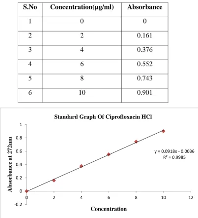
Ciprofloxacin HCl can be estimated spectrometrically at 272 nm as it obeys Beer’s – Lambert’s law limit is the range of 2-20 µg/ml.
7.2.1. Preparation of 0.1 N Hydrochloric acid (pH 1.2):
8.5 ml of concentrated hydrochloric acid was taken and then diluted with distilled water up to 1000 ml (0.1N HCl).
A. Stock Solution
100 mg of Ciprofloxacin HCL was dissolved in 100 ml of 0.1N HCl. So, as to get a stock solution of 1000 g/ml concentration.
B. Standard Solution
10 ml of stock solution was made to 100 ml with 0.1N HCl thus giving a concentration of 100 g/ml. Aliquot of standard drug solution ranging from 0.2 ml to 1 ml were transferred in to 10 ml volumetric flask and were diluted up to the mark with 0.1N HCl. Thus the final concentration ranges from 2-10 g/ml. Absorbance of each solution was measured at 272 nm against 0.1N HCl as a blank. A plot of concentrations of drug vs. absorbance was plotted.
7.
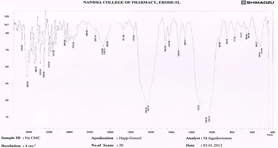
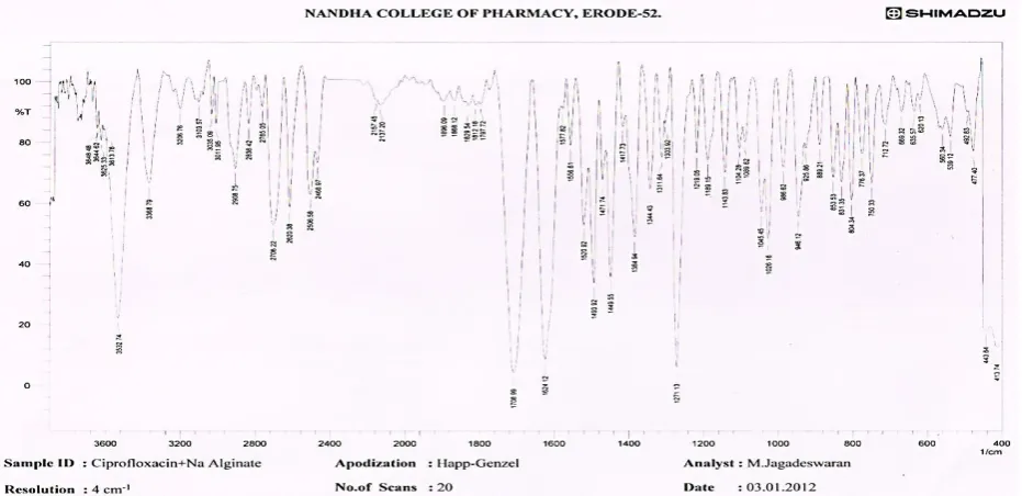
3. Drug-excipients interaction study by
FTIR Spectrum
45:
Department of pharmaceutics, Nandha college of pharmacy. Page 40
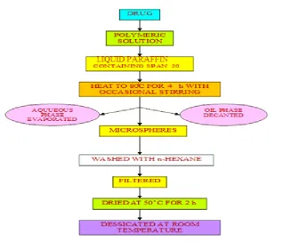
Emulsification Solvent evaporation technique
Dissolve the coated substance in a volatile solvent (immiscible with liquid Manufacturing Vehicle, LMV)
Dissolve or disperse the core material in coating polymer solution with agitation. Disperse the core coat mixture in LMV with continuous agitation.
Heat the whole mixture to evaporate the solvent for the polymer. Cross linking with glutaraldehyde, formaldehyde etc.,
7.4. Emulsification Solvent Evaporation Method for Preparation of
Mucoadhesive microspheres
46, 51:
Department of pharmaceutics, Nandha college of pharmacy. Page 41
[image:50.595.145.483.418.699.2]Composition of Mucoadhesive Microspheres of Ciprofloxacin HCl:
Table: 3
Sl. No Formulations Drug (mg)
Sodium CMC
(mg)
Sodium Alginate
(mg)
HPMC (mg)
Drug Ratio
1 F1 500 500 --- --- 1:1
2 F2 500 1000 --- --- 1:2
3 F3 500 --- 500 --- 1:1
4 F4 500 --- 1000 --- 1:2
5 F5 500 --- --- 500 1:1