The Role of the Prefrontal Cortex in the Expression of Impulsive- and
Premeditated-Aggression
by
Sarah Haberle BA (Hons)
Submitted in fulfilment of the requirements for the Degree of Doctor of Philosophy (Clinical Psychology)
School of Psychology University of Tasmania
i
diploma by the University or any other institution, except by way of background information and duly acknowledged in the thesis. To the best of my knowledge and belief, this thesis contains no material previously published or written by another person except where due acknowledgement is made in the text of the thesis, nor does the thesis contain any material that infringes copyright.
__________________________________ Date: _______________ Sarah Haberle
The research associated with this thesis abides by the international and Australian codes on human and animal experimentation, the guidelines by the
Australian Government’s office of the Gene Technology Regulator and the rulings of the Safety, Ethics and Institutional Biosafety Committees of the University.
__________________________________ Date: _______________ Sarah Haberle
This thesis may be made available for loan and limited copying in accordance with the Copyright Act 1968.
ii
The notion that there is a relationship between frontal lobe damage and aggressive behaviour has been recognised in the clinical literature for over 50 years. However, although there is evidence for an association between general brain dysfunction and aggression, there is little evidence pertaining to subclinical impairment and the propensity for aggressive behaviour. Further to this, given the functionally heterogeneity of the prefrontal cortex, it is vital to delineate the specific roles of the dorsolateral, orbitofrontal and medial aspects of the prefrontal cortex in the expression of aggression.
Two forms of aggression are distinguished: reactive, impulsive-aggression and goal-directed premeditated aggression. While impulsive-aggression is typically described as an emotionally-charged aggressive response characterised by a lack of control, premeditated aggression is considered to be a planned and controlled
aggressive display that is instrumental in nature. The qualitative differences between these subtypes of aggression suggest distinct neuropsychological differences
mediating the likelihood of their display.
The aim of this thesis was to clarify the role of the prefrontal cortex in subclinical impulsive-aggression and premeditated aggression. More specifically, possible executive functioning deficits mediated by the dorsolateral prefrontal cortex, and emotion recognition, impulsivity, and response reversal capabilities mediated by the orbitofrontal cortex were explored. Participants included university
undergraduate students identified as having high levels of trait aggression, classified as either predominantly impulsive, or predominantly premeditated in nature.
iii
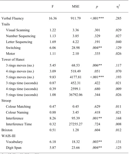
impulsive-aggressive individuals performed significantly poorer on measures of cognitive flexibility, planning, problem-solving, and flexibility of verbal thought processes.
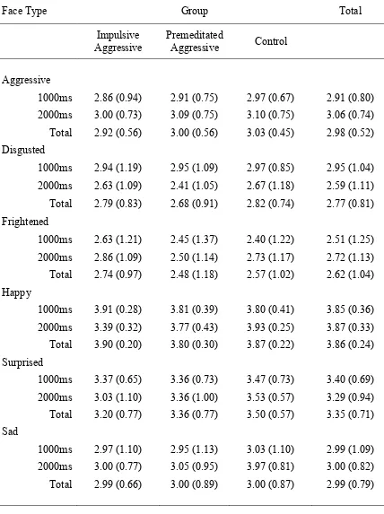
Experiment 2 (n=87) sought to identify possible deficits in interpretations of facial expressions of emotion and hostile attribution biases. Contrary to expectations, the results indicated that while impulsive- and premeditated-aggressive individuals do not incorrectly interpret emotional expressions, premeditated-aggressive
individuals attributed greater levels of aggression to neutral faces.
Experiment 3 (n=87) investigated functions of the orbitofrontal cortex, namely impulsivity, response reversal, and decision-making capabilities. No differences between impulsive-aggressive and premeditated-aggressive individuals were found on any of these measures suggesting negligible involvement of the orbitofrontal cortex in subclinical aggression.
Overall, the results from this thesis suggest distinct neuropsychological processes in individuals who display predominantly impulsive-aggressive behaviour compared to those who display predominantly premeditated-aggression. While impulsive-aggression may result from executive dysfunction pertaining to the
iv
v Abstract
Acknowledgements List of Tables List of Figures
Chapter 1. Overview of the Thesis p. 1
Chapter 2. Impulsive- and Premeditated-Aggression: A Review of
the Literature p. 6
2.1 Aggression p. 6
2.2 Distinguishing between impulsive- and premeditated-
aggression p. 7
2.2.1 Psychobiological evidence p. 11
2.2.2 Psychophysiological evidence p. 12
2.2.3 Neuropsychological evidence p. 14
2.2.4 Psychopathy p. 15
2.3 Importance of distinguishing between impulsive- and
premeditated-aggression p. 17
Chapter 3. Prefrontal Cortex & Aggression p. 20
3.1 The prefrontal cortex p. 22
3.2 Prefrontal divisions p. 24
3.3 Prefrontal dysfunction and aggression p. 28
3.3.1 Lesion studies p. 28
3.3.2 Neuroimaging and clinical neurological findings p. 32 3.4 The specific roles of the orbitofrontal and dorsolateral
prefrontal cortex in aggression p. 35
3.5 The role of the prefrontal cortex in impulsive- and
premeditated-aggression p. 37
3.6 Subcortical structures p. 40
3.7 Conclusion p. 41
Chapter 4. Rationale p. 42
Chapter 5. Study 1: Executive Functioning p. 46
5.1 Neuroanatomy of executive functions p. 47
5.2 Executive functioning measures p. 50
5.2.1 Verbal Fluency Test p. 50
5.2.2 Trail Making Test p. 51
5.2.3 Tower of Hanoi p. 52
vi
research p. 55
5.4 Executive functioning and impulsive- and premeditated-
Aggression p. 57
5.5 The relationship between executive functioning deficits and
aggression p. 60
5.6 Limitations of other studies p. 62
5.7 Aim and hypotheses p. 64
5.8 Method p. 65
5.8.1 Participants p. 65
5.8.2 Materials p. 70
5.8.2.1 Questionnaires p. 70
5.8.2.2 Executive function measures p. 73 5.8.2.3 Wechsler Adult Intelligence Scale –
Third Edition p. 77
5.8.3 Procedure p. 78
5.9 Results p. 80
5.9.1 Participants p. 80
5.9.2 Executive function measures p. 82
5.9.3 Wechsler Adult Intelligence Scale – Third Edition p. 83
5.10 Discussion p. 85
5.10.1 Verbal Fluency Test p. 87
5.10.2 Trail Making Test p. 88
5.10.3 Tower of Hanoi p. 90
5.10.4 Stroop Colour-Word Interference Task p. 92
5.10.5 The Brixton Test p. 95
5.10.6 Personality measures p. 96
5.10.7 Psychopathy and Antisocial Personality Disorder p. 98 5.10.8 The link between executive functioning deficits and
impulsive-aggression p. 100
5.10.9 Conclusion p. 103
Chapter 6. Study 2: Emotion Recognition and Aggression Attribution p. 107 6.1 Neural systems involved in emotion recognition p. 109 6.2 Separable neural systems for different emotional expressions p. 113 6.3 The relationship between emotion recognition and aggression p. 115
6.4 Aim and hypotheses p. 124
6.5 Method p. 125
6.5.1 Participants p. 125
6.5.2 Materials p. 127
6.5.2.1 Questionnaires p. 127
6.5.2.2 Facial recognition tasks p. 128 6.5.2.3 Wechsler Adult Intelligence Scale –
Third Edition p. 129
6.5.3 Procedure p. 129
6.6 Results p. 131
6.6.1 Participants p. 131
vii
6.7 Discussion p. 150
6.7.1 Emotion recognition task p. 150
6.7.2 Aggression rating task p. 155
6.7.3 Personality measures p. 157
6.7.4 The link between emotion recognition and aggression p. 157
6.7.5 Conclusion p. 159
Chapter 7. Study 3: Inhibition, Response Reversal, and
Decision-Making p. 161
7.1 Inhibition p. 161
7.1.1 Inhibition and the frontal lobes p. 164
7.1.2 Inhibition measures p. 166
7.1.3 Inhibition and aggression p. 169
7.2 Response reversal p. 171
7.2.1 Response reversal and the frontal lobes p. 172
7.2.2 Response reversal measures p. 174
7.2.3 Response reversal and aggression p. 177
7.3 Decision-making p. 180
7.3.1 Decision-making and the frontal lobes p. 181
7.3.2 Decision-making measures p. 181
7.3.3 Decision-making and aggression p. 184
7.3.4 Somatic marker hypothesis p. 186
7.4 Impulsivity, response reversal, decision-making, and aggression p. 187
7.5 Aim and hypotheses p. 188
7.6 Method p. 189
7.6.1 Participants p. 189
7.6.2 Materials p. 189
7.6.2.1 Questionnaires p. 189
7.6.2.2 Inhibition, response reversal, and
decision-making tasks p. 189
7.6.2.3 Wechsler Adult Intelligence Scale –
Third Edition p. 191
7.6.3 Procedure p. 191
7.7 Results p. 195
7.7.1 Stop Signal Task p. 195
7.7.2 Intra/Extra Dimensional Set Shift task p. 196
7.7.3 Cambridge Gambling Task p. 198
7.8 Discussion p. 202
7.8.1 Stop Signal Task p. 203
7.8.2 Intra/Extra Dimensional Set Shift task p. 207
7.8.3 Cambridge Gambling Task p. 210
7.8.4 Limitations and directions for future research p. 212
viii
8.1 Overview of findings p. 215
8.1.1 Executive functioning p. 215
8.1.2 Emotion recognition and aggression attribution p. 218 8.1.3 Inhibition, response reversal, and decision-making p. 219
8.2 Theoretical implications p. 220
8.2.1 Inhibition p. 222
8.2.2 Premeditated-aggression p. 224
8.3 Clinical implications p. 226
8.4 Limitations p. 228
8.5 Conclusion p. 229
References p. 231
Appendices p. 307
Appendix A: HREC approval letter p. 308
Appendix B: Information sheet for Study 1 p. 310
Appendix C: Consent form for Study 1 p. 312
Appendix D: Facial stimuli from Ekman and Friesen’s (1976)
collection used for the emotion recognition task p. 313 Appendix E: Facial stimuli from Ekman and Friesen’s (1976)
collection used for the aggression rating task p. 314 Appendix F: Information sheet for Study 2 and Study 3 p. 315 Appendix G: Consent form for Study 2 and Study 3 p. 317
ix Chapter 5
Study 1: Executive Functioning
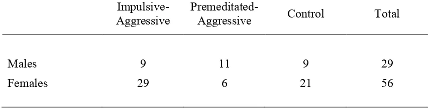
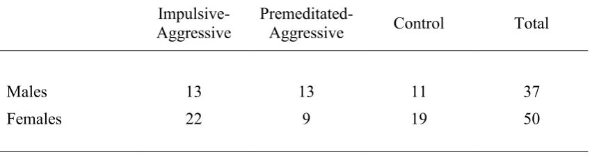
Table 5.1 The Impulsive-Premeditated Aggression Scale p. 67 Table 5.2 Number of males and females in the three participant
groups and total sample p. 68
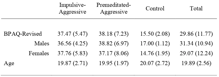
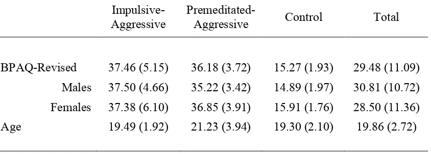
Table 5.3 Mean (and standard deviation) scores on the Aggression Questionnaire – Short Form and ages for the three
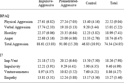
participant groups and total sample p. 69 Table 5.4 Means (and standard deviations) for the Aggression
Questionnaire – Full Scale and I7 Impulsivity
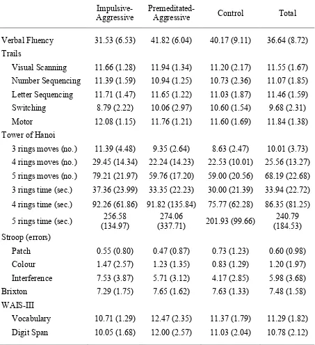
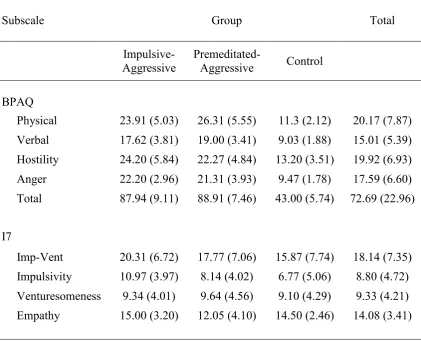
Questionnaire for the three participant groups p. 81 Table 5.5 Means (and standard deviations) for the three participant
groups on the executive function and WAIS-III measures p. 84 Table 5.6 Results of ANOVAs for the executive function and
WAIS-III measures p. 85
Chapter 6
Study 2: Emotion Recognition and Aggression Attribution Table 6.1 Number of males and females in the three participant
groups and total sample p. 126
Table 6.2 Mean (and standard deviations) scores on the Aggression Questionnaire – Short Form and ages for the three
participant groups and total sample p. 127 Table 6.3 Means (and standard deviations) for the subscales of the
Aggression Questionnaire – Full Scale and I7 Impulsivity
Questionnaire for the three participant groups p. 132 Table 6.4 Mean (and standard deviations) number of correct
responses (maximum = 4) on the emotion recognition task
x
type p. 135
Table 6.6 Mean (and standard deviations) frequency of responses to the neutral face on the emotion recognition task for the
three participant groups p. 137
Table 6.7 ANOVA results for responses to the neutral face for the main effect of participant group at the 1000ms and 2000ms
stimulus durations p. 138
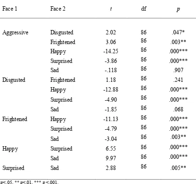
Table 6.8 Paired samples t-test results for frequency of response for
each face type when interpreting the neutral face p. 139 Table 6.9 Mean (and standard deviations) reaction times on the
emotion recognition task for the three participant groups p. 140-141 Table 6.10 Paired samples t-test results for reaction time for each
face type p. 142
Table 6.11 Mean (and standard deviations) responses for the three
participant groups on the aggressive rating task p. 144 Table 6.12 Mean (and standard deviations) reaction times for the
three participant groups on the aggressive rating task p. 145 Table 6.13 Paired samples t-test results for face type at the 1000ms
stimulus duration p. 148
Table 6.14 Paired samples t-test results for face type at the 2000ms
stimulus duration p. 149
Table 6.15 Means (and standard deviations) for Vocabulary and
Digit Span for the three participant groups p. 150
Chapter 7
Study 3: Inhibition, Response Reversal, and Decision-Making Table 7.1 Means (and standard deviations) for the Stop Signal Task
for the three participant groups p. 196 Table 7.2 Means (and standard deviations) on the Intra-Extra
Dimensional Set Shift task for the three participant groups p. 198 Table 7.3 Results of univariate ANOVAs for the Intra-Extra
xi
Gambling Task for the three participant groups p. 201 Table 7.5 Results of univariate ANOVAs for the Cambridge
xii Chapter 6
Study 2: Emotion Recognition and Aggression Attribution Figure 6.1 Mean number of correct responses (maximum = 4) for each
face type at the 1000ms and 2000ms stimulus durations p. 136 Figure 6.2 Reaction times for each face type by each group at the
1000ms stimulus duration p. 147
Figure 6.3 Reaction times for each face type by each group at the
Chapter 1 Overview of the Thesis
While the underpinnings of human aggression are clearly multifactorial, including political, socioeconomic, cultural, and psychological factors, it is also clear that some forms of aggression, either impulsive or premeditated in nature, have an underlying neurobiology that is only just beginning to be understood. In this research the
neurobiology of aggression is addressed, specifically the role of the prefrontal cortex in the expression of both impulsive- and premeditated-aggression.
A significant body of evidence indicates that the likelihood of acting aggressively is related to the functional capacity of the frontal lobe. Using neuroimaging techniques, studies of violent offenders have consistently shown abnormalities in frontal lobe structures in individuals who have histories of violence (e.g., Raine, Lencz, Bihrle, LaCasse & Colletti, 2000; Raine et al., 1998).
Additionally, lesion studies (e.g., Damasio, Grabowski, Frank, Galaburda & Damasio, 1994) and neuropsychological studies (e.g., Stanford, Greve & Gerstle, 1997) have provided evidence of the relationship between prefrontal impairment and the
propensity for aggressive behaviour. Unfortunately, however, the above studies have placed little emphasis on the separable regions of the prefrontal cortex. This is despite the fact that neuropsychological data strongly suggesting that only medial and
orbitofrontal regions of the prefrontal cortex are involved in mediating aggression, while dorsolateral prefrontal cortex has little role (Grafman et al., 1996). The prefrontal cortex is functionally and anatomically heterogeneous and thus the
Many researchers have suggested that the relationship between prefrontal abnormalities and likelihood of aggression is mediated by the failure to adaptively use executive functions (Giancola, 2000). As outlined in Chapter 5, executive functions is a broad term used to describe those abilities which allow an individual to respond to situations in a flexible manner, creating and adapting plans, and not being governed exclusively by external stimuli (Hoaken, Allaby, & Earle, 2007). Such abilities are presumed to be mediated predominantly by the dorsolateral region of the prefrontal cortex.
A further ability linked to the prefrontal cortex is the ability to correctly interpret emotional facial expressions. More specifically, patients with orbitofrontal cortex lesions are impaired in their ability to recognise facial expressions, particularly anger (Blair & Cipolotti, 2000; Hornak, Rolls & Wade, 1996). Neuroimaging studies support these findings, demonstrating activation in the orbitofrontal cortex by
negative emotional expressions; in particular, anger, but also fear and disgust (Blair, Morris, Frith, Perrett & Dolan, 1999; Kesler-West et al., 2001). As described in Chapter 6, aberrations in the ability to identify facial expressions may result in the generation of inappropriate social responses, such as reacting aggressively to
ambiguous social situations (Dodge, Laird, Lochman & Zelli, 2002). This hypothesis is based on Dodge (1986) who proposed that the accurate interpretation of social stimuli must be completed for prosocial behaviour to be manifested.
adapt to rapidly changing contexts (Happaney, Zelazo & Stuss, 2004). Furthermore, an inability to suppress previously rewarded responses due to inhibitory deficits, will in turn lead to inappropriate social responses.
In research on aggression, it is vital to distinguish between impulsive- and premeditated-aggression. Impulsive-aggression is more reactive in nature and
displayed without a self-generated goal. Premeditated-aggression, in contrast, appears to occur without provocation, is proactive, and is seen as a means to gain a valued outcome. This heterogeneity between impulsive- and premeditated-aggression suggests distinct cognitive mechanisms responsible for their display.
Both the animal and human neuropsychological literature suggests that the prefrontal cortex is involved in the modulation of impulsive-aggression (Anderson, Bechara, Damasio, Tranel & Damasio, 1999; Gregg & Siegel, 2001). Certainly, damage to the medial frontal and orbitofrontal cortex is associated with increased risk for the display of impulsive-aggression in humans whether the lesion occurs in childhood (Anderson et al., 1999) or adulthood (Grafman et al., 1996). More specifically, individuals with orbitofrontal cortex lesions are typically described as disinhibited, socially inappropriate, impulsive, irresponsible, and as often
misinterpreting others‟ moods (Rolls et al., 1994). In addition, there are considerable neuroimaging data assessing the neural functioning of patients with impulsive-aggression. These data have revealed reduced prefrontal functioning in patients presenting with impulsive-aggression (Søderstrom, Tullberg, Wikkelso, Ekholm & Forsman, 2000). Interestingly, this reduced prefrontal functioning is not observed in those with predominantly premeditated-aggression (Raine et al., 1998).
premeditated-aggression, do not present with deficits on neuropsychological measures which pertain predominantly to the dorsolateral prefrontal cortex (Mitchell, Colledge, Leonard & Blair, 2002). Psychopathic individuals‟ high level of
premeditated-aggression is thus completely unlike that of patients with orbitofrontal cortex lesions (Cornell et al., 1996). It is likely then that such individuals show elevated levels of premeditated-aggression because they have been reinforced, and not punished, for committing such behaviour in the past (Blair, 2004). Such aversive conditioning has been shown to be mediated by the amygdala. In support of this, an MRI study by Tiihonen et al. (2000) found a strong negative correlation between level of psychopathy and amygdala volume.
However, while there is clear evidence of amygdala involvement in
premeditated-aggression through its role in aversive conditioning and instrumental learning, the orbitofrontal cortex may also be involved through its role in response reversal and extinction. That is, changing a response to a stimulus when the
reinforcement contingencies change (Dias, Robbins & Roberts, 1996a). Moreover, the orbitofrontal cortex has been linked to decision-making when knowledge about potential positive and negative results is necessary to guide behavioural responding (Rogers et al., 1999b). On tasks such as the Intradimensional/Extradimensional (ID/ED) Set Shift Task which involves response reversal, adult psychopathic individuals show impairment (Mitchell et al., 2002). This suggests possible orbitofrontal dysfunction in individuals presenting with marked premeditated aggression.
gambling, schizophrenia, and bipolar disorders, all of which may or may not involve an aggressive component. Frontal lobe functions have been implicated in all of these comorbid conditions, however frontal lobe deficits have also been found in disorders not readily associated with antisocial behaviour, such as obsessive-compulsive disorder. The question thus lies in whether there is a single common component of these disorders given that studies on such populations have been inconsistent with regard to demonstrated neuropsychological deficits.
Through an investigation of prefrontal functioning in impulsive- and
premeditated-aggressive individuals, the current study aimed to answer the following research questions:
1. Do impulsive-aggressive and premeditated-aggressive individuals perform differently on measures of executive functioning known to relate to dorsolateral prefrontal cortex functioning?
2. Do impulsive-aggressive and premeditated-aggressive individuals differ in their recognition of emotions in faces?
3. Do impulsive-aggressive and premeditated-aggressive individuals demonstrate a hostile attributional bias in their interpretation of neutral facial expressions or overestimate the level of aggression in aggressive faces?
Chapter 2
Impulsive- and Premeditated-Aggression – A Review of the Literature
2.1 Aggression
Aggression can be defined as “any behaviour directed toward another individual that is carried out with the proximate (immediate) intent to cause harm” (Bushman & Anderson, 2001, p. 274). The aforementioned definition of aggression encompasses a variety of behaviours, which can range from verbal to relational to physical (Crick & Grotpeter, 1995). For example, a widely used definition of
aggression is behaviour deliberately aimed at harming people and/or objects (Dodge, 1991). In this definition, harm has implicitly been defined as hurting someone physically. However, other forms of harm, such as psychological harm (e.g.,
humiliating), and relational harm (e.g., malevolent gossip), are just as important when discussing the notion of aggression.
The aforementioned definition of aggression does not assume that all harmful behaviours are aggressive, rather, there are many instances in which harmful
behaviours are prosocial. For example, the possible pain caused by a dentist to their patient is not aggressive because the proximate intent of the dentist is to help rather than hurt the individual. Similarly, both physical aggression in the context of self-defence and the selective use of verbal aggression by politicians, for example, are adaptive. Aggression, on the contrary, is problematic when it is a habitual behavioural pattern (Bushman & Anderson, 2001).
theoretical constructs of anger and hostility continue to be used in lieu of aggression and it is therefore necessary to elucidate the conceptual distinctions between these constructs by reviewing their operational definitions. As noted above, aggression refers to a behavioural process that includes the goal of inflicting harm to another individual or object. In contrast, anger is conceptualised as an emotional state that can vary in intensity, from mild annoyances to rage (Spielberger, Jacobs, Russell & Crane, 1983). Moreover, the experience of anger lacks a specific goal (Berkowitz, 1993) and is not necessary for aggression to occur. Unlike aggression or anger, hostility is an attitudinal or cognitive construct comprised of enduring cognitions that involve negative interpretations of the environment. As such, once hostile attitudes are verbally or physically expressed, they may be more appropriately labelled as aggression. Aggressive behaviour also needs to be distinguished from antisocial behaviour. Antisocial behaviour is defined as behaviour by which people are disadvantaged and basic forms and norms are violated (Merk, de Castro, Koops, & Matthys, 2005). Examples of such behaviour are lying, stealing and truancy. Aggressive behaviour, then, is a specific form of antisocial behaviour (Kempes, Matthys, de Vries & van Engeland, 2005).
Caprara and colleagues (Caprara et al., 1985) suggest that the use of such concepts should be restricted to the use of two primary concepts: aggressiveness and aggression. According to their view, aggressiveness refers to a personality
characteristic, while aggression refers to the aggressive behaviours manifested.
2.2 Distinguishing between impulsive- and premeditated-aggression
can be classified in a number of ways, for example, by the target of aggression (e.g., self-directed or directed towards another individual or object), mode of aggression (e.g., physical or verbal, direct or indirect), or cause of aggression (e.g., medical) (Siever, 2008). Although many individuals display more than one subtype of aggression (Barker, Tremblay, Nagin, Vitaro, & Lacourse, 2006), and correlations often exist among subtypes of aggression (Kempes et al., 2005), two distinct subtypes of aggressive behaviour consistently emerge; an affective or impulsive type, and a predatory or premeditated type. Characteristically, these two subtypes of aggression are distinguished by several features, but primarily by the amount of behavioural control exhibited during the incident.
Impulsive-aggression is described as a reactive or emotionally charged aggressive response characterised by a loss of behavioural control (Barratt, 1991; Raine et al., 1998). These aggressive acts are unplanned and spontaneous in nature and are either unprovoked or out of proportion to the provocation.
Impulsive-aggression is usually accompanied by an agitated or irritated mood, poor modulation of physiological arousal and loss of behavioural control (Houston, Stanford,
Villemarette-Pittman, Conklin & Helfritz, 2003). Interpersonal communication is often non-adaptive during the agitated state and information processing appears to be inefficient (Elliot, 1992). This subtype of aggression can result in sudden, heightened, or inappropriate aggressive responses, and probably accounts for most societal
(Barratt, 1994). Barratt proposes that the personality traits of impulsiveness and anger/hostility are related to most impulsive-aggressive acts.
The theoretical roots of impulsive-aggression lie in the frustration-aggression model (Berkowitz, 1993). According to this theory, aggression is displayed as a consequence of frustration, actual or perceived threat, and heightened arousal in the form of anger. Aggression is displayed in reaction to aversive events with the subjective experiences of the individual central to the situation. The subjective experience of, for example, feeling threatened and not necessarily being threatened is a principal concept in this theory. Frustration may not immediately lead to aggression, but generate such emotions as anger, which can then augment the readiness to display aggression (Merk et al., 2005).
Premeditated-aggression, on the other hand, is considered a purposeful, controlled aggressive display that is usually instrumental in nature. These acts require forethought and planning and are generally executed with low autonomic arousal (Stanford, Houston, Villemarette-Pittman, & Greve, 2003b). Premeditated-aggressive acts are carried out with a high degree of behavioural control and are directed toward a goal, such as external reinforcers (e.g., money oriented) or intimidation (Dodge & Coie, 1987; Hubbard, Dodge, Cillessen, Coie, & Schwartz, 2001; Vitiello, Behar, Hunt, Stoff, & Ricciuti, 1990).
present or where aggression is viewed in a more positive light (Kingsbury, Lambert, & Hendrickse, 1997). A deficit in the ability to experience or anticipate remorse or the aversive outcomes increases the risk of premeditated-aggression, which appears to be the case in aggressive individuals with psychopathy who have difficulty anticipating and experiencing negative feelings of remorse or guilt (Hare, 1999).
Although there has been some criticism of the dichotomous method of characterising aggressive behaviour (see Bushman & Anderson, 2001; Parrott & Giancola, 2007), a dichotomous approach is supported by a number of important distinctions between individuals who express predominantly impulsive- or
predominantly premeditated-aggression. More specifically, researchers have found that impulsive-aggressive individuals experience disruptions across a variety of domains including verbal ability and intelligence, physiological reactivity, biological function and treatment response (Barratt et al., 1997b; Coccaro, 1992). In contrast, individuals demonstrating premeditated-aggression tend to have more circumscribed disturbances on measures of personality (Barratt et al., 1997a; Stanford et al., 2003b).
2.2.1 Psychobiological evidence
The neurotransmitter that has received the most attention in regards to aggressive behaviour is serotonin. Serotonin facilitates prefrontal cortical regions, such as the orbitofrontal cortex and anterior cingulate cortex that are involved in modulating and often suppressing the emergence of aggressive behaviours (Siever, 2008). In both humans and animals, it appears that serotonin is primarily associated with impulsive-aggression in comparison to premeditated-aggression, and it appears that its effects may be receptor specific (Miczek, 1987; Shaikh, De Lanerolle, & Siegel, 1997). In humans, a relationship between decreased serotonergic function and impulsive-aggressive behaviour has consistently been demonstrated using a number of strategies, including central neurochemical measures (CSF 5-HIAA; Linnoila et al., 1983; Roy, Adinoff and Linnoila, 1988; Virkkunen, De Jong, Bartko, Goodwin, & Linnoila, 1989a), platelet binding (Kent et al., 1988), prolactin response
Liu & Coccaro, 1994; fluoxetine; Coccaro, Kavoussi & Hauger, 1997) and regional metabolic activity in response to serotonergic agonist (m-CPP; New et al., 2002). Barratt, Kent, Bryant and Felthous (1991) found that phenytoin reduces the frequency of aggressive acts. Replication studies have shown that phenytoin may reduce
incidences of impulsive-aggression, but not premeditated-aggression (Barratt et al., 1997b; Barratt, Felthous, Kent, Liebman & Coates 2000). Such findings support the hypothesis that impulsive- and premeditated-aggression have different underlying biological substrates that respond differently to pharmacologic agents with specific modes of action.
2.2.2 Psychophysiological evidence
Psychophysiological techniques also provide a practical measure of neuropsychological functioning, however while a substantial literature exists on autonomic correlates of antisocial behaviour (Raine, 2002a, 2000b), there are a limited number of studies which have compared these measures in groups whose aggression was explicitly classified according to an impulsive-premeditated scheme. Pitts (1997) measured heart rate in aggressive children whose behaviour was
described as being more emotional, angry and unstable (impulsive). Finally, a more recent study of aggressive children indicated a significant increase in skin
conductance reactivity in those rated high in reactive aggression during a laboratory-based measure of induced anger (Hubbard et al., 2002). Again, those individuals exhibiting primarily reactive or impulsive aggression responded differently autonomically than those deemed more proactive or premeditated.
Electroencephalography (EEG) abnormalities are also present among those who engage in impulsive-aggression. For example, Drake, Hietter and Pakalnis (1992) found a greater incidence of EEG slowing in a group of patients described as having episodic dyscontrol syndrome as compared to depressed patients and controls. Abnormalities in P1 amplitude have been reported in impulsive-aggressive college students (Houston & Stanford, 2001) and youths characterised by explosive
aggressive behaviours (Bars, Heyrend, Simpson & Munger, 2001). In addition, adults classified as impulsive-aggressive exhibit decreased P1-N1-P2 latency (Houston & Stanford, 2001), reduced P3 amplitude (Barratt et al., 1997b; Gerstle, Mathias & Stanford 1998; Mathias & Stanford, 1999), increased P3 latency (Mathias & Stanford, 1999), and reduced amplitude and increased latency on the late positive potential, a purported measure of emotional processing (Conklin & Stanford, 2002). These
differences reflect a number of sensory and information processing deficits specific to impulsive-aggression, as well as preliminary evidence for emotional processing impairment.
Volkow et al. (1995), using positron emission tomography (PET), found that psychiatric patients with a history of repetitive, purposeless violent behaviour showed significantly lower relative metabolic values in medial temporal and prefrontal
affective (impulsive) murderers have significantly reduced prefrontal activation when compared to predatory (premeditated) murderers and controls.
The literature regarding premeditated-aggression though sparse is consistent. Individuals who engage in acts of premeditated-aggression show few differences from non-aggressive controls on psychophysiological measures, including P3 (Barratt et al., 1997b; Stanford et al., 2003b). Stanford et al. found that the P3 latency difference did approach significance (p = .06), suggesting a trend toward a longer P3 latency in the premeditated-aggressive group. Such prolonged P3 latency has been linked to increased attitudinal hostility (Bond & Surguy, 2000). Thus, the high levels of anger/hostility evidenced in the premeditated-aggressive group may have played a role in the latency trend observed in the sample.
2.2.3 Neuropsychological evidence
While small in number, neuropsychological studies comparing modes of aggression have established a correlation between increased impulsive-aggression and decreased executive functioning, while few deficits have been found in those who are premeditated in their aggressive behaviour (Houston et al., 2003). Dolan and
Stanford and Greve (2002) found that verbal deficiencies varied according to executive demands of the task in a sample of impulsive-aggressive college students.
In the first study to compare premeditated-aggressive subjects with controls on a variety of neuropsychological tests, Stanford et al. (2003b) found no significant differences except for a single subscale of the Wisconsin Card Sorting Task (WCST), where the premeditated group exhibited greater failure to maintain set than controls. In contrast, there were pronounced differences on a range of personality measures, including impulsivity, verbal and physical aggression, anger, hostility, psychoticism and neuroticism. The authors concluded that the difference between the premeditated-aggressive group and controls was a result of an impulsive personality style rather than a significant cognitive deficit.
In summary, neuropsychological assessment has shown a clear link between impulsive-aggressive behaviour and problems in executive functioning, while few if any deficits have been demonstrated in premeditated-aggressive individuals.
2.2.4 Psychopathy
The concept of psychopathy has provided some utility in further distinguishing between impulsive- and premeditated-aggression. Psychopathy refers to a
constellation of personality and behavioural characteristics marked by low baseline arousal, dishonesty, absence of remorse, empathy, and conscience, antisocial
behaviour, and impersonal relationships (Hare, 2003). Interpersonally, they are often described as grandiose, arrogant, callous, superficial and manipulative (Hare, 1999).
and predatory violence than non-psychopathic criminals (Cornell et al., 1996; Serin, 1991). Williamson, Hare and Wong (1987) found that incarcerated psychopaths had higher rates (45.2%) than incarcerated non-psychopaths (14.6%) of committing their crime for material gain (i.e., proactive in nature), and that non-psychopaths had higher rates (31.7 vs. 2.4%) of emotional arousal leading to their offences (i.e., impulsive in nature). Likewise, Cornell et al. (1996) found premeditated-aggressive offenders could be distinguished from non-premeditated offenders by higher total psychopathy specifically concerning: pathological lying; manipulative actions; lack of empathy; parasitic lifestyle; irresponsibility; criminal versatility; and superficiality. The authors concluded that “the link between psychopathy and instrumental violence supports the distinction between instrumental and reactive violence, and raised the possibility that the presence of instrumental violence could be an associated characteristic of
psychopathic offenders” (p. 790). Similarly, Woodworth and Porter (2002), in a study of 125 homicide offenders, found that the great majority of homicides committed by psychopaths were instrumental (i.e., premeditated), whereas only 48.4% of homicides committed by non-psychopaths were instrumental. Meloy (1988) theorised that a predisposition to engage in premeditated violence in psychopaths would be due to their low levels of autonomic arousal and reactivity, their disidentification with the victim, their emotional detachment and their lack of empathy.
problems, Christian, Frick, Hill and Tyler (1997) found that a subgroup of children exhibiting symptoms of Oppositional Defiant Disorder (ODD) and Conduct Disorder (CD) and callous and unemotional traits differed from those without such traits in the number and variety of conduct problems. Pardini, Lochman and Frick (2003) found that the presence of callous and unemotional traits in adjudicated juveniles was associated with the use of aggression to obtain rewards and dominate (i.e.,
premeditated). Frick et al. (2003), in a sample of non-referred children, found that children demonstrating conduct problems and callous and unemotional traits were more likely to demonstrate high levels of proactive aggression than those without these traits, whose aggression was predominantly impulsive. It thus appears that it is the presence of callous and unemotional traits that distinguish this subgroup and its associated problems.
2.3 Importance of distinguishing between impulsive- and premeditated
aggression
There is evidence in both children and adults that impulsive- and
These findings are in line with previous studies that have found proactive, but not reactive, aggression to be predictive of ODD, CD and externalising problems (e.g., Conner, Steingard, Anderson & Melloni, 2003; Pulkkinen, 1996; Vitaro et al., 1998). Given that individuals diagnosed with APD must have evidence of CD before the age of 15, early onset behavioural problems leading to adult antisocial behaviour was prevalent in the premeditated group. This provides evidence that premeditated-aggressive individuals may have increased personality psychopathology and be at increased risk for early aggressive and antisocial behaviours relative to impulsive-aggressive or non-impulsive-aggressive individuals. The fact that CD was a distinguishable factor between groups is of significance due to the fact that CD is stable over time (Bassarath, 2001a; Kazdin, 2000), and is associated with criminal behaviour and substance abuse (Hser, Grella, Collins & Teruya, 2003; Mueser et al., 2006; Tcheremissine & Lieving, 2006).
Heilbrun et al. (1978) found that murderers whose violence was classified as impulsive were more likely to fail on parole that those whose murders were
While distinguishing between these forms of aggression can not only lead to a better theoretical understanding of aggression (Coie & Dodge, 1998; Poulin & Boivin, 2000; Vitiello & Stoff, 1997), it can also to better prognostication. Such a distinction is also assumed to lead to the development of more specific interventions and
Chapter 3
Prefrontal Cortex & Aggression
Research into the antecedents of violence and aggression indicates that there are many factors which contribute to the development of these behaviours. It is important to note that while there are general predictors of violent and aggressive behaviour, no single theory can account for causation in all situations. It is accepted that the causes of aggression are multi-faceted and that neurological deficit may be a factor in only a small percentage of those who demonstrate such behaviour. However, given that aggression – like any behaviour – ultimately derives from the normal and abnormal operations of the brain, closer examination of the aspects of brain structure and function relevant to aggressive behaviour are required.
Numerous studies, in both animals and humans, have supported an association between abnormalities in brain function and aggressive and violent behaviour (Filley et al., 2001; Golden, Jackson, Peterson-Rohne & Gontkovsky, 1996; Krakowski, 2003). Case studies of patients with neurological disorders or those who have suffered traumatic brain injury provide provocative insights into which brain regions, when damaged, might predispose to irresponsible, aggressive behaviour.
Psychophysiological and neuropsychological assessments have also demonstrated that violent and/or aggressive individuals have lower brain functioning than controls, including lower verbal ability and diminished executive functioning (Barratt et al., 1997b; Dolan & Park, 2002; Hoaken et al., 2007).
antisocial behaviours. More specifically, as reviewed by Davidson and colleagues, a circuit that includes several regions of the prefrontal cortex, the amygdala,
hippocampus, hypothalamus, anterior cingulate cortex, ventral striatum, and other interconnected structures has been implicated in various aspects of emotion regulation and affective style (Davidson & Irwin, 1999; Davidson, Jackson & Kalin, 2000a). Emotion regulation includes those processes which amplify, attenuate, or maintain an emotion, and thus incorporates the expression of aggressive behaviours. Related to this is evidence which suggests that individuals who are vulnerable to faulty
regulation of negative emotion may be at increased risk for aggression and/or violent behaviour (Davidson, Putnam & Larson, 2000b).
3.1.1 The prefrontal cortex
The frontal cortex encompasses the brain areas anterior to the central sulcus and comprises approximately one third of the cerebral cortex. The frontal cortex can be divided into three principle regions: the primary motor cortex, the prefrontal
cortex, and the limbic cortex (Duke & Kaszniak, 2000). The prefrontal cortex refers to the most anterior regions of the frontal lobes and it is functionally and anatomically heterogeneous (Fuster, 2001). The prefrontal cortex has a rich supply of connections with other neural regions. Cortically, it is connected with association cortex in the temporal, parietal and occipital lobes, and subcortically with the hippocampus, amygdala, thalamus, hypothalamus, subthalamus, septum, striatum, pons, and mesencephalon (Fuster, 2001; Pandya & Barnes, 1987). Given that the prefrontal cortex is connected to more brain regions that any other cortical region, its position allows the integration of information processed at lower levels, including input from the limbic circuits, as well as being the major target of the basal
ganglia-thalamocortical circuits (Royall et al., 2002).
The prefrontal cortex, along with its underlying subcortical regions, is extensively interconnected with the major sensory and motor systems of the brain. Connections from the posterior cortical areas, particularly areas of multimodal convergence, bring information regarding the external environment. Subcortical pathways, including the amygdala, hippocampus, midbrain area, and thalamus, bring details about internal states (Duke & Kaszniak, 2000).
from the prefrontal cortex to the amygdala have also been identified (Afifi &
Bergman, 1998). Output projections to the amygdala are both excitory and inhibitory in nature. However, damage to the prefrontal cortex results in an overactivation of the amygdala, suggesting that the effect of the prefrontal cortex on the amygdala is predominantly inhibitory (Gerwitz, Falls & Davis, 1997; Morgan, Romanski & LeDoux, 1993). With regard to the functionality of this connection, lesions to the prefrontal cortex in rats reduce the prefrontal inhibitory action on the amygdala, resulting in an increased difficulty in the extinction of aversive responses (Morgan et al., 1993), as well as impairing the ability to anticipate future negative consequences (Bechara, Tranel, Damasio & Damasio, 1996). Therefore, it appears that the
prefrontal cortex plays an important role in regulating the acquisition of new
responses, and the extinction of aversive responses (Lopez, Vazquez & Olson, 2004). The prefrontal cortex also plays a central role in many aspects of social
cognition (Rilling et al., 2002), including perspective taking (Frith & Frith, 1999), and also in the regulation of emotions such as aggression (see Blair, 2004 for review). Early descriptions of frontal lobe syndromes arose from several 19th century
investigators, described in a number of reviews (Damasio et al., 1994; Macmillan, 2002; Tranel, Anderson & Benton, 1994), highlighting the changes displayed in social behaviour, personality, and emotional regulation that occurred after frontal lobe pathology. Subsequent investigators continued to elaborate on the nature and extent of these deficits, their causes and management (Miller & Cummings, 1999; Stuss & Knight, 2002), firmly establishing a vital role for the frontal lobes, particularly the prefrontal cortex, in such processes.
knowledge to regulate behaviour; (2) impaired ability to handle sequential behaviour; (3) impaired ability to establish or change a mental set; (4) impaired ability to
maintain a mental set; (5) impaired ability to monitor personal behaviour; and (6) attitudes of apathy.
3.2 Prefrontal divisions
For clinical purposes, the prefrontal cortex can be divided into three distinct neuroanatomical regions: 1) dorsolateral prefrontal cortex (Brodmann‟s areas 9, 10, 46); 2) medial prefrontal cortex (including the functionally related anterior cingulate cortex and Brodmann‟s area 24); and 3) orbital prefrontal cortex (Brodmann‟s areas 11 and 12), corresponding to the most inferior and ventral parts of the prefrontal cortex (behind the eyes, or orbits). Both medial prefrontal and orbitofrontal are part of a frontostriatal circuit that has strong connections to the amygdala and other parts of the limbic system. Consequently, these regions are anatomically well suited for the integration of affective and non-affective information, and for the regulation of appetitive/motivated responses. Functionally, these regions are often considered together, as when researchers focus on effects of damage to ventromedial prefrontal cortex (Happaney et al., 2004).
The prefrontal cortex is a heterogeneous region of the brain and the three principal frontal-subcortical circuits are involved in cognitive, emotional, and
motivational processes. The primary focus of the current research will be on the roles of the dorsolateral and orbital divisions of the prefrontal cortex, which manifest quite distinct anatomical and functional properties (Fuster, 1989; Stuss & Benson, 1986).
premotor areas. The dorsolateral circuit then connects to the dorsolateral part of the globus pallidus and rostral substantia nigra reticulate, and continues to the
parvocellular area of the medial dorsal and ventral anterior portions of the thalamus. Projections from the thalamus back to the dorsolateral prefrontal circuit close the circuit (Cummings, 1993).
Functionally, the high-level cognitive abilities mediated by the dorsolateral prefrontal cortex and its connections are those referred to as „executive functions‟, including cognitive flexibility, temporal ordering of events, planning, monitoring and inhibiting pre-programmed behaviour, set-shifting, working memory and concept formation (Smith & Jonides, 1999). According to Cummings (1995), dysfunction in the dorsolateral prefrontal circuit is associated with circuit-specific problems
including decreased verbal fluency, perseveration, difficulty shifting set, poor
recall/retrieval of information, reduced mental control, limited abstraction ability, and poor response inhibition. However, while patients with lesions restricted to this region are concrete and perseverative and show impairments in reasoning and mental
flexibility (Benton, 1986), they typically demonstrate intact perception, calculation, language abilities and storage of memories (Duke & Kaszniak, 2000).
The orbitofrontal cortex occupies the ventral region of the prefrontal cortex (Kringelbach & Rolls, 2004), which is reciprocally connected with the amygdala (Ghashghaei & Barbas, 2002). The orbitofrontal cortex projects to the ventromedial caudate nucleus, which receives input from other cortical association areas and brainstem regions, and has open interconnections with the dorsolateral prefrontal cortex, the temporal pole, and the amygdala (Davis & Whalen, 2001). The
in which the identity and also the reward value of odours are represented. The orbitofrontal cortex also receives information about the sight of objects from the temporal lobe cortical visual areas (Rolls, 1999).
The orbitofrontal-subcortical circuit is said to underlie social behaviour and appears to play a critical role in the representation of the reward value of a stimulus and the way in which this representation guides goal-directed behaviour (Rolls, 1999). Lesions specific to the circuit have been found to result in marked changes in
personality, including disinhibition, impulsivity, and antisocial behaviour, and irritability and lability are often prominent (Cummings, 1995). Some of the changes may be related to difficulty in the learning and reversal of stimulus-reinforcement associations, and thus the correction of behavioural responses when they are no longer appropriate due to changes in reinforcement contingencies (Rolls, 2004; Hornak et al., 2004). Indeed, investigations in macaques have shown that lesions to the orbitofrontal cortex impair reversal learning (Dias et al., 1996a). Consistent with this, the
orbitofrontal cortex is activated by monetary rewards and punishments, and the magnitude of the reinforcers (O‟Doherty, Kringelbach, Rolls, Hornak & Andrews, 2001). The visual input to neurons in the orbitofrontal cortex is in many cases the reinforcement association of visual stimuli, one of which is information about faces. Such facial stimuli convey information that is important in social reinforcement (Rolls, 2004).
motivation. Lesions to this region often produce apathy, lack of motivation, decreased social interaction, and psychomotor retardation (Sbordone, 2000).
The ventromedial prefrontal region includes the medial and varying sectors of the lateral orbitofrontal cortex, encompassing Brodmann‟s areas 25, lower 24, 32, and medial aspect of 11, 12, and 10, and the white matter subjacent to all of these areas (Bechara, 2004). Patients with bilateral lesions of the ventromedial cortex develop severe impairments in personal and social decision-making, in spite of otherwise largely preserved intellectual abilities. Following damage to this region of the prefrontal cortex, patients develop difficulties in daily and future planning, and difficulties in choosing friends and activities (Bechara, Damasio & Damasio, 2000a; Bechara, Tranel & Damasio, 2002).
The identification of these adjacent circuits provides insight as to the
similarities of behavioural changes caused by lesions to different brain regions. Whilst focal lesions to the areas of the prefrontal cortex have led to what have been labelled “frontal lobe syndrome”, the involvement of multiple circuits in subcortical lesions has resulted in variable behavioural manifestations (Cummings, 1995). For example, studies of lesions to the globus pallidus have described patients with marked changes in personality and reduced activity levels with memory and executive function deficits, but with normal intelligence and language abilities (e.g., Strub, 1989).
In summary, the frontal–subcortical circuits are extensively connected to each other at the level of the frontal lobes. The circuits are discrete in subcortical regions. The dorsolateral circuit, because of its neuroanatomy, is uniquely able to integrate information from all three frontal–subcortical circuits. Here, the integrated
3.3 Prefrontal dysfunction and aggression
Frontal lobe dysfunction in particular, has been invoked to explain the actions of individuals convicted of violent crimes, who appear to fail to inhibit impulsive, trivially motivated, or habitual aggression. Case studies as far back as 1935 have reported the onset of antisocial personality traits after frontal lobe injury (Blumer & Benson, 1975). Such cases typically involve damage to the orbitofrontal cortex, which clinical observations have associated with poor impulse control, explosive aggressive outbursts, inappropriate verbal lewdness, jocularity, and lack of interpersonal
sensitivity (Duffy & Campbell, 1994). This dysregulation of affect and behaviour may occur while cognitive, motor, and sensory functioning remains relatively intact
(Mesulam, 1986).
3.3.1 Lesion studies
Research on individuals who have suffered traumatic brain injury is of key importance in investigating the neural substrates of aggressive behaviour. The critical role of the prefrontal cortex in aggressive behaviour was initially recognised by case reports that prefrontal brain lesions could result in the emergence of antisocial behaviours or psychopathic traits in previously normal subjects (Damasio, Tranel & Damasio, 1990). A prime example of this disinhibition is found in the often cited case of Phineas Gage, a dependable and responsible stable railroad worker who was injured by a tamping rod that penetrated his skull through his orbital frontal cortex. After the accident he became irresponsible and impulsive, despite preserved general cognitive and motor skills (Damasio et al., 1994).
EEG. Results indicated that 60% of the patients displayed disinhibited behaviour with affective lability and 10% displayed violent outbursts. However, the results of this study are difficult to interpret given that, in addition to having a frontal lobe pathology, all of the patients had at least one psychiatric diagnosis, and the exact neuroanatomical location of the pathology for each patient was not reported. In another study, Heinrichs (1989) found that a frontal cortical lesion was the best predictor of violent behaviour in a sample of 45 neuropsychiatric patients. Again, many of the patients in this study had other psychiatric diagnoses and the exact anatomical locations of the frontal neuropathologies were not specified.
Further data from neurological case reports have provided much useful information regarding the relationship between prefrontal cortical functioning and aggression. Thompson (1970) reported a case of a 33-year-old male with a history of violent behaviour subsequent to a head injury at the age of 12. A
pneumencephalography revealed bilateral cortical atrophy in the prefrontal regions. Price et al. (1990) studied the adult behaviour patterns of two patients who acquired brain damage during childhood. While both patients developed relatively normally until the damage was sustained, following the damage these patients displayed an inability to respond to punishment or delay gratification, irresponsibility, sexual promiscuity, grand larceny, drug involvement, angry outbursts, arson, suspected rape, and physical violence. Although the exact location of the lesions in both cases is equivocal, neurological and neuropsychological examination indicated bilateral lesions in the prefrontal cortex.
of bizarre speech, sexual disinhibition, disobeying parental orders, and verbal and physical aggression. An EEG revealed activity in the frontal lobes and
neuropsychological tests demonstrated deficits on prefrontal tests involving attention, alternation between tasks, performance on mazes, response inhibition, and
distractibility. In a similar case, Eslinger and Damasio (1985) noted personality changes in patient EVR subsequent to surgical ablation of the orbital and mesial areas of the prefrontal cortex. Following the surgery, while his level of intelligence was above average, EVR began to engage in what the authors termed „sociopathic behaviour‟, including difficulties in decision-making, adjustment problems, poor judgement, and employment problems. The patient performed well on prefrontal cortical tests such as the WCST which the authors attributed to the fact that the dorsolateral prefrontal regions and superior mesial regions were left undamaged.
Meyers, Berman, Scheibe and Hayman (1992) noted similar behavioural sequelae involving disinhibition, poor judgement, and irresponsibility subsequent to surgical damage to the left orbital prefrontal cortex in a 33-year-old male.
Interestingly, this patient performed in the above average range on prefrontal cortical tests such as the WCST; however this is again likely due to the preservation of the dorsolateral prefrontal cortex. These findings reflect those of Phineas Gage described earlier, who subsequent to his injury, was described as being untrustworthy,
irresponsibly, and disrespectful. Again, the majority of the neural damage was located in the orbital and mesial prefrontal regions, whereas the dorsolateral area was found to be spared (Damasio et al., 1994).
within the frontal lobes. As a result, these data do not provide evidence to implicate more specifically the dorsolateral or orbital prefrontal regions in the expression of aggressive behaviour. The remaining two reports indicate that their patients had lesions in the orbital prefrontal cortex and not the dorsolateral area.
Other case studies of patients who have sustained damage to the orbitofrontal region, such as EVR, resemble Gage in manifesting a behavioural profile that has been referred to as „acquired sociopathy‟ (Saver & Damasio, 1991; Tranel, 1994; Meyers et al., 1992; Blair & Cipolotti, 2000). Damasio et al. (1994) describe acquired sociopathy as a reactive, emotionally driven violence toward a person that is related to emotional inhibitory dyscontrol. Although showing minimal impairments on standard neuropsychological tests of intelligence and executive functions, these patients display marked deficits in real life tasks involving judgement, awareness of socially appropriate conduct, and the capacity to assess future consequences (Bechara et al., 2000b).
Blair and Cipolotti (2000) reported on JS who sustained damage to the orbitofrontal cortex and some damage to the left amygdala. Premorbidly, JS was described as being a quiet, withdrawn person who was never aggressive. Following the damage, JS showed unpredictable, impulsive-aggression and violence, and demonstrated deficits in the recognition of facial expression, particularly in the recognition of anger and disgust. He also produced significantly lower skin conductance responses (SCR) to the anger and disgust expressions compared with comparison groups.
twenties, who suffered early damage to orbital and lateral sectors of the prefrontal cortex. Both exhibited a significant deficit in moral reasoning, a history of verbal and physical aggression, and intermittent, explosive bursts of anger. A further study of two adults who sustained frontal lobe injury in childhood suggests that early damage to orbitofrontal regions may lead to a “comportmental learning disability” that closely resembles sociopathy and includes a diminished capacity to inhibit violence (Price et al., 1990).
Further evidence implicating the orbitofrontal regions comes from a large retrospective study of Vietnam veterans with penetrating head injuries, which found that ventromedial frontal and orbitofrontal lesions, as assessed by computed
tomography scans, specifically increased the risk of aggressive and violent behaviour. (Grafman et al.,1996). Data have also been reported showing higher rates of antisocial behaviour (including stealing, physical assault and sexual comments or advances) in patients with frontotemporal dementia, even when compared with equally cognitively impaired patients with Alzheimer‟s disease (Miller, Darby, Benson, Cummings, & Miller, 1997; Stip, 1995).
3.3.2 Neuroimaging and clinical neurological findings
Brain imaging studies are now beginning to confirm the role of the prefrontal cortex in modulating and controlling violence in humans. Reviews of brain imaging studies of violent and psychopathic populations completed by Raine (1993), Mills and Raine (1994), Raine and Buchsbaum (1996), Henry and Moffitt (1997), and Bufkin and Luttrell (2005), while showing some variability across studies, concur in
Most recently, in their review of 17 neuroimaging studies, Bufkin and Luttrell (2005) found that the areas associated with aggressive and/or violent behaviour, particularly impulsive acts, are located in the prefrontal cortex and the medial
temporal regions. Of the 17 studies reviewed, 14 specifically examined possible links between frontal lobe pathology and aggressive and/or violent behaviour. In the 10 single photon emission computed tomography (SPECT) and PET studies, 100% reported deficits in either prefrontal (8 of 10 studies) or frontal (2 of 10 studies) functioning in aggressive, violent and/or antisocial groups compared to
non-aggressive patients or healthy controls. Analyses of specific regions in the prefrontal cortex revealed that individuals who were aggressive and/or violent had significantly lower prefrontal activity in the orbitofrontal cortex (4 of 10 studies), anterior medial cortex (2 of 10 studies) and/or superior frontal cortex (1 of 10 studies). In the four MRI studies, half reported decreased grey matter volume in prefrontal or frontal regions, and 25% reported non-specific white matter abnormalities, not localised to the frontal cortex.
Initial studies demonstrate anterior brain dysfunction in individuals with a history of violence. Goyer et al. (1994), using PET in an auditory activation condition, showed that an increased number of aggressive impulsive acts were associated with reduced glucose in the anterior medial and left anterior orbitofrontal frontal cortex of 17 personality disordered patients. Volkow and colleagues in two PET studies which compared forensic psychiatric patients with normal controls, documented decreased frontal cortical blood flow or metabolism associated with „repetitive‟ and
In a PET study evaluating responses to the probe metachlorophenylpiperazine, decrements in the lateral, medial and orbital frontal cortices were found at baseline in men with a history of physical aggression and in the orbital frontal cortex for both men and women with a history of physical aggression (New et al., 2009).
Furthermore, a series of studies demonstrated that while normal subjects show increased relative glucose metabolism in orbitofrontal cortex and anterior cingulate gyrus following acute serotonergic stimulation, impulsive-aggressive personality disordered patients show decreased relative metabolism in this area (New et al., 2002; Siever et al., 1999; Soloff et al., 2003). These studies suggest that orbitofrontal and adjacent regions may exert an inhibitory influence on aggression, perhaps through a serotonergic mechanism.
Interictal episodes of impulsive aggression have also been observed in patients with temporal lobe epilepsy. Such patients who display episodes of impulsive
aggression have a highly significant reduction (approximately 17%) in left prefrontal gray matter compared with temporal lobe epilepsy patients with no history of
aggression or controls (Woermann et al., 2000).
who commit planned, predatory murder or those who committed affective, impulsive murder. The impulsive murderers showed reductions in lateral prefrontal metabolism compared with controls, whereas the predatory group did not (Raine et al., 1998). Findings from a more recent structural MRI study indicated that individuals with a diagnosis of APD recruited from the community showed an 11% reduction in the volume of gray matter in the prefrontal cortex, compared to both normal controls and a substance dependence control group (Raine et al., 2000).
A number of studies have also found abnormal frontal EEG activity, as well as diminished frontal event related potentials (ERP), correlating with antisocial
personality disorder or histories of aggression (Bauer, O‟Connor & Hesselbrock, 1994; Bernat, Hall, Steffen, & Patrick, 2007; Finn, Ramsey & Earleywine, 2000; O‟Connor, Bauer, Tasman & Hesselbrock, 1994).
3.4 The specific roles of the orbitofrontal and dorsolateral prefrontal cortex in
aggression
Unfortunately, however, the above studies have placed little emphasis on considering the separable regions of frontal cortex. Data based on acquired damage to the prefrontal cortex implicate the ventromedial and orbitofrontal regions of the prefrontal cortex (Damasio et al., 1994; Grafman et al., 1996). Alternatively, impairments in the dorsolateral region, which is critically involved in cognitive flexibility and response perseveration, cannot be ruled out because repetitive
aggressive behaviour can be conceptualised as perseverative, unmodifiable behaviour in response to repeatable punishment. One of few studies to dissociate functional regions of the prefrontal cortex with regard to aggression was conducted by Goyer et al. (1994) who found lower normalised cerebral blood flow (CBF) in lateral
orbitofrontal cortex that correlated with a history of reactive aggression.
The parallels between the effects of orbitofrontal lesions on social behaviour and the symptoms of antisocial disorders are prominent. In addition to the classic case of Phineas Gage, further studies of individuals with orbitofrontal cortex lesions have described these patients as disinhibited, socially inappropriate, misinterpreting others‟ moods, impulsive, unconcerned with the consequences of their actions, irresponsible in everyday life, lack insight, and show a poor sense of initiative (Rolls et al., 1994). Based on these findings, a logical prediction is that the orbitofrontal cortex activity in response to provocation may be attenuated in certain individuals, predisposing to aggression and violence (Davidson et al., 2000b).
Several hypotheses about the role of the orbitofrontal cortex in behaviour regulation have been developed. One of these, the „somatic markers hypothesis‟ suggests that ventromedial frontal lobe lesions impair the capacity to consider emotions when making a decision (Damasio, 1996). Alternate and often
lobe lesions lies in self-reflective awareness (Stuss, Gow & Hetherington, 1992), perspective taking (Stuss, Gallup & Alexander, 2001b), social schema knowledge (Grafman et al., 1996), the ability to respond appropriately to social reinforcers (Rolls et al., 1994), and the ability to make inferences about the mental state of others
(„Theory of Mind‟; Stone, Baron-Cohen & Knight, 1998).
3.5 The role of the prefrontal cortex in impulsive- and premeditated-aggression
Despite increasing support for the impulsive-premeditated aggression distinction as outlined in the previous chapter, surprisingly little is known about the neurophysiological and neuroanatomical factors that characterise these subtypes of aggression in humans. Indications can nevertheless be gained from the literature on the cortical and subcortical mechanisms thought to be involved in aggression and violence per se.
At a cortical level, it has been proposed that abnormalities of the anterior cingulate cortex and orbitofrontal cortex, regions of the ventromedial prefrontal cortex, alone or in combination with abnormalities of the amygdala, underlie the hyperarousal and dyscontrol states seen in impulsive aggressors (Best, Williams & Coccaro, 2002; Blair, 2004; Davidson et al., 2000b; New et al., 2002, 2004). Blair and Davidson et al. proposed that the anterior cingulate cortex and orbitofrontal cortex are normally activated during anger arousal via serotonergic mechanisms and exert inhibitory influence over aggressive emotional responding via mechanisms including inhibition of the amygdala, hypothalamus, and brainstem periaqueductal gray.