Copyright © 2001, American Society for Microbiology. All Rights Reserved.
Simian Virus 40 Vp1 DNA-Binding Domain Is Functionally Separable
from the Overlapping Nuclear Localization Signal and Is Required
for Effective Virion Formation and Full Viability
PEGGY P. LI, AKIRA NAKANISHI, DOROTHY SHUM, PETER C.-K. SUN, ADLER M. SALAZAR,
CESAR F. FERNANDEZ, SZE-WAI CHAN,ANDHARUMI KASAMATSU*
Department of Molecular, Cell and Developmental Biology and Molecular Biology Institute, University of California at Los Angeles, Los Angeles, California 90095
Received 19 January 2001/Accepted 17 May 2001
A DNA-binding domain (DBD) was identified on simian virus 40 (SV40) major capsid protein Vp1, and the domain’s function in the SV40 life cycle was examined. The DBD was mapped by assaying various recombinant Vp1 proteins for DNA binding in vitro. The carboxy-terminal 58-residue truncated Vp1⌬C58 pentamer bound DNA with aKdof 1.8ⴛ10ⴚ9M in terms of the protein pentamer, while full-length Vp1 and
carboxy-terminal-17-truncated Vp1⌬C17 had comparable apparentKds of 5.3ⴛ10ⴚ9to 7.3ⴛ10ⴚ9M in terms of the protein
monomers. Previously identified on Vp1 was a nuclear localization signal (NLS) consisting of two N-terminal basic clusters, NLS1 (4-KRK-6) and NLS2 (15-KKPK-18). Vp1⌬C58 pentamers harboring multiple-point mutations in NLS1 (NLSm1), NLS2 (NLSm2), or both basic clusters (NLSm1䡠2) had progressively decreased DNA-binding activity, down to 0.7% of the Vp1⌬C58 level for NLSm1䡠2 Vp1. These data, along with those of N-terminally truncated proteins, placed the DBD in overlap with the bipartite NLS. The role of the Vp1 DBD during infection was investigated by taking advantage of NLS phenotypic complementation (N. Ishii, A. Nakanishi, M. Yamada, M. H. Macalalad, and H. Kasamatsu, J. Virol. 68:8209–8216, 1994), in which an NLS-defective Vp1 could localize to the nucleus in the presence of wild-type minor capsid proteins Vp2 and Vp3. This approach made it possible to dissect the role of the bifunctional Vp1 NLS-DBD in virion assembly in the nucleus. Mutants of the viable nonoverlaping SV40 (NO-SV40) DNA NLSm1, NLSm2, and NLSm1䡠2 replicated normally following transfection into host cells and produced capsid proteins at normal levels. All mutant Vp1s were able to interact with Vp3 in vitro. The mutants NLSm1 and NLSm1䡠2 were nonviable, and the mutant Vp1s unexpectedly failed to localize to the nucleus though Vp2 and Vp3 did, suggesting that the mutated NLS1 acted as a dominant signal for the cytoplasmic localization of Vp1. Mutant NLSm2, for which the mutant Vp1’s nuclear localization defect was complemented by Vp2 and Vp3, displayed a 5,000-fold reduced viability. Analysis of NLSm2 DNA-transfected cell lysate revealed a 10-fold reduction in the level of DNase I-protected viral DNA, and yet virion-like particles were found among the DNase I-resistant material. Collec-tive results support a role for Vp1 NLS2-DBD2 in the assembly of virion particles. The results also suggest that this determinant can function in the infection of new cells.
The virion particle of simian virus 40 (SV40), like those of other papovaviruses, packages the double-stranded circular vi-ral DNA in an icosahedvi-rally symmetric capsid. The SV40 cap-sid is composed of 72 pentamers of the major capcap-sid protein Vp1, and the pentameric units are tied together by the car-boxy-terminal arms which extend into neighboring pentamers (20, 27). The amino-terminal 15 amino acids of Vp1 are not visible in the crystal structure due to disorder but probably extend into the virion core to interact with the viral minichro-mosome (20), which consists of the viral DNA and the four cellular core histones. A lower-resolution view of murine poly-omavirus virions identifies molecules of the minor capsid pro-teins Vp2 and Vp3 (Vp2/3) as prongs that extend from the minichromosome core into the axial cavities of Vp1 pentamers (10). Though much is known about the structure of the virion, how virions are assembled in the cell nucleus during productive
infection is still not well defined. Presumed to be important for the assembly is the interaction of the capsid proteins with DNA and with histones. Since all capsid proteins of SV40 bind DNA (4, 26), but only Vp1 of polyomavirus does (2, 22), the inter-actions involved in the assembly process can be different even among members of the papovavirus family. Defining a func-tional DNA-binding domain (DBD) for the interaction of each capsid protein with viral DNA may unravel the unique process for the assembly of individual viruses.
SV40 assembly is thought to consist of two phases. In the “subvirion” assembly phase, pentamerized Vp1 associates with Vp2/3 in the cytoplasm soon after the proteins’ synthesis, and they are transported to the nucleus as such subvirion com-plexes (9, 21). All of the SV40 Vp1, Vp2, and Vp3 harbor nuclear localization signals (NLSs) (3, 13). Consistent with the capsid proteins’ interaction prior to nuclear transport, wild-type Vp2/3 could rescue the nuclear localization of an NLS-defective Vp1 and vice versa (13). This phenotypic comple-mentation would prove useful in our functional dissection of the Vp1 DBD (see below). The second phase of “virion” as-sembly begins once the capsid proteins enter the nucleus, the site of viral DNA replication and packaging. In a stepwise
* Corresponding author. Mailing address: Molecular Biology Insti-tute, 456 Boyer Hall, University of California at Los Angeles, 611 E. Charles E. Young Dr., Box 951570, Los Angeles, CA 90095-1570. Phone: (310) 825-3048. Fax: (310) 206-7286. E-mail: harumi_K@mbi .ucla.edu.
7321
on November 9, 2019 by guest
http://jvi.asm.org/
model for virion formation, the capsid proteins are sequen-tially added to and arranged on the viral minichromosome, resulting in the condensation and packaging of the viral DNA and the formation of the capsid (1, 5, 15). All three SV40 capsid proteins can bind DNA nonspecifically (4, 26). The cooperative binding of Vp3 and the transcription factor Sp1 to
ses, the encapsidation signal of SV40 DNA, has been proposed to provide packaging specificity by nucleating the capsid pro-teins’ addition to the viral minichromosome (6, 11, 23). The DNA-binding and protein-interactive functions of individual capsid proteins may collectively contribute to the packaging or virion assembly process. In this study, we mapped the DBD of SV40 Vp1 and found that it overlaps with the previously iden-tified, amino-terminal bipartite NLS (14), which comprises two clusters of basic residues, 4-lysine-arginine-lysine-6 and 15-lysine-lysine-proline-lysine-18. We also examined whether this DBD is important for the formation of infectious virions in the viral life cycle. Despite the overlap of the DBD with the NLS, the application of phenotypic complementation has allowed us to identify at least the second basic cluster, represented by mutant NLSm2, as a determinant for nuclear virion assembly. Our results are consistent with an important role of the Vp1 DNA-binding activity in the proper packaging of virions.
MATERIALS AND METHODS
Construction of plasmids.Subcloning was performed using standard tech-niques (25). All mutagenized nucleotides were confirmed by double-stranded DNA sequencing, as were the absence of unwanted mutations within the Vp1 gene. In the DNA sequences below, mutated SV40 nucleotides are given in lowercase letters, and relevant restriction sites are underlined. Vp1 amino acids are numbered from the alanine of the second codon.
A series of pQE-Vp1 plasmids were used to express Vp1 as carboxy-terminally
(histidine)6 (H6)-tagged proteins (see Fig. 1E for diagrams). First,
pQE-XbaMCS was made from pQE60 (Qiagen) by knocking out theXbaI site and
inserting a linker throughNcoI andBgIII sites to introduceXbaI andBamHI sites in the multicloning region. To express the first 344 Vp1 residues
(Vp1⌬C17), pQE-Vp1-⌬C17 was constructed by subcloning the 1,040-bpXba
I-to-BamHI fragments of pSV-Vp1 (13) into pQE-XbaMCS. To express the first
303 Vp1 residues (Vp1⌬C58) of which cysteines 104 and 254 are changed into
alanines (to minimize possible oxidative cross-linking of Vp1 during protein
isolation), pQE-Vp1-2CA-⌬C58 was constructed by inserting the 953-bpXba
I-to-BamHI fragment of pBS-Vp1-2CA-⌬C58 into pQE-XbaMCS.
pBS-Vp1-2CA-⌬C58 was made by inserting into pBS-Vp1-⌬C58 (19) the 495-bpXbaI-to-PstI
fragment from pBS-Vp1-C104A (19) and the 458-bpPstI-to-BamHI fragment
from pBS-Vp1-C254A (19). To express the N-terminal mutant counterparts of
⌬C17 or⌬C58 Vp1s, pQE-Vp1-⌬C17 or pQE-Vp1-2CA-⌬C58 was inserted with
the 206-bp or smallerXbaI-to-AflII fragments from pSV-Vp1 -p567, -p81 (14), -p25 (14), -d20, and -d50 to yield the N-terminal mutant counterparts NLSm1,
NLSm2, NLSm1䡠2 (see Table 1),⌬N(2–21), and⌬N(2–51) (see Fig. 1E).
pSV-Vp1 -p567, -d20, and -d50 were made from pSV-pSV-Vp1 by site-directed
mutagen-esis using theSacI antisense primer (5⬘-CAAGAATTCGAGCTCGCCCAACT
TG-3⬘) and either the p567-XbaI sense primer (5⬘-CAGGTCCATGGTCTAGA
ATGAAGATGGCCCCAACAAAcgGAAAcGGAAGTTGTCCAGGGGCAG
CTCCCAA-3⬘), the d20-XbaI sense primer (5⬘-CAGGTCCATGGTCTAGAAT
GAAGATGGCCCAAGTGCCAAAGCTCGTCAT-3⬘), or the d50-XbaI sense
primer (5⬘-CAGGTCCATGGTCTAGAATGAAGATGGCCAATCCTCAAAT
GGGCAATCC-3⬘).
pSG5-Vp1⌬C58-GFP was used for the transient mammalian cell expression of
fluorescently tagged Vp1 fusion proteins. The proteins consist of the first 303 Vp1 amino acids connected to the red-shifted green fluorescsent protein (GFP) by a flexible linker, which is made up of three repeats of (glycine)4-serine [(G4S)3].
First, pSG5-XNAB was made from pSG5 (Stratagene) by knocking out theXbaI
site and inserting intoEcoRI andBamHI sites a linker that introduces unique
XbaI,NotI,AgeI, andBsrGI sites. pSG5-Vp1⌬C58-GFP was then constructed by inserting three fragments into pSG5-XNAB: the 925-bpXbaI-to-NotI Vp1 frag-ment from pBS-Vp1-⌬C58, the 85-bpNotI-to-AgeI (G4S)3fragment from
pETC-64M5-NotAge, and the 722-bp AgeI-to-BsrGI GFP fragment from pEGFP-1
(Clontech). pETC-64M5-NotAge was made from pETC-64M5LH15His (17) by inserting aNotI linker throughKpnI andNheI sites and inserting anAgeI linker throughEcoRI andSacI sites.
The NLSm1, NLSm2, or NLSm1䡠2 derivatives of pBS-Vp1 (19), of
pSG5-Vp1⌬C58-GFP, and of the viral plasmid NO-pSV40 (13) were obtained by
sub-stituting the 206-bpXbaI-to-AflII fragments from pSV-Vp1 -p567, -p567, -p81, or -p25, respectively. NO-SV40 DNAs were prepared from respective NO-pSV40 plasmids byBamHI digestion and ligation as previously described (13). pBS-Vp1 plasmids were used for the in vitro synthesis of Vp1 proteins from T7 promoter-driven coding sequences.
Preparation of recombinant proteins.GST (glutathioneS-transferase)-Vp1 was expressed from pGEX-Vp1, purified, and cleaved with factor Xaas described previously (7).
For H6-tagged Vp1s, XL1-Blue Escherichia colicells harboring individual
pQE-Vp1 plasmids were induced at the early log phase with 0.2 to 0.5 mM IPTG for 6 h at 30°C or for 24 h at 20°C. H6 proteins were purified using the Talon metal affinity resin (Clontech) according to the supplier’s protocols as follows.
Vp1⌬C17 and N-terminal-mutant⌬C17 proteins were purified according to the
denaturing-buffer protocol, and the eluted protein in 20 mM Tris-Cl (pH 8.0)– 100 mM NaCl–8 M urea–100 mM imidazole was dialyzed against changes of
20 mM Tris-Cl (pH 8.0)–10 mM NaCl–0.1 mM-mercaptoethanol in which
the concentration of urea was decreased from 8 M to none. The dialysate was
removed of insoluble materials by a 15-min centrifugation at 15,000⫻gand
concentrated using a Microcon-30 concentrator (Amicon). Except for⌬
N(2–51)-⌬C58, which failed to express to useful quantities in bacteria, Vp1⌬C58 and
N-terminal-mutant⌬C58 proteins were prepared as follows. Each protein was
purified from lysozyme- and DNase I-treated, sonicated bacterial lysate accord-ing to the native-buffer protocol, and the pentamer fraction was isolated by sedimenting 150 to 300g of the eluted protein, in 20 mM Tris-Cl (pH 8.0)–100 mM NaCl–100 mM imidazole, through a 10.5-ml, 5 to 20% continuous sucrose gradient in the same buffer without imidazole for 23 h at 35,000 rpm at 4°C in an SW41 rotor. Fifteen fractions were collected from the bottom of the gradient, and two to three peak pentamer fractions (see Fig. 1C), containing 50 to 75% of the total input protein, were pooled for use.
All protein preparations were quantitated by sodium dodecyl sulfate-poly-acrylamide gel electrophoresis (SDS-PAGE) and Coomassie blue staining and were stored in aliquots at 0.1 to 1 mg/ml at⫺70°C.
DNA-binding assays.Southwestern blots were performed as previously de-scribed (without dithiothreitol in the blotting buffer) (4) using nick-translated, 32P-labeled SV40 DNA (4) and 0.4 to 2.0g of cleaved GST-Vp1 or⌬C17 Vp1s. Filter-binding assays (4) were performed using nick-translated,32P-labeled SV40
or pBR322 DNA for cleaved GST-Vp1 and⌬C17 Vp1s, or using a 326-bp,
32P-labeled PCR fragment of SV40 DNA (see below) for⌬C58 pentamers. Each
binding experiment used at least seven protein concentrations, and an apparent
Kdwas determined from the binding curve as the protein monomer or pentamer
concentration at which 50% of the DNA probe was retained on the filter. An averageKdvalue from three experiments was tabulated along with the standard
deviation.
The 326-bp labeled fragment, corresponding to SV40 nucleotides 1670 to 1996, was amplified by PCR in a 20-l reaction containing 1 ng of NO-pSV40 as template; 10 pmol each of sense and antisense primers; SV40 nucleotides 1670 to 1689 and 1996 to 1976; 120Ci of [␣-32P]dATP (ICN; specific activity, 3,000 Ci/mmol); 150 nM each of dCTP, dGTP, and dTTP; 37 nM cold dATP; and 5 U
ofTaqDNA polymerase (Gibco-BRL) in 1⫻Mg2⫹-free PCR buffer
(Gibco-BRL) supplemented with 2 mM MgCl2. The reaction was brought to 94°C for
1 min and cycled 35 times through 94°C for 45 s, 60°C for 45 s, and 72°C for
2 min, and the approximately 1 g of PCR product was purified using the
Qiaquick PCR Purification Kit (Qiagen).
Immunofluorescence and plaque assays.TC7 cells were nuclearly microin-jected with NO-SV40 DNAs and analyzed for the localization of virally encoded Vp1 and Vp2/3 by indirect immunofluorescence microscopy (3, 7) or analyzed for plaque formation (29) with the modification that a total of five agar overlays were performed, and cells were visualized on day 25. Plaque assays were also performed by transfecting CV-1 cells with NO-SV40 DNAs and infecting the harvested cell lysates onto new cells as described previously (19). To examine the localization of wild-type or mutant Vp1s expressed alone, cells were transfected
as before (19) with pSG5-Vp1⌬C58-GFP DNAs, fixed at 24 h posttransfection
with 3.7% formaldehyde in phosphate-buffered saline, and processed for anti-Vp1 immunofluorescence as before (3, 7). The autofluorescence of GFP was also recorded.
Analyses for viral DNA replication, capsid protein production, and viral DNA packaging.To examine viral DNA replication, CV-1 cells on 60-mm dishes were transfected with NO-SV40 DNAs as described above and harvested at various
on November 9, 2019 by guest
http://jvi.asm.org/
hours posttransfection for the extraction of total viral DNAs by the Hirt method (12). One-tenth of each DNA preparation was analyzed by Southern slot blot using radiolabeled SV40 DNA as a probe. Individual slots were excised from the membrane and quantitated for radioactivity in a liquid scintilation counter.
To examine the steady-state levels of virally encoded capsid proteins, CV-1 cells on a 60-mm dish were transfected with a 4:1 molar mixture of each
NO-SV40 DNA and pmiwZ (28), which expresses-galactosidase under the control
of a complex of Rous sarcoma virus and-actin promoter-enhancers. The cells
were harvested at 72 h posttransfection, and aliquots of cells containing equal
-galactosidase activities as determined from the -Galactosidase Assay Kit
(Stratagene) were analyzed by anti-Vp1 and anti-Vp3 Western blots using poly-clonal rabbit anti-Vp1 and anti-Vp3 sera (16) and the Enhanced Chemilumines-cence Western Blotting System (Amersham-Pharmacia).
To examine viral DNA packaging, NO-SV40 DNAs-transfected CV-1 cells were harvested at 72 h posttransfection and analyzed as described previously (19). Briefly, cells from each 150-mm dish were lysed by sonication in 0.5 ml of hypotonic buffer, and the total viral DNA and DNase I-resistant viral DNA were extracted from aliquots of the cell lysate. After the extracted viral DNAs were linearized withKpnI, and the proportion of DNase I-resistant to total viral DNA was determined by Southern blot and phosphorimaging. A value of 65 to 80% was obtained for wild-type NO-SV40 transfected lysate. To examine the presence of virions or virion-like particles, DNase I-treated lysates were sedimented through 5 to 32% sucrose gradient, and the resulting fractions were analyzed for total viral DNA by Southern blot as before (19), as well as analyzed for Vp1 by Western blot using polyclonal rabbit anti-Vp1 serum and125I-labeled protein A detection as described elsewhere (21). Note that the amount of DNase I used in the above assays was several times higher than was needed to completely degrade free viral DNA and yet did not noticeably affect the integrity of wild-type virion particles (19).
Vp3 interaction assay.The GST-Vp3 fusion protein and its preparation from the soluble fraction of the bacterial lysate by binding to glutathione affinity resin have been described (4).35S-labeled Vp1 proteins were synthesized by in vitro transcription and translation of pBS-Vp1 plasmids as described previously (19). For the interaction assay, 5l of a slurry of glutathione resin bound with 10 pmol of GST-Vp3 was extensively washed and then reacted for 30 min at room temperature with 50 fmol of35S-labeled wild-type or NLS-mutant Vp1 in 400l of a buffer (20 mM HEPES [pH 7.5], 150 mM NaCl, 1 mM EDTA, 0.5% Triton X-100, 0.1% sodium deoxycholate, 0.1% SDS, 1 mM phenylmethylsulfonyl flu-oride, and 10g of aprotinin, 10g of leupeptin, 10g of pepstatin, and 50 ng of ethidium bromide per ml). The resin was collected and extensively washed with the same buffer without ethidium bromide, and the bound proteins were analyzed by SDS-PAGE and fluorography using the Amplify reagent (Amer-sham-Pharmacia). In control assays, the same amount of resin-bound GST, expressed from pGEX-3X (Amersham-Pharmacia)-transformed bacteria, was used instead of resin-bound GST-Vp3.
RESULTS
DNA binding by recombinant SV40 Vp1 proteins.We used two types of in vitro assays to examine the DNA-binding abil-ities ofE. coli-expressed recombinant Vp1s. In the Southwest-ern assay, proteins resolved by SDS-PAGE were transferred to nitrocellulose and renatured on the membrane before probing with labeled DNA. GST-Vp1 was inactive in DNA binding (Fig. 1A, GST-Vp1 band in lanes 1 and 3) unless the Vp1 moiety (Fig. 1A, Vp1ⴱband in lanes 1 and 3) was freed by proteolytic cleavage at its amino-terminal junction with GST. Vp1⌬C17, with a natural N terminus and a C-terminal poly-histidine tag, was active in Southwestern assay (lanes 2 and 4). To estimate the DNA binding affinities of GST-Vp1-derived Vp1 and Vp1⌬C17, a solution-phase filter-binding assay was used. Both proteins had dissociation constants (Kds) in the range of 5.3⫻10⫺9to 7.3⫻10⫺9M for interacting with either SV40 or pBR322 DNA (Fig. 1E). The recombinant Vp1’s ap-parent lack of DNA-sequence specificity (Fig. 1E) is in agree-ment with a previous report (26).
Since Vp1⌬C17 was found mostly in inclusion bodies in bacteria, its purification required urea denaturation and
sub-sequent renaturation steps. We sought to improve recombi-nant Vp1 design and preparation in the following ways. First, recombinant SV40 Vp1s can be expected to exist as pentamers or their complexes, similar to bacterially purified polyomavirus Vp1 (24). Deleting up to about 58 amino acids from the C terminus should eliminate the assembly of SV40 Vp1 pentam-ers into higher complexes without affecting pentamerization. Second, urea-denatured proteins may not regain proper fold-ing and tend to aggregate upon the denaturant’s removal. It would be preferable to use native buffer conditions throughout purification. Third, Vp1 pentamers may also aggregate through disulfide linkages, such as those between cysteines 104 ob-served in virion crystal structure (27) or perhaps artifactual ones involving cysteine 254. Eliminating these cysteines should minimize such cross-links. Hence, we expressed the C-termi-nal-58 truncated Vp1⌬C58 in which cysteines 104 and 254 are replaced by alanines, purified it under native buffer conditions, and isolated the pentamer fraction in a sucrose gradient (Fig. 1C). To derive aKdthat is more likely to reflect the one-to-one stoichiometric interaction between the pentamer and DNA, the filter-binding assay was performed using 326-bp, PCR-generated SV40 DNA probe rather than nick-translated DNA, which may consist of fragments that are longer or heteroge-neous in length. Under these conditions the Kdmeasured for Vp1⌬C58 was 1.8 (⫾0.2)⫻ 10⫺9 M in terms of the protein pentamer (Fig. 1D and E). Thus, the DNA-binding ability of Vp1 as a homogeneous, C-terminal-58 truncated pentamer was comparable to those of full-length Vp1 and Vp1⌬C17. These results indicated that the Vp1 C terminus does not mediate DNA binding and led us to investigate whether the Vp1 DBD lies in the N terminus.
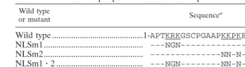
The Vp1 DBD overlaps with the bipartite Vp1 NLS.The N terminus of Vp1 is likely to harbor a DBD because of its proximity to the core of the virion (20) and by analogy to the mapped polyomavirus Vp1 DBD (22). It is also the location for the previously identified bipartite NLS (14), which consists of two clusters of basic residues. We will refer to the first cluster, 4-lysine-arginine-lysine-6, as NLS1 and to the second cluster, 15-lysine-lysine-proline-lysine-18, as NLS2 (Table 1). To test whether these basic clusters also mediate Vp1 DNA binding, sets of multiple-point mutations (NLSm1, NLSm2, and NLSm1䡠2) and N-terminal truncations [⌬N(2–21) and⌬N(2– 51)] were introduced into Vp1⌬C17 and Vp1⌬C58, and DNA-binding assays were performed. In NLSm1, NLS1 is mutated into asparagine-glycine-asparagine; in NLSm2, NLS2 is changed to asparagine-asparagine-proline-asparagine; and in NLSm1䡠
2, both clusters are correspondingly changed (Table 1). In
⌬N(2–21) or⌬N(2–51), residues 2 to 21 or residues 2 to 51, respectively, were deleted. Among the mutant⌬C17 proteins, NLSm2 had partial activity in the Southwestern blot, while NLSm1䡠2 and both N-terminal deleion mutants were inactive (Fig. 1B), suggesting that the basic clusters were necessary for the DNA binding. For the N-terminal mutant⌬C58 proteins, the Kds were determined with purified pentamers as before. The multiple-point mutant⌬C58 proteins sedimented in the sucrose gradients with a similar profile as Vp1⌬C58 (data not shown), while the N(2–21)-⌬C58 pentamer sedimented some-what slower than Vp1⌬C58, an expected result for the addi-tional deletion (Fig. 1C). We found that mutants NLSm1, NLSm2,⌬N(2–21), and NLSm1䡠2 bound DNA with
on November 9, 2019 by guest
http://jvi.asm.org/
FIG. 1. DNA binding by recombinant Vp1 proteins. (A and B) Southwestern blots. Protein samples were resolved on SDS–10% polyacrylamide gels and either stained with Coomassie blue (A, lanes 1 and 2; B, lanes 1 to 5) or electrotransferred to nitrocellulose and probed with nick-translated32P-labeled SV40 DNA (10 to 12 ng/ml) (A, lanes 3 and 4; B, lanes 6 to 10). In panel A, 0.5g of Vp1 (Vp1ⴱ, lanes 1 and 3) as a cleavage
product of GST-Vp1, or 0.5g of Vp1⌬C17 (lanes 2 and 4), were used. Intact GST-Vp1 (GST-Vp1) and the GST moiety (GSTⴱ) were also present in the samples of lanes 1 and 3. In panel B, 2g of Vp1⌬C17 (lanes 1 and 6) or of the following N-terminal mutant⌬C17 proteins were used: NLSm2 (lanes 2 and 7), NLSm1䡠2 (lanes 3 and 8),⌬N(2–21) (lanes 4 and 9), and⌬N(2–51) (lanes 5 and 10). Four bars to the left of each Coomassie gel mark the positions (from top to bottom) for molecular mass standards of 110, 74, 45, and 26 kilodaltons. (C) Isolation of pentameric Vp1⌬C58 and⌬N(2–21)-⌬C58. Sedimentation through sucrose gradients was performed as described in Materials and Methods, and an aliquot
on November 9, 2019 by guest
http://jvi.asm.org/
sively decreased affinities; their Kds in terms of the protein pentamer, 4.4 (⫾0.6)⫻10⫺9, 1.3 (⫾0.2)⫻10⫺8, 4.8 (⫾0.9)⫻ 10⫺8, and 2.6 (⫾0.4)⫻ 10⫺7, respectively, correspond to 41, 14, 4, and 0.7% of the Vp1⌬C58 activity, respectively (Fig. 1D and E). AKdwas not determined for⌬N(2–51)-C58 because of difficulty with its bacterial expression, but its ⌬C17 counter-part,⌬N(2–51)-⌬C17, was found not to have sufficient DNA-binding affinity for Kdmeasurement (Fig. 1D and E). These results indicate that NLS1 and NLS2—that is, NLS1-DBD1 and NLS2-DBD2—represent essential parts of the Vp1 DBD, with the latter part contributing more to DNA binding than the former part. The N-terminal 22-to-51 region also appears to harbor a small fraction of total binding activity.
The NLS function of the N-terminal basic clusters was con-firmed in cells transfected with pSG5-Vp1⌬C58-GFP, for which the fusion protein of Vp1 N-terminal 303 residues and GFP was expressed from the SV40 early promoter in the ab-sence of Vp2/3 and the tumor antigens. Both the Vp1 immu-nofluorescence (Fig. 2) and the GFP autofluorescence (data not shown) indicated that, while wild-type Vp1⌬C58-GFP lo-calized effectively to the nucleus (Fig. 2a), the mutant coun-terpart NLSm1, NLSm2, and NLSm1䡠2 proteins largely ac-cumulated in the cytoplasm (Fig. 2b, c, and d). A mostly cytoplasmic localization pattern was also observed for full-length mutant Vp1s expressed from pSV-Vp1-p81 (NLSm2) and -p25 (NLSm1 䡠 2), in which the tumor antigen coding sequences are present, and from pSG5-Vp1-p25 (14). There-fore, the two basic clusters within N-terminal 21 serve as the NLS of Vp1. The collective results indicate that the DBD of Vp1 overlaps with the NLS, sharing the requirement for at least the two basic amino acid clusters.
Importance of Vp1 NLS-DBD for viability.To test the role of the overlapping Vp1 DBD and NLS in the viral life cycle, we examined the viability of nonoverlapping SV40 (NO-SV40) genomes into which NLSm1, NLSm2, and NLSm1䡠2 muta-tions had been introduced. NO-SV40 is a viable SV40 DNA containing all of the regulatory sequences, the early genes, and spatially separated Vp2/3 and Vp1 coding sequences (13). Two types of viability assays were performed, one by microinjection of the viral DNA and the other by infection of the viral DNA-transfected cell lysate. Mutants NLSm1 and NLSm1䡠2 failed to produce plaques in the microinjection assay, and the infec-tion assay confirmed NLSm1 to be incapable of producing infectious particles (Table 2). Compared with wild-type NO-SV40, mutant NLSm2 formed smaller plaques in the
microin-jection assay and formed plaques that were 5,000 times fewer, as well as smaller, in the infection assay (Table 2).
To determine which stages of the viral life cycle were af-fected by the Vp1 NLS mutations, we examined the state of various viral processes, beginning with viral DNA replica-tion and capsid protein producreplica-tion. The amount of intracellu-lar viral DNA, quantitated by Southern slot blot, increased steadily with increasing times posttransfection for the wild type and the three Vp1 NLS-mutant DNAs (Fig. 3A). Comparable amounts of Vp1, Vp2, or Vp3 were detected by Western blot in cell lysates from transfection with either wild-type or the three mutant viral DNAs (Fig. 3B). Thus, defects other than those in viral DNA replication or capsid protein production were responsible for the abolished or greatly reduced viabili-ties of the Vp1 NLS-DBD mutants.
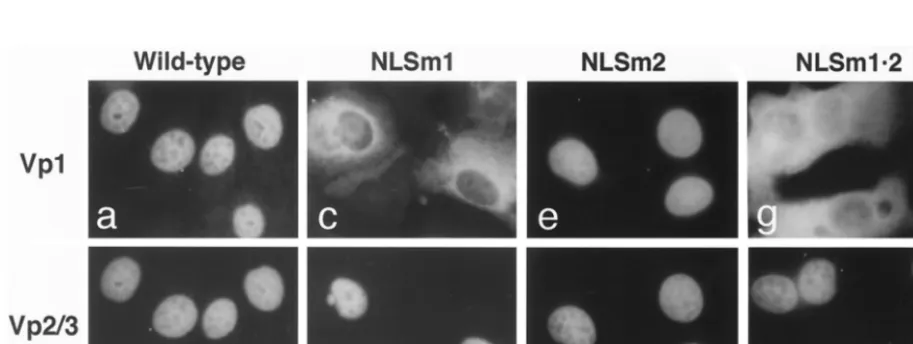
[image:5.612.310.552.81.148.2]Nuclear localization defect of mutant Vp1s expressed by NO-SV40-NLSm1 and-NLSm1䡠2.We next examined the NO-SV40 mutants for the subcellular distribution of the capsid proteins. Since mutations in either or both of NLS1 and NLS2 impaired the nuclear localization of a Vp1 fusion protein (Fig. 2), at least two different scenarios are possible when the mu-tant Vp1s were present along with Vp2/3 in cells introduced with the mutant viral DNAs. One is that the intact NLSs of wild-type Vp2/3 can complement the defective NLS of Vp1, as observed for NO-SV40-Vp1⌬N5 (13), and the mutant Vp1 is piggybacked to the nucleus by Vp2/3. This phenotype was seen for mutant NLSm2. Both the mutant Vp1 and the Vp2/3 were nuclearly localized (Fig. 4e and f), as were the capsid proteins expressed by wild-type NO-SV40 (Fig. 4a and b). The second scenario is that NLS complementation fails to occur, for ex-ample, because cytoplasmic Vp1-Vp2/3 interaction is somehow blocked; as a result, Vp2/3, but not the mutant Vp1, enters the nucleus. This phenotype was observed for mutants NLSm1 and NLSm1䡠2, whose mutant Vp1s remained in the cytoplasm
TABLE 1. Multiple-point mutants of the Vp1 N terminus Wild type
or mutant Sequencea
Wild type ...1-APTKRKGSCPGAAPKKPKEPV-21
NLSm1...
---NGN---NLSm2...
---NN-N---NLSm1䡠2 ...
---NGN---NN-N---aUnderlining indicates the two constituent basic clusters of the bipartite NLS,
NLS1, and NLS2. Dashes are used to denote the same amino acids as wild-type Vp1.
from each of the 15 fractions was analyzed by SDS-PAGE and Coomassie blue staining. In the profiles shown, twice as much protein was sedimented for⌬N(2–21)-⌬C58 (lower panel) than for Vp1⌬C58 (upper panel). Six bars to the left of each gel mark the positions for six molecular mass standards of 100, 71, 44, 28, 19, and 14 kilodaltons. Pentamers were found in fractions 8 and 9 for Vp1⌬C58 and in fractions 8 through 10 for⌬N(2–21)-⌬C58. NLSm1⌬C58, NLSm2⌬C58, and NLSm1䡠2⌬C58 gave sedimentation profiles similar to that of Vp1⌬C58. (D) Solution-phase DNA binding. Filter-binding assays were performed by incubating various concentrations of each protein with 32P-labeled DNA, and the
percentages of the input radiolabel that was retained on nitrocellulose membrane upon filtration were determined. Average values from three experiments are shown with error bars for the binding of a 326-bp PCR-derived SV40 fragment by pentameric Vp1⌬C58 or its N-terminal mutant derivatives. Values from one experiment are shown for the binding of nick-translated SV40 DNA by ⌬N(2–51)-⌬C17 whose monomeric concentrations are given in parentheses. Dissociation constants (Kds) were determined as molar protein concentrations at 50% DNA retention.
(E) Summary of DNA-binding activities for recombinant Vp1s. For GST-Vp1-derived Vp1 and for Vp1⌬C17, the apparentKdwas given in protein
monomer concentration for the binding of nick-translated SV40 (SV) or pBR322 (pBR) DNA. For⌬C58 proteins, an averageKdalong with the
standard deviation was given in the protein pentamer concentration for the binding of a 326-bp SV40 fragment. The relative activity is the reciprocal ofKdmade relative to that of the Vp1⌬C58Kd, which was taken to be 100%. An “X” on the schematic protein diagram represents the
mutation of an N-terminal basic cluster; a dot beneath residues 104 and 254 indicates their mutation from cysteines into alanines, n.d., not done.
on November 9, 2019 by guest
http://jvi.asm.org/
despite the nuclear localization of Vp2/3 (Fig. 4c, d, g, and h). Thus, the major defect of nonviable mutants NLSm1 and NLSm1䡠2 appears to lie in the inability of the mutant Vp1s produced to localize to the nucleus. This defect could arise from the mutant Vp1s’ lack of intrinsic Vp2/3-interactive abil-ity or from the mutant Vp1s’ inabilabil-ity to reach a cytoplasmic site necessary for the Vp1-Vp2/3 interaction. The former pos-sibility is examined below.
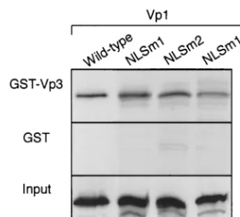
In vitro interaction of mutant Vp1s with Vp3.To determine if the N-terminal mutant Vp1 proteins had the intrinsic ability to associate with Vp3, an in vitro interaction assay was per-formed. In vitro-transcribed and-translated,35S-labeled NLSm1, NLSm2, and NLSm1䡠2 mutant Vp1s bound resin-immobilized GST-Vp3, as did wild-type Vp1, whereas little of the Vp1s bound to GST alone (Fig. 5). Thus, the intrinsic interaction between Vp1 and Vp2/3 was not affected by the Vp1 N-terminal muta-tions.
Nuclear virion assembly defect of NO-SV40-NLSm2.Since all capsid proteins of mutant NLSm2 were produced in normal quantities and could localize to the nucleus, and the mutant DNA replicated normally, we tested whether the mutant could form virion-like particles. When viral DNA-transfected cell lysates were treated with DNase I, only 6.9% of the total mu-tant DNA remained, compared with 71% of the total wild-type DNA. Thus, mutant NLSm2 either packaged only 1/10 the amount of viral DNA as wild-type NO-SV40 or packaged the viral DNA mostly in a manner that left the DNA susceptible to DNase I digestion.
To determine if the protected mutant DNA was packaged in a particle form, the DNase I-resistant materials from wild-type and NLSm2 DNA-transfected lysates were examined by sedi-mentation through sucrose gradients. To adjust for the 10-fold-reduced amount of the nuclease-resistant mutant viral DNA, 10 times as much nuclease-treated NLSm2 sample as the
cor-responding wild-type sample was sedimented, and equal ali-quots of wild-type and NLSm2 sucrose fractions were analyzed for viral DNA by Southern blot (Fig. 6). Since Vp1, whether wild type or mutant, would quantitatively remain after the nuclease treatment, it would be much more abundant in the 10-times-larger NLSm2 sample than in the wild-type sample. Accordingly, one-fifth as much of the NLSm2 fractions than the wild-type fractions was analyzed by anti-Vp1 Western blot (Fig. 6). The distribution profiles showed that wild-type viral DNA and Vp1 were present mostly in fractions 1 through 9 as well as 17 (Fig. 6A and C). About 42% of the wild-type DNA resided in fractions 3 through 5, which corresponded to the sedimentation location of purified virions (Fig. 6C). The dis-tributions of NLSm2 DNA and the mutant Vp1 were some-what broader than their wild-type counterparts, with compar-atively more of both mutant DNA and Vp1 present in fractions 7 through 9 (Fig. 6B and C). Nonetheless, about 30% of the mutant viral DNA was present in the expected particle frac-tions 3 through 5 (Fig. 6C). These results indicated that mutant NLSm2 could form virion-like particles, though at a reduced level. Taken together, these results point to a reduced level of viral DNA packaging by mutant NLSm2 and hence a role for NLS2-DBD2 in the virion assembly process.
DISCUSSION
[image:6.612.83.263.70.265.2]In this study, we mapped within the N-terminal 21 amino acids of SV40 Vp1, a DBD that accounted for most (96 to 99%) of the in vitro-measured Vp1 DNA-binding activity. This DBD overlaps the two basic clusters, NLS1-DBD1 and NLS2-DBD2, of the bipartite Vp1 NLS (14). Both basic clusters are required for full DNA-binding activity, though the second clus-ter (15-lysine-lysine-proline-lysine-18) is more important for the binding than the first cluster (4-lysine-arginine-lysine-6). The biological functions of the overlapping DBD and NLS were then dissected in vivo using mutant NO-SV40 viral ge-nomes with the application of the NLS phenotypic comple-mentation we have previously established (13). Nonviable mul-tiple-point mutants NLSm1 and NLSm1䡠2 were exceptional in that their mutant Vp1s were unable to enter the nucleus de-spite the presence of Vp2/3, though the mutant Vp1s’ ability to interact with Vp3 in vitro was not affected. For a multiple-point mutant of NLS2-DBD2, NLSm2, a greatly decreased viability was correlated with a decreased level of virion packaging but not with any defects in viral DNA replication or capsid protein production, interaction, or nuclear localization. Our results
FIG. 2. Subcellular localization of NLS-mutant Vp1⌬C58-GFP pro-teins. Cells transfected with wild-type (a), NLSm1 (b), NLSm2 (c), and NLSm1.2 mutant pSG5-Vp1⌬C58-GFP were fixed and stained with guinea pig anti-Vp1, followed by rhodamine-labeled anti-guinea pig antibody. Photographs of the rhodamine fluorescence are shown. The GFP autofluorescence in each case gave an essentially identical pat-tern, although the intensity was less than the corresponding Vp1 im-munofluorescence.
TABLE 2. Plaque-forming abilities of NO-SV40 Vp1 NLS-DBD mutants
NO-SV40 Formation ofplaquesa
Titer in transfected cell lysate (PFU/ml)c
Mean plaque
diam (mm)c
⫾SD
Wild-type Yes 1.8⫻108 5.0⫾1.2
NLSm1 No 0d
NLSm2 Yesb 3.5⫻104 0.8⫾0.3
NLSm1.2 No ND
aIn microinjection assay.
bThe average plaque diameter was ca. one-fourth that of the wild type.
cDetermined from infection-type plaque assay. ND, not done.
dNo plaques were detected in 0.1 ml of transfected cell lysate.
on November 9, 2019 by guest
http://jvi.asm.org/
[image:6.612.312.551.620.694.2]demonstrated, for the first time, that NLS2-DBD2 is important for the formation of infectious particles and that the Vp1 DNA-binding function is likely to have an essential role in this process. The N-terminal location of the DBD is consistent with the crystallographic structure of the virion in which the Vp1 N terminus is oriented toward the minichromosomal core and is disordered (20).
To obtain a Kdfor homogeneous Vp1 pentamers and to favor a one-to-one molar pentamer-DNA interaction, we used the purified Vp1⌬C58 pentamer, mutated in cysteines 104 and 254 to minimize artifactual pentamer-pentamer aggregation, and a short (326-bp) DNA probe. AKdof 1.8⫻ 10⫺9M in terms of the protein pentamer was obtained. Two other re-combinant Vp1s, cleaved GST-Vp1 and Vp1⌬C17, had appar-entKds 5.3⫻10⫺9to 7.3⫻10⫺9M in terms of the protein monomers, which are roughly comparable to the Vp1⌬C58 pentamer value and reinforce the reliability of our assays. Whereas the DBD of SV40 Vp1 comprises NLS1 and NLS2,
that of the related polyomavirus Vp1 consists of a single basic cluster, 1-alanine-proline-lysine-arginine-lysine-5 (2, 22), with a sequence resemblance to that of NLS1-DBD1. For SV40, it is NLS2-DBD2 (represented by mutant NLSm2) that was at-tributed with a larger share of DNA-binding activity and a role in in vivo virion packaging. Whether mutating the polyomavi-rus Vp1 DBD impairs polyomavipolyomavi-rus particle packaging would make an interesting comparison. The reported apparentKdfor DNA binding by polyomavirus Vp1 is 1-2⫻10⫺11M in term of the protein monomer (22). The difference between this value and the values for SV40 Vp1 may reflect either a com-paratively higher DNA-binding affinity of polyomavirus Vp1 or differences in the assay methods employed.
[image:7.612.113.499.74.220.2]The DBD and the bipartite NLS of SV40 Vp1 overlap within the N-terminal 21 residues. A number of other proteins, in-cluding polyomavirus Vp1, influenza virus matrix protein M1, and proteins with bHLH/bZIP, zinc finger, and homeobox domains, are also known to harbor a DNA- or RNA-binding
FIG. 3. DNA replication and capsid protein production by N-terminal Vp1 mutants. (A) Time course of viral DNA replication. Cells transfected with each NO-SV40 DNA were harvested at the indicated time points, and the total intracellular viral DNA was extracted and probed with nick-translated SV40 DNA in a Southern slot blot. The radioactivities of the individual slots were counted and plotted against time. (B) Levels of capsid proteins. Cells transfected with individual NO-SV40 DNAs were analyzed by immunoblotting with anti-Vp1 (upper panel) or anti-Vp3 (lower panel) antibodies. The amount of cells analyzed was adjusted for transfection efficiency as measured by the activity of-galactosidase expressed from pmiwZ, which was cotransfected with each NO-SV40 DNA. Bands corresponding to Vp1, Vp2, and Vp3 are indicated at the right.
FIG. 4. Subcellular localization of capsid proteins expressed from Vp1 N-terminal-mutant NO-SV40 DNAs. Cells were nuclearly microinjected with individual NO-SV40 DNAs, cultured for 24 h, fixed, and doubly stained with guinea pig anti-Vp1 (a, c, e, and g) and rabbit anti-Vp3 (b, d, f, and h), followed by rhodamine-labeled (a, c, e, and g) or fluorescein-labeled (b, d, f, and h) secondary antibodies. The same cells are shown in panels a and b, c and d, e and f, and g and h.
on November 9, 2019 by guest
http://jvi.asm.org/
[image:7.612.75.532.517.689.2]domain in overlap with an NLS (18). This overlapping or bifunctional domain arrangement is not surprising, since clus-tered basic residues typify many known NLSs and nucleic acid-binding domains (8, 18). Nevertheless, we were able to distin-guish the two functions in our analysis (see below). Our data, however, do not rule out a possibility that Vp1 amino acids other than the first 21 residues also make an additional
con-tribution to DNA binding. Such residues would conceivably be at the base of the Vp1 pentamer. This question is not ad-dressed in this study.
The functional duality of the Vp1 N-terminal domain can potentially complicate the determination of how an individual domain function contributes to viral viability. Yet by applying NLS phenotypic complementation, we found that the pheno-type of viral mutant NLSm2 was consistent with a role for the Vp1 DBD in nuclear virion assembly. This mutant was via-ble but had a 5,000-fold-lowered infectious titer and notably smaller plaque sizes. The pattern of capsid protein nuclear localization is consistent with the reported functional comple-mentation of NLSs, in which an NLS-defective Vp1 (e.g., Vp1⌬N5) accumulated in the nucleus in the presence of wild-type Vp2/3 as a result of the capsid proteins’ association in the cytoplasm prior to nuclear entry (13). Given this effective complementation of NLSm2 Vp1 nuclear localization, the nor-mal levels of viral DNA replication and capsid protein produc-tion, and the normal ability of the mutant Vp1 to interact with Vp3 in vitro, the mutant’s reduced viability is likely the result of the mutant Vp1’s impaired DNA-binding ability. There are at least two major ways a compromised Vp1 DNA-binding function can lead to a lower viral infectivity. First, impaired Vp1 DNA binding could affect the efficiency of DNA packag-ing in the nucleus and reduce number of particles produced. Second, the mutant virion-like particles assembled may be less stable or less effective at cell entry or nuclear targeting steps in a new round of infection. Our NLSm2 results reflect defects
FIG. 5. Interaction of N-terminal-mutant Vp1s with Vp3. In vitro-transcribed and -translated,35S-labeled Vp1 from each pBS-Vp1 DNA
was mixed with the resin to which either GST-Vp3 (first panel) or GST (second panel) had been immobilized. The resin-bound proteins were analyzed by SDS-PAGE and fluorography. The amounts of input35
[image:8.612.85.257.69.225.2]S-labeled Vp1s used for the pull-down experiments are also shown (third panel).
FIG. 6. Virion particle formation by NO-SV40-NLSm2. (A and B) Sonicated lysate prepared from wild-type (Wt, A) or mutant NLSm2 (NLSm2, B) NO-SV40 DNA transfected cells was treated with DNase I, and aliquots containing equivalent amounts of DNase I-resistant viral DNAs (60l for wild-type raised to 600l with buffer and 600 l for NLSm2) were sedimented through 5 to 32% sucrose gradients and fractionated from the bottom into 17 fractions. Five-sixths of each fraction was analyzed for viral DNA by Southern blot (upper panels). One-sixth of each wild-type fraction or one-thirtieth of each NLSm2 fraction was also analyzed for Vp1 by Western blot (lower panels). An arrowhead points to fraction 4, the peak fraction for viral DNA and Vp1 from purified virions sedimented in a parallel gradient. (C) Distribution of post-DNase I viral DNA and Vp1 in sucrose fractions. Wild-type (upper plot) and NLSm2 (lower plot). For each Southern or Western profile, the radioactivity per lane of the DNase I-resistant viral DNA or Vp1, obtained by phosphorimager quantitation of the observed band(s), was made relative to the total radioactivity of the DNA or Vp1 for all 17 fractions, which was taken to be 100%. The percentages were plotted against the fraction number.
on November 9, 2019 by guest
http://jvi.asm.org/
possibly at both stages of the viral life cycle. The observed 10-fold reduction in the packaging of the viral DNA (Fig. 6) would certainly lower the yield of potentially infectious parti-cles. In view of the 5,000-fold-lowered overall viability, it is also likely that the particles that did form were less infectious than wild-type particles in the next infection cycle. Further experi-ments would be needed to address the defect of mutant NLSm2 particles during reinfection.
Distinctly different from the apparent defect of mutant NLSm2 is the intriguing phenotype of mutants NLSm1 and NLSm1䡠2, which harbor mutations in the first basic cluster, NLS1. In contrast to NO-SV40-NLSm2 (above) and -Vp1⌬N5 (13), these two nonviable mutants produced cytoplasmically localized mutant Vp1s despite the nuclear localization of Vp2/3, indicating that the defective NLSs of the mutant Vp1s were not complemented by the presence of wild-type Vp2/3 NLS. This phenotype also indicates that Vp2/3 could enter the nucleus independently of the mutant Vp1s. Two lines of evi-dence support the hypothesis that the mutated NLS1 acts as a dominant signal for the cytoplasmic retention of NLSm1 and NLSm1䡠2 Vp1s. First, since all mutant Vp1 proteins were made in the mutant DNAs-transfected cells (Fig. 3B) and could interact with Vp3 in vitro (Fig. 5), the lack of comple-mentation suggests that mutant Vp1s were prevented from interacting with Vp2/3 in vivo. Second, deleting NLS1 as in the case of NO-SV40-Vp1⌬N5 actually restored NLS complemen-tation (13). Thus, the evidence points to the presence of an unidentified function, aside from those of nuclear localization and DNA binding, in NLS1-DBD1. That is, NLS1 may define a process in the cytoplasmic Vp1 biosynthetic pathway that precedes the association and the nuclear import of Vp1 and Vp2/3. We observed that Vp1 bearing single arginine 5-to-glycine point mutation (also part of the NLSm1 mutations) exhibited a cytoskeleton-like localization pattern when ex-pressed from a pSG5-based plasmid (N. Ishii and N. Minami, unpublished results) but showed a diffuse cytoplasmic staining when expressed from pSV-Vp1-p1 (14), from which the tumor antigens were also expressed. The suggestive role of cytoskel-etal elements in Vp1’s nuclear transport and association with Vp2/3 remains to be determined.
In summary, our study has mapped a DBD on SV40 Vp1 and has found that the DNA-binding contribution from NLS2-DBD2 plays a role during virion assembly. This Vp1 region may also function during next-cycle infection. DBDs have now been defined for all three SV40 capsid proteins. These do-mains are expected to function in conjunction with other known and as-yet-unknown interactive domains during the packaging of viral DNA.
ACKNOWLEDGMENTS P.P.L. and A.N. contributed equally to this work.
We thank Hiroshi Morioka of Hokkaido University, Sapporo, Ja-pan, for supplying the pETC-64M5LH15His plasmid DNA. We also thank Mary A. Tran for assistance in constructing pQE-Vp1-2CA-⌬C58.
This work was supported by Public Health Service grant CA50574 from the National Institutes of Health (NIH) and by a grant from the UCLA Academic Senate. P.P.L. was supported by a predoctoral fel-lowship from USPHS National Research Service Award GM07185. A.M.S. and C.F.F. were supported in part by undergraduate fellow-ships from, respectively, NIH Minority Scientist Development
gram GM55052, and NIH Minority Access to Research Careers pro-gram GM08563.
REFERENCES
1.Blasquez, V., S. Beecher, and M. Bina.1983. Simian virus 40 morphogenetic pathway. J. Biol. Chem.258:8477–8484.
2.Chang, D., X. Cai, and R. A. Consigli.1993. Characterization of the DNA binding properties of polyomavirus capsid proteins. J. Virol.67:6327–6331. 3.Clever, J., and H. Kasamatsu.1991. Simian virus 40 Vp2/3 small structural
proteins harbor their own nuclear transport signal. Virology181:78–90. 4.Clever, J., D. A. Dean, and H. Kasamatsu.1993. Identification of a
DNA-binding domain in simian virus 40 capsid proteins Vp2 and Vp3. J. Biol.
Chem.268:20877–20883.
5.Coca-Prados, M., and M.-T. Hsu.1979. Intracellular forms of simian virus-40 nucleoprotein complexes. II. Biochemical and electron microscopic analysis of simian virus 40 virion assembly. J. Virol.31:199–208.
6.Dalyot-Herman, N., O. Ben-nun-Shaul, A. Gordon-Shaag, and A. Oppen-heim.1996. The simian virus 40 packaging signalsesis composed of redun-dant DNA elements which are partly interchangeable. J. Mol. Biol.259:69–80. 7.Dean, D. A., P. P. Li, L. M. Lee, and H. Kasamatsu.1995. Essential role of the Vp2 and Vp3 DNA-binding domain in simian virus 40 morphogenesis. J. Virol.69:1115–1121.
8.Dingwall, C., and R. A. Laskey.1991. Nuclear targeting sequences—a con-sensus? Trends Biochem. Sci.16:478–481.
9.Gharakhanian, E., J. Takahashi, J. Clever, and H. Kasamatsu.1988. In vitro assay for protein-protein interaction: carboxyl-terminal 40 residues of simian virus 40 structural protein Vp1 contain a determinant for interaction with
Vp1. Proc. Natl. Acad. Sci. USA85:6607–6611.
10. Griffith, J. P., D. L. Griffith, I. Rayment, W. T. Murakami, and D. L. D. Caspar.1992. Inside polyomavirus at 25-A˚ resolution. Nature355:652–654. 11. Gordon-Shaag, A., O. Ben-nun-Shaul, H. Kasamatsu, A. B. Oppenheim, and A. Oppenheim.1998. The SV40 capsid protein Vp3 cooperates with the cellular transcription factor Sp1 in DNA-binding and in regulating viral promoter activity. J. Mol. Biol.275:187–195.
12. Hirt, B.1967. Selective extraction of polyoma DNA from infected mouse cell cultures. J. Mol. Biol.26:365–369.
13. Ishii, N., A. Nakanishi, M. Yamada, M. H. Macalalad, and H. Kasamatsu.
1994. Functional complementation of nuclear targeting-defective mutants of simian virus 40 structural proteins. J. Virol.68:8209–8216.
14. Ishii, N., N. Minami, E. Y. Chen, A. L. Medina, M. M. Chico, and H. Kasamatsu.1996. Analysis of a nuclear localization signal of simian virus 40 major capsid protein Vp1. J. Virol.70:1317–1322.
15. Jakobovits, E. B., and Y. Aloni.1980. Isolation and characterization of var-ious forms of simian virus 40 DNA-protein complexes. Virology102:107–118. 16. Kasamatsu, H., and A. Nehorayan.1979. Intracellular localization of viral
polypeptides during simian virus 40 infection. J. Virol.32:648–660. 17. Kobayashi, H., H. Morioka, K. Tobisawa, T. Torizawa, K. Kato, I. Shimada,
O. Nikaido, J. D. Stewart, and E. Ohtsuka.1999. Probing the interaction between a high-affinity single-chain Fv and a pyrimidine(6–4)pyrimidone photodimer by site-directed mutagenesis. Biochemistry38:532–539. 18. LaCasse, E. C., and Y. A. Lefebvre.1995. Nuclear localization signals overlap
DNA- or RNA-binding domains in nucleic acid-binding proteins. Nucleic Acids Res.23:1647–1656.
19. Li, P. P., A. Nakanishi, M. A. Tran, A. M. Salazar, R. C. Liddington, and H. Kasamatsu.2000. The role of SV40 Vp1 cysteines in virion infectivity. J. Virol.74:11388–11393.
20. Liddington, R. C., Y. Yan, J. Moulai, R. Sahli, T. L. Benjamin, and S. C. Harrison.1991. Structure of simian virus 40 at a 3.8-A˚ resolution. Nature
354:278–284.
21. Lin, W., T. Hata, and H. Kasamatsu.1984. Subcellular distribution of viral structural proteins during simian virus 40 infection. J. Virol.50:363–371. 22. Moreland, R. B., L. Montross, and R. L. Garcea.1991. Characterization of
the DNA-binding properties of the polyomavirus capsid protein Vp1. J. Vi-rol.65:1168–1176.
23. Oppenheim, A., Z. Sandalon, A. Peleg, O. Shaul, S. Nicolis, and S. Otto-lenghi.1992. Acis-acting DNA signal for encapsidation of simian virus 40. J. Virol.66:5320–5328.
24. Salunke, D. M., L. D. Caspar, and R. L. Garcea.1986. Self-assembly of purified polyomavirus capsid protein Vp1. Cell46:895–904.
25. Sambrook, J., E. F. Fritsch, and T. Maniatis.1989. Molecular cloning: a laboratory manual, 2nd ed. Cold Spring Harbor Laboratory, Cold Spring Harbor, N.Y.
26. Soussi, T.1986. DNA-binding properties of the major structural protein of simian virus 40. J. Virol.59:740–742.
27. Stehle, T., S. J. Gamblin, Y. Yan, and S. C. Harrison.1996. The structure of simian virus 40 refined at a 3.1-A˚ resolution. Structure4:165–182. 28. Suemori, H., Y. Kadodawa, K. Goto, I. Araki, H. Kondoh, and N. Nakatsuji.
1990. A mouse embryonic stem cell line showing pluripotency of differenti-ation in early embryos and ubiquitous-galactosidase expression. Cell Dif-fer. Dev.29:181–186.
29. Yamada, M., and H. Kasamatsu.1993. Role of nuclear pore complex in simian virus 40 nuclear targeting. J. Virol.67:119–130.