The Effect of Handwriting on Cortical Excitability:
A TMS study
Lillian Brinken
A report submitted as a partial requirement for
the degree of Bachelor of Science with Honours at the
University of Tasmania, 2015.
I declare that this report is my own original work and that contributions of other have been
duly acknowledged.
Acknowledgements
First and foremost, thanks go to my supervisor, Dr. Mike Garry, for his continued patience
and support throughout the very long journey that was this thesis. Thank you for always
being available to field my questions and concerns and sort out any unexpected glitches in the
TMS lab or doubts I had about this project. Thank you also to all of my excellent participants
who so generously donated their time and cortices for the glory of science, research
participation credits and the chance to win some gift vouchers. Your willingness to take part
in what must have seemed a somewhat tedious and at times slightly uncomfortable
experiment has been greatly appreciated.
Thanks also to the staff and other students of the Human Movement and Neuroscience
laboratory for trouble-shooting tips and making it a happier place to spend time in. Finally,
also, last but definitely not least, thank you so much to my Mum for your unwavering support
Table of Contents
Title Page
Abstract………...1
Introduction……….2
Transcranial Magnetic Stimulation……….5
Response Variability………...5
Metaplasticity………8
SICI………10
Overlearned Tasks………..11
Rationale, Aims and Hypotheses………15
Method………16
Participants………..16
Materials and Apparatus………..16
Procedure………..17
Design and Data Analysis………...18
Results……….19
MEPs………...19
SICI……….23
Discussion………28
Limitations………..32
Implications and Future Research………34
Conclusions………..35
References………..37
List of Tables
Table Title Page
Table 1. Means and standard deviations of MEPs recorded from 20
the FDI muscle across different conditions
Table 2. Means and standard deviations of MEPs recorded from 21
the APB muscle across different conditions.
Table 3. Pairwise comparisons of MEP amplitude recorded at 23
different intensities and time points at the FDI muscle.
Table 4. Pairwise comparisons of MEP amplitude recorded at 23
different intensities at time points at the APB muscle.
Table 5. Mean Log SICI for FDI across different times and intensities 24
Table 6. Pairwise comparisons of log SICI ratios of the FDI muscle 25
Table 7. Means and standard deviations of log SICI ratios across time 26
and intensity in APB.
Table 8. Pairwise comparisons of log SICI ratios of the APB muscle 26
Table 9. Mean co-efficient of variation across the different conditions 27
The Effect of Handwriting on Cortical Excitability:
A TMS Study.
Lillian Brinken
Abstract
The current study investigated the effect of a handwriting task on cortical excitability in the
primary motor cortex using transcranial magnetic stimulation (TMS). Seventeen participants
(10 female) took part in a single session during which the amplitude of motor evoked
potentials (MEPs) was measured in response to single and paired pulse TMS stimulation.
Measurements were taken at baseline, immediately after a handwriting task and again 15
minutes after task completion. It was hypothesised that the handwriting task would cause a
change to cortical excitability in the form of an increase in facilitation and a decrease in
inhibition, as demonstrated by greater mean MEP amplitude and decreased short interval
cortical inhibition (SICI) ratios, respectively. This study failed to detect a significant effect
of handwriting on cortical excitability. Whether this is due to the absence of an overt effect,
a methodological shortcoming associated with the exploratory nature of this study or a
random fluctuation is unclear. The main implication of this study is that overlearned tasks
such as handwriting represent a currently under-investigated area and further research would
A large proportion of the cognitive and motor behaviours in which we engage on a daily basis
consist of overlearned tasks. These skills, such as walking, speaking, driving or handwriting
were initially challenging when novel, but once acquired, can be carried out with minimal
conscious effort (Willingham, 1998). Performance fluctuates very little as a function of
practice (Ungerleider et al., 2002) and activities are often executed simultaneously or in
conjunction with more cognitively demanding skills (Logan, 1979; Poldrack et al. 2005).
Given the extent to which we engage in overlearned tasks, surprisingly little is known
about how these activities might induce or interact with neuroplastic changes to cortical
excitability (CE) in the motor cortex. Although a great deal of transcranial magnetic
stimulation (TMS) research focuses on motor learning and CE, the neural basis of
overlearning everyday tasks and the effect of engaging in these tasks on CE is largely
overlooked. This is in part due to an historical focus in research on the effect of learning
novel or highly contrived tasks such as finger sequences or forced abductions (eg. Butefisch
et al. 2000; Koeneke et al. 2006; Stefan et al. 2006;) .
Novel-task paradigms offer increased experimental control by eliminating potential
effects of differential expertise, thus allowing causal inferences to be made about the
relationship between CE and motor learning. As studying how people learn or overlearn
everyday motor skills would require greater complexity in terms of research design, a blind
spot seems to have evolved in terms of researching the effects of everyday tasks and CE more
generally. While it seems likely that some of the observations associated with learning novel,
abstracted tasks might also hold true for everyday tasks, it is also quite possible that
generalisability of these findings to overlearned motor behaviour could be limited.
Decades of TMS research have robustly established that motor activity associated
Garry & Thomson, 2009; Karabanov et al., 2012; Pascual-Leone, Grafman & Hallett, 1994)
but the extent to which similar changes are evoked by motor activity associated with
overlearned tasks is currently unknown. If an overlearned task such as handwriting were to
affect CE, this would be potentially problematic in terms of current TMS protocols, which
generally treat participants arriving at laboratories as blank cortical slates and do not control
for engagement in overlearned tasks prior to research participation. The potential concern in
this practice is that cortical activity is continuous and CE is inherently dynamic, which, taken
together means that participants already have a history of synaptic activity prior to engaging
in any research. If overlearned tasks such as handwriting have the capacity to modulate CE
in a similar way to novel motor tasks due to their shared physical demands, it stands to reason
that overlearned tasks might also have the capacity to interact with the induction of
subsequent neuroplasticity as novel or abstracted motor tasks have been observed to do (e.g.
Goldsworthy et al. 2014; Iezzi et al., 2008; Rosenkranz, Kacar & Rothwell, 2007). If indeed
overlearned tasks do have the capacity to modulate metaplastic mechanisms, it would be
necessary to reconsider current TMS methodologies and potentially control for overlearned
tasks.
Metaplasticity refers to activity-dependent mechanisms which regulate the expression
of synaptic plasticity within neural networks (Abraham, 2008). In other words,
metaplasticity describes processes that control the amount or direction of synaptic plasticity
which can be induced by subsequent plasticity induction protocols after a given history of
synaptic activation. Many metaplastic mechanisms appear to be guided by the principle of
homeostasis, ensuring that neural networks maintain an adaptive level of dynamic flexibility
by setting limits on the amount of long-term-potentiation (LTP) or long term depression
Homeostatic metaplasticity is described by the Bienenstock-Cooper-Munro theory of
bi-directional synaptic plasticity (Bienenstock et al. 1982), which maintains that synaptic
plasticity is bi-directional (i.e. there is a possibility of evoking either LTP or LTD) and that
the threshold for inducing either effect at any given time varies as a function of previous
post-synaptic activity. According to this model, a history of high frequency activity causes an
increase in the threshold for induction of LTP and a decrease in threshold for LTD induction,
whereas low frequency synaptic activity will cause a reverse effect, allowing LTP to be
induced at a lower threshold, and increasing the threshold for LTD. As such, metaplastic
effects cannot be observed immediately after the activity that causes them, but rather, only
when plasticity is subsequently induced.
The idea that overlearned tasks undertaken prior to research participation might cause
overt changes to CE or influence subsequent plasticity induction seems plausible given that
many of the tasks observed to cause changes to CE in a laboratory setting share some
parameters with overlearned tasks (e.g. Byblow & Stinear, 2006; Garry, Kamen & Nordstrom
2004; Goldsworthy et al. 2014). A few rare studies on overlearned tasks and CE support this
hypothesis, particularly when tasks have a linguistic component (Fililpovic et al. 2008; Lo &
Fook-Chong, 2004; Papathanasiou et al. 2003).
The frequency with which humans engage in handwriting, an overlearned task with a
linguistic and fine motor component is high, especially among students, a group likely to be
heavily represented as participants in TMS research given the reliance on undergraduate
student populations for research participation. Levels of response-variability observed in
TMS research are also commonly high, and while this is likely to be related to the
physiological complexity of the neural circuitry underpinning the modulation of CE, it may
also be driven by participants’ differential history of engagement in overlearned tasks such as
TMS
Transcranial magnetic stimulation refers to a non-invasive technique by which an electrical
field is created in neural tissue in response to magnetic pulses emitted by a wire coil placed
on the outside of the scalp (Kobayashi & Pascual-Leone, 2003). This electrical field causes a
depolarisation of neurons, and if stimulation is of sufficient intensity, the generation of action
potentials. When TMS is applied to the motor cortex, excitation of pyramidal neurons in the
corticospinal tract results in a volley of waves being conducted along the spinal cord causing
motor evoked potentials (MEPs) that can be detected and measured in the corresponding
anatomical area on the contralateral side of the body, providing an index of corticospinal
excitability (CE) (Bashir et al. 2010). Corticospinal excitability refers to how responsive the
corticospinal circuit is to stimulation at any given time. In other words, CE describes the
relationship between the input and output of the circuit and is often assessed by placing
electrodes on a target muscle in the hand, locating the representation of that muscle in the
contralateral primary motor cortex (M1) and measuring the effects of stimulation on the
output of the target muscle. Varying the pattern of TMS pulses in terms of timing and
intensity can allow the characterisation of different components of the neural circuitry which
comprise CE.
Response Variability
High levels of both inter- and intra-subject variability are commonly found in MEP
amplitudes recorded in TMS research and despite considerable research into influential
factors, the source of a large amount of this variability remains poorly understood (Bestmann
& Krakauer 2015; Choudhury et al. 2011; Hamada et al. 2013). Some broad trends have
emerged, such as a tendency towards decreased intra-subject variability at higher levels of
motor threshold (Cuypers, Thijs & Meesen, 2004) or an increase in inter-subject variability
among women and older adults (Pitcher, Ogsden & Miles 2003), but much variability
remains unaccounted for.
A recent review of the uses and interpretations of MEPs by Bestmann and Krakauer
(2015) emphasises the physiological complexity of MEPs elicited via TMS, noting that MEPs
can be considered a summation of cortico-spinal, intra-cortical and trans-cortical
contributions to excitability and that the respective influences of these elements (and
therefore any potential modulators) can be difficult to quantify. MEPs have been observed to
comprise a series of cortico-spinal volleys called direct waves (D-waves) and indirect waves
(I-waves) which are characterised by different generators and latencies (Hamada et al. 2013).
Di Lazzaro and Ziemann (2013) suggest that the characteristics of these MEP components are
best described at the most basic level by a canonical microcircuit model of cortical
input-output, featuring excitatory pyramidal neurons in layers II and III (P2 and P3) as well as the
large, fast-conducting pyramidal tract neurons (PTNs) in layer V, and a network of inhibitory
interneurons. According to this model, D-waves, which are the first volley of excitation to
descend the spinal cord are generated by direct stimulation of the axons of PTNs in the white
matter. Early I-waves (I1) are believed to be generated by mono-synaptic, excitatory inputs
of P2 and P3 neurons onto PTNs as a result of axonal stimulation by the TMS pulse and later
I-waves are generated by circuits involving reciprocal excitation between P2, P3 and PTNs as
well as modulation by networks of inhibitory GABAergic interneurons.
Given that this is a complex circuit and that MEPs can be modulated by different
components of this circuit, Bestmann and Krakauer (2015) emphasise that caution needs to be
taken in interpreting the drivers of any changes on the basis of MEP amplitude alone. It also
needs to be borne in mind that this is not a closed circuit, and there is evidence to support the
potentially reflecting cognitive processes occurring elsewhere in the brain (e.g. Klein-Flügge
& Bestmann 2012; Klein-Flügge et al. 2013). This may be particularly true of changes to
MEPs in response to tasks featuring a linguistic component. For example, Papathanasiou et
al (2003) observed increased CE during a visual search task involving a linguistic component
in which participants were required to be physically inactive. Bilateral measurement of the
first dorsal interosseous (FDI) muscles revealed larger MEPs in the right rather than the left
hand, possibly associated with the predominant lateralisation of language to the left
hemisphere. Their finding emphasises the potential of modulation of CE in the motor cortex
via upstream changes in cortical activity associated with cognition. However, their study
examined CE during task execution, so it remains unclear as to whether any changes to CE
endured beyond task completion. If cognitively induced changes to CE of a linguistic origin
were to persist post task, this might have profound implications for future TMS protocols as
researchers would need to consider the possibility that baseline measurements of CE (such as
resting motor threshold, which is commonly used to determine stimulus intensity for
subsequent measures) might be affected by prior engagement in reading or writing (for
example, consent forms or information sheets associated with research participation, or
indeed activities undertaken prior to participation).
Another related factor influencing response variability is that due to the dynamic
nature of neural circuits, patterns of excitation and inhibition are liable to change depending
on a participant’s history of synaptic activation (Ridding & Ziemann, 2010). This means that
the same experimental protocols can evoke different responses at different times within a
single participant (eg. Rosenkranz, Kacar & Rothwell, 2007), and that differences observed
between multiple participants might have the potential to reflect the influence of extraneous
variables relating to synaptic activity prior to testing rather than the independent variable
Metaplasticity
Homeostatic metaplasticity at a systems level has been observed in the human primary motor
cortex, as demonstrated by changes in CE measured by MEPs (eg. Fricke et al.; 2011;
Goldsworthy et al. 2014; Murakami et al. 2012). These metaplastic effects have been found
in both inhibitory and excitatory circuits by pairing different combinations of continuous and
intermittent transcranial magnetic theta burst stimulation (cTBS and iTBS) and comparing
their respective input-output curves (IO) using TMS to evoke MEPs and calculate IO of SICI
(Murakami et al., 2012).
In excitatory corticospinal circuits Murakami et al. observed predictable baseline IO
responses following non-primed iTBS and cTBS (increased and decreased IO of MEPs,
respectively) and a homeostatic metaplastic effect when pairing identical protocols as
demonstrated by a decrease in the magnitude of plasticity induced by the second protocol of
each pair relative to baseline and pairing of non-identical protocols. The authors also found
evidence of homeostatic metaplasticity in inhibitory circuits, with decreases in IO of SICI
observed following paired excitatory protocols and increases in IO of SICI after paired
inhibitory protocol. Measuring metaplastic effects by comparing different combinations of
plasticity-inducing stimulation protocols in the motor cortex provides something of an
analogue for naturally occurring LTP and LTD-like processes and homeostatic metaplastic
effects can also be observed in response to or in interaction with motor activity (eg. Jung &
Ziemann, 2009; Stefan et al. 2006).
Additionally, there is considerable evidence of interactions between or modulations of
different stimulation protocols or stimulation protocols and motor activity of a
non-homeostatic nature. For example, Iezzi et al (2008) found that the introduction of a phasic
than just a suppression of the extent of facilitation and inhibition). Other studies, such as
Rosenkranz, Kacar and Rothwell (2007) have observed changes in the interaction between
motor activity and plasticity induction depending on the phase of motor learning, with a
reversal in polarity of response to paired-associative-stimulation (PAS). When applied to
cortex which has not been primed, PAS with a 25ms inter-stimulus interval (PAS25)
produces an LTP-like effect, however, Rosenkranz found PAS25 following motor activity led
to an LTD-like effect during early stages of motor learning which was not observed following
motor activity in later stages of motor learning.
Studies looking at metaplastic effects in the motor cortex evidently vary profoundly in
terms of both the protocols used and also the nature of the relationships observed.
Inter-subject response variability to plasticity induction protocols within studies in this field is also
often quite high (eg. Hamada et al. 2013; Stefan et al. 2006). A recent study by Goldsworthy
et al. (2014) suggested that prior activation of target hand muscles may lead to increased
inter-subject variability following plasticity induction. This finding has implications for the
interpretation of studies looking at metaplastic effects because increases in variability can be
understood as a decrease in net effect when analysed on a group level, which may be
interpreted as a metaplastic suppression of LTP or LTD-like changes. This also has
implications for methodologies by future studies in this field that use TMS to measure
changes in CE because tonic contractions such as those used by Goldworthy et al. are
frequently employed to establish active motor threshold in order to adjust stimulus intensity,
or to aid the location of the cortical representation of a target hand area. Finally, this finding
highlights the necessity of controlling for participants’ activity prior to participating in TMS
SICI
Short latency or short-interval intra-cortical inhibition (SICI) is a widely used TMS technique
which measures levels of inhibition within the cortex by networks of inhibitory interneurons
via a paired-pulse paradigm consisting of an initial conditioning pulse followed by a test
pulse (Bashir et al. 2010). The conditioning pulse activates the circuit of inhibitory
interneurons and the test pulse, delivered 1-6ms later, evokes an MEP which is measured and
when compared to single-pulse MEPs, can provide an index of the inhibitory effect of the
interneuron circuit (Di Lazarro et al. 1998). These inhibitory circuits are maximally activated
at levels of stimulation below the resting motor threshold (RMT), so the conditioning pulse is
usually set at between 60-80% of RMT, with a supra-threshold test pulse following. Resting
motor threshold refers to the level of stimulation at which an MEP can be evoked from an
individual when muscles are at rest. This is usually determined as the intensity at which an
MEP with an amplitude >50mV can be measured in 50% of trials. Cortical inhibition as
measured by SICI has been observed to affect later components of the test MEPs (indirect
waves caused by synaptic facilitation) rather than the early components caused by direct
stimulation of the axons of pyramidal tract neurons by the TMS pulse itself (direct waves)
(Di Lazarro et al. 1998). Studies using pharmacological adjuncts have observed these
inhibitory circuits to be GABAergic, most likely mediated by GABA-a (Rothwell et al.
2009).
Decreases in inhibition are often observed to accompany neurological conditions in a
clinical setting but are also associated with motor learning in a research context (Rothwell et
al. 2009). In a motor learning context, disinhibition is thought to play a role in long term
potentiation (LTP) in M1 by facilitating neuroplastic change at a synaptic level, as well as
potentially unmasking existing excitatory inputs onto pyramidal tract neurons
excitability failed to locate any studies which measured changes to SICI in response to
engaging in an overlearned task. However, a study by Filipovic et al. (2008) measured
cortical silent period (SP) (another index of cortical inhibition) and observed an effect of
disinhibition during a handwriting task which was interpreted as being associated with the
linguistic demands of the task.
Silent period refers to the phenomenon by which electromyographic (EMG) activity
associated with the constant voluntary contraction of a muscle ceases for a few hundred
milliseconds following the delivery of a TMS pulse above resting motor threshold (Bashir et
al. 2010). The silent period is usually measured from the end of the MEP evoked by the TMS
pulse until the point at which EMG activity caused by the continued contraction resumes
Both spinal and cortical inhibitory mechanisms are believed to contribute to different stages
of SP, with inhibitory spinal mechanisms affecting the earlier, direct waves and cortical
networks of inhibitory interneurons affecting the later, indirect waves (Chen, Lozano &
Ashby, 1999)
Evidence of disinhibition from studies looking at SP during linguistic tasks (Filipovic
et al. 2008; Lo & Fook-Chong, 2004, Papathanasiou et al., 2004), along with the decreases in
SICI observed in response to fine motor tasks which feature similar movements to
handwriting and rely on the same effectors (eg. Garry, Kamen & Nordstrom, 2004) makes it
seems plausible that handwriting might cause a decrease in SICI and given the lack of
literature on this subject, worthy of investigation.
Overlearned tasks
Overlearned motor skills consist of movement sequences which are largely automated, can be
conducted with minimal conscious effort and are not vulnerable to decreases in proficiency if
looking at changes in CE associated with motor learning focuses on novel tasks, usually over
a short period of time, either within a single session or over a period of a week or two (eg.
Bütefisch et al., 2000; Pascual-Leone et al. 1994; Koeneke et al., 2006). Observations of
disinhibition or increased MEP amplitude immediately following learning within a single
session are common when comparing post-task to baseline levels (eg. Garry, Kamen &
Nordstrom, 2004; Ziemann et al. 2001; Bütefisch et al., 2000). There is also some evidence to
suggest that patterns of excitation and activation change over subsequent sessions as
proficiency increases, for example, Pascual-Leone, Grafman and Hallett (1994) found that
areas in M1 representing muscles involved in a finger sequencing task increased in size and
showed increased CE associated with behavioural gains during the learning process. These
cortical changes abated as participants’ knowledge of the sequence became automated. The
time course of these changes led the authors to suggest that changes observed during the
learning phase were consistent with the notion of unmasking existing connections and
increasing synaptic efficacy associated with LTP and that the flexibility surrounding cortical
modulation during learning could lead to structural changes in intra-cortical and subcortical
networks as skills become overlearned.
Similarly, evidence from imaging studies show behavioural changes correlate with
patterns of activation as motor learning occurs over time (e.g. Penhune & Doyon, 2002;
Puttemans, Wenderoth & Swinnen, 2005). In their 2002 review of studies using neural
imaging to investigate motor skill learning, Ungerleider, Doyon and Karni propose that early
stages of learning involve rapid, dynamic increases in activity in cortical frontal lobe areas,
the striatum and the cerebellum with some involvement of the primary motor area, but that
over a period of weeks this gives rise to a slower re-organisation of the primary motor area,
overlearning motor skills involves the recruitment of additional neurons in M1 to overlapping
sequence-specific local networks within representation areas.
The idea of longer-term motor learning being underpinned by structural and
functional changes in M1 is also supported by neurophysiological evidence taken from
animal studies such as Rioult-Pedotti et al. (1998), who observed developments in horizontal
networks of pyramidal cells in layers II/III of the primary motor cortex of rats after five days
of practicing a skilled reaching task. Rioult-Pedotti et al. reported increases in amplitude of
local field potential recordings and decreased propensity for LTP induction in vitro in the
affected cortical areas, which was interpreted as indicative of LTP having occurred and
increased the threshold for the induction of subsequent LTP. Increased synaptogenesis in
layer V accompanied by increased size of representational area has also been observed in a
similar paradigm by Kleim et al (2002), suggesting a clear association between behaviour,
cortical excitability and changes to cell morphology in M1 which would be consistent with
LTP.
There seems to be considerable evidence showing that the process of becoming
proficient at a motor skill is associated with changes to M1 as learning occurs and also more
enduring changes which persist past once task execution has ceased. In other words, the level
of proficiency attained during learning is associated with differential patterns of cortical
activation, cortical excitability and morphological changes in the motor cortex. The presence
of these effects in studies of novel tasks further underscores the apparent lack of research
conducted into the neurophysiological substrate of overlearned tasks we engage in on a daily
basis, such as writing (Filipovic et al. 2008).
The very basic, abstracted, novel tasks commonly used to observe learning from a
experimental control; findings are not confounded by differential levels of expertise and
lengthy periods of skill development are not necessary for high levels of proficiency to be
achieved. However, this means that very little is known about the effect of engaging in
everyday motor tasks on the motor cortex, or how the development of new skills might
interact with those already attained. One rare study, by Balas et al. (2007) looked at how
engaging in an overlearned writing task (as compared to a writing task in an unfamiliar
alphabet) could interfere with consolidation when learning a novel finger opposition
sequence. The authors found a significant interference effect of the overlearned writing task
on offline-gains (improvements in performance occurring between practice sessions) when
participants were tested 24 hours later, which was not observed in the unfamiliar writing task
condition or the control group. This is an interesting result insofar as dominant explanations
of interference suggest that it is likely to occur when tasks are similar, making task
parameters set in the first task vulnerable to supersession by those set in the second task
(Shadmehr & Holcomb, 1997).
The findings of Balas et al. (2007) led the authors to suggest that practicing sequences
of movement with very different attributes can nonetheless lead to interference if the tasks
share a cortical representation area and potentially recruit neurons to specific local networks
from the same pool, which would be a possibility in this scenario given that representation
areas in M1 have been observed to be involved in executing novel tasks as well as coding for
well-learned sequences (Ungerleider et al., 2002). On a practical level, Balas et al. (2007)
suggested that these finding should be taken into account in therapeutic or experimental
context in order to avoid potential disruption of motor learning.
Rationale, Aims and Hypotheses of the current study
There is currently a dearth of literature examining the effects of overlearned tasks
such as handwriting on CE. There is some evidence of changes to CE during the execution of
a handwriting task (Filipovic et al. 2008), but to date, we have been unable to find any
investigation of the potential for handwriting to induce changes to CE which endure beyond
task completion. Given that handwriting has been observed to interact with motor learning
(Balas et al. 2007) and also features fine motor movements which have been observed to
induce longer lasting changes to CE (Garry, Kamen & Nordstrom, 2004), it seems plausible
that handwriting might cause a neuroplastic modulation of CE.
As TMS protocols rarely control for activities undertaken prior to research
participation, there is a distinct possibility that if there was an effect of handwriting, this
might influence subsequent measurements of changes in CE unbeknownst to researchers.
This is likely to be especially relevant and worthy of further investigation given that a high
proportion of participants are students and may frequently engage in handwriting prior to
research participation. Also, because TMS research often reveals high levels of response
variability and recent evidence suggests that prior motor activity may increase variability
following plasticity induction (Goldsworthy et al. 2014), there would be major
methodological implications if an effect of handwriting were to emerge.
The present study aims to address the current lack of literature on overlearned tasks
and CE by measuring the effect of a short handwriting task on MEP amplitude and SICI.
This investigation is exploratory in nature and will have the capacity to measure only an overt
change, not any priming or metaplastic effects. It is hypothesised that the handwriting task
will cause a significant increase in excitability in the primary motor cortex, as evidenced by
significant effect of disinhibition, shown by a reduction in the difference between single and
paired pulse MEPs.
Method
Participants
Seventeen participants (10 female) were recruited through advertisements at the University of
Tasmania as part of a larger study after an initial calculation indicated that a sample of 20
participants, would be sufficient to detect a moderate effect size of d = .66 as calculated by
G*Power 3 (Faul, Erdfelder, Lang & Buchner, 2007). Prospective participants underwent a
brief medical screening and those with contraindications for TMS were excluded (see
Appendix D for contraindications). Participants were aged between eighteen and forty-five
years in order to be able to give consent and to exclude potential confounds due to changes in
cortical excitability associated with increased age (Fujiyama et al. 2012). The mean age was
31.06 years (SD = 6.54). Participants were all right-handed to avoid variability associated
with cerebral dominance among the left-handed (Isaacs et al., 2006) Course credit was
offered to eligible students, and all participants were put in to a draw to win gift vouchers.
Apparatus and Materials
Two Magstim 2002 stimulators (Magstim Co., Whitland, UK) were used to apply TMS
through a single figure of eight coil attached to a BiStim module. Ag/AgCl electrodes were
used to record electromyographic activity from the first dorsal interosseous (FDI) and the
abductor pollicis brevis (APB) muscles. Recordings were amplified and band-pass filtered
using CED1902 amplifiers (Cambridge Electronics Design, Cambridge, UK) before being
Signal 4.0 software (Cambridge Electronics Design, Cambridge, UK). Participants
completed the writing task using a ball point pen and paper and a text exemplar of 300 words
printed on an A4 sheet in 12 point Times New Roman (See Appendix F).
Procedure
The following procedures were approved by the Tasmanian Social Sciences Human Research
Ethics Committee (See appendix A). Participants underwent a short medical screening (see
Appendix D) , were briefed as to the procedure and informed consent was obtained (see
Appendix C for consent form). Participants were seated, the skin above the FDI and APB
muscles was abraded to prevent impedance and electrodes were attached in a belly-tendon
montage. The area of left motor cortex at which maximal MEPs could be obtained from the
right FDI by stimulating moderately above threshold was located by moving the coil around
the M1 hand area in small steps until maximal MEPs were consistently evoked. The coil was
then placed at an angle with the handle at 45 ̊ from the midline and facing backwards to
induce currents in a posterior-to-anterior direction across the central sulcus and the position
of the coil was marked on participants’ scalps using a felt-tipped marker to ensure a
consistent coil position across trials.
Resting Motor Threshold (RMT) was established by beginning stimulation in this
location at a suprathreshold intensity and then reducing intensity in increments of 2% of the
maximum of the simulator output until no MEPs could be elicited over five consecutive
pulses. Stimulation intensity was then increased in steps of 1% and the lowest intensity at
which MEPS (with an amplitude of at least 50µV) were recorded in three of five consecutive
pulses was considered the RMT (Garry et al. 2009). Cortical excitability was measured via
times. Two measurements were taken at baseline, prior to the writing task, another
immediately afterwards (post 0) and finally at 15 minutes post-task (post 15).
SICI was measured with ten paired pulses at each stimulation intensity. A
conditioning pulse of 70% and test pulses at intensities between 120-140% of RMT (in 10%
steps) with an interstimulus interval of 3ms. Ten single TMS pulses were also delivered at
each intensity to obtain an MEP recruitment curve. Silent period was measured during a
voluntary contraction of FDI and APB (at approximately 10% of maximal force) with 20
single pulses per measurement interval at a stimulus intensity of 130% of RMT. The
measurement of silent period as well as the administration of single and paired pulses was
conducted in a randomised sequence to avoid any order effects.
Design and Data Analysis
The current study used a within-subjects, repeated-measures design to examine the effect of
the independent variable of handwriting on the dependent variable of cortical excitability.
Cortical excitability was operationalised as mean peak-to-peak MEP amplitude in microvolts
(mV) and SICI, which was measured as the ratio of the conditioned MEP (paired pulse
stimulation) to the test MEP (single pulse stimulation). Measurements were taken just prior
to; immediately following and at 15 minutes after completing a hand-writing task. Two sets
of baseline measurements were taken for each participant in order to increase the reliability of
estimates of SICI and MEP amplitude at rest. This was not feasible for post-task
measurements given the time taken to administer two sequences of pulses immediately
post-task would not have allowed for measurements to be taken at 15 minutes post-post-task.
Data for one participant was excluded on the grounds of high levels of background EMG
discarded after it was established that in a large proportion of sweeps, there was insufficient
evidence of background muscle activity to be able to discern the end of the silent period.
Single and paired-pulse MEPs were analysed using two separate repeated-measures
ANOVAs for each muscle. SICI was analysed using a 3 (time: mean baseline, post 0 and
post 15 minutes) x 3 (intensity: 120%, 130% and 140% of resting motor threshold) way
ANOVA. MEP was analysed using a 3 (time: mean baseline, post 0, post 15 minutes) x 2
(tms type: test or conditioned) x 3 (intensity: 120%, 130% and 140% of resting motor
threshold) way ANOVA. Both significant and non-significant main effects are reported, as
are significant interaction effects. Multivariate tests were used, so sphericity is not reported.
Because SICI ratios were not normally distributed, SICI was transformed to log SICI prior to
analysis.
Results
MEP amplitude
As would be expected, mean amplitude of MEPs recorded at both the FDI and APB muscles
was greater in the single pulse condition compared with the paired pulse condition, and
increased at higher levels of stimulation intensity. In the FDI muscle, mean MEP amplitude
in both the single and paired pulse conditions was greater 15 minutes following the writing
task than measurements at baseline and immediately after the writing task; however, MEP
amplitude immediately following the writing task was lower than at baseline in both TMS
condition (see Table 1 for means and standard deviations). In the APB muscle, mean MEP
amplitude was lowest at baseline and increased with each measurement in both the paired and
single pulse conditions (see Table 2 for means and standard deviations). Significant main
Table 1
Means and standard deviations of MEPs recorded from the FDI muscle across different
conditions. .
Intensity Baseline
Conditioned
Post 0 Post 15 Baseline
Test
Post0 Post 15
120% 0.55
(0.58) 0.68 (1.13) 0.58 (0.53) 1.53 (1.43) 1.49 (1.20) 1.65 (1.41)
130% 1.24
(1.47) 0.81 (0.77) 1.17 (1.41) 2.47 (2.37) 2.08 (1.67) 2.38 (1.71)
140% 1.60
(1.55) 1.64 (1.68) 2.06 (2.00) 2.79 (2.22) 2.90 (2.32) 3.47 (2.62)
Note: Standard deviations are presented in parentheses. Stimulation Intensity is expressed as
a percentage of RMT and all MEPs are in millivolts.
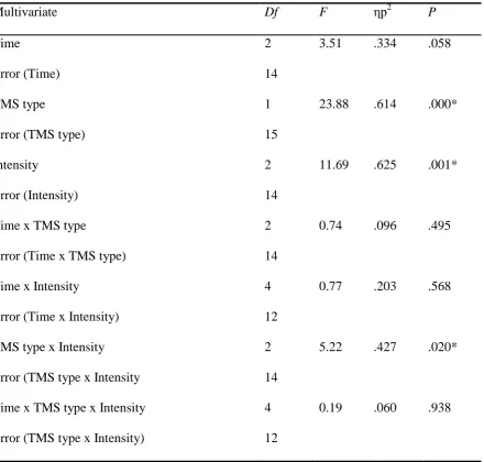
In the FDI muscle, the main effect of TMS intensity was highly significant, F (2,14) =
11.69, p = .001, ηp2 = .625, as was the main effect of TMS type (conditioned vs test pulse), F
(1, 15) = 23.88, p = >.001, ηp2 = .614. There was also a main effect of time which was
approaching significance, F (2, 14) = 3.51, p = .058, ηp2
= .334 and a significant interaction
effect between TMS type * TMS intensity, F (2, 14) = 5.22, p = .02 ηp2
= .427. None of the
other interaction terms were significant. Multivariate data can be found in Appendix G
Bonferroni corrected pairwise-comparisons showed that the three levels of stimulation
intensity were all significantly different from each other and that there was a significant
minutes afterwards but that neither of the post task measurements differed significantly from
[image:28.595.67.528.274.496.2]mean baseline (See Table 3 ).
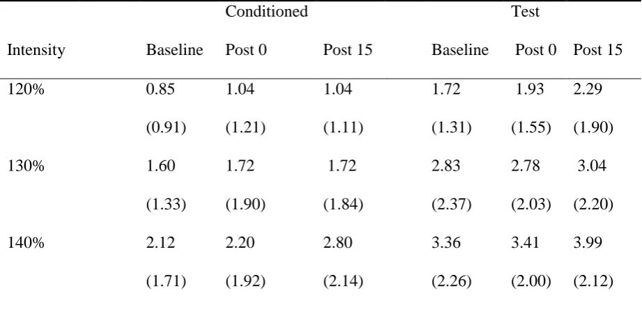
Table 2
Means and standard deviations of MEPs recorded from the APB muscle across different
conditions.
Intensity Baseline
Conditioned
Post 0 Post 15 Baseline
Test
Post 0 Post 15
120% 0.85
(0.91) 1.04 (1.21) 1.04 (1.11) 1.72 (1.31) 1.93 (1.55) 2.29 (1.90)
130% 1.60
(1.33) 1.72 (1.90) 1.72 (1.84) 2.83 (2.37) 2.78 (2.03) 3.04 (2.20)
140% 2.12
(1.71) 2.20 (1.92) 2.80 (2.14) 3.36 (2.26) 3.41 (2.00) 3.99 (2.12)
Note: Standard deviations are presented in parentheses. Stimulation Intensity is expressed as
a percentage of RMT and all MEPs are in millivolts.
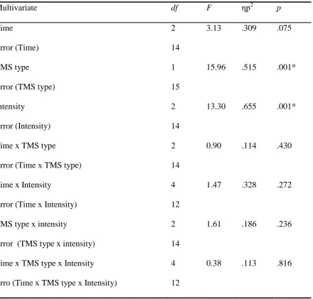
Analysis of MEP amplitude in the APB muscle yielded a similar picture; there were
significant main effects of TMS intensity, F (2, 14) = 13.3, p = .001, ηp2
= .655 and TMS
type F (1, 15) = 15.96, p = .001, ηp2
= .515, with a main effect of time which could be
described as approaching significance F (2, 12) = 3.13, p = .075, ηp2
=.309, however, no
comparisons with a Bonferroni correction suggested that there were no significant differences
in amplitude between measurements taken at baseline, following task completion or 15
minutes later. Once again, significant differences were observed in all comparisons of the
[image:29.595.66.525.338.580.2]three stimulation intensities (see Table 4).
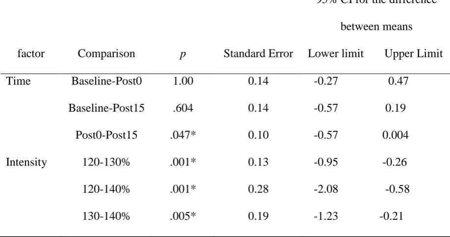
Table 3.
Pairwise comparisons of MEP amplitude recorded at 3 different intensities at 3 time points at
the FDI muscle.
factor Comparison p Standard Error
95% CI for the difference
between means
Lower limit Upper Limit
Time Baseline-Post0 1.00 0.14 -0.27 0.47
Baseline-Post15 .604 0.14 -0.57 0.19
Post0-Post15 .047* 0.10 -0.57 0.004
Intensity 120-130% .001* 0.13 -0.95 -0.26
120-140% .001* 0.28 -2.08 -0.58
130-140% .005* 0.19 -1.23 -0.21
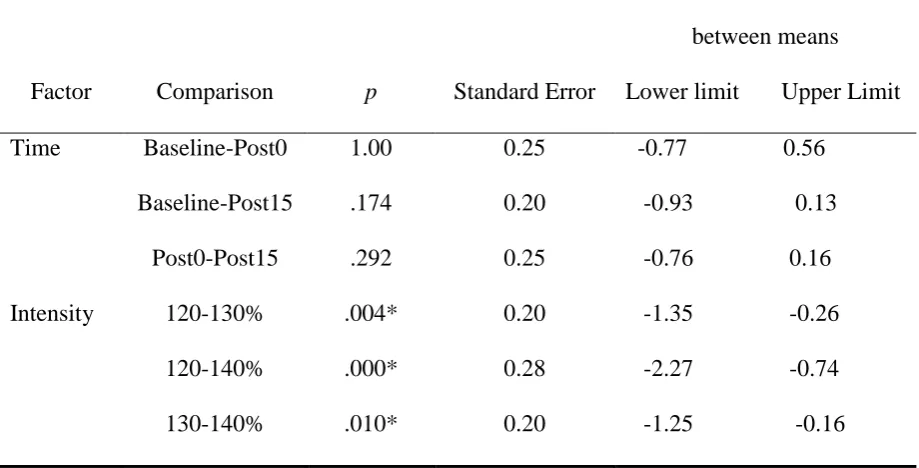
Table 4.
Pairwise comparisons of MEP amplitude recorded at 3 different intensities at 3 time points at
the APB muscle.
Factor Comparison p Standard Error
95% CI for the difference
between means
Lower limit Upper Limit
Time Baseline-Post0 1.00 0.25 -0.77 0.56
Baseline-Post15 .174 0.20 -0.93 0.13
Post0-Post15 .292 0.25 -0.76 0.16
Intensity 120-130% .004* 0.20 -1.35 -0.26
120-140% .000* 0.28 -2.27 -0.74
130-140% .010* 0.20 -1.25 -0.16
Note: p values have been Bonferroni corrected. Asterisks denote significant p values.
SICI ratios
Mean Log SICI ratios of MEPs recorded from the FDI muscle showed a pattern of decreasing
inhibition as intensity increased, and an increase in inhibition over time, with maximum
inhibition observed 15 minutes after task completion (see Table 5). However, the 3x3 way
ANOVA revealed non-significant main effects of intensity, F (2,14) = 3.49, p = .059, ηp2
=
.33, and time, F (2,14) = .60, p = .56, ηp2
=.08, although intensity could be construed as
Multivariate data can be found in Appendix G. Bonferroni corrected pairwise comparisons
revealed significant differences in effect of intensity on inhibition in stimulation at 120%
versus 140%, of resting motor threshold, and but no significant differences between 120%
and 130% or 130% compared with 140% (see Table 6). No significant differences were
[image:31.595.71.518.329.524.2]observed between any of the pairwise comparisons of the different levels of time.
Table 5
Mean Log SICI for FDI across different times and intensities.
Stimulation intensity Mean baseline Post 0 minutes Post 15 minutes
120% -0.42
(0.28)
-0.45
(0.41)
-0.47
(0.29)
130% -0.39
(0.34)
-0.43
(0.32)
-0.42
(0.35)
140% -0.29
(0.27)
-0.31
(0.21)
-0.32
(0.22)
Note: Standard deviations are presented in parentheses. Stimulation Intensity is expressed as
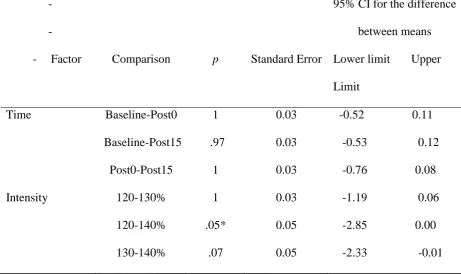
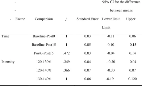
Table 6
Pairwise comparisons of log SICI ratios of the FDI muscle
-- Factor Comparison p Standard Error
95% CI for the difference
between means
Lower limit Upper
Limit
Time Baseline-Post0 1 0.03 -0.52 0.11
Baseline-Post15 .97 0.03 -0.53 0.12
Post0-Post15 1 0.03 -0.76 0.08
Intensity 120-130% 1 0.03 -1.19 0.06
120-140% .05* 0.05 -2.85 0.00
130-140% .07 0.05 -2.33 -0.01
Note: p values have been Bonferroni corrected. Asterisks denote significant p values.
At the APB muscle, SICI ratios showed a similar pattern to the FDI muscle, in terms
of decreased mean Log SICI at higher stimulation intensities, indicating lower levels of
inhibition at higher intensities but in contrast to the FDI, inhibition at the APB muscle
decreased over time (see table 7),.
The 3x3 ANOVA showed these trends were not indicative of a significant main effect
of intensity (F (2, 14) = 1.84, p = .57, ηp2 = .208) or a significant main effect of time (F (2,
14) = 1.26, p = .31, ηp2 = .153 ) (see Appendix G). Once again, the time*intensity
interaction was also found to be non-significant. Bonferroni corrected pairwise comparisons
Table 7
Means and standard deviations of log SICI across time and intensity in APB.
Stimulation intensity Mean baseline Post 0 minutes Post 15 minutes
120% -0.40
(0.38)
-0.34
(0.38)
-0.45
(0.41)
130% -0.30
(0.34)
-0.30
(0.30)
-0.35
(0.37)
140% -0.28
(0.23)
-0.29
(0.30)
-0.27
(0.31)
Note: Standard deviations are presented in parentheses. Stimulation Intensity is expressed as
a percentage of RMT
Table 8
Pairwise comparisons of log SICI ratios of the APB muscle
-- Factor Comparison p Standard Error
95% CI for the difference
between means
Lower limit Upper
Limit
Time Baseline-Post0 1 0.03 -0.11 0.06
Baseline-Post15 1 0.05 -0.10 0.15
Post0-Post15 .472 0.03 -0.04 0.14
Intensity 120-130% .249 0.04 - 0.20 0.04
120-140% .366 0.07 -0.30 0.07
130-140% 1 0.06 -0.19 0.120
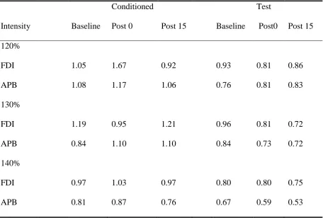
[image:33.595.67.528.477.755.2]Variability
The co-efficient of variation (CV) was calculated for each condition by dividing the standard
deviation (SD) of the group at each intensity, time point and tms type by its respective mean.
Dispersion of MEP amplitudes around the group mean was generally quite high, with means
and SDs often having a similar value. Higher mean CVs were observed in paired pulse trials
compared with single pulse trials, and an inverse relationship between CVs and stimulation
[image:34.595.71.528.431.739.2]intensity was observed (see table 9).
Table 9
Mean co-efficient of variation across the different conditions from the APB and FDI muscle.
Intensity Baseline
Conditioned
Post 0 Post 15 Baseline
Test
Post0 Post 15
120% FDI APB 1.05 1.08 1.67 1.17 0.92 1.06 0.93 0.76 0.81 0.81 0.86 0.83 130% FDI APB 1.19 0.84 0.95 1.10 1.21 1.10 0.96 0.84 0.81 0.73 0.72 0.72 140% FDI APB 0.97 0.81 1.03 0.87 0.97 0.76 0.80 0.67 0.80 0.59 0.75 0.53
Discussion
This study aimed to explore the possibility that engaging in a handwriting task might cause
an overt change to cortical excitability. It was hypothesised that a change in CE would be
induced, and that this would manifest as increased MEP amplitude and a decrease in SICI
ratios measured at both the FDI and APB muscles. None of these hypotheses were supported
by significant main effects or interactions. Although mean amplitude of both single and
paired pulse MEPs showed cortical excitability was at its highest 15 minutes after completing
the writing task at both the FDI and APB muscles, the absence of a significant main effect of
time at the designated alpha level means this observation cannot be justifiably interpreted as
the result of the handwriting task rather than chance fluctuations. Similarly, no causal
inferences can be made on the basis of the results of the analysis of SICI in relation to time as
there were neither any significant main effects, nor clear trends apparent in the pattern of
mean SICI ratios of MEPs from the two muscles.
The significant main effects of TMS intensity and TMS type on MEP amplitude
which were observed were expected given that previous research has fairly robustly
established the correlation between input intensity and output amplitude in the corticospinal
circuit (e.g. Choudhury et al. 2011; Darling, Wolf & Butler, 2006; Pitcher, Ogston & Marsh,
2003;) as well as the inhibitory effect of paired pulse stimulation on MEP amplitude (e.g.
Rothwell, Thompson & Kujirai 2009). However, these effects can be considered ancillary as
neither relate directly to the hypotheses. As the current study failed to detect an overt effect
of handwriting on CE at the determined significance level, the finding can be considered a
null result. Because this study was exploratory by nature and the hypotheses were not
It is entirely plausible that the current study failed to detect an effect because there was no
effect to be found. In other words, handwriting does not have any influence on CE. While
engaging in novel fine motor tasks (Garry, Kamen & Nordstrom, 2004) or abstracted thumb
abduction tasks (Buetefisch et al., 2000) causes enduring changes to CE, it is possible that
despite having similar physical demands, handwriting tasks do not have a similar effect due
to some other difference between these tasks and handwriting. One possibility is that
changes to CE observed in motor learning could be a reflection of task novelty rather than the
physical requirements of the task. Perhaps the changes to CE observed following novel tasks
represent a neural flexibility which allows for novel task parameters to be more easily
encoded. This would be adaptive in the early stages of learning, but might undermine the
stability of automated skills if it were to continue to be induced simply by motor movement.
The idea that changed CE itself might be indicative of a change in flexibility which
facilitates motor learning is conditionally supported by some studies reporting enhanced
motor performance following the induction of plasticity by non-invasive brain stimulation
techniques (Takeuchi & Izumi, 2015). Depending on the timing of plasticity induction and
motor learning, Takeuchi & Izumi suggest that inducing changes in CE can either have a
homeostatic effect (induction of an increase or a decrease in CE causes an effect of the
opposite direction on learning) or a synergic effect (induction of a change in CE causes an
effect of the same direction on learning). Synergic effects are more likely to occur when
plasticity induction and motor learning are simultaneous or separated by a short period of
time. Synergic effects are consistent with the idea that CE might index neural flexibility or
associated with novel motor learning because of the directional association between CE and
learning of novel tasks (e.g. Teo et al., 2011).
The finding of increased size and CE of representation areas in M1 associated with early
qualitative change in the neural production of motor behaviour associated with learning. The
authors observed these changes only during the early learning phase of a novel motor task,
and as the task became overlearned, these changes subsided. This would be highly consistent
with an interpretation of the current results that suggests that there was no effect of
handwriting on CE because overlearned tasks do not in fact cause any change to CE.
Imaging studies can also be interpreted as supporting the notion that M1 activity in response
to motor movements might vary as a function of task novelty or learning status rather than the
movement parameters of the task itself, for example, Puttemans et al. (2005) observed an
increase in M1 activation during initial learning, but this subsided when automaticity was
achieved. While imaging cortical activation is by no means an analogue of CE, it is possible
that the corresponding decreases in activation and CE in terms of skill learning and
performance improvement are epiphenomenal processes associated with a change in the
nature of neural basis for the production of motor movements as learning occurs.
The fact that this study did not find an overt effect of handwriting on MEP amplitude or
SICI does not preclude the possibility that the synaptic activity associated with handwriting
might cause a metaplastic change to subsequent plasticity induction. In order to investigate
the metaplastic potential of handwriting, it would be necessary to use quite a different
research paradigm to that employed by the current study. Rather than using TMS to assess
CE at baseline and then different time points post task, the protocol would most likely assess
CE first after a handwriting task and then again following the use of a non-invasive brain
stimulation (NIBS) technique. A change in CE following induction would be considered
indicative of plasticity induction, and finding a difference between groups (or between
sessions, in a within-subjects design) defined by the presence or absence of a handwriting
task would be understood as an activity-dependent modulation of plasticity induction (or a
associative stimulation (PAS) which can either cause facilitation or depression depending on
the inter-stimulus interval (PAS25 and PAS10, respectively); repetitive TMS (rTMS) which
leads to facilitation at low frequencies (<1Hz) and depression at higher frequencies (>5Hz);
theta burst stimulation, which induces facilitation when stimulation is intermittent (iTBS) and
depression when continuous (cTBS) and finally transcranial direct current stimulation (tDCS)
which varies in effect depending on the placement of anodal and cathodal electrodes on the
scalp (Ridding & Ziemann, 2010).
The literature on the relationship between motor activity and plasticity induction at a
systems-level is somewhat disparate to say the least. Motor activity is credited with causing a
range of different effects, including suppression of the usual facilitatory effect of PAS25 but
no change to the inhibitory effect of PAS10 (Stefan et al. 2006); reversal of the facilitatory
effect of PAS25 to an effect of inhibition (Rosenkranz, Kacar & Rothwell, 2007); reversal of
facilitatory effect of cTBS to inhibition (Gentner, et al., 2008); decrease in the extent of
plasticity in the expected directions following iTBS and cTBS (Huang, 2008); reversal of the
expected effects of iTBS and cTBS (Iezzi et al., 2008) and increased inter-subject variability
in the effect of inhibition following cTBS (Goldworthy, 2014). There is also a raft of
different effects observed when plasticity induction precedes motor activity (Takeuchi &
Izumi, 2015) and in paradigms featuring different timings and combinations of NIBS
(Ridding & Ziemann, 2010).
While it is likely that the large array of effects observed in relation to NIBS
techniques reflects the complexity of the processes underpinning them, it also seems possible
that, in the absence of a clear, overarching theory of metaplasticity in terms of a
neurophysiological substrate, this field may be vulnerable to an over-reliance on
significance-testing when interpreting data. Sometimes significant results occur randomly, and
unexpected significant results with post facto interpretations declaring the presence of effects
not previously considered or documented.
Sometimes non-significant results also occur randomly, and as such, another possible
explanation of the current pattern of results would be that handwriting does affect CE, but
that the current study was unable to detect this effect either due to a purely random
fluctuation (which would most likely not occur again if the study were replicated) or perhaps
due to some kind of a methodological error or mistake in data collection or experimental
design. Given the exploratory nature of the current study, and the corresponding lack of
literature in this area, the methodology employed could be construed as somewhat arbitrary at
times. Although utmost effort was taken to ensure that there was an empirical basis for all
techniques used, the novelty of this paradigm meant that in some instances, there was no
option other than to base a parameter on previous research which may not have been valid in
this context. For example, the five minute length of the handwriting task was based on
timeframes used in the induction of changes to CE by fine motor tasks in previous research
(e.g. Caramia et al., 2000, Garry, Kamen & Nordstrom, 2004; Rossi, Triggs & Eisenschenk,
1999). While fine motor activity is inarguably an important component of handwriting, when
designing an exploratory research paradigm, it is difficult to infer the relative importance of
other aspects of handwriting, such as its cognitive or linguistic demands, or status as an
overlearned task, which might also have an influence on the time course of induction of
neuroplastic changes.
Limitations
One possible limitation of the current study was the number of single and paired pulse
MEPs evoked and measured at each time point and intensity. Although it is common to
& Miles, 2002; Ziemann et al., 2001), because of the high rates of inter and intra subject
variability it might be advisable to measure more in order to increase reliability of estimates
of CE. Research by Cuypers, Thijs and Meesen (2014) looking at the optimisation of single
pulse TMS protocols in an inactive state suggests that measurement of at least 30 MEPs is
required in order to have a 99% chance of landing within the 95% confidence interval of CE.
However, as it takes time to deliver TMS and measure MEPs, increasing the number of
MEPs measured might make measurement at certain time points post-task untenable, as the
time taken to measure MEPs exceeds the interval between the designated time points. For
example, the current study measured 10 MEPs per pulse type (paired and single) at each
intensity plus a silent period at each time point (baseline, post 0 and post 15). This meant that
at post 0, 80 MEPs were measured in total and this took around 10 minutes. Measuring three
times as many MEPs would presumably cause the measurements taken at post 0 to run for
more than 15 minutes and consequently some temporal resolution would be forgone. This is
something of a catch-22 situation, where researchers must negotiate a trade-off between
reliably measuring MEP amplitude and charting the time course of CE. Another possible
option for decreasing variability in MEP amplitude might be to measure MEPs when the
muscle of interest is slightly contracted. Darling, Wolf and Butler (2006) found that
background muscle contractions of between 5-10% of maximum contraction caused a
significant decrease in MEP variability.
An additional potential limitation of the current study may have been insufficient
measurement intervals. For example, Caramia et al. (2000) found that under some
circumstances, the time course of changes to CE can vary over a period of up to 30 minutes
following task completion, with facilitation of MEP amplitude potentially only beginning at
15 minutes post task. Buetefisch et al. (2000) also observed a continuation of task-related
decision to take ultimate MEP measurements at 15 minutes post-task might have prevented
the current study from detecting effects which could plausibly have occurred after this point.
Implications and future research:
Given the dearth of previous research on overlearned tasks and cortical excitability, it seems
clear that more research should be conducted in this field. The lack of a cohesive body of
literature in this area makes interpretation of the absence of a main effect of time in the
current study less straight forward, and thus directions for future research less specific. It is
possible that the current study failed to detect an overt effect of handwriting because there
was no effect to be detected but is also possible that the current study failed to detect an effect
which was present, either randomly or because the current study paradigm did not have the
requisite sensitivity to detect an effect. Conducting more research in this area and varying
different parameters in terms of: length of handwriting task; timing of measurement post task;
number of MEPs per condition and intensity of stimulation would undoubtedly elucidate the
relationship between handwriting and overt effects on CE and would provide a context for the
interpretation of the current study.
If the current study failed to detect an effect of handwriting on CE because handwriting
cannot change CE, the implications of this would be effectively nil. There would be no
reason to review research protocols in terms of controlling for handwriting behaviour prior to
research participation, and any incidental handwriting involved in consent forms or medical
screening should not be of concern. However, the design of the current study meant that it
would only have been possible to detect an overt effect. If the current study did not detect an
effect because indeed there was no overt effect, this would not necessarily have any bearing
The capacity of handwriting to influence metaplastic mechanisms remains
unchartered water. As metaplastic effects have been observed following simple, tonic
contractions of the hand muscles (Goldworthy et al. 2014), it seems plausible that a more
complex task such as handwriting might also have the potential to influence plasticity
induction. However, the design of the current study meant that any results pertaining to an
overt effect of handwriting on CE would not have had any functional significance for
metaplastic research; the only implications the current study could have had for research in
this direction is the possibility of piquing interest in metaplastic effects of overlearned tasks
more generally.
Conclusions
The basis of the rationale for this study was the dearth of literature on overlearned tasks and
cortical excitability combined with the potential for major methodological implications were
an effect to be detected. Given the exploratory nature of this study, the hypotheses were
somewhat arbitrary and ultimately were not supported by observation as there was no clear
emergence of an effect of time on MEP amplitude or SICI as indices of CE. It remains
unclear as to whether this indicates an absence of effect, or the scope of this particular study
was not sufficient to detect an effect. The trending towards significance of the main effect of
time on MEP amplitude might provide tentative support for the presence of an effect of
handwriting on CE, but the results are far from conclusive. Further investigation in this area,
perhaps varying timing and task parameters would shed light on the findings of the current
study. As the absence of overt effects such as those which might have been detected in the
present study has little physiological bearing on whether or not handwriting or other
overlearned everyday tasks might influence subsequent plasticity induction, there is no reason
current finding can be seen to represent a small and inconclusive piece of a larger puzzle
References
Abraham, W. C. (2008). Metaplasticity: tuning synapses and networks for plasticity. Nature Reviews
Neuroscience, 9(5), 387-387.
Balas, M., Roitenberg, N., Giladi, N., & Karni, A. (2007). When practice does not make perfect:
well-practiced handwriting interferes with the consolidation phase gains in learning a
movement sequence. Experimental brain research, 178(4), 499-508.
Bashir, S., Mizrahi, I., Weaver, K., Fregni, F., & Pascual-Leone, A. (2010). Assessment and
modulation of neural plasticity in rehabilitation with transcranial magnetic stimulation.
PM&R, 2(12), S253-S268.
Bestmann, S., & Krakauer, J. W. (2015). The uses and interpretations of the motor-evoked potential
for understanding behaviour. Experimental brain research, 1-11.
Bienenstock, E. L., Cooper, L. N., & Munro, P. W. (1982). Theory for the development of neuron
selectivity: orientation specificity and binocular interaction in visual cortex. The Journal of
Bütefisch, C. M., Davis, B. C., Wise, S. P., Sawaki, L., Kopylev, L., Classen, J., & Cohen, L. G.
(2000). Mechanisms of use-dependent plasticity in the human motor cortex. Proceedings of
the national academy of sciences, 97(7), 3661-3665.
Byblow, W. D., & Stinear, C. M. (2006). Modulation of short-latency intracortical inhibition in
human primary motor cortex during synchronised versus syncopated finger movements.
Experimental brain research, 168(1-2), 287-293.
Caramia, M. D., Scalise, A., Gordon, R., Michalewski, H. J., & Starr, A. (2000). Delayed facilitation
of motor cortical excitability following repetitive finger movements. Clinical
neurophysiology, 111(9), 1654-1660.
Chen, R., Lozano, A. M., & Ashby, P. (1999). Mechanism of the silent period following transcranial
magnetic stimulation evidence from epidural recordings. Experimental brain research,
128(4), 539-542.
Choudhury, K. R., Boyle, L., Burke, M., Lombard, W., Ryan, S., & McNamara, B. (2011). Intra
subject variation and correlation of motor potentials evoked by transcranial magnetic
stimulation. Irish journal of medical science, 180(4), 873-880.
Cuypers, K., Thijs, H., & Meesen, R. L. (2014). Optimization of the transcranial magnetic
stimulation protocol by defining a reliable estimate for corticospinal excitability. PloS one,
9(1), e86380.
Darling, W. G., Wolf, S. L., & Butler, A. J. (2006). Variability of motor potentials evoked by
transcranial magnetic stimulation depends on muscle activation. Experimental brain
research, 174(2), 376-385.
Di Lazzaro, V., Restuccia, D., Oliviero, A., Profice, P., Ferrara, L., Insola, A., Mazzone, P., Tonali,