CORRELATION BETWEEN TIMING OF ASV
ADMINISTRATION AND COMPLICATIONS IN SNAKE
BITES
-
AN ANALYTICAL STUDY
Dissertation submitted for
MD Degree (Branch-I) General Medicine
March 2010
The Tamil Nadu Dr.M.G.R. Medical University
Chennai, Tamil Nadu
CERTIFICATE
This is to certify that this dissertation titled ‘CORRELATION BETWEEN TIMING OF ASV ADMINISTRATION AND COMPLICATIONS IN SNAKE BITES- AN ANALYTICAL STUDY’
submitted by DR.S.THIRUMURUGAN to the faculty of General Medicine,
The Tamil Nadu Dr.M.G.R. Medical University, Chennai in partial fulfillment
of the requirement for the award of MD degree Branch I (General Medicine) is
a bonafide research work carried out by him under our direct supervision and
guidance.
Dr.Moses K Daniel, MD., Dr.A.Ayyappan, MD.,
Professor of Medicine Professor and HOD
Chief II Medical Unit Department of Medicine
Department of Medicine Madurai Medical College
Madurai Medical College Madurai
DECLARATION
I, Dr.S.THIRUMURUGAN, solemnly declare that the dissertation
titled ‘CORRELATION BETWEEN TIMING OF ASV
ADMINISTRATION AND COMPLICATIONS IN SNAKE BITES- AN ANALYTICAL STUDY’ has been prepared by me.
This is submitted to the Tamil Nadu Dr.M.G.R. Medical University,
Chennai in partial fulfillment of the requirement for the award of MD degree
Branch I (General Medicine).
Place :
ACKNOWLEDGEMENT
I express my sincere thanks to The Dean for permitting me to use the facilities of Madurai Medical College and Govt. Rajaji Hospital to conduct this
study.
I will ever remain in gratitude to my chief Dr.Moses K Daniel, MD., Prof of Medicine, not only for guiding me through the study, but also for being
my mentor and source of inspiration during the period of my postgraduate
training.
My professor and Head of the Department Dr.A.Ayyappan, MD has always guided me by example and valuable words of advice through the
conduct of the study and also during my postgraduate course. My sincere thanks
to him.
I express my heartfelt thanks to my Assistant Professor Dr.David Pradeep Kumar, MD., Dr.K.Senthil, MD and Dr.P.Ganeshbabu, MD for their valuable support and guidance throughout my study.
My family and friends have stood by me during my times of need.
Their help and support have been valuable to the study.
I would grossly fail in my duty if I fail to mention here of my patients
who have ungrudgingly borne the pain and discomfort of investigations. I
cannot but pray for their speedy recovery and place this study as a tribute to
them and to the numerous others likely affected.
CONTENTS
PAGE NO.
1. INTRODUCTION 1
2. REVIEW OF LITERATURE 5
3. AIMS AND OBJECTIVES 31
4. MATERIALS AND METHODS 32
5. RESULTS 36
6. DISCUSSION 46
7. CONCLUSION 54
APPENDIX
1. BIBLIOGRAPHY
2. PROFORMA
3. MASTER CHART
1.
INTRODUCTION
Snake bites contribute to health problem in India and continue to be a
major medical concern. India alone contributes to 81,000 envenomations and
11,000 deaths annually. It appears that every 10 seconds one individual is
envenomed and one among four dies due to snake bite. Snake bite is an
occupational disease of farmers (rice), plantation workers (rubber, coffee),
herdsmen, hunters, snake handlers, fishermen and fish farmers.
In Tamil Nadu the total number of snake bite cases admitted (and
expired) in the secondary care hospitals alone during 2005 - 2006 and 2006
-2007 were 19321(85) and 20677(75) respectively. The total number of ASV
vials used in these hospitals during the respective periods was 94481 and 96800.
Over all analysis revealed that the snakebites and ASV usage in West, North,
East, Central, South zone of Tamil Nadu were 13, 17, 20, 24 and 26%
respectively.
Snakebites are observed all over the country with a rural / urban ratio of
9:1. They are more common during monsoon and post monsoon seasons.
Snakebites are seen often among agricultural workers and among those going to
the forest. The male / female ratio among the victims is approximately 3:2.
Majority are young and their age is between 25 to 44 years. Most of the bites
There are more than 3000 species of snakes in the world. For the purpose of clinical practice, snakes are classified into poisonous (venomous) and
non-poisonous (nonvenomous) snakes. Poisonous snakes common in India are
classified into these families and they are
• Elapidae [Cobra group] • Viperidae [Viper group]
• Hydrophidae [Sea snake group]
For many decades, the concept of the “Big 4” snakes of medical
importance has reflected the view that 4 species and responsible for Indian
snakebite mortality. They are the Indian cobra (Naja naja), the Common Krait
(Bungarus caeruleus), the Russell’s viper (Daboia russelii) and the Saw scaled viper (Echis carinatus). However, recently another species, the Hump-nosed pit viper (Hypnale hypnale), has been found to be capable of causing lethal
envenomation, and that this problem had been concealed by systematic
misidentification of this species as the saw-scaled viper. The concept of the “Big
4” snakes has failed to include all currently known snakes of medical
Anti snake venom (ASV):
ASV neutralizes the circulating venom only and no amount of ASV will
neutralize or combine with venom once the venom is attached or adsorbed to
the target organs. Currently available ASV in India is polyvalent i.e., it is
effective against all the four common species; Russells Viper (Daboia russelii),
Common Cobra (Naja naja), Common Krait (Bungarus caeruleus) and Saw
Scaled Viper (Echis carinatus). Indian ASV is a F(ab)2 product derived from horse serum and has a halflife of 26-95 hours. Though it is purified, it still can
be immunogenic.
In India ASV is manufactured by Bengal Chemicals & Pharmaceuticals, Kolkata; Bharat Serums, Mumbai; Biological Evans,
Hyderabad; Central Research Institute, Kausali; Haffkins Pharmaceuticals,
Mumbai; King Institute of preventive medicine, Chennai; Serum Institute,
Pune and Vins bio-products, Hyderabad.
The approximate fatal doses of venom of the poisonous snakes are as
follows:
The approximate quantity of venom neutralized by 1ml of polyvalent
ASV is given below:
1.Russell’s Viper :0.60mg 2.Saw scaled Viper :0.45mg 3.Cobra :0.60mg 4.Krait :0.45mg
ASV can save many of the complications and death due to snake bites if given in time. However superstitions, lack of prompt medical access, late
reporting to health care system and cost of ASV delays the administration of
ASV. Some authors have postulated that the renal abnormality correlates well
with late onset of treatment and that early ASV administration prevents renal
damage.
A detailed clinical study correlating the development of complications
with timing of ASV administration was needed. Therefore present study was
undertaken in inpatients admitted with snake bite in Govt. Rajaji hospital,
Madurai to study the use of ASV as an early intervention and to study the
relationship of late administration of ASV due to late arrival of patient to the
2. REVIEW OF THE LITERATURE
2.1 CLASSIFICATION:
There are two important groups (families) of venomous snakes in
South-East Asia –Elapidae have short permanently erect fangs. This family includes the cobras, king cobra, kraits, coral snakes and the sea snakes. The most important species, from a medical point of view, include the following:
Cobras: N naja Common spectacled Indian cobra (Genus Naja) N oxiana North Indian or Oxus cobra N kaouthia Monocellate cobra
N philippinensis Philippine cobra
N atra Chinese cobra
Spitting cobras: N siamensis N sumatrana
N sputatrix
King cobra: Ophiophagus hannah
Kraits: B caeruleus Common krait
(Genus Bungarus) B candidus Malayan krait B multicinctus Chinese krait B fasciatus Banded krait
Figure 2.1 Common types of Snakes in India
Viperidae have long fangs which are normally folded up against the upper jaw but, when the snake strikes, are erected. There are two subgroups, the
typical vipers (Viperinae) and the pit vipers (Crotalinae). The Crotalinae
have a special sense organ, the pit organ, to detect their warm-blooded prey. This is situated between the nostril and the eye.
Medically important species in
South-East Asia are:
Typical vipers: Daboia russelii
Russell’s vipers
Echis carinatus Saw-scaled or carpet vipers E sochureki
Pit vipers: Calloselasma rhodostoma Malayan pit viper
Hypnale hypnale Hump-nosed viper
Green pit vipers or bamboo vipers: (Genus Trimeresurus)
T albolabris White-lipped green pit viper
T gramineus Indian bamboo viper
T mucrosquamatus Chinese habu
T purpureomaculatus Mangrove pit viper
T stejnegeri Chinese bamboo viper
Common Krait Common Cobra
2.2 THE VENOM APPARATUS:
Venomous snakes of medical importance have a pair of enlarged teeth,
the fangs, at the front of their upper jaw. These fangs contain a venom channel (like a hypodermic needle) or groove, along which venom can be introduced
deep into the tissues of their natural prey. If a human is bitten, venom is usually
injected subcutaneously or intramuscularly.
2.3 IDENTIFICATION OF VENOMOUS SNAKES:
Identification of poisonous snakes is complex (involves counting of
scales) and not definitive (the identification of pre or post maxillary teeth) and
of no use to a doctor in a medical situation. What is important therefore is to
focus on the key aspects of identification that enable the medical professional to
rapidly identify whether they are dealing with a venomous species, and what
that species might be.
The following features can be used to identify the poisonous snakes:
1. Pupil size: The pupil of harmless snakes is round. Poisonous snakes have elliptical pupils.
2. Pit: Poisonous snakes have a pit, heat sensitive organ situated between the eye and nostril, which detects warm blooded prey. Harmless snakes do not have
Figure 2.2 Venomous Snakes identification
3. Scale arrangements: The underside scales of a venomous snake’s tail go all the way across in a single row from anal plate. The very tip of the tail may have
two scale rows. Nonpoisonous snakes have two rows of scales from the vent to
the end of the tail.
4. Head shape: Venomous snakes have a triangular or spider-shaped head.
5. Distinctive sounds: Russell’s viper will produce blowing hiss sound and saw-scaled viper will produce grating rasp sound.
2.4 COMPOSITION OF VENOM:
Snake venoms contain more than 20 different constituents, mainly
proteins, including enzymes and polypeptide toxins. The following venom
constituents cause important clinical effects:
(i) Procoagulant enzymes (Viperidae) that stimulate blood clotting but result in incoagulable blood. Eventually, and sometimes within 30 minutes of the bite,
the levels of clotting factors have been so depleted (consumption coagulopathy)
that the blood will not clot. Russell’s viper venom affects factors V, X, platelets
and protein C while saw-scaled viper venom activates prothrombin and
plasminogen.
(iii) Cytolytic or necrotic toxins - these digestive hydrolases (proteolytic enzymes and phospholipases A), polypeptide toxins and other factors increase
permeability resulting in local swelling.
(iv) Haemolytic and myolytic phospholipases A2 - these enzymes damage cell membranes, endothelium, skeletal muscle, nerve and red blood cells.
(v)Pre-synaptic neurotoxins (Krait and Russell’s viper) β-bungarotoxin, crotoxin and taipoxin that contain phospholipases A2 that damage nerve
endings, initially releasing acetylcholine transmitter, then interfering with
release.
(vi) Post-synaptic neurotoxins (Elapidae) - these polypeptides such as α-bungarotoxin and cobratoxin compete with acetylcholine for receptors in the
neuromuscular junction and lead to curare-like paralysis.
2.5 QUANTITY OF VENOM INJECTED AT A BITE:
This is very variable, depending on the species and size of the snake, the
mechanical efficiency of the bite, whether one or two fangs penetrated the skin
and whether there were repeated strikes. The snake may be able to control
whether or not venom is injected.
About 50% of bites by Malayan pit vipers and Russell’s vipers, 30% of
bites by cobras and 5-10% of bites by saw-scaled vipers do not result in any
symptoms or signs of envenoming (Dry bites). Snakes do not exhaust their store
of venom, even after several strikes, and they are no less venomous after eating
specimens of the same species, the venom of smaller, younger vipers may be
richer in some dangerous components, such as those affecting haemostasis.
2.6 HOW DO SNAKE BITES HAPPEN?
In South-East Asia, snake bite is an occupational hazard of rice
farmers; rubber, coffee and other plantation workers; fishermen and those who
handle snakes. Most snake bites happen when the snake is trodden on, either in
the dark or in undergrowth, by someone who is bare-footed or wearing only
sandals. Some bites occur when the snake (usually a krait) comes in to the home
at night in search of its prey (other snakes, lizards, frogs, mice) and someone
sleeping on the floor rolls over onto the snake in their sleep.
2.7 SYMPTOMS AND SIGNS:
Some people who are bitten by snakes or suspect or imagine that they have been bitten, may develop quite striking symptoms and signs, even when no venom has been injected. This results from fear of the consequences of a real venomous bite. Anxious people may overbreathe so that they develop pins and
2.7.1 GENERAL SYMPTOMS AND SIGNS OF VIPERINE ENVENOMATION:
Local effects:
• Swelling and local pain with or without erythema
• Tender enlargement of local lymph nodes
• Bruising and local inflammation
• Fang marks
Systemic effects:
• Bleeding from the gingival sulci and other orifices, epistaxis, petechiae,
purpura and ecchymoses.
• Renal failure in cases of Russell’s viper and sea snake due to acute
tubular necrosis secondary to prolonged hypotension, hypovolemia, DIC, direct
toxic effect of venom on the renal tubule, hemoglobinuria, myoglobinuria and
hyperkalaemia.
• Hypotension resulting from hypovolaemia, direct vasodilation and direct
effect on the myocardium,cardiac arrhythmias and pulmonary oedema.
• Muscle pain indicating rhabdomyolysis.
• Parotid swelling, conjunctival oedema, sub-conjunctival haemorrhage.
2.7.2 GENERAL SYMPTOMS AND SIGNS OF ELAPID ENVENOMATION
Local effects:
• Swelling and local pain with or without erythema (Cobra).
• Local necrosis and / or blistering / bullae (Cobra).
Neurotoxic effects:
• Descending paralysis, initially of muscles innervated by the cranial
nerves, commencing with ptosis, diplopia, or ophthalmoplegia. There may be
some involvement of the senses of taste and smell.
• Problems of vision, breathing and speech.
• Paralysis of jaw and tongue may lead to upper airway obstruction and
aspiration of pooled secretions because of the patient’s inability to swallow.
• Numbness around the lips and mouth, progressing to bulbar paralysis
and respiratory failure.
• Hypoxia due to inadequate ventilation can cause cyanosis, altered
sensoriun and coma.
• Krait bites often present in early morning with paralysis that can be
Figure-2.3 Cellulitis and blisters Figure-2.4 Ptosis in neurotoxicity
2.8 LONG TERM COMPLICATIONS (SEQUELAE) OF SNAKE BITE:
At the site of the bite, chronic ulceration, infection, osteomyelitis or
arthritis may persist causing severe physical disability. Malignant
transformation may occur in skin ulcers after a number of years. Chronic renal
failure occurs after bilateral cortical necrosis (Russell’s viper bites) and chronic
panhypopituitarism or diabetes insipidus after Russell’s viper bites. Chronic
neurological deficit is seen in the few patients who survive intracranial
haemorrhages (Viperidae).
Table 2.1 Snakes, clinical aspects and therapeutic response:
Features Cobra
s
Kraits Russell’s viper Saw scaled viper Hump nosed viper Local Pain /Tissue
Damage
Yes No Yes Yes Yes
Ptosis / Neurological Signs
Yes Yes Yes No No
Haemostatic abnormalities
No No Yes Yes Yes
Renal Complications No No Yes No Yes
Response to Neostigmine
Yes No No No No
Response to ASV Yes Yes Yes Yes No
2.9 INVESTIGATIONS:
The 20 Minutes Whole Blood Clotting Test (20WBCT) is considered as
the most reliable test for coagulation. It is significantly superior to the ‘capillary
tube’ method and is the preferred method of choice in snakebite. If the 20WBCT
is normal in a suspected case of poisonous snakebites, the test should be carried
out every 30 minutes from admission for three hours and then hourly after that.
If incoagulable blood is discovered, the 6 hourly cycle will then be adopted to
test for the requirement of repeat doses of ASV. This is due to the inability of
the liver to replace clotting factors under 6 hrs.
The test is done as follows:
• Place a few mls of freshly sampled venous blood in a small glass vessel
• Leave undisturbed for 20 minutes at ambient temperature
• Tip the vessel once
• If the blood is still liquid (unclotted) and runs out, the patient has
hypofibrinogenaemia as a result of venom-induced consumption coagulopathy.
• Warning! If the vessel used for the test is not made of ordinary glass, or
if it has been used before and cleaned with detergent, its wall may not stimulate
(ii) Other Useful Tests:
a) Haemoglobin concentration/haematocrit: A transient increase indicates haemoconcentration resulting due to increase in capillary permeability,
blood loss or intravascular haemolysis.
b) Platelet count: This may be decreased in victims of viper bites. c) White blood cell count: An early neutrophil leucocytosis is present.
d) Blood film: Fragmented red cells (“helmet cell”, schistocytes) are seen when there is microangiopathic haemolysis.
e) Plasma/serum may be pinkish or brownish if there is gross haemoglobinaemia or myoglobinaemia.
f) Biochemical abnormalities: Aminotransferases and muscle enzymes (creatine kinase, aldolase etc) will be elevated if there is severe local
damage or generalised muscle damage. Bilirubin is elevated following
massive extravasation of blood. Creatinine and urea levels are raised in
the renal failure. Early hyperkalaemia may be seen following extensive
rhabdomyolysis.
g) Arterial blood gases and pH may show evidence of respiratory failure and acidaemia (respiratory or metabolic acidosis).
h) Urine examination: Microscopy will confirm whether there are
2.10 MANAGEMENT OF SNAKE BITE: 2.10.1First aid: ‘do it “RIGHT”’
R. = Reassure the patient.
(70% of all snakebites are from non-venomous species. Only 50% of bites by
venomous species actually envenomate the patient)
I = Immobilize in the same way as a fractured limb.
(Use bandages or cloth to hold the splints, not to block the blood supply or
apply pressure. Do not apply any compression in the form of tight ligatures,
they don’t work and can be dangerous!)
G. H. = Get to Hospital Immediately.
(Traditional remedies have NO PROVEN benefit in treating snakebite).
T= Tell the doctor of any telltale signs and symptoms such as ptosis that manifest on the way to hospital.
2.10.2 Pressure Immobilization Method (PIM):
PIM was developed in Australia in 1974 by Sutherland and gained some
supporters on television. Further work done by Howarth demonstrated that the
pressure, to be effective, was different in the lower and upper limbs. The upper
limb pressure was 40-70mm of Mercury; the lower limb was 55-70mm of
mercury. In addition, pressure bandages should not be used where there is a risk
In short, pressure-immobilization should be used only in cases where
the offending snake is reliably identified and has a primarily neurotoxic venom,
the rescuer is skilled in pressure-wrap application, and the victim can be carried
to medical care—an uncommon combination of conditions. Initial research has
suggested that a ‘Pressure Pad or Monash Technique’ may have some benefit in the first aid treatment of snakebite.
2.10.3 Indications for ASV:
ASV treatment is recommended if and when a patient with proven or
suspected snake bite develops one or more of the following signs.
Systemic envenomation:
a) Haemostatic abnormalities: spontaneous systemic bleeding,
coagulopathy (20WBCT) or thrombocytopenia
b) Neurotoxic signs: ptosis, external ophthalmoplegia, paralysis, etc
c) Cardiovascular abnormalities: hypotension, shock, cardiac arrhythmia
and abnormal ECG
d) Acute renal failure: oliguria/anuria and rising blood creatinine/ urea
e) Haemoglobinuria/myoglobinuria: dark brown urine, urine dipsticks,
other evidence of intravascular haemolysis or generalised
rhabdomyolysis
Local envenomation:
a)Local swelling involving more than half of the bitten limb (in the absence
of a tourniquet) and swelling after bites on the digits (toes and especially
fingers)
b)Rapid extension of swelling (for example beyond the wrist or ankle
within a few hours of bites on the hands or feet)
c)Development of an enlarged tender lymph node draining the bitten limb
ASV treatment should be given as soon as it is indicated. It may reverse systemic envenoming even when this has persisted for several days or, in the
case of haemostatic abnormalities, for two or more weeks. However, when there are signs of local envenoming, without systemic envenoming, ASV will be effective only if it can be given within the first few hours after the bite (WHO
guidelines).
2.10.4 Precautions before ASV:
There is no absolute contraindication to ASV. In the absence of any
prophylactic regimen that has proved effective in clinical trials, the high risk
patients with strong history of atopic diseases may be pre-treated empirically with subcutaneous adrenaline (0.25mg of 0.1%), intravenous antihistamines (both anti-H1, such as promethazine; and anti- H2, such as ranitidine) and
2.10.5 Administration of ASV:
Two methods of administration are recommended:
(1) Intravenous “push” injection: Reconstituted freeze-dried antivenom or liquid antivenom is given by slow intravenous injection (not more than 2 ml/minute).
(2) Intravenous infusion: Reconstituted freeze-dried or liquid antivenom is
diluted in approximately 5-10 ml of isotonic fluid per kg body weight (ie 250-500 ml of isotonic saline or 5% dextrose in case of adults) and is infused at a constant rate over a period of about one hour.
Local administration of ASV at the site of the bite is not recommended
as it is extremely painful, may increase intracompartmental pressure and has not
been effective. Intramuscular injection of ASV reach blood very slowly. Other
disadvantages are the pain and haematoma formation. ASV must never be given
by the intramuscular route if it could be given intravenously.
Situations in which intramuscular administration might be considered:
(i) At a peripheral first aid station, before a patient with obvious envenomation is put in an ambulance for a journey to hospital that may last several hours
(ii) On an expedition exploring a remote area very far from medical care
(iii) When intravenous access has proved impossible
Under these unusual circumstances, the dose of ASV should be divided
maximum of 5-10 ml should be given at each site by deep intramuscular
injection followed by massage to aid absorption.
ASV should never be injected into the gluteal region (upper outer
quadrant of the buttock) as absorption is exceptionally slow and unreliable and
there is always the danger of sciatic nerve damage when the injection is given
by an inexperienced operator.
2.10.6 Dose of antivenom:
Snakes inject the same dose of venom into children and adults. Children
must therefore be given exactly the same dose of ASV as adults. In practice, the
choice of an initial dose of ASV is usually empirical. For neurotoxic / anti haemostatic envenomation, 8 to 10 vials of ASV is recommended to be administered as initial dose.
Repeat Doses of ASV:
If on reassessment after 1 - 2hrs the initial dose has been unsuccessful in
reducing the symptoms / if the symptoms have worsened / if the patient has
gone into respiratory failure then a further dose should be administered. This
dose should be the same as the initial dose, and then ASV is discontinued. 20 vials is the maximum dose of ASV that should be given to a neurotoxically envenomed patient. Once a patient in respiratory failure, has received 20 vials
of ASV and is supported on a ventilator, ASV therapy should be stopped.
In the case of anti haemostatic envenomation, the ASV strategy will be
will be given until the next clotting test is carried out. This is due to the inability
of the liver to replace clotting factors within 6 hours. After 6 hours a further
coagulation test should be performed and a further dose should be administered
in the event of abnormal test. Clotting tests and repeat doses of ASV should
continue on a 6 hourly pattern until coagulation is restored. The repeat dose
should be 5 -10 vials of ASV i.e., half to one full dose of the original amount. The most logical approach is to administer the same dose again, as was
administered initially.
The normal guidelines are to administer ASV every 6 hours until
coagulation has been restored. However, what should the clinician do after say,
30 vials have been administered and the coagulation abnormality persists? At
this point the clinician should consider whether the continued administration of
ASV is serving any purpose, particularly in the absence of proven systemic
bleeding. At this stage the use of Fresh Frozen Plasma (FFP), cryoprecipitate
(fibrinogen, factor VIII) fresh whole blood, thrombocytes or coagulation factors
2.10.7 ASV reactions:
A proportion of patients, usually more than 20%, develop a reaction
either early (within a few hours) or late (5 days or more) after being given ASV. A) Early anaphylactic reactions: Usually within 10-180 minutes of starting ASV, the patient begins to itch and develops urticaria, dry cough, fever, nausea, vomiting, abdominal colic, diarrhoea and tachycardia. A minority of these
patients develop severe life-threatening anaphylaxis: hypotension, bronchospasm and angio-oedema. These reactions are not truly “allergic”. They are not IgE-mediated but by complement activation or direct stimulation of mast
cells or basophils by antivenom protein are more likely mechanisms.
b) Pyrogenic (endotoxin) reactions: Usually develop 1-2 hours after treatment. Symptoms include shaking chills (rigors), fever, vasodilatation and a fall in
blood pressure. These reactions are caused by pyrogen contamination during the manufacturing process.
c) Late (serum sickness type) reactions: develop 1-12 (mean 7) days after treatment. Clinical features include fever, nausea, vomiting, diarrhoea, itching,
2.10.8 Treatment for ASV reactions:
(i) Discontinue ASV
(ii) Administer Inj. Adrenaline 0.5ml of 1:1000 IM, (Adults).
(If after 10 to 15 minutes the patient’s condition has not improved or is
worsening, a second dose of 0.5 ml of Adrenaline IM is given. This can be
repeated for a third and final occasion but in the vast majority of reactions 2
doses of Adrenaline will be sufficient).
(iii) Start an adrenaline infusion if the patient remains shocked, (preferably via a
central venous line), commencing at 0.25 microgram/kg/minute, and titrating as
required to restore blood pressure.
(iv) Antihistamines: Administer both H1 receptor blockers Chlorpheniramine
maleate 10 - 20mg as IV / IM and H2 receptor blockers Ranitidine 1mg/kg
(v)Administer Corticosteroids intravenously: Hydrocortisone 2 - 6mg/kg or
Dexamethasone 0.1 - 0.4mg/kg.
(vi) Late (serum sickness) reactions usually respond to a 5-day course of oral antihistamine (Chlorpheniramine: adults 2 mg six hourly). Patients who fail to
2.10.9 Recovery Phase:
If an adequate dose of ASV has been administered, the following
responses may be seen:
a) Spontaneous systemic bleeding such as gum bleeding usually stops within 15
to 30 minutes.
b) Blood coagulability is usually restored in 6 hours (20WBCT).
c) Post synaptic neurotoxic envenoming such as the Cobra may begin to
improve as early as 30 minutes after ASV, but can take several hours.
d) Presynaptic neurotoxic envenoming such as the Krait usually takes a
considerable time to improve reflecting the need for the body to generate new
acetylcholine emitters.
e) Active haemolysis and rhabdomyolysis may cease within a few hours and the
urine returns to its normal colour during the course of treatment.
f) In patients with shock, blood pressure may increase after 30 minutes while on
treatment.
2.10.10 Recurrence of systemic envenoming:
In patients envenomed by vipers, after an initial response to ASV
(cessation of bleeding, restoration of blood coagulability), signs of systemic
This is attributable to:
(1) Continuing absorption of venom from the “depot” at the site of the bite
(2) A redistribution of venom from the tissues into the vascular space, as the
result of ASV treatment.
Criteria for repeating the initial dose of ASV
a) Persistence or recurrence of blood incoagulability after 6 hours or
bleeding after 1-2 hr.
b) Deteriorating neurotoxic or cardiovascular signs after 1-2 hr.
2.10.11 Treatment of the complications:
(1) Neurotoxic envenomation and respiratory paralysis:
Assisted ventilation has proved effective. Acetylcholinesterase inhibitors (e.g., edrophonium and neostigmine) may promote neurologic improvement in
patients bitten by snakes with postsynaptic neurotoxins.
Anticholinesterase (“Tensilon”/Edrophonium) test:
1. Patients with clear, objective evidence of neurotoxicity after snakebite (e.g., ptosis or inability to maintain upward gaze) should receive a trial of edrophonium (if available) or neostigmine.
a. Pretreat with atropine: 0.6 mg IV (children, 0.02 mg/kg) b. Follow with:
Edrophonium: 10 mg IV (children, 0.25 mg/kg) or
2. If objective improvement is evident at 5 min, continue neostigmine at a dose of 0.5 mg (children, 0.01 mg/kg) every 30 min as needed, with continued administration of atropine by continuous infusion of 0.6 mg over 8 h (children, 0.02 mg/kg over 8 h).
3. If edrophonium chloride is not available, any other anticholinesterases
(distigmine, pyridostigmine, ambenomium) can be used for this assessment but a longer period of observation will be needed (up to one hour).
4. Maintain vigilance regarding aspiration risk, and secure the airway with endotracheal intubation as needed.
(2) Hypotension and shock:
Ideally, hypotension should be treated with plasma expanders (colloids
or crystalloids) with observation of the central venous pressure (jugular venous
pressure). Excessive volume replacement may cause pulmonary oedema. In
patients with evidence of a generalised increase in capillary permeability, a
selective vasoconstrictor such as dopamine may be given by intravenous
infusion, preferably into a central vein (starting dose 2.5-5 μg/kg/minute).
In victims of Russell’s viper bites in South India, acute pituitary and
pituitary and adrenals may contribute to shock. Hydrocortisone is effective in
these cases.
(3) Renal failure:
Most, but not all, patients with acute renal failure are oliguric, defined
as a urine output of less than 400 ml/day or less than 20 ml/hour. If the patient is
hypovolaemic, indicated by supine or postural hypotension, empty neck veins,
sunken eyeballs, loss of skin turgor and dryness of mucosae, proceed as follows:
(a) Establish intravenous access
(b) Insert a urethral catheter
(c) Determine the central venous pressure.
(d) Fluid challenge: depending on the initial state of hydration/dehydration, an adult patient can be given two litres of isotonic saline over one hour or, until the
jugular venous pressure has risen to 8-10 cm above the sternal angle. The fluid
challenge must be stopped immediately if pulmonary oedema develops. If the
urine output does not improve, try furosamide challenge.
(f) Furosamide challenge: 100 mg of furosamide is injected slowly (4-5 mg/minute). If this does not induce a urine output of 40 ml/hour, give a second
dose of furosamide, 200 mg. If urine output does not improve, try mannitol
challenge.
dangerous fluid and electrolyte imbalance. An improvement in urine output to
more than 40 ml/hr or more than 1 litre/day is considered satisfactory.
(h) Conservative management: If the urine output does not improve, despite these challenges, no further diuretics should be given and fluid intake should be
restricted to a total of the previous day’s output plus “insensible losses”
(500-1000 ml/day). If possible, the patient should be referred to a renal unit.
(i) Biochemical monitoring: Serum potassium, urea, creatinine and, if possible, pH, bicarbonate, calcium and phosphate should be monitored frequently.
(j) Dialysis.
Prevention of renal damage in patients with myoglobinuria or
haemoglobinuria is by
• Correcting hypovolaemia and maintain saline diuresis (if possible)
• Correcting severe acidosis with bicarbonate
• Giving a single infusion of 20% mannitol (200 ml over 20 minutes)
In the diuretic phase of renal failure urine output increases. The patient
may become polyuric and volume depleted so that salt and water must be
carefully replaced. Hypokalaemia may develop, in which case a diet rich in
Persisting renal dysfunction:
In India, 20-25% of patients referred to renal units with acute renal failure
following Russell’s viper bite suffered oliguria for more than 4 weeks
suggesting the possibility of bilateral renal cortical necrosis. Patients with
patchy cortical necrosis show delayed and partial recovery of renal function but
those with diffuse cortical necrosis require regular maintenance dialysis and
eventual renal transplantation.
(4) Haemostatic disturbances:
In cases of severe bleeding or imminent urgent surgery, restoration of
coagulability and platelet function can be accelerated by giving fresh frozen
plasma, cryoprecipitate, fresh whole blood or platelet concentrates. Heparin and
antifibrinolytic agents are not effective.
(5) Bacterial infections:
Infection at the time of the bite with organisms from the snake’s venom
and buccal cavity is common. In this case, a prophylactic course of penicillin
(or erythromycin for penicillin-hypersensitive patients) and a single dose of
gentamicin, together with a booster dose of tetanus toxoid is recommended.
Interference with the wound creates a risk of secondary bacterial infection and
justifies the use of broad spectrum antibiotics (eg. amoxicillin or a
(6) Compartment syndrome:
The clinical features of a compartmental syndrome are
• Disproportionately severe pain
• Pain on passive stretching of intra compartmental muscles
• Hypoaesthesia of areas of skin supplied by underlying nerves
• Obvious tension and tenderness of the compartment on palpation
Detection of arterial pulses by palpation or doppler ultrasound probes, does not
exclude intra compartmental ischaemia. The most reliable test is to measure
intra compartmental pressure directly through a cannula introduced into the
compartment. Intracompartmental pressures exceeding 40 mmHg (less in
children) may carry a risk of ischaemic necrosis. However, fasciotomy should
not be contemplated until haemostatic abnormalities have been corrected.
In one study which was conducted by Narvencar K., JAPI., 2006 Sep;54:717-9 showed that incidence of complications was directly proportional to the duration of venom in the blood prior to neutralization by ASV due to late
arrival of patient at hospital. The early institution of ASV is beneficial in
preventing complications in the systemic envenomation.
In another study by S. R. Vijet et al., JIPMER concluded that renal abnormality correlated well with the degree of coagulation abnormality when
left untreated due to late arrival at the hospital. Early administration of ASV
3. AIMS AND OBJECTIVES:
The present study was undertaken to study the relationship between the
time of anti-snake venom (ASV) administration due to late arrival of patient at
4. MATERIAL AND METHODS:
Study design: Analytical study
Place: Department of Medicine, Govt. Rajaji Hospital, Madurai.
Period: 6 months (July- December 2008)
Collaborating departments: Department of Biochemistry
Madurai Medical College
Madurai.
Department of Nephrology
Madurai Medical College
Madurai.
Sample size: 164
Selection of the study subjects: 164 patients admitted with snake bite in the
medical wards, Govt. Rajaji Hospital from July to December 2008 formed the
study group.
Inclusion criteria:
1.Local signs of envenomation:
• Swelling and local pain with or without erythema
• Tender enlargement of local lymph nodes
• Local inflammation
• Local necrosis and / or blistering / bullae
2.Haematological signs of envenomation
• Haemostatic abnormalities: spontaneous systemic bleeding,
coagulopathy (20WBCT) or thrombocytopenia
• Cardiovascular abnormalities: hypotension, shock, cardiac
arrhythmia and abnormal ECG
• Acute renal failure: oliguria/anuria and rising blood creatinine and
urea
• Haemoglobinuria/myoglobinuria: dark brown urine, urine dipsticks,
other evidence of intravascular haemolysis or generalised
rhabdomyolysis
3.Neurological signs of envenomation
• Ptosis, diplopia, dysphagia and dysphonia
• Muscle paralysis/weakness
• Respiratory distress
• Confusion
Exclusion criteria:
1 Persons who did not show any signs of envenomation
Investigations done:
1) 20 minute Whole Blood Clotting Time (20WBCT)
2) Blood sugar, urea and serum creatinine
3) Urine for albumin, sugar and deposits
4) Complete blood count
5) Liver function tests
6) Ultrasonogram (for selected patients)
Complications noted:
a) Acute renal failure (serum creatinine >1.5 mg/dl or oliguria <400ml/day )
b) Disseminated intravascular coagulation or primary fibrinolysis
c) Compartment syndrome
d) Gangrene of the bitten part
e) Cellulitis/necrosis that needed debridement
f) Shock
g) Sepsis
h) Acute respiratory distress syndrome
i) Neurological paralysis requiring ventilatory support
Treatment used:
i. Anti snake venom (ASV)
ii. Antibiotics
iv. Wound debridement and fasciotomy
v. Hemodialysis for acute renal failure
vi. Artificial ventilation for respiratory failure
vii. Supportive management
Consent: Informed consent was obtained
Ethical committee approval: Obtained
Financial support: Nil
Conflict of interest: Nil
Statistical tool used: The information collected regarding all the selected cases were recorded in a Master Chart. Data analysis was done with the help of
computer using Epidemiological Information Package (EPI 2008).
Using this software range, frequencies, percentages, means, standard
deviations, chi square and 'p' values were calculated. Kruskul Wallis chi-square
test was used to test the significance of difference between quantitative
variables and Yate’s test for qualitative variables. A 'p' value less than 0.05 is
5. RESULTS
[image:42.595.69.395.172.358.2]In our study, 164 patients were studied. The following results were obtained.
Table-5.1 Age distribution
Age group Total cases
No. Percentage
Upto 20 yrs 25 15.2
21-30 yrs 39 23.8
31-40 yrs 39 23.8
41-50 yrs 34 20.7
>50 yrs 27 16.5
Total 164 100
Range 13-70 yrs
Mean 36.7 yrs
SD 14.2 yrs
In our study, among the total 164 patients 15.2% of patients (25) were
below 20 years of age, 23.8% of patients (39) belonged to 21-30 years of age,
23.8% of patients (39) belonged to 31-40 years of age, 20.7% of patients (34)
belonged to 41-50 years of age and 16.5% of patients (27) were in the age group
of above 50 years. Most of the patients were in the age group of 21-50 years.
Figure 5.1 Age Distribution Table-5.2 Sex distribution
Sex Cases
No. Percentage
Male 104 63.4
Female 60 36.6
Total 164 100
[image:42.595.71.343.615.718.2]As seen in table-5.2 in our study, of the total 164 patients 63.4% of
patients (104) were males and 36.6% of the patients (60) were females. Males
[image:43.595.66.342.200.286.2]were more commonly bitten by snakes.
Table-5.3 Place of the snake bite
Bite place Cases
No. Percentage
House 40 24.4
Field 124 75.6
Total 164 100
Among the 164 patients of our study, as in the table-5.3, 75.6% of the snake
bites (124) occurred in their working fields and 24.4% of the snake bites (40)
occurred in their houses. More number of snake bites occurred in the fields.
Figure 5.2 Sex Distribution
[image:43.595.69.432.599.716.2]Figure 5.3 Place of Snake bite
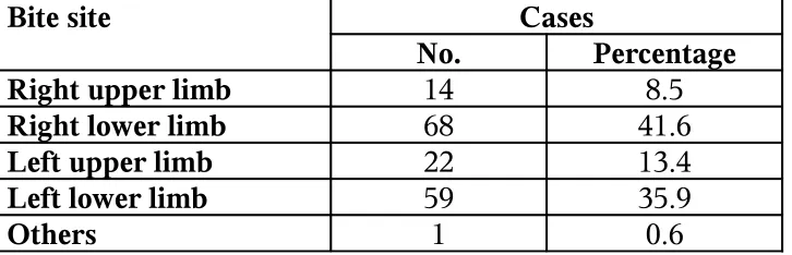
Table-5.4 Site of the snake bite
Bite site Cases
No. Percentage
Right upper limb 14 8.5
Right lower limb 68 41.6
Left upper limb 22 13.4
Left lower limb 59 35.9
In our study as in table-5.4, 41.6% of snake bites (68) occurred in right
lower limb, 35.9% of the bites (59) occurred in left lower limb, 13.4% of the
bites (22) occurred in left upper limb, 8.5% of the bites (14) occurred in right
upper limb and one case reported in the forehead.
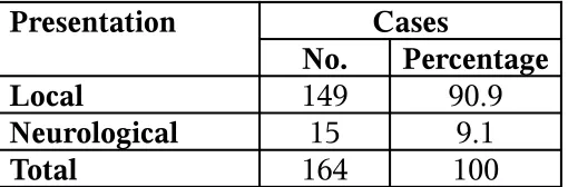
Table-5.5 Type of presentation
Presentation Cases
No. Percentage
Local 149 90.9
Neurological 15 9.1
[image:44.595.68.323.299.383.2]Total 164 100
Figure 5.4 Site of Snake bite
Figure 5.5 Type of presentation
In this study, 90.9% of the patients (149) presented to the hospital with
the local manifestations of envenomation and 9.1% of the patients (15)
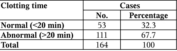
Table-5.6 Clotting time abnormalities
Clotting time Cases
No. Percentage
Normal (<20 min) 53 32.3
Abnormal (>20 min) 111 67.7
Total 164 100
Among the 164 patients in this study, 67.7% of the patients (111)
presented with prolonged clotting time and 32.3% of the patients (53) presented
with normal clotting time.
[image:45.595.69.387.341.481.2]Figure 5.6 Clotting abnormalities Table-5.7 Supportive treatment
Supportive treatment Cases
No. Percentage
Blood transfusion 4 2.4
Dialysis 14 8.5
Fasciotomy 4 2.4
Ventilator support 11 6.7
Nil 131 79.9
Total 164 100
In our study, 8.5% of the patients (14) of the total 164 needed dialysis for
the treatment of renal failure. 6.7% of the patients (11) needed ventilator support
for respiratory failure, 2.4% of the patients (4) needed blood transfusion and
2.4% of the patients needed fasciotomy for the treatment of the compartment
syndrome. Among the 164 patients, 79.9% of the patients (131) recovered
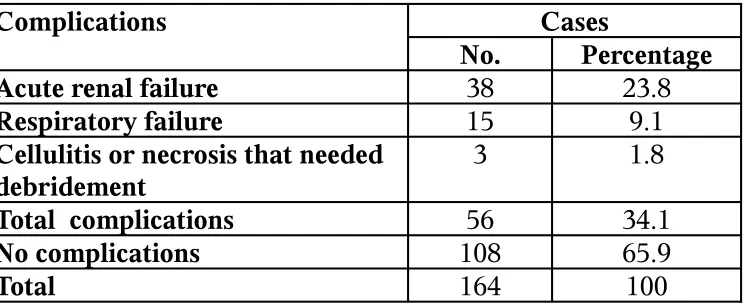
Figure 5.7 Supportive treatment Table-5.8 Complications
Complications Cases
No. Percentage
Acute renal failure 38 23.8
Respiratory failure 15 9.1
Cellulitis or necrosis that needed debridement
3 1.8
Total complications 56 34.1
No complications 108 65.9
Total 164 100
As seen in the table-5.8 in our study, 65.9% of the patients (108) recovered
well without any complications. In the remaining 34.1% of the patients (56),
23.8% of the patients (38) developed acute renal failure, 9.1% of the patients
(15) developed respiratory failure and 1.8% of the patients (3) developed
cellulitis or necrosis that needed debridement.
[image:46.595.69.434.512.698.2]Figure 5.8 Complications Table-5.9 Bite to needle time
Bite to needle time Cases
No. Percentage
0-4 hrs 66 40.2
4-8 hrs 63 38.4
8-12 hrs 16 9.8
12-24 hrs 15 9.1
More than 24 hrs 4 2.4
Total 164 100
Range 1-58 hrs
Mean 7.03 hrs
The bite to needle time (time between snake bite and administration of
ASV) varied between 1 and 58 hours in our study. The bite to needle time was
0-4 hours in 40.2% of the patients (66); 4-8 hours in 38.4% of the patients (63);
8-12 hours in 9.8% of the patients; 12-24 hours in 9.1% of the patients (15) and
more than 24 hours in 2.4% patients (4). The mean bite to needle time was 7.03
hours. The standard deviation was 8.03 hours.
[image:47.595.69.540.326.561.2]Figure 5.9 Bite to Needle time
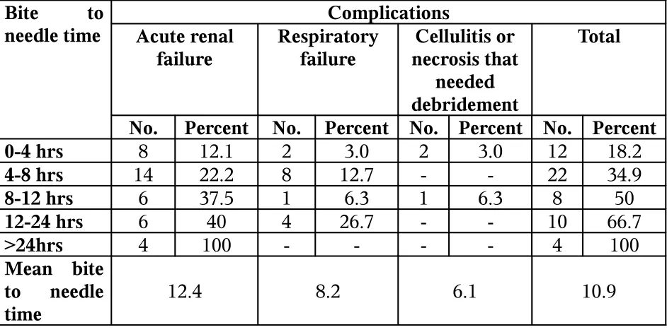
Table-5.10 Bite to needle time and complications
Bite to needle time
Complications Acute renal
failure
Respiratory failure
Cellulitis or necrosis that
needed debridement
Total
No. Percent No. Percent No. Percent No. Percent
0-4 hrs 8 12.1 2 3.0 2 3.0 12 18.2
4-8 hrs 14 22.2 8 12.7 - - 22 34.9
8-12 hrs 6 37.5 1 6.3 1 6.3 8 50
12-24 hrs 6 40 4 26.7 - - 10 66.7
>24hrs 4 100 - - - - 4 100
Mean bite to needle time
12.4 8.2 6.1 10.9
The table-5.10 showed the occurrence of various complications in the
relation to the bite to needle time. In the group with bite to needle time of 0-4
hours, 12.1% of the patients (8) had acute renal failure, 3% of the patients (2)
had respiratory failure and 3% of the patients (2) had cellulitis that needed
Figure 5.10 Bite to needle time and complications
In the group with bite to needle time of 4-8 hours, 22.2% of the patients
(14) had acute renal failure and 12.7% of the patients (8) had respiratory failure.
Among the group with bite to needle time of 8-12 hours, 37.5% of the patients
(6) had acute renal failure, 6.3% of the patients (one) had respiratory failure and
6.3% of the patients (one) had cellulitis that needed debridement. In the age
group with bite to needle time of 12-24 hours, 40% of the patients had acute
renal failure and 26.7% of the patients had respiratory failure. In the group with
bite to needle time of more than 24 hours, all patients (100%) had acute renal
[image:48.595.69.520.423.623.2]failure.
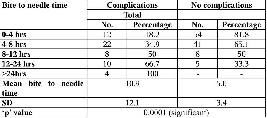
Table-5.11 Correlation between bite to needle time and complications
Bite to needle time Complications No complications
Total
No. Percentage No. Percentage
0-4 hrs 12 18.2 54 81.8
4-8 hrs 22 34.9 41 65.1
8-12 hrs 8 50 8 50
12-24 hrs 10 66.7 5 33.3
>24hrs 4 100 -
-Mean bite to needle time
10.9 5.0
SD 12.1 3.4
‘p’ value 0.0001 (significant)
In our study the bite to needle time was well correlated with the
complications as shown in the table-5.11. In this study among the group with
bite to needle time of 0-4 hours, 81.8% of the patients (54) had no
complications and 18.2% of the patients (12) had complications.
In the group with bite to needle time of 4-8 hours, 65.1% of the patients
(41) had no complications and 34.9% of the patients (22) had complications.
Among the group with bite to needle time of 8-12 hours, 50% (8) had
complications and 50% (8) had no complications. In the group with bite to
needle time of 12-24 hours, 66.7% of the patients (10) had complications and
33.3% of the patients (5) had no complications. In the group with bite to needle
time of more than 24 hours, 100% of the patients (4) had complications.
The mean bite to needle time was 10.9 hours in the group with
complications and 5 hours in the group without complications. This is
6. DISCUSSION
All the patients with history of snake bite were considered for the study
and 164 patients were selected for our study. These patients had undergone
various investigations like clotting time (20WBCT), blood sugar, urea, serum
creatinine, urine for albumin, sugar and deposits, complete blood count, liver
function tests and ultra sonogram (for selected patients). ASV was administered
for those patients indicated. The indications were
Systemic envenomation:
a) Haemostatic abnormalities: spontaneous systemic bleeding, coagulopathy
(20WBCT) or thrombocytopenia
b) Neurotoxic signs: ptosis, external ophthalmoplegia, paralysis etc
c) Cardiovascular abnormalities: hypotension, shock, cardiac arrhythmia
and abnormal ECG
d) Acute renal failure: oliguria/anuria and rising blood creatinine/ urea
e) Haemoglobinuria/myoglobinuria: dark brown urine, urine dipsticks, other
evidence of intravascular haemolysis or generalised rhabdomyolysis
Local envenomation:
a) Local swelling involving more than half of the bitten limb (in the
absence of a tourniquet) and swelling after bites on the digits (toes and
especially fingers)
b) Rapid extension of swelling (for example beyond the wrist or ankle
within a few hours of bites on the hands or feet)
c) Development of an enlarged tender lymph node draining the bitten
limb
The presence of any adverse reactions were noted and treated accordingly.
Clotting time was repeated every 6 hours for those who presented with
hemotoxicity and treated with ASV as indicated in the review of literature. The
complications due to the snake bite due to the late arrival and late
administration of ASV were noted. The bite to needle time which is the time
between the time of snake bite and time of ASV administration were noted and
the parameters were analyzed.
In our study, 164 patients were selected for study. In these patients,
63.4% of the patients (104) were males and 36.6% of the patients were females.
The high incidence of snake bites in males is probably due to their
lifestyles and occupational exposures as farmers or herdsmen, while most
females in our state are usually housewives or office workers, thus less prone
for snake bites.
In our study as seen in table-5.1, the incidence of snake bite occurred
commonly in the age group of 20-50 years. 16.7% of cases seen in the ages of
above 50 years. In the study by Narvencar K., J Assoc Physicians India., 2006 Sep;54:717-9 showed that the maximum number of cases (66%) were in the age group of 11-40 years, while only 8% were above the age of 60 years. This
was similar to the observation of Thomas G Glass who found 74% incidence in the age of 10-70 years while only 2% were above 70 years The high incidence
in the age group of 11-40 years is again because of occupational exposure, this
being the productive age group.
Among the study population, as seen in the table-5.3, 75.6% of the
snake bites occured in their working fields and 24.4% of the snake bites occured
in their houses. The high number of snake bites in the fields is related to the
working place, agricultural land, the prey base of the snake (that is frogs
and rats).The rice fields, which harbour millions of rats attract a lot of
snakes. Humans go into the field every morning and come out in the
encounter between farmer and snake is very high. As more areas are
inhabited at the periphery of towns, even there the chances of human /
snake interaction increase.
In this study, 41.6% of snake bites occurred in right lower limb, 35.9% of
the bites occurred in left lower limb, 13.4% of the bites occurred in left upper
limb and 8.5% of the bites occurred in right upper limb. The manual working
nature of our people is responsible for the increased number of bites in the
lower limbs.
In our study, 90.9% of the patients presented to the hospital with the
local manifestations of the envenomation which includes local cellulitis,
regional lymphadenitis, local bruises and hematological abnormalities like
prolonged clotting time, acute renal failure and coagulopathy and 9.1% of the
patients presented with neurotoxic manifestations. 67.7% of the patients
presented with prolonged clotting time and 32.3% of the patients presented with
normal clotting time.
The complications due to late administration because of late arrival of
the patients were noted. The complications noted were acute renal failure
(serum creatinine >1.5 mg/dl or oliguria <400ml/day); disseminated
intravascular coagulation or primary fibrinolysis; compartment syndrome,
gangrene, cellulitis/necrosis that needed debridement; shock; sepsis; acute
respiratory distress syndrome; neurological paralysis requiring ventilatory
As seen in the table-5.8 in our study, 65.9% of the patients recovered
well without any complications. In the remaining 34.1% of the patients (56),
23.8% of the patients (38) developed acute renal failure, 9.1% of the patients
(15) developed respiratory failure and 1.8% of the patients (3) developed
cellulitis or necrosis that needed debridement. In the study by Narvencar K., J Assoc Physicians India., 2006 Sep;54:717-9, among the 50 patients, 30 cases recovered well without any complications and 20 cases resulted in
complications.
The bite to needle time varied between 1 and 58 hours in our study. The
bite to needle time was 0-4 hours in 40.2% of the patients (66); 4-8 hours in
38.4% of the patients (63); 8-12 hours in 9.8% of the patients; 12-24 hours in
9.1% of the patients (15) and more than 24 hours in 2.4% patients (4). In
Narvencar K., J Assoc Physicians India., 2006 Sep;54:717-9 study among the 50 patients, 36 patients came to the hospital within 6 hours; seven patients in
6-24 hours; five patients in 1-3 days and two patients came to the hospital after 3
days.
The late arrival of the patients are due to poor knowledge about the snake
bite, their belief in the traditional methods such as application of tourniquet,
cutting (incision) and suction, washing the wound, snake stone or other methods
which are useless and harmful and delay in transporting the patients from the
In our study among the group with bite to needle time of 0-4 hours,
81.8% of the patients (54) had no complications and 18.2% of the patients (12)
had complications. In the group with bite to needle time of 4-8 hours, 65.1% of
the patients (41) had no complications and 34.9% of the patients (22) had
complications. Among the group with bite to needle time of 8-12 hours, 50% (8)
had complications and 50% (8) had no complications. In the group with bite to
needle time of 12-24 hours, 66.7% of the patients (10) had complications and
33.3% of the patients (5) had no complications. In the group with bite to needle
time of more than 24 hours, all patients (4) had complications. Chi-square test
was used to find the significance and that is found to be statistically significant
(‘p’ value -0.0001) in this study.
The study by Narvencar K., J Assoc Physicians India., 2006 Sep;54:717
showed that the incidence of complications was directly proportional to the
timing of ASV administration. The late administration resulted in more
complications. In his study, the complications were less in the population who
presented to the hospital as early as within 6 hours (26 cases were complicated
and 10 cases were uncomplicated). Among the seven patients with bite to needle
time of 6-24 hours four cases were complicated and three were uncomplicated.
In the group with bite to needle time of 1-3 days, 80% of the patients (4) got
complications and 20% (one) recovered without any complications. All the
patients who presented late with bite to needle time of more than 3 days had for
This finding is similar to the observation made by Vijeth SR et al.,
JIPMER that the incidence of complications was directly proportional to the duration of venom in the blood prior to neutralization by ASV. This fact is also
proved by Ash T et al and Thomas L et al who documented a positive correlation between severity of renal failure and increased time interval between
bite and ASV administration.
One study by K. Sam and M. Khan et al., The Internet Journal
of Emergency Medicine., 2009 Vol. 5 showed that snake bite severity
scores were directly proportional to the time elapsed between snake bite
instance and hospitalisation time. Those patients who were admitted late
had higher number of complications like renal failure (52%), breathing
difficulty (42%), cellulitis (40%), abnormal PT and APTT in 42% and 39%
of cases respectively. Mortality rate was the highest (16%) and higher
morbidity and squeal were observed among patients (18%) who were
admitted after 24 hours of envenomation. Majority (64%) of those
admitted after 13-18 hours seemed to have moderate severity with life
threatening symptoms, while those patients (82%), who were admitted
The fact that the incidence of complications was directly proportional to
the duration of venom in the blood prior to its neutralization by ASV due to late
arrival of the patient at hospital, point to the possibility or direct toxicity of the
venom on organ systems of the body. Based on the findings of present study, we
suggest that the early institution of ASV is beneficial in preventing
complications however severe the systemic envenomation. The delay in ASV
administration could increase the incidence of complications and morbidity as
7. CONCLUSION
The incidence of complications is directly proportional to the duration of
venom in the blood prior to neutralization by ASV due to late arrival of
patient at hospital.
The early institution of ASV is beneficial in preventing complications
BIBLIOGRAPHY
1. S.Vijaya kumar, M.Kamatchi, P. Thirumalaikolundusubramanian., Handbook on treatment guidelines for snake bite and scorpion sting, Tamil
Nadu Health Systems Project, Health and Family Welfare Department,
Government of Tamil Nadu, Chennai, 2008.
2. Guidelines for the Clinical Management of Snake Bite in the South-East
Asia Region, Reprint of the 1999 edition written and edited for
SEAMEOTROPMED – Regional Centre for Tropical Medicine, Mahidol
University, and edited by David A Warrell with contributions by an international panel of experts.
3. Narvencar K ., Correlation between timing of ASV administration and
complications in Snake Bites; J Assoc Physicians India., 2006 Sep;54:717-9
4. Paul S.Auerbach, Robert L. Norris, Poisoning, drug over dosage and envenomation, Disorders caused by reptile bites and marine animal
exposures, Harrison’s Internal Medicine; 17th edition; volume II.
5. D.A Warrel, Animal toxins; Manson’s Tropical diseases; chapter 32
6. Pradeep Bombery, Poisoning and toxicology, Snake bites and arthropod envenomation; API Textbook of Medicine; 8th edition; volume 2
7. A.L Jones, Poisoning, Envenomation; Snake bites; Davidson’s Principles and Practice of Medicine, 20th edition.
9. Rodney D. Adam, John B. Sullivan, Venomous Snake Bites, Cecil Textbook of Medicine, 21st edition.
10. Richard F Clark, Snake Bite, Poisoning and drug overdose; 5th edition
11. K. Sam, M. Khan, S. Peerally, P.G. Kumar & P.G. Rao: Snake-bite Envenomation: A Comprehensive Evaluation of Severity, Treatment and
Outcome in a tertiary Care South Indian Hospital. The Internet Journal of
Emergency Medicine, 2009 Volume 5 Number 1.
12. Vijeth SR, Dutta TK, Shahapurkar J. Correlation of renal status with haematological profile in viperine bite, JIPMER, Am J Trop Med Hyg
1997;56:168 -70.
13. Dutta TK, Ghotekar LH. Rational use of anti snake venom.In : Apicon Medicine Update 1998;8:760-65.
14. Hansdak SG, Lallar KS, Pokharel P, Shyangwa P, Kakri P, Koirala S. A clinic epidemiological study of snake bite in Nepal. Tropical Doctor 1998; 28:223-6.
15. Warrell, D.A., 1999. Guidelines for the clinical management of snake bites in the South East Asian regions. Southeast Asian Journal of Tropical
Medicine and Public Health 30,1-84.
16. Theakston, R.D.G., and Warrell, D.A., 2000. Crisis in snake antivenom supply for Africa. Lancet 356 (9247), 2104.
17.Tariang DD, Philip PJ, Alexander G, Macaden S, Jeyaseelan L, Peter JV,