Copyright © 1997, American Society for Microbiology
The Nef Protein of Human Immunodeficiency Virus Type 1
Enhances Serine Phosphorylation of the Viral Matrix
SIMON SWINGLER,1PHILIPPE GALLAY,1DIANA CAMAUR,1JINPING SONG,1ARIE ABO,2ANDDIDIER TRONO1*
Infectious Disease Laboratory, The Salk Institute for Biological Studies, La Jolla, California 92037,1
and Onyx Pharmaceuticals, Richmond, California 948062
Received 27 September 1996/Accepted 12 March 1997
The human immunodeficiency virus type 1 matrix (MA) protein is phosphorylated during virion maturation on its C-terminal tyrosine and on several serine residues. Whereas MA tyrosine phosphorylation facilitates viral nuclear import, the significance of MA serine phosphorylation remains unclear. Here, we report that MA serine but not tyrosine phosphorylation is strongly enhanced by Nef. Mutations that abrogated the membrane association of Nef and its ability to bind a cellular serine/threonine kinase greatly diminished the extent of virion MA serine phosphorylation. Correspondingly, a protein kinase coimmunoprecipitated with Nef could phosphorylate MA on serine in vitro, producing a phosphopeptide pattern reminiscent of that of virion MA. Recombinant p21-activated kinase hPAK65, a recently proposed relative of the Nef-associated kinase, achieved a comparable result. Taken together, these data suggest that MA is a target of the Nef-associated serine kinase.
Lentiviruses are distinguished among retroviridae by the presence of genes encoding accessory proteins that fulfill im-portant functions in the control of viral replication (48, 49). The primate lentiviruses are further characterized by a unique open reading frame at the distal end of the viral genome, nef. The crucial role of Nef was initially revealed by studies in the simian immunodeficiency virus-rhesus macaque model of AIDS pathogenesis, where it was found to be essential for high levels of viral replication and disease progression in adult an-imals (25). More recently, nef-defective strains have been im-plicated in some cases of long-term survival to human immu-nodeficiency virus type 1 (HIV-1) infection in humans (11, 27). The 206-amino-acid HIV-1 Nef protein is produced in abun-dance from the earliest stage of viral gene expression (26, 28). Myristoylated at its N terminus, Nef is predominantly cytoplas-mic and associates with membranes and cytoskeletal elements (15, 24, 34). In tissue culture, Nef affects both the kinetics of virus replication and the biology of the host cell. Nef increases HIV-1 infectivity by acting at the stage of particle production to promote steps that immediately follow viral entry. This influence is particularly noticeable following the inoculation of quiescent primary lymphocytes and their subsequent activation (9, 33, 47). It reflects increased rates of viral DNA synthesis, apparently due to improved properties of the uncoated viral nucleoprotein complex rather than to a stimulation of the enzymatic activity of reverse transcriptase per se (2, 10, 44).
In parallel, Nef alters several cellular functions. First, it downregulates the surface expression of CD4 (22, 23) and of major histocompatibility complex class I molecules (45). This modulation occurs posttranscriptionally through mechanisms that include enhanced endocytosis (1, 31, 37, 43, 45). Nef also affects cellular activation pathways, particularly in T lymphoid cells (4, 35, 46). This may at least partly reflect its interaction with cellular protein kinases (38, 40), including a serine/threo-nine kinase that can be coimmunoprecipitated with Nef in a wide variety of cells (40). The membrane association of Nef, as
well as a critical arginine in its central domain, is required for recruiting this activity (41). Although the Nef-associated serine/threonine kinase has not yet been formally identified, recent experiments suggest that it belongs to the p21-activated kinase (PAK) family (29, 36, 42).
Matrix (MA) is the N-terminally myristoylated cleavage product of the Gag polyprotein precursor by the viral protease (12). An essential structural element of retroviruses, MA gov-erns HIV-1 assembly by recruiting either directly or indirectly the other components of the virion at the plasma membrane. Mutations in HIV-1 MA can block assembly, prevent the in-corporation of envelope into virions, or affect steps of the viral life cycle that immediately follow viral entry (7, 13, 14, 16–21, 30, 50, 51, 53–56). During particle formation, a small subset of HIV-1 MA undergoes phosphorylation on serine and tyrosine (6, 12, 18). Tyrosine phosphorylation occurs on the C-terminal residue of MA and triggers the binding of MA to integrase (18, 19). This allows for the recruitment of MA into the virion core and subsequently into the uncoated viral nucleoprotein com-plex (19). A nuclear localization signal in MA is then recog-nized by the cell nuclear import machinery, which directs the viral nucleoprotein complex to the nucleopore (7, 18–20, 51). The present study was originated to examine the modalities of MA serine phosphorylation. We found that this modifica-tion, but not MA tyrosine phosphorylamodifica-tion, is strongly en-hanced by Nef. In addition, mutations that abrogated the bind-ing of Nef to a protein serine kinase diminished the extent of MA serine phosphorylation. Finally, MA could be an in vitro substrate for recombinant p21-activated kinase hPAK65, a proposed relative of the Nef-associated kinase.
MATERIALS AND METHODS
DNA constructions.The HIV-1HXB2-derived R7 proviral construct was
previ-ously described (26); R9 (previprevi-ously called R8 in reference 19) was obtained by cloning a BssHII-BamHI fragment, including the MA coding sequence, from
HIV-1NL4-3into R7. The Nef-defective cloneDNef contains a frame-shift
mu-tation at nucleotide 100 and the myristoylation-defective NefG2A, a
glycine-to-alanine change at position 2 of the nef open reading frame (1). NefRR106AAwas
constructed by PCR-mediated mutagenesis, and its sequence was verified by
dideoxy sequencing. Recombinant HIV-1HXB2MA carrying an N-terminal
his-tidine tag was produced in Escherichia coli by using the vector pET-15b (Nova-gen) and purified according to the manufacturer’s instructions as previously described (18).
* Corresponding author. Mailing address: Infectious Disease Labo-ratory, The Salk Institute for Biological Studies, 10010 N. Torrey Pines Rd., La Jolla, CA 92037. Phone: (619) 554-0869. Fax: (619) 453-7760. E-mail: [email protected].
4372
on November 9, 2019 by guest
http://jvi.asm.org/
Radiolabelling and biochemical procedures.CEM lymphoblastoma cells, ob-tained originally from Peter Nara, and SupT1 cells, obob-tained from James Hoxie through the NIH AIDS Research and Reference Reagent Program, were main-tained in RPMI 1640 medium. Methods used for radiolabelling virions with
[32P]orthophosphate and for radioimmunoprecipitations, in vitro kinase assays,
and Western blotting have been previously described (18). Phosphoamino acid and tryptic phosphopeptide analyses were performed as described by Boyle et al. (5). For the examination of the de novo phosphorylation of HIV MA in target
cells, 53106CEM cells were radiolabelled for 24 h with 25 mCi of [32P]H
3PO4
prior to infection with concentrated viruses (1 to 2mg of p24) in a small volume
of complete RPMI 1640 (1 to 2ml) containing 20mg of DEAE-Dextran/ml for
4 h. After three washes with phosphate-buffered saline to remove unadsorbed virions, cells were lysed in single detergent lysis buffer (39), and the nuclear pellet was extracted further with a high salt buffer (18). Both extracts were pooled and standardized by p24 enzyme-linked immunosorbent assay before immunopre-cipitation with HIV-1 MA-specific antibodies.
Kinase assays with hPAK65 and PKC.Recombinant myc-tagged hPAK65 (1
to 2mg) (32) bound to protein G-Sepharose conjugated with monoclonal Myc
antibody was washed once and incubated in 40ml of kinase buffer (50 mM
Tris-HCl [pH 7.5], 100 mM NaCl, 10 mM MgCl2, 1 mM MnCl2) containing 1 to
2mg of CDC42Hs previously loaded with GTP and 5mg of myelin basic protein
(MBP) or recombinant MA. The reaction was initiated by adding 10ml of kinase
buffer containing 50mM ATP and 5mCi of [g-32P]ATP and incubated for 20 min
at 30°C. The reaction was stopped by adding 10 ml of 53sodium dodecyl
sulfate-polyacrylamide gel electrophoresis (SDS-PAGE) sample buffer and boil-ing for 5 min. Samples were applied to a an SDS–14% PAGE gel, transferred to a polyvinylidene difluoride membrane, stained with Coomassie blue, destained, dried, and exposed to X-ray film for 1 to 2 h. Phosphorylated bands were excised and subjected to phosphoamino acid and tryptic phosphopeptide analyses as previously described (5). MA phosphorylation mediated by protein kinase C (PKC) was similarly analyzed, using recombinant PKC (Upstate Biotechnology, Inc.) as the enzyme and histone H1 (Boehringer Mannheim) as a control sub-strate.
RESULTS
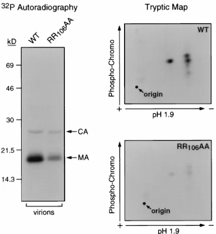
Virion MA serine but not tyrosine phosphorylation is en-hanced by myristoylated Nef. The potential dependence of MA serine phosphorylation on other viral gene products was investigated. Virions purified from the supernatant of CEM cells infected with a nef-defective (DNef) HIV-1 clone, or with one encoding a nonmyristoylated version of Nef (NefG2A), contained strikingly lower amounts of phosphorylated MA than wild-type virions (Fig. 1A). Of note, this difference was less apparent when examining MA molecules extracted from virus producer cells (data not shown). Tryptic peptide mapping studies revealed three major tryptic phosphopeptides of high chromatographic mobility in wild-type MA (Fig. 1B, left). These peptides contained exclusively phosphoserine, while phosphotyrosine was detected in minor phosphopeptides of low mobility, as previously described (data not shown) (18). MA molecules from nef-defective virions exhibited decreased amounts of all three of the major phosphoserine-specific pep-tides (Fig. 1B, right). In contrast, Nef did not influence the C-terminal tyrosine phosphorylation of MA (Fig. 1C), nor the phosphorylation of CA, another serine phosphoprotein of HIV-1 virions (data not shown). Multiple analyses performed on virions produced from either CEM or SupT1 T lymphoid cells confirmed that Nef stimulates the incorporation of phos-phate at all the major serine phosphoacceptor sites of MA (data not shown). In contrast, no significant difference was noted in the levels of MA serine phosphorylation when viruses were obtained from the supernatant of transfected 293(T) hu-man kidney cells (data not shown). Of note, the lack of Nef effect on MA tyrosine phosphorylation correlates with our previous observation that this modification can occur when MA is expressed in the absence of any other viral protein (18). A Nef mutant that fails to associate with a cellular serine kinase exhibits decreased levels of MA serine phosphorylation. The membrane association of Nef is important for recruiting a cellular serine/threonine kinase, the binding of which also de-pends on an arginine residue at position 106 of HIV-1 Nef (29,
[image:2.612.318.556.73.485.2]41, 42). We previously reported that a Nef mutant in which this and the upstream arginine are changed to alanine (NefRR106AA) is stably expressed and 50% as efficient as wild-type Nef for CD4 downregulation (3). As previously reported (29, 41, 42), this mu-tant was defective for the binding of a serine kinase activity (data not shown). The levels of virion MA serine phosphory-lation were significantly decreased at all major serine phos-phoacceptor sites in an HIV-1 derivative producing the NefRR106AA protein (Fig. 2). In contrast, mutating the Nef arginine dipeptide did not affect CA phosphorylation (Fig. 2) or MA tyrosine phosphorylation (data not shown). FIG. 1. Serine but not tyrosine phosphorylation of HIV-1 virion MA is stim-ulated by Nef. (A) Lysates of HIV-1 (R9) virions, expressing either wild-type (WT), truncated (DNef), or nonmyristoylated (NefG2A) versions of Nef, pro-duced in CEM cells in the presence of [32P]orthophosphate were immunopre-cipitated with anti-MA antibodies; products were analyzed by SDS-PAGE and autoradiography (left) or MA-specific Western blotting (right). In the Western blot, the high-molecular-weight band corresponds to the antibody light chain. (B) MA molecules from wild-type and DNef virions were subjected to a tryptic phosphopeptide analysis. Unlabelled arrows point to major phosphoserine-con-taining peptides. Phosphotyrosine-specific peptides are poorly visible due to low intensity of signal. (C) Lysates of the indicated virions and infected CEM cells were subjected to Western blotting with anti-phosphotyrosine (left) or a mixture of MA- and CA-specific (right) antisera.
on November 9, 2019 by guest
http://jvi.asm.org/
Similar results were obtained when virions produced from Sup T1 cells were examined (data not shown). The association of Nef with a serine kinase thus appears to promote MA serine phosphorylation.
Postentry MA serine phosphorylation is Nef independent. After virion internalization, MA is the substrate of additional phosphorylation events (6). When CEM cells labelled with orthophosphate were infected with HIV-1, a significant level of de novo phosphorylation of MA occurred within 4 h (Fig. 3A), whereas CA did not incorporate any phosphate (data not shown). MA molecules immunoprecipitated from CEM cells inoculated with either wild-type orDNef viruses exhibited sim-ilar levels of de novo MA phosphorylation (Fig. 3A), indicating that Nef does not influence this process. Phosphoamino acid analyses revealed that in target cells phosphate was added only on serine residues (Fig. 3B). Finally, tryptic maps of MA mol-ecules phosphorylated immediately after infection of CEM cells with either wild-type or nef-defective virions confirmed that Nef did not significantly affect this event (data not shown). MA serine kinase activity can be coimmunoprecipitated with Nef. The above results incriminated the Nef-associated serine kinase as a potential mediator of MA phosphorylation. To explore this issue further, wild-type and myristoylation-defective versions of Nef were immunoprecipitated from cyto-plasmic extracts of stably expressing CEM cells, and the result-ing immune complexes were used in an in vitro kinase assay with or without recombinant MA as substrate. The products of the reaction were resolved by SDS-PAGE and autoradiogra-phy. As previously described (41), a 62-kDa phosphoprotein was observed in kinase reactions performed on immunopre-cipitates obtained from cells producing wild-type but not non-myristoylated Nef (Fig. 4A, top). When recombinant MA was added to these reactions, phosphorylation of the Gag product
was observed with wild-type Nef immunoprecipitates at levels that were significantly higher than those which arose nonspe-cifically with immunoprecipitates from both control and non-myristoylated Nef-expressing cells (Fig. 4A, middle). In this context, MA phosphorylation occurred only on serine residues (Fig. 4B) and produced a tryptic phosphopeptide map compa-rable to that observed with virion MA (Fig. 4C). Of note, the pattern obtained with recombinant MA, which was derived from HIV-1HXB2, differed slightly from that yielded by MA molecules purified from R9 virions, derived from HIV-1NL4-3 (compare Fig. 1 and 4). A similar difference was observed when comparing MA from R7 and R9 virions (compare Fig. 1 and 5). This most likely reflects small differences in the NL4-3 and HXB2 MA sequences, which probably affect the sensitivity of the proteins to digestion by trypsin. However, the Nef de-pendence of MA serine phosphorylation was observed for both R7 and R9 virions (data not shown).
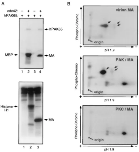
[image:3.612.72.287.69.302.2]The human serine kinase PAK65 can phosphorylate MA in vitro. Recent results indicate that the Nef-associated serine/ threonine kinase is related to the p21-activated kinase (PAK) family (29, 36, 42). Based on this premise, the ability of recom-binant human PAK65 (32) to phosphorylate MA in an in vitro kinase assay was evaluated (Fig. 5). When PAK was activated by incubation with the small GTP-binding protein cdc42, it autophosphorylated and could use MBP as a substrate (Fig. 5A, lane 2). Activated PAK also efficiently mediated the serine phosphorylation of recombinant MA (Fig. 5A, lane 4), yielding a tryptic phosphopeptide pattern reminiscent of that observed with virion-associated MA (Fig. 5B), even though additional phosphopeptides were observed, perhaps reflecting the ex-treme efficiency and thereby partial lack of specificity of the in vitro kinase assay. Several other recombinant protein kinases were tested for their ability to phosphorylate MA in vitro. FIG. 2. A Nef variant defective for kinase binding fails to stimulate virion
MA serine phosphorylation. Lysates of HIV-1 (R9) derivatives expressing wild-type (WT) or diarginine mutated (RR106AA) versions of Nef produced from [32P]orthophosphate-labelled CEM cells were immunoprecipitated with anti-CA and anti-MA antisera; products were analyzed by SDS-PAGE and autoradiog-raphy (left) and by two-dimensional tryptic phosphopeptide mapping (right). The similar levels of phosphorylated CA in lanes corresponding to the wild-type and mutant viruses demonstrate that equal amounts of material were loaded.
FIG. 3. MA serine phosphorylation in target cells is Nef independent. CEM cells incubated with [32P]orthophosphate were acutely infected with wild-type (WT) and nef-defective (DNef) R9 virions. MA molecules immunoprecipitated 4 h later were analyzed by SDS-PAGE and autoradiography (A) and by phos-phoamino acid analysis (B). Only phosphoserine was detected, even after pro-longed exposure. The data shown here are representative of two independent experiments.
on November 9, 2019 by guest
http://jvi.asm.org/
Mitogen-activated protein kinase was inactive, whereas protein kinase A (PKA) and casein kinase II did phosphorylate MA but induced phosphopeptide patterns that markedly differed from that observed with MA molecules purified from virions (data not shown). PKC, which has been suggested to mediate MA phosphorylation (8, 52), could also use the viral protein as a substrate. However, PKC targeted phosphoacceptor sites completely distinct from the ones modified in virion MA (Fig. 5C). These data, while not formally identifying the MA serine kinase, lend further support to a model in which the Nef-associated kinase is the mediator of MA serine phosphoryla-tion and is related to hPAK65.
DISCUSSION
This study reveals that an accessory protein of HIV-1, Nef, is involved in stimulating the serine phosphorylation of one of the virus structural components, MA. Whereas the levels of phosphoserine-containing MA were significantly decreased in
nef-defective virions, Nef did not stimulate MA tyrosine
phos-phorylation nor the additional serine phosphos-phorylation of MA that takes place after viral entry.
Although the mechanism by which Nef enhances MA serine phosphorylation is yet undefined, several lines of evidence incriminated the Nef-associated cellular serine kinase. First, mutations in Nef which prevent the recruitment of this kinase,
such as ones targeting the myristoylation signal or a critical arginine in Nef, also impaired MA serine phosphorylation. Second, a cellular protein kinase activity that coimmunopre-cipitates with Nef could phosphorylate MA on serine in vitro, by using the same major phosphoacceptors as observed in vivo. Finally, recombinant hPAK65, recently proposed to be related to the Nef-associated kinase, could use MA as an in vitro substrate, producing a phosphopeptide pattern reminiscent of, although not identical to, that observed in MA molecules pu-rified from HIV-1 particles. In contrast, several other recom-binant protein kinases failed to exhibit such an ability, includ-ing mitogen-activated protein kinase, casein kinase II, PKA, and PKC. Of note, it was previously demonstrated that PKC can phosphorylate MA on serine in vitro and in transfected COS cells (8, 52). Although our analyses confirmed that PKC can use MA as a substrate, they also revealed that the resulting phosphopeptide pattern differs markedly from that observed with virion MA.
Because MA kinase activity can be detected in virions (6, 18), we asked whether the particle incorporation of this en-zyme was Nef dependent. When highly purified wild-type,
[image:4.612.316.556.69.331.2]DNef, or NefRR106AAviruses were used as sources of kinase activity, recombinant MA was efficiently phosphorylated on serine and tyrosine, with no discernable difference between the viruses (data not shown). Also, similar levels of phosphoryla-tion were noted on endogenous viral MA molecules immuno-precipitated after performing an in vitro kinase reaction on purified virions (data not shown). Although it is possible that FIG. 4. In vitro serine phosphorylation of HIV-1 MA by a Nef-associated
[image:4.612.64.289.70.328.2]kinase. (A) Lysates from CEM cells constitutively expressing nonmyristoylated (lane 1) or wild-type (lanes 2 and 3) forms of Nef or from control CEM cells (lane 4) were immunoprecipitated with Nef-specific antiserum (3). Immune complexes were subjected to an in vitro kinase assay with [g-32P]ATP without (top) or with (middle) addition of recombinant MA. After SDS-PAGE and autoradiography, a 62-kDa phosphoprotein (top, arrow) and MA kinase activity (middle) were detected in precipitates from wild-type Nef-expressing cells (lanes 2 and 3). A Western blot analysis (bottom) controlled for Nef production. (B) Two-dimensional phosphoamino acid analysis of32P-labelled MA from panel A demonstrates that only serine is phosphorylated. (C) Two-dimensional tryptic phosphopeptide analysis of32P-labelled MA from panel A reveals a pattern comparable to that observed in virions. Unlabelled arrows indicate phosphopep-tides.
FIG. 5. In vitro serine phosphorylation of HIV-1 MA by hPAK65 and PKC. (A) Top: In vitro phosphorylation of recombinant MA by cdc42-activated (lane 4) but not by unactivated (lane 3) human PAK65. Reactions were done in the presence of [g-32P]ATP and analyzed by SDS-PAGE and autoradiography. MBP was used as a positive control substrate for activated hPAK65 (lanes 1 and 2). Bottom: PKC-mediated in vitro phosphorylation assay, similarly analyzed, using histone H1 (lane 2) or recombinant HIV-1 MA (lane 3) as substrates; lane 1 contained no substrate. (B) Two-dimensional tryptic phosphopeptide analysis of 32P-labelled MA purified from R7 virions (top) or phosphorylated in vitro with hPAK65 (middle) or PKC (bottom). Unlabelled arrows indicate phosphopep-tides.
on November 9, 2019 by guest
http://jvi.asm.org/
the virion-associated serine kinase activity detected in these assays does not correspond to the enzyme responsible for mod-ifying MA in vivo, these results suggest that Nef does not act by recruiting the MA serine kinase in virions. Instead, Nef could activate this enzyme in the context of assembling particles, promote its interaction with MA, or even stimulate the pref-erential incorporation of serine-phosphorylated MA molecules into virions.
While MA tyrosine phosphorylation has been shown to fa-cilitate HIV-1 nuclear import (6, 18, 19), the functional signif-icance of MA serine phosphorylation remains unclear. The treatment of virus producer cells with the serine/threonine kinase inhibitor H7 was recently shown to decrease markedly the infectivity of HIV-1 in dividing as well as nondividing cells (6). This effect correlated with reduced levels of MA translo-cation into the nucleus of target cells. Because a virus express-ing a nonmyristoylated form of MA was unaffected by the drug, it was proposed that serine phosphorylation triggers the mem-brane release of MA, hence allowing the viral nucleoprotein complex to migrate first to the cytosol and then to the nucleus. Nevertheless, these results must be interpreted with caution, because inhibitors of serine/threonine kinases are likely to interfere with the phosphorylation of CA, another major phos-phoprotein of HIV-1 virions (12) (Fig. 2) which plays an im-portant role during the early steps of infection (50).
It is tempting to propose that the serine phosphorylation of MA might contribute to the positive effect of Nef on HIV-1 replication. However, HIV-1 mutants expressing the NefRR106AA variant were found to be only moderately impaired in single-round infectivity assays, when produced either from 293 or from CEM cells, as they exhibited infectious titers that were approximately 30% those of wild type, that is, from 3- to 30-fold higher than those of nef-deleted viruses (data not shown). Of note, MA phosphoserine-defective variants could exhibit a phenotype distinct from that of Nef-mutated virions, because some degree of MA serine phosphorylation is con-served in the absence of Nef. Our analyses have allowed us so far to detect at least five phosphoserine residues in HIV-1 MA, although their exact mapping is still in progress. Ultimately, the identification of these phosphoacceptors will be required to elucidate fully the role of MA serine phosphorylation, as well as to explore further the connection between this process and the observed effect of Nef on HIV-1 replication.
ACKNOWLEDGMENTS
We thank T. Hunter and J. Meisenhelder for many helpful sugges-tions during these studies, as well as V. Stitt and L. Barden for the artwork.
This work was supported by postdoctoral fellowships from the Hoff-man Foundation and the Swiss National Foundation to S.S. and P.G., respectively, by an NIH training grant to D.C., and by grants RO1 AI37510 and AI34306 from the NIH to D.T.
REFERENCES
1. Aiken, C., J. Konner, N. R. Landau, M. E. Lenburg, and D. Trono. 1994. Nef induces CD4 endocytosis: requirement for a critical dileucine motif in the membrane-proximal CD4 cytoplasmic domain. Cell 76:853–864.
2. Aiken, C., and D. Trono. 1995. Nef stimulates human immunodeficiency virus type 1 proviral DNA synthesis. J. Virol. 69:5046–5056.
3. Aiken, C., L. Krause, Y.-L. Chen, and D. Trono. 1996. Mutational analysis of HIV-1 Nef: identification of two mutants that are temperature-sensitive for CD4 downregulation. Virology 217:293–300.
4. Baur, A. S., E. T. Sawai, P. Dazin, W. J. Fantl, C. Cheng-Mayer, and B. M.
Peterlin.1994. HIV-1 Nef leads to inhibition or activation of T-cells depend-ing on its intracellular location. Immunity 1:373–384.
5. Boyle, W. J., P. Van Der Geer, and T. Hunter. 1991. Phosphopeptide map-ping and phosphoamino acid analysis by two-dimensional separation on thin-layer cellulose plates. Methods Enzymol. 201:110–256.
6. Bukrinskaya, A. G., A. Ghropade, N. K. Heinzinger, T. E. Smithgall, R. E.
Lewis, and M. Stevenson.1996. Phosphorylation-dependent human immu-nodeficiency virus type 1 infection and nuclear targeting of viral DNA. Proc. Natl. Acad. Sci. USA 93:367–371.
7. Bukrinsky, M. I., S. Haggerty, M. P. Dempsey, N. Sharova, A. Adzhubei, L.
Spitz, P. Lewis, D. Goldfarb, M. Emerman, and M. Stevenson.1993. A nuclear localisation signal within HIV-1 matrix protein that governs infection of non-dividing cells. Nature 365:666–669.
8. Burnette, B., G. Yu, and R. L. Felsted. 1993. Phosphorylation of HIV-1 Gag proteins by protein kinase C. J. Biol. Chem. 268:8698–8703.
9. Chowers, M. Y., C. A. Spina, T. J. Kwoh, N. J. S. Fitch, D. D. Richman, and
J. C. Guatelli.1994. Optimal infectivity in vitro of human immunodeficiency virus type 1 requires an intact nef gene. J. Virol. 68:2906–2914.
10. Chowers, M. Y., M. W. Pandori, C. A. Spina, D. D. Richman, and J. C.
Guatelli.1995. The growth advantage conferred by HIV-1 nef is determined at the level of viral DNA formation and is independent of CD4 downregu-lation. Virology 212:451–457.
11. Deacon, N. J., A. Tsykin, A. Solomon, K. Smith, M. Ludford-Menting, D. J.
Hooker, D. A. McPhee, A. L. Greenway, A. Ellett, C. Chatfield, V. A. Lawson, S. Crowe, A. Maerz, S. Sonza, J. Learmont, J. S. Sullivan, A. Cunningham, D. Dwyer, D. Dowton, J. Mills.1995. Genomic structure of an attenuated quasi species of HIV-1 from a blood transfusion donor and recipients. Science 270:988–991.
12. Di Marzo Veronese, F., T. D. Copeland, S. Oroszlan, R. C. Gallo, and M. C.
Sarngadharan.1988. Biochemical and immunological analysis of human immunodeficiency virus gag gene products p17 and p24. J. Virol. 62:795–801. 13. Dorfman, T., F. Mammano, W. A. Haseltine, and H. Gottlinger. 1994. Role of the matrix protein in the virion association of the human immunodefi-ciency virus type 1 envelope glycoprotein. J. Virol. 68:1689–1696. 14. Fa¨cke, M., A. Janetzko, R. L. Shoeman, and H.-G. Kra¨usslich. 1993. A large
deletion in the matrix domain of the human immunodeficiency virus gag gene redirects virus particle assembly from the plasma membrane to the endo-plasmic reticulum. J. Virol. 67:4972–4980.
15. Franchini, G., M. Robert-Guroff, N. Grayeb, and F. Wong-Staal. 1986.
Cy-toplasmic localization of the HTLV III 39orf in cultured T cells. Virology
155:593–599.
16. Freed, E. O., J. M. Orenstein, A. J. Buckler-White, and M. A. Martin. 1994. Single amino acid changes in the human immunodeficiency virus type 1 matrix protein block virus particle formation. J. Virol. 68:5311–5320. 17. Freed, E. O., and M. A. Martin. 1995. Virion incorporation of envelope
glycoproteins with long but not short cytoplasmic tails is blocked by specific, single amino acid substitutions in the human immunodeficiency virus type 1 matrix. J. Virol. 69:1984–1989.
18. Gallay, P., S. Swingler, C. Aiken, and D. Trono. 1995. HIV-1 infection of non-dividing cells: C-terminal phosphorylation of the viral matrix protein is a key regulator. Cell 80:379–388.
19. Gallay, P., S. Swingler, J. Song, F. Bushman, and D. Trono. 1995. HIV nuclear import is governed by the phosphotyrosine-mediated binding of matrix to the core domain of integrase. Cell 83:569–576.
20. Gallay, P., V. Stitt, C. Mundy, M. Oettinger, and D. Trono. 1996. Role of the karyopherin pathway in human immunodeficiency virus type 1 nuclear im-port. J. Virol. 70:1027–1032.
21. Gallina, A., G. Mantoan, G. Rindi, and G. Milanesi. 1994. Influence of MA internal sequences, but not of the myristylated N-terminus sequence, on the budding site of HIV-1 Gag protein. Biochem. Biophys. Res. Commun. 204: 1031–1038.
22. Garcia, J. V., and A. D. Miller. 1991. Serine phosphorylation-independent downregulation of cell-surface CD4 by nef. Nature 350:508–511. 23. Guy, B., M. P. Kieny, Y. Riviere, C. Le Peuch, K. Dott, M. Girard, L.
Montagnier, and J. P. Lecocq.1987. HIV F/39orf encodes a phosphorylated
GTP-binding protein resembling an oncogene product. Nature 330:266–269. 24. Kaminchik, J., R. Margalit, S. Yaish, H. Drummer, B. Amit, N. Sarver, M.
Gorecki, and A. Panet.1994. Cellular distribution of HIV type 1 Nef protein: identification of domains in Nef required for association with membrane and detergent-insoluble cellular matrix. AIDS Res. Hum. Retroviruses 10:1003– 1010.
25. Kestler, H. W., III, D. J. Ringler, K. Mori, D. L. Panicali, P. K. Sehgal, M. D.
Daniel, and R. C. Desrosiers.1991. Importance of the nef gene for mainte-nance of high viral loads and for development of AIDS. Cell 65:651–662. 26. Kim, S., R. Byrn, J. Groopman, and D. Baltimore. 1989. Temporal aspects of
DNA and RNA synthesis during human immunodeficiency virus infection: evidence for differential gene expression. J. Virol. 63:3708–3713. 27. Kirchhoff, F., T. C. Greenough, D. B. Brettler, J. L. Sullivan, and R. C.
Desrosiers.1995. Absence of intact nef sequences in a long-term survivor with nonprogressive HIV-1 infection. N. Engl. J. Med. 332:228–232. 28. Klotman, M. E., S. Kim, A. Buchbinder, A. DeRossi, D. Baltimore, and E.
Wong-Staal.1991. Kinetics of expression of multiply spliced RNA in early human immunodeficiency virus type 1 infection of lymphocytes and mono-cytes. Proc. Natl. Acad. Sci. USA 88:5011–5015.
29. Lu, X., W. Xiaoning, A. Plemenitas, H. Yu, E. T. Sawai, and B. M. Peterlin. 1996. Cdc42 and Rac1 are implicated in the activation of the Nef-associated kinase and replication of HIV. Curr. Biol. 6:1677–1684.
30. Mammano, F., E. Kondo, J. Sodroski, A. Bukovsky, and H. G. Go¨ttlinger.
on November 9, 2019 by guest
http://jvi.asm.org/
1995. Rescue of human immunodeficiency virus type 1 matrix protein mu-tants by envelope glycoproteins with short cytoplasmic domains. J. Virol.
69:3824–3830.
31. Mangasarian, A., M. Foti, C. Aiken, D. Chin, J.-L. Carpentier, and D. Trono. 1997. The HIV-1 Nef protein acts as a connector with sorting pathways in the Golgi and at the plasma membrane. Immunity 6:67–77.
32. Martin, G. A., G. Bollag, F. McCormick, and A. Abo. 1995. A novel serine kinase activated by rac1/CDC42Hs-dependent autophosphorylation is re-lated to PAK65 and STE20. EMBO J. 14:1970–1978.
33. Miller, M. D., M. T. Warmerdam, I. Gaston, W. C. Greene, and M. B.
Feinberg.1994. The human immunodeficiency virus type 1 nef gene product: a positive factor for viral infection and replication in primary lymphocytes and macrophages. J. Exp. Med. 179:101–113.
34. Niederman, T. M., W. R. Hastings, and L. Ratner. 1993. Myristoylation-enhanced binding of HIV-1 Nef to T cell skeletal matrix. Virology 197:420– 425.
35. Niederman, T. M. J., J. V. Garcia, W. R. Hastings, S. Luria, and L. Ratner.
1992. Human immunodeficiency virus type 1 Nef protein inhibits NF-kB
induction in human T cells. J. Virol. 66:6213–6219.
36. Nunn, M. F., and J. W. Marsh. 1996. Human immunodeficiency virus-1 Nef associates with a member of the p21-activated kinase (PAK) family. J. Virol.
70:6157–6161.
37. Rhee, S. S., and J. W. Marsh. 1994. Human immunodeficiency type 1 Nef-induced down-modulation of CD4 is due to rapid internalization and deg-radation of surface CD4. J. Virol. 68:5156–5163.
38. Saksela, K., G. Cheng, and D. Baltimore. 1995. Proline-rich (PxxP) motifs in HIV-1 Nef bind to SH3 domains of a subset of Src kinases and are required
for the enhanced growth of Nef1viruses but not down-regulation of CD4.
EMBO J. 14:484–491.
39. Sambrook, J., E. F. Fritsch, and T. Maniatis. 1989. Molecular cloning: a laboratory manual, 2nd ed. Cold Spring Harbor Laboratory Press, Cold Spring Harbor, N.Y.
40. Sawai, E. T., A. S. Baur, H. Struble, B. M. Peterlin, J. A. Levy, and C.
Cheng-Mayer.1994. Human immunodeficiency virus type 1 Nef associates with a cellular serine kinase in T lymphocytes. Proc. Natl. Acad. Sci. USA
91:1539–1543.
41. Sawai, E. T., A. S. Baur, B. M. Peterlin, J. A. Levy, and C. Cheng-Mayer. 1995. A conserved domain and membrane targeting of Nef from HIV and SIV are required for association with a cellular serine kinase activity. J. Biol. Chem. 270:15307–15314.
42. Sawai, E. T., I. H. Khan, P. M. Montbriand, M. B. Peterlin, C. Cheng-Mayer,
and P. A. Luciw.1996. Activation of PAK by HIV and SIV Nef: importance for AIDS in rhesus macaques. Curr. Biol. 6:1519–1527.
43. Schwartz, O., A. Dautry-Varsat, B. Goud, V. Marechal, A. Subtil, J.-M.
Heard, and O. Danos.1995. Human immunodeficiency virus type 1 Nef induces accumulation of CD4 in early endosomes. J. Virol. 69:528–533. 44. Schwartz, O., V. Marechal, O. Danos, and J.-M. Heard. 1995. Human
im-munodeficiency virus type 1 Nef increases the efficiency of reverse transcrip-tion in the infected cell. J. Virol. 69:4053–4059.
45. Schwartz, O., V. Marechal, S. Le Gall, F. Lemonnier, and J.-M. Heard. 1996. Endocytosis of major histocompatibility complex class I molecules is induced by HIV-1 Nef protein. Nat. Med. 2:338–342.
46. Skowronski, J., D. Parks, and R. Mariani. 1993. Altered T cell activation and development in transgenic mice expressing the HIV-1 nef gene. EMBO J.
12:703–713.
47. Spina, C. A., T. J. Kwoh, M. Y. Chowers, J. C. Guatelli, and D. D. Richman. 1994. The importance of Nef in the induction of human immunodeficiency virus type 1 replication from primary quiescent CD4 lymphocytes. J. Exp. Med. 179:115–123.
48. Subbramanian, R. A., and E. A. Cohen. 1994. Molecular biology of the human immunodeficiency virus accessory proteins. J. Virol. 68:6831–6835. 49. Trono, D. 1995. HIV accessory proteins: leading roles for the supporting
cast. Cell 82:189–192.
50. Trono, D., M. B. Feinberg, and D. Baltimore. 1989. HIV-1 Gag mutants can dominantly interfere with the replication of the wild-type virus. Cell 59:113– 120.
51. Von Schwedler, U., R. S. Kornbluth, and D. Trono. 1994. The nuclear localization signal of the matrix protein of human immunodeficiency virus type 1 allows the establishment of infection in macrophages and quiescent T lymphocytes. Proc. Natl. Acad. Sci. USA 91:6992–6996.
52. Yu, G., F. S. Shen, S. Sturch, A. Aquino, R. I. Glazer, and R. Felsted. 1995. Regulation of HIV-1 gag protein subcellular localization by protein kinase C. J. Biol. Chem. 270:4792–4796.
53. Yu, X., Q.-C. Yu, T.-H. Lee, and M. Essex. 1992. The matrix protein of human immunodeficiency virus type 1 is required for incorporation of viral envelope protein into mature virions. J. Virol. 66:4966–4971.
54. Yu, X., Q.-C. Yu, T.-H. Lee, and M. Essex. 1992. The C terminus of human immunodeficiency virus type 1 matrix protein is involved in early steps of the virus life cycle. J. Virol. 66:5667–5670.
55. Yuan, X., X. Yu, T.-H. Lee, and M. Essex. 1993. Mutations in the N-terminal region of human immunodeficiency virus type 1 matrix protein block intra-cellular transport of the Gag precursor. J. Virol. 67:6387–6394.
56. Zhou, W., L. J. Parent, J. W. Wills, and M. Resh. 1994. Identification of a membrane-binding domain within the amino-terminal region of human im-munodeficiency virus type 1 Gag protein which interacts with acidic
phos-pholipids. J. Virol. 68:2556–2569.