RESEARCH
Increased BST2 expression during simian
immunodeficiency virus infection is not
a determinant of disease progression
in rhesus monkeys
Bianka Mussil
1, Aneela Javed
1,2, Katharina Töpfer
1, Ulrike Sauermann
1and Sieghart Sopper
3,4*Abstract
Background: Bone marrow stromal cell antigen 2 (BST2), also known as tetherin, HM1.24 or CD317 represents a type 2 integral membrane protein, which has been described to restrict the production of some enveloped viruses by inhibiting the virus release from the cell surface. This innate antiviral mechanism is counteracted by the HIV-1 viral factor Vpu, targeting BST2 for cellular degradation. Since antiviral BST2 activity has been mainly confirmed by in vitro data, we investigated its role in vivo on the disease progression using the SIV/macaque model for AIDS. We deter-mined BST2 expression in PBMC and leukocyte subsets of uninfected and SIV-infected rhesus macaques by real-time PCR and flow cytometry and correlated it with disease progression and viral load.
Results: Compared to pre-infection levels, we found increased BST2 expression in PBMC, purified CD4+ lymphocytes and CD14+ monocytes of SIV-infected animals, which correlated with viral load. Highest BST2 levels were found in progressors and lowest levels comparable to uninfected macaques were observed in long-term non-progressors (LTNPs). During acute viremia, BST2 mRNA increased in parallel with MX1, a prototype interferon-stimulated gene. This association was maintained during the whole disease course.
Conclusion: The detected relationship between BST2 expression and viral load as well as with MX1 indicate a common regulation by the interferon response and suggest rather limited influence of BST2 in vivo on the disease outcome.
Keywords: BST2, Rhesus macaque, SIV, MX1, LTNPs, Real-time PCR, PBMC
© 2015 Mussil et al. This article is distributed under the terms of the Creative Commons Attribution 4.0 International License (http://creativecommons.org/licenses/by/4.0/), which permits unrestricted use, distribution, and reproduction in any medium, provided you give appropriate credit to the original author(s) and the source, provide a link to the Creative Commons license, and indicate if changes were made. The Creative Commons Public Domain Dedication waiver (http://creativecommons.org/ publicdomain/zero/1.0/) applies to the data made available in this article, unless otherwise stated.
Methods
Ethics statement
Rhesus macaques (Macaca mulatta) of Indian origin of either sex were housed at the German Primate Cen-tre under conditions according to the German Animal Welfare act complying with the European Union guide-lines on the use of non-human primates for biomedi-cal research. All animals studies were approved by an external ethics committee authorized by the Lower Saxony State Office for Consumer Protection and Food
Safety (project licenses 509.42502/08-04.03, 33.14-4502-04-017/07, 33.14.42502-04-072/08 and 33.14-42502-04-10/0037 issued by the same State Office. Rhesus macaques were kept under permanent medical super-vision by veterinarians and animal caretakers. This includes measures of animal welfare and amelioration of suffering such as a 12:12 light dark schedule, a tem-perature between 18 and 23 °C with a humidity range of 50–60 % and provision with fresh air. Macaques were fed twice a day with dry food supplemented with fresh fruits and constant water access was provided. Environ-mental enhancement was realized by putting rings and perches into the cages, by task-oriented feeding meth-ods (e.g. treats, vegetables or fruits frozen in ice cubes,
Open Access
*Correspondence: [email protected]
food puzzle), and by playing music. In cases of suffer-ing, macaques were euthanized in accordance to a scor-ing system describscor-ing termination criteria as approved by the external ethics committee and corresponding to the IACUC endpoint guidelines. The scoring system considers weight loss, defecation anomalies, water and food consumption, behavior (attention/alertness, move-ment disorders), respiration, size of lymph nodes, and the hemogram. Animals were euthanized by an overdose of Pentobarbital-Natrium (Narcoren®, Merial, Hallberg-moos, Germany) under anesthesia.
Viral challenge and sample collection
For virus inoculation, animals received a deep anesthe-sia by intramuscular injection (i.m.) of a mixture of keta-mine, xylazine, and atropine. Animals were challenged either tonsillar or intravenously (i.v.) with SIVmac239 [1] or intrarectally (i.r.) or intravenously (i.v.) with a SIV-mac251-derived virus stock [1, 2], both prepared in rhe-sus monkey peripheral blood mononuclear cells (PBMC). Blood samples were collected of monkeys, sedated i.m. with 10 mg ketamine per kg body weight.
Animals
A total of 153 animals involved in different experiments were used for this study. For whole blood analysis, blood was taken from 17 macaques before and 24 weeks after low-dose i.r. infection with SIVmac251. For compari-son between relative BST2 mRNA expression and BST2 protein expression, 32 animals infected with SIVmac251 either via i.r. or i.v. routes were investigated. Another 35 infected macaques (20 with SIVmac251 and 15 with SIV-mac239) were grouped according to their clinical stage in progressors and LTNPs. The 26 progressor animals dis-played viral loads above 1E + 04 RNA copies/ml plasma and up to 18 progressor animals were investigated between 11 and 148 wpi when clinically still asympto-matic. Of them, 13 monkeys were euthanized and ana-lysed between 22 and 138 wpi when first signs of AIDS appeared, as judged from clinical as well as necropsy and histopathological findings, i.e. anorexia, incurable diar-rhoea, Pneumocystis jirovecii infection or neurological dysfunction. Another nine monkeys survived for more than 3 years post infection in the absence of any signs of immunodeficiency with a viral load below 1E + 04 RNA copies/ml and were termed as LTNPs. In a longitudinal study, two animals were inoculated intravenously with 100 TCID50, three animals with 10 TCID50 and two ani-mals with 1 TCID50 of SIVmac251 as part of an in vivo titration experiment aimed at defining the in vivo infec-tive dose of a new monkey PBMC-derived virus stock. This SIVmac251 challenge virus was prepared on PHA-stimulated PBMC from several monkeys. Supernatants
were harvested and pooled. After filtration, aliquots were prepared and stored at liquid nitrogen and the TCID50 was determined on C8166 cells.
Lymphocyte isolation
Peripheral blood was obtained by venipuncture and peripheral blood mononuclear cells (PBMC) were iso-lated via ficoll-paque gradient centrifugation (lymphocyte separation medium, PAA laboratories, Pasching, Aus-tria). CD4+ T-cells and CD14+ monocytes were enriched
from fresh PBMC by positive selection using magnetic beads (Miltenyi Biotec, Bergisch-Gladbach, Germany) and monoclonal antibodies to either CD4 or CD14. Purity of isolated CD4+ T-cells and CD14+ monocytes
was assessed by flow cytometry. Only CD4+ T-cells and
CD14+ monocytes with purity above 90 % were used for
downstream applications.
Flow cytometric analysis of BST2 surface expression
Surface BST2 expression was detected on different sub-sets of leukocytes, defined by CD3-Alexa700, anti-CD4-Pacific Blue, anti-CD8-V500, anti-CD14-PerCP (all from BD Biosciences, Heidelberg, Germany) and anti-CD45-FITC (Miltenyi, Bergisch-Gladbach, Ger-many), by using an anti-BST2-APC conjugated antibody (RS38E, Biolegend). Labeled cells were fixed with 3 % for-malin and analyzed on a BD LSRII flow cytometer (BD Biosciences, Heidelberg, Germany). The data files were evaluated using FlowJo Version 8.7 (Tree Star, Ashland, USA). Median fluorescence intensity (MFI) for granulo-cytes, monocytes and lymphocytes were determined.
Induction of BST2 with type I interferon
PBMC from three healthy animals were stimulated with 10, 50 and 100 ng Human Interferon Alpha A (Alpha 2a) (PBL Biomedical Laboratories) for 16 h. Fold induction of BST2 mRNA expression was calculated using GAPDH as internal control.
Quantification of plasma IFN‑alpha
Blood samples were obtained from 18 uninfected rhe-sus macaques 24 h after intramuscular inoculation with 1011 particles of a replication incompetent adenoviral
vector construct or 109 PFU of a Fowlpox construct.
at 405 nm with a 550 microplate reader (Bio-Rad Labo-ratories, Hercules, CA, USA). The detection limit of the ELISA was 10 pg/ml.
RNA isolation and cDNA synthesis
Total cellular RNA was isolated from 2 × 10⁶–5 × 10⁶
cells with RNeasy Plus Mini Kit (Qiagen, Hilden, Ger-many), except for whole blood samples, where RNA was isolated with PAXgene Blood RNA Kit (Qiagen, Hilden, Germany). Purified RNA was quantified by measur-ing the optical density at 260 nm (OD260). All samples
showed an OD260/OD280 ratio of 1.9 or above. RNA
qual-ity was randomly checked by Agilent 2100 Bioanalyzer (Agilent Technologies, Böblingen, Germany) show-ing RIN values (RNA Integrity Number) of at least 8.0. Synthesis of cDNA was performed using SuperScript III First-Strand Synthesis System for RT-PCR kit (Invitrogen GmbH, Karlsruhe, Germany) according to the manufac-turer protocol.
Quantification of BST2 and MX1 mRNA
Quantification of BST2 and MX1 mRNA levels in leuko-cytes were performed by real-time PCR assay using SYBR Green (Qiagen, Hilden, Germany) chemistry with primers designed to uniquely amplify BST2 (Genbank accession number NM_001161666) and MX1 (Genbank accession number EF101561). Following primers (Sigma, Hamburg, Germany) were used: BST2 forward, 5′-GACGAAA GAAAGTGGAGGAGCTT-3′ (nt 335–57); BST2 reverse, 5′-TCTCTTCTCAGTCGCTCCACCT-3′ (nt 428–407); MX1 forward, 5′-AGGAGTTGCCCTTCCCAGA-3′
(nt 295–313); MX1 reverse, 5′-TCGTTCACAAGT TTCTTCAGTTTCA-3′ (nt 372–348). MX1 primers were taken from Abel [3]. Glyceraldehyde-3-phosphate dehydrogenase (GAPDH), Genbank accession num-ber XM_001105471) was used as a house keeping gene with following primers taken from Rodriguez-Jimenez [4] forward, 5′-CCTGCACCACCAACTGCTTA-3′ (nt 525–544) and reverse 5′-CATGAGTCCTTCCACGA TACCA-3′ (nt 598–577). PCR reactions were performed in Micro Amp optical tubes or plates (Applied Biosys-tems GmbH, Darmstadt). Each 25 µl reaction mixture contained 12.5 µl 2 × QuantiTect SYBR Green PCR mas-ter mix (Qiagen, Hilden, Germany), 1 µl of each 10 µM primer, and 2 µl cDNA products. Samples were amplified by TaqMan-based real-time PCR on an ABI Prism 7500 sequence detection system (Applied Biosystems GmbH, Darmstadt) with one cycle at 95 °C (15 min) followed by 40 cycles at 95 °C (15 s) and 55 °C (1 min). The calcu-lated efficiency for all primers was determined by dilu-tion experiments and was from 97 to 98 %, thus target sequences were amplified with similar efficiency. All sam-ples were run at least in duplicates. Results were analysed
by Sequence Detection Software (Applied Biosystems GmbH, Darmstadt). Relative mRNA expression levels were calculated by using the comparative cycle threshold (Ct) method. BST2 and MX1 gene expression were nor-malized to endogenous control GAPDH mRNA. Rela-tive mRNA expression levels were determined using the formula: 100 × 2−ΔCt. Fold induction (n-fold of baseline)
was calculated by normalization to individual pre-values.
Quantification of plasma viral RNA
Isolation of viral RNA was performed from plasma sam-ples according to the MagAttract Virus Mini M48 pro-tocol (Qiagen, Hilden, Germany). Purified SIV RNA was quantified with TaqMan-based real-time PCR on an ABI-Prism 7500 sequence detection system (Applied Biosys-tems GmbH, Darmstadt) as described [5]. Amplified viral RNA was calculated as SIV-RNA copies per millilitre plasma.
Statistics
The statistical analyses were done with GraphPad Prism version 5.00 for Windows, GraphPad Software, San Diego, CA, USA. For multiple comparisons Kruskal– Wallis test with Dunn’s correction was used. For comparative interpretation between two groups the non-parametric two-tailed Mann–Whitney’s U test was used. For correlation the nonparametric two tailed Spearman test was performed. p values below 0.05 were regarded as significant.
Background
Mammalian cells encode a variety of proteins capa-ble of interfering with retroviral replication and caus-ing intrinsic inhibition of retroviral infection [6]. BST2/ CD317/HM1.24 has recently been described as one of these restriction factors, also named tetherin because of its ability to directly tether HIV-1 virions to infected cell membranes and thus potently inhibiting the release of budding virions [7–9]. Tethered particles are internalized and targeted for lysosomal [10, 11] or proteasomal deg-radation [12]. Consistent with a tethering mechanism, immuno-EM and biochemical studies identified BST2 on cell surface tethered virions as well as on particles teth-ered to each other [9, 13]. Biochemical analyses further demonstrated that BST2 forms cysteine-linked dimers modified by N-linked glycosylation [14–16].
glycosyl-phosphatidylinositol (GPI) anchor incorpo-rated in cholesterol-rich lipid rafts. BST2 is continuously cycling between the cell surface and internal membranes, including the trans-Golgi network [15, 16, 19, 20].
BST2 is expressed on a wide variety of cell types [17,
21] and has been described to potentially restrict the release of many enveloped viruses from the surface of infected cells, including members of the retrovirus, are-navirus, filovirus, herpesvirus and flavivirus families [22–26]. In order to circumvent BST2-mediated antiviral restriction, many viruses express BST2 antagonists [7, 8,
22, 27, 28]. Human immunodeficiency virus type 1 (HIV-1) Vpu and HIV-2 Env counteract human BST2 [29–32]. Most simian immunodeficiency viruses (SIVs) do not encode Vpu and use Nef to antagonize BST2 [27, 28, 33].
The BST2 promotor contains an interferon regulatory factor (IRF) binding site and it is well established that BST2 expression is strongly induced by type I interferons (IFNs) [34–37]. BST2 was also linked with innate viral sensing. Some studies demonstrate NFkappaB activation after BST2 cross-linking resulting in proinflammmatory immune responses [38–40]. Furthermore, BST2 binds to immu-noglobulin-like transcript 7 (ILT7) expressed on pDCs supposedly acting as a negative regulator of interferon pro-duction by these cells [41, 42]. These findings were ques-tioned by recent studies showing that activated mature pDCs downregulate ILT7 in vitro, indicating BST2-ILT7 cross-linking on immature circulating pDCs may function as a homeostatic regulatory mechanism rather than a nega-tive feedback for activated mature pDCs [43].
Only little is known about the impact of BST2 during retroviral disease course. In this study, we investigated the role of BST2 in the SIV rhesus macaque model, which represents currently the best animal model to study HIV infection and AIDS pathogenesis [44, 45]. BST2 mRNA transcription levels were quantified in cross-sectional and longitudinal analyses and its surface protein expres-sion was determined in PBMC and in SIV target cells, CD4+ T lymphocytes and CD14+ monocytes. Our results
show increased BST2 expression in SIV-infected pro-gressors animals, whereas in long term non propro-gressors (LTNPs) only low BST2 levels were detected. A direct correlation between BST2 expression and viral load as well as with type I interferon response as evidenced by MX1 mRNA expression was found. Our data suggest that BST2 is part of the antiviral interferon response but has a limited influence on disease progression.
Results
BST2 is increased in SIV‑infected macaques and correlates with viral load
In order to investigate the impact of SIV infection on BST2 transcription, relative BST2 mRNA levels in whole
blood of 17 rhesus macaques were determined before and 24 weeks after infection with SIVmac251 using an intrarectal (i.r.) repeated low dose inoculation scheme. RNA was isolated using the PAXgene Blood RNA Kit and BST2 mRNA was quantified by real time PCR relative to the housekeeping gene GAPDH. Relative BST2 mRNA levels were significantly increased after infection (Fig. 1a, p < 0.0001). Compared to individual pre-infection levels, the relative BST2 mRNA levels at 24 weeks post infec-tion (wpi) were on average about threefold higher. Higher BST2 values in infected animals were significantly asso-ciated with higher plasma viral loads at 24 wpi (Fig. 1b, p < 0.05). Interestingly, this correlation was even better after normalizing relative BST2 mRNA expression levels to their individual pre-infection values (Fig. 1c, p < 0.01).
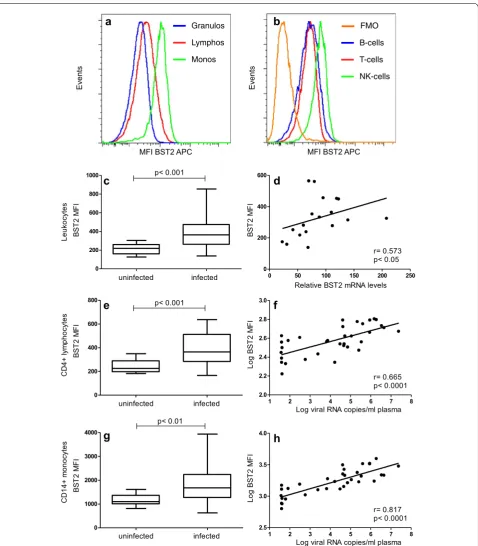
We next investigated whether the increased relative BST2 mRNA levels 24 weeks after infection also trans-lated into increased BST2 protein expression. To this end, 32 SIV-infected monkeys from two additional exper-iments were analysed. Twenty animals had been repeat-edly infected i.r. with low doses of SIVmac251, another 12 monkeys were infected intravenously (i.v.) with 100 TCID50 of the same viral stock. Again, BST2 mRNA lev-els were significantly increased compared to pre-infec-tion levels (p < 0.01) and correlated significantly with viral load (p < 0.05) (data not shown). Surface expression of BST2 was then determined in these monkeys by flow cytometry and compared with the one of twelve unin-fected monkeys analysed the same day. Surface expres-sion of BST2, shown as median fluorescence intensity (MFI) of allophycocyanin (APC), differs in uninfected animals between major leukocyte subsets (Fig. 2a; MFI granulocytes 179 ± 16.5, monocytes 1164 ± 87, lym-phocytes 338 ± 30). Among lymphocyte subsets, B-cells (MFI 360 ± 44) and T-cells (MFI 308 ± 22) showed simi-lar BST2 expression, whereas NK-cells (MFI 822 ± 64) showed the highest expression (Fig. 2b and Additional file 1: Figure S1A).
Surface expression of BST2 on total leukocytes was significantly increased in blood of infected animals com-pared to uninfected monkeys (Fig. 2c, p < 0.001). Moreo-ver, protein expression correlated well with the respective BST2 mRNA levels in SIV-infected monkeys (Fig. 2d, p < 0.05). A similar picture was seen when investigat-ing BST2 expression in CD4+ lymphocytes and
mono-cytes, the major target cells of SIV. Surface expression of BST2 was significantly higher in infected monkeys than in uninfected controls (Fig. 2e, p < 0.001 and Fig. 2g, p < 0.01) and BST2 expression on these cell populations correlated significantly with plasma viral load (Fig. 2f, p < 0.0001 and H, p < 0.0001). Despite different normal levels in monocytes and CD4+ lymphocytes, the relative
Also for other leukocyte populations, granulocytes, B cells and CD8 + T cells, increased BST2 levels and cor-relations between BST2 expression and plasma viral load were observed (Additional file 1: Figure S1A–H). NK cells showed also higher BST2 expression but no correla-tion with viral load was seen (Addicorrela-tional file 1: Figure S1I and J). In summary, SIV infection resulted in increased levels of BST2 in all leukocyte subsets tested, which cor-related with SIV RNA copy numbers.
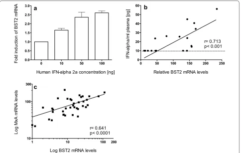
BST2 is regulated by type I interferons in vitro and in vivo
Human BST2 expression can be influenced by type I interferons in vitro and in vivo [34–37]. Similarly, a dose-dependent increase in BST2 transcription by IFN-alpha in macaque PBMC in vitro was found (Fig. 3a). Further-more, a direct correlation between BST2 transcription levels and in vivo plasma interferon-alpha levels exists (Fig. 3b). However, the application of the IFN-alpha assay is restricted due to its low sensitivity. Therefore, the influ-ence of type I interferons on BST2 expression in vivo was assessed by quantifying the transcription levels of MX1, which has long been used as a reliable marker for type I interferon bioactivity, but may be influenced by other cytokines as well. We speculated that the increased levels of BST2 found in infected animals were associated with the interferon response known to be elevated in these animals [46, 47]. MX1 levels were assessed in a total of 38 monkeys at 24 wpi. Indeed, MX1 levels in whole blood correlated well with BST2 (Fig. 3c, p < 0.0001) indicating BST2 can be regulated by type I interferons in vivo.
BST2 is increased in progressors but not in LTNPs
To evaluate the role of BST2 on disease course, we used stored material from previous experiments where sur-vival times of SIV-infected monkeys were known. To this end, 35 animals infected intravenously (20 with SIV-mac251, 15 with SIVmac239) were grouped according to their survival time and viral load. A total of 26 macaques displayed a progressive course of infection with viral load above 1 × 10⁴ copies/ml. Among them, 13 animals had
developed AIDS-like symptoms at time samples were analysed. Another nine monkeys, which had survived for more than 3 years post infection in the absence of any signs of immunodeficiency and with a viral load below 1 × 10⁴ copies/ml were termed LTNPs. Samples obtained
from nine uninfected animals during the same time frame, were used as negative control group.
In this experiment, RNA was isolated from Ficoll-separated PBMC. Similar to the first experiments where whole blood was investigated, infected animals with
a
b
c
Fig. 1 Whole blood BST2 mRNA levels in SIV-infected macaques. BST2 mRNA levels in whole blood of 17 rhesus macaques were determined using PAXgene Blood RNA Kit and real time PCR before and at 24 weeks after infection (wpi). a BST2 mRNA levels were com-pared between uninfected (circles) and SIVmac251 infected animals
(squares). BST2 mRNA levels are shown in copy numbers per 100
[image:5.595.56.293.87.551.2]B-cells
T-cells
NK-cells
b
a
MFI BST2 APC
Events
0 200 400 600 800 1000
0 200 400 600 800
0 1000 2000 3000 4000
0 50 100 150 200 250
0 200 400 600
1 2 3 4 5 6 7 8
2.0 2.2 2.4 2.6 2.8 3.0
1 2 3 4 5 6 7 8
2.5 3.0 3.5 4.0
IF
M
2T
SB
IF
M
2T
SBB
ST
2
MF
I
d
c
f
e
h
g
infected
r= 0.573 p< 0.05
Relative BST2 mRNA levels
Log viral RNA copies/ml plasma r= 0.665 p< 0.0001
r= 0.817 p< 0.0001
BS
T2
MF
I
Log
BS
T2
MF
I
Log
BS
T2
MF
I
Log viral RNA copies/ml plasma
Leuk
ocyt
es
CD
4+
ly
mp
ho
cyt
es
CD1
4+
mo
noc
yt
es
p< 0.001
p< 0.001
p< 0.01 uninfected
infected uninfected
infected uninfected
MFI BST2 APC
Events
Granulos
Lymphos
Monos
FMO
Fig. 2 BST2 mRNA and protein expression in uninfected and SIVmac251-infected macaques. Representative flow cytometric analyses of BST2 surface expression of an uninfected animal shown as histogram for leukocyte populations (a) and histogram for lymphocyte subsets including a fluorescence minus one (FMO) control (b). BST2 surface expression, displayed as median fluorescence intensity (MFI) on all leukocytes (c), on CD4+ lymphocytes (e) and on CD14+ monocytes (g) is illustrated for 12 uninfected and 32 SIVmac251 infected animals at 24 wpi. Box plots depict median and quartiles while whiskers show range. Group comparisons were calculated using two-tailed Mann–Whitney’s U test. d Whole blood BST2 mRNA levels correlate with BST2 surface protein expression. BST2 mRNA levels are depicted as copy numbers per 100 copies of GAPDH. BST2 surface expression on CD4+ lymphocytes (f) and CD14+ monocytes (h) correlates with plasma viral load. Each data point represents one individual animal.
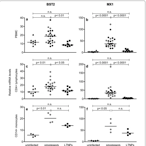
[image:6.595.60.539.87.634.2]progressive disease showed increased BST2 mRNA levels in PBMC. However this was statistically not sig-nificant (Fig. 4a, p = 0.052). No differences in BST2 or MX1 expression levels were observed between asymp-tomatic progressive animals (Fig. 4a–d; empty circles) and those with AIDS-like symptoms (Fig. 4a–d; filled circles). BST2 RNA levels were comparable between SIVmac239- and SIVmac251-infected macaques (data not shown). As in the previous experiments, BST2 levels correlated significantly with viral load (data not shown). Moreover, analysis of magnetically purified target populations for SIV revealed significantly higher BST2 mRNA levels also in CD4+ lymphocytes (Fig. 4c) and in
CD14+ monocytes of progressor animals (Fig. 4e),
cor-roborating the flow cytometric data and demonstrating for the first time a correlation between BST2 expression and disease progression in SIV-infected macaques.
In LTNPs however, BST2 levels were comparable to uninfected monkeys both in bulk PBMC as well as in purified CD4+ lymphocytes (Fig. 4a, c). Only for CD14+
monocytes, a trend to higher BST2 levels in LTNPs was observed, not reaching statistical significance (Fig. 4e). BST2 levels (Fig. 4, left panels) were in good accordance with MX1 levels (Fig. 4, right panels) in all cell popula-tions and disease progression groups, again supporting the notion that BST2 is mainly regulated by type I inter-ferons in vivo.
Kinetics of BST2 transcription during early infection
Our results from the cross-sectional studies suggest an induction of BST2 together with MX1 during the asymp-tomatic phase of infection. In order to determine more precisely the kinetics upon infection, BST2 and MX1 tran-scription were assessed during the 1st weeks of infection
0 10 50 100
0.0 0.5 1.0 1.5 2.0 2.5 3.0
1 10 100
10 100
200 300
a
Fo
ld
in
du
ct
io
n
of
BS
T2
mR
NA
Human IFN-alpha 2a concentration [ng]
c
Log
Mx
A
m
RNA
le
ve
ls
Log BST2 mRNA levels r= 0.641 p< 0.0001
0 50 100 150 200 250
0 10 20 30 40 50 60
b
IF
N-al
pha/
ml
pl
as
ma
[p
g]
Relative BST2 mRNA levels r= 0.713 p< 0.001
[image:7.595.58.543.89.396.2]in seven animals infected with different doses of SIVmac 251MPBMC as part of an in vivo titration study. All ani-mals, except one of those inoculated with the lowest dose, became infected and showed a typical course of plasma viral load (Fig. 5a). This experiment was terminated early
after infection and macaques were euthanized at prede-termined time points between six and 30 wpi without signs of AIDS. Figure 5 shows the kinetics of BST2 (B) and MX1 transcription (C) for PBMC in these macaques normalized to the mean of three independently measured
0 50 100 150
0 10
20 30 40
0 10 20 30 40 50
1
X
M
2
T
S
B
uninfected progressors LTNPs
a
b
d
c
f
e
Relative mRNA levels
PBM
C
CD4+ lymphocytes
CD14+ monocyte
s
p< 0.01
p< 0.01 p< 0.05
0 10 20
30 p< 0.01
p< 0.0001
0 50 100
150 p< 0.05
p< 0.0001
p< 0.0001 p< 0.0001
uninfected progressors LTNPs
0 50 100 150 200
n.s.
n.s. n.s.
n.s. n.s.
n.s. n.s.
n.s. n.s.
[image:8.595.58.542.88.569.2]a
b
d
c
e
f
g
h
[image:9.595.62.539.87.586.2]individual pre-infection values. The inoculated macaque that remained uninfected served as control. One week after infection, we observed a simultaneous increase of BST2 and MX1 transcripts in PBMC compared to pre-infection values, which reached a maximum at 10 days post infec-tion (dpi) (Fig. 5b, c). This happened shortly before peak viremia, which occurred 2 weeks after infection (Fig. 5a). In the single animal, which remained uninfected after inocu-lation, these variations were not seen. Later in the course of infection the transcription of BST2 and MX1 remained significantly elevated above pre-infection values (Fig. 5b, p < 0.05 and Fig. 5c, p < 0.05). This parallel kinetics of BST2 and MX1 expression, and the similar transcription pat-terns of both genes analysed cross-sectionally (Fig. 4) sug-gest common regulatory mechanisms for BST2 and MX1 already during acute SIV infection. Indeed, BST2 and MX1 mRNA expression levels are directly correlated at ten to 14 dpi in PBMC (Fig. 5d, p < 0.0001).
Furthermore, four animals that became LTNPs and 10 macaques that displayed progressive disease course were compared with respect to their viral load, relative BST2 and relative MX1 mRNA expression levels at acute SIV infection (Fig. 5e–g). Future LTNPs showed significantly lower BST2 and MX1 mRNA expression levels at 2 wpi (Fig. 5f, g) than the future progressors, probably attribut-able to reduced viral loads (Fig. 5e). These differences in BST2 expression between LTNPs and progressors were preserved later during chronic infection. Moreover, no relationship between relative pre-infection BST2 mRNA levels and viral load at 2 wpi was observed (Fig. 5h). In summary, individual differences in BST2 levels before infection do not influence early viral replication and higher levels of BST2 during peak viremia are not associ-ated with a favourable disease course.
Discussion
The present study investigated the role of BST2 dur-ing the natural course of retroviral infection in the SIV/ macaque model for AIDS. In line with previous studies using human cells [35, 48, 49], we found surface expres-sion of BST2 on all blood leukocyte populations, with highest levels on CD14+ monocytes [50]. Compared to
uninfected animals, BST2 was increased in SIV-infected rhesus macaques both at the RNA and the protein level in all leukocyte subsets investigated, including CD4+ T
lymphocytes and CD14+ monocytes, the major target
cells of SIV infection (compare Figs. 2, 4). Longitudinal analyses revealed that BST2 levels steeply increase fol-lowing i.v. infection to reach a maximum shortly before peak viremia, later dropping to levels still higher than before infection, corroborating a recent study where macaques were infected by the rectal route with a lym-photropic viral clone [50]. Similarly, higher levels of BST2
were found in HIV-infected patients within the first 4 weeks of infection compared to chronic infection [35]. In extension to these previous studies, we were also able to study for the first time the relationship between BST2 levels and disease course by combining data from several infection experiments. In all experiments and at different phases of infection, BST2 transcription but also protein expression was positively correlated with plasma viral load. Although, compared to acute viremia, low BST2 levels in the chronic phase, and even lower BST2 levels in patients under ART, were observed before, such a direct correlation between viral load and BST2 levels has not been reported yet, probably due to the small number of individuals studied [35, 36, 50]. Interestingly, BST2 levels were significantly lower in LTNPs than in progressors but in the same range as in uninfected monkeys, reminiscent to HIV-infected patients after antiretroviral therapy [35].
BST2 has not only been shown to counteract viral rep-lication in vitro, its importance in vivo is demonstrated by several lines of evidence for a strong selective pres-sure on immunodeficiency viruses to inhibit BST2. For example the switch from Nef as simian BST2 to Vpu as human BST2 antagonist is thought to present an impor-tant step during the evolution of HIV from a zoonosis to a pandemic virus [33]. Vice versa recent success in adapt-ing HIV to macaques was associated with mutations ren-dering vpu capable of antagonizing macaque BST2 [51]. Similarly earlier studies demonstrated that some HIV Vpu proteins are also able to inhibit rhesus BST2 to some extent and that Vpu-deficient SHIVs are less pathogenic in vivo [52]. Moreover, by extensively analyzing Vpu vari-ants isolated from several HIV-patients at different time points of infection it was recently shown that the function of Vpu remains preserved despite high variability even within an individual, indicating that BST2 function has to be kept at bay during acute and chronic stages of infection [53]. In our model, rhesus BST2 is antagonized by SIVmac Nef [27, 28, 54]. So far selective pressure on Nef activity against BST2 has not been demonstrated, and although BST2 antagonism is genetically separable from other Nef functions [54] the pathogenicity of such mutants has not been assessed in vivo. As BST2 has been described to prevent virion release [7–9] and in the light of the strong selective pressure on the virus to counteract BST2 activity, higher BST2 expression should result in lower plasma viral RNA levels. This was not the case in our study suggesting that the antagonistic effect of SIV was sufficient to even counteract modestly increased BST2 levels. Along this line, it was shown that anti BST2 activity was not different between variants isolated from HIV-infected patients with either rapid or LTNP disease course [53].
limited in vivo [55]. Moreover, there is growing evidence that BST2 does not actually restrict HIV-1 replication but simply shifts virus spread from a free to a cell-to-cell transmission mode [56–59]. Such a cell-to-cell spread of HIV-1 has been described as an efficient viral dissemination mechanism, being more rapid [60, 61], and even interferon insensitive [62]. This suggests that, once the infection is established, increased BST2 expression may favour viral dissemination by escaping host immune response [63].
BST2 is regulated by exogenous cues like type I inter-ferons and intrinsic signal cascades after sensing infec-tions through TLRs, both convening at transactivation of BST2 through IRFs [37, 48, 64]. As BST2 was uniformly upregulated on all leukocytes and not only on infected cells it is clear that soluble factors are responsible for the increased levels in SIV-infected macaques. In vitro, BST2 transcription seems to be specifically controlled by IFN-alpha as other cytokines such as IL-6, TNF-alpha did not influence BST2 expression [35]. Thus it comes as no surprise that our results showed a strong correla-tion throughout different phases of the infeccorrela-tion between BST2 levels and MX1, a prototype interferon stimulated gene (ISG), complementing a previous study, which demonstrated similar kinetics of BST2 expression and IFN-alpha levels in plasma in the acute phase but did not find a correlation between the two parameters [50]. One possible reason for this discrepancy is that IFN-alpha in plasma becomes undetectable by ELISA in the chronic phase of the infection and is therefore less sen-sitive for determining the interferon response [65] than measuring ISG like MX1 as in our work or ISG15 [36]. In agreement with results from present study, Aamer and colleagues also observed a maximum BST2 and MX1 expression during acute infection [66]. Strong induction of BST2 together with other restriction factors has also been observed in vivo after treatment of HIV/HCV-coin-fected patients with pegylated IFN-alpha/ribavirin (IFN-alpha/riba) exhibiting a good correlation with ISG15 [36]. The only other cytokine known to directly enhance BST2 transcription is IL-27 [49]. It is not yet known whether this cytokine plays a role in vivo as there are conflicting data on IL-27 levels in HIV patients [43, 51]. Because IL-27 also stimulates MX1 transcription [67] it is not possible to differentiate between the effects of IFN-alpha and IL-27 in our study.
Considering the clear association between viral load and BST2 levels throughout the infection and the lower expression of BST2 in LTNPs as early as 2wpi, there is little evidence that BST2 has a positive effect on disease progression in the SIV model for AIDS. This is further underlined by the fact that there was no relationship between pre-infection BST2 mRNA levels and early viral
load. In addition, no impact of pre-viremia BST2 levels with either the extent of viral load reduction after peak viremia or post acute virus RNA levels in plasma was found (data not shown).
On the other hand, human BST2 also exhibits signal transduction properties leading to NFkappaB activation and secretion of proinflammatory cytokines [38] and it has been hypothesized that this may lead to a positive feedback loop perpetuating increased BST2 expression [50]. However, the signaling trait seems to be an evo-lutionary recent acquisition and macaque BST2 lacks this activity [38]. Thus we think that, at least in the SIV macaque model, signaling through other pattern rec-ognition receptors is more important for keeping BST2 levels increased. In summary, its expression levels seem to just reflect the immune activation associated with the ongoing viral replication. This is in clear contrast to other restriction factors like the APOBEC3G and -3F deami-nases, for which we previously reported a negative corre-lation with plasma viral load and higher levels in LTNPs [47]. Indeed, in those animals, where we had the oppor-tunity to measure both restriction factors, we observed a trend for a negative correlation between APOBEC3G, APOBEC3F and BST2 (data not shown). Only during acute infection with the dominant influence of the type I interferon response both antiretroviral factors were positively correlated. This clearly demonstrates that BST2 and the APOBEC3 deaminases are regulated dif-ferently in vivo and reflects results from in vitro studies where, in addition to IFN-alpha, numerous cytokines (IL-2; IL-15 [53], IL-27 [67], IL-32 [68] affect transcription of APOBEC3 genes, while BST2 transcription is mainly stimulated by IFN and to some extent by IL-27 [49]. Deciphering the mechanisms responsible for the differ-ent regulation in vivo might help to shape the antiretrovi-ral response by restriction factors in a favorable way.
infection under certain circumstances. For example dur-ing initial infection locally produced IFN may increase BST2 expression on surrounding cells and thus limit early spread of the virus. Such an effect might be most relevant in situations where the viral antagonists of BST2 function are missing, for example after infection with nef-deleted SIV, where mechanisms acting before peak viremia deter-mine disease progression [69]. Given the low number of animals investigated during this early time point and the fact that we used blood cells to determine BST2 and ISG levels, our present study was likely not powered to detect such subtle effects. There is however recent evidence that the interferon response indeed influences very early events during infection. As an example, treatment of SIV-infected animals with a type I interferon receptor antag-onist blunted the infection induced BST2 induction, led to increased viral replication during acute infection and accelerated disease progression [54]. Conversely, treat-ment of macaques with pegylated IFN-alpha 2a, starting 1 week before challenge and proceeded 4 weeks after sys-temic infection, increased BST2 levels and the number of i.r. low dose challenges necessary to achieve systemic infection [54]. As these treatments also modulated other restriction factors the contribution of BST2 to the posi-tive effects of the interferon response remains uncertain. There is however some evidence that BST2 may indeed help to reduce viral replication in the chronic phase of HIV-infection. IFN-alpha/riba treatment of ART naïve HIV-patients increased BST2 levels and transiently reduced plasma viral load [36]. Of the interferon induced restriction factors investigated (APOBEC3G, APOBEC3F, BST2), only BST2 exhibited a significant correlation with reduction of plasma viral load. In addition, in two of nine patients mutations developed in the Vpu sequence during treatment, which were associated with enhanced down-regulation of BST2.
Taken together, variations of BST2 levels seem to only have measurable effects on viral replication at supraphys-iological levels induced by pharmacological treatment. However, the observed mutations leading to increased resistance of HIV against BST2 will limit the long term effect of such a treatment. In addition, prolonged treat-ment with pegylated IFN-alpha 2a results in desensiti-zation of the interferon response [54] further curtail the clinical benefit of such a treatment. In situations however, where viral replication is low for example after interrup-tion of ART [52] BST2 together with other interferon induced mechanisms may help to hold the viral replica-tion at bay.
Conclusions
By detecting an association between elevated BST2 expres-sion with plasma viral load and only low BST2 levels in
LTNPs, our results indicate rather limited effects of BST2 in vivo on the disease outcome in the SIV macaque model for AIDS. During acute viremia, BST2 mRNA levels increased in parallel with the prototype interferon-stimu-lated gene MX1. This correlation was maintained during the entire disease course and suggests BST2 expression as a part of the antiviral interferon alpha response reflecting immune activation associated with ongoing viral replica-tion. Future investigations are necessary to evaluate a pos-sible role of BST2 in limiting viral spread during early SIV/ HIV infection and to assess its potential impact on shifting from a cell-free to a cell-to-cell virus transmission.
Abbreviations
Ad: adenoviral vector; AIDS: acquired immune deficiency syndrome; APC: allophycocyanin; APOBEC: apolipoprotein B mRNA editing enzyme, catalytic polypeptide-like; ART: antiretroviral therapy; BST2: bone marrow stromal cell antigen 2; cDNA: complementary deoxyribonucleic acid; Ct: cycle threshold; Dpi: days post infection; ELISA: enzyme-linked immunosorbent assay; Env: envelope; FITC: fluorescein isothiocyanate; GAPDH: glyceraldehyde-3-phos-phate dehydrogenase; GPI: glycosyl-phosphatidylinositol; HCV: hepatitis C virus; HIV: human immunodeficiency virus; IACUC: institutional animal care and use committees; IFNs: interferons; IL: interleukin; ILT7: immunoglobulin-like transcript 7; i.m.: intramusculary; immuno-EM: immuno electron micros-copy; i.r.: intrarectally; IRF: interferon regulatory factor; ISG: interferon stimu-lated gene; i.v.: intravenously; LP-BM5: murine leukemia virus isolate; LTNPs: long-term non-progressors; MFI: median fluorescence intensity; Ml: millilitre; Mo-MLV: moloney murine leukemia virus; MPBMC: monkey peripheral blood mononuclear cells; mRNA: messenger ribonucleic acid; MX1: myxovirus (influ-enza virus) resistance 1; Nef: negative regulatory factor; NFkappaB: nuclear fac-tor kappa-light-chain-enhancer of activated B cells; NK-cells: natural killer cells; p p: value; PBMC: peripheral blood mononuclear cells; PCR: polymerase chain reaction; pDCs: plasmacytoid dendritic cells; PerCP: peridinin chlorophyll; PFU: plaque-forming units; PHA: phytohemagglutinin; R: Spearman’s correlation coefficient; SHIV: simian/human immunodeficiency virus; SIV: simian immu-nodeficiency virus; TCID50: 50 % tissue culture infective dose; TLRs: toll-like receptors; TNF: alpha tumor necrosis factor alpha; Vpu: viral protein unique; Wpi: weeks post infection; VSV-G: vesicular stomatitis virus G protein.
Authors’ contributions
BM performed most experiments, analyzed most of the data and drafted the manuscript. AJ carried out PCR on whole blood specimen, participated in the data analysis and revised the manuscript. KT performed flow cytometric analysis. US determined the viral load, performed the interferon ELISA and revised the manuscript. SS conceived the study, participated in its design and coordination and helped to draft the manuscript. All authors read and approved the final manuscript.
Additional file
Author details
1 Unit of Infection Models, German Primate Centre, Goettingen, Germany. 2 Department of Healthcare Biotechnology, Atta-ur-Rahman School of Applied Biosciences (ASAB), National University of Science and Technology (NUST), H12, Islamabad, Pakistan. 3 Department of Hematology and Oncology, Medical University Innsbruck, ZVG 7G5 009A, Anichstr. 35, 6020 Innsbruck, Austria. 4 Tyrolean Cancer Research Institute, Innsbruck, Austria.
Acknowledgements
We thank C. Stahl-Hennig, M. Franz, K. Raue, N. Stolte-Leeb for animal infection and clinical assessment, K. Mätz-Rensing for pathological analysis, and T. Egg-ers, G. Marschhausen, H. Mascher and P. Müller for the animal care.
Competing interests
The authors declare that they have no competing interests.
Received: 12 May 2015 Accepted: 23 October 2015
References
1. Kestler H, Kodama T, Ringler D, Marthas M, Pedersen N, Lackner A, et al. Induction of AIDS in rhesus monkeys by molecularly cloned simian immunodeficiency virus. Science. 1990;248(4959):1109–12. 2. Stahl-Hennig C, Voss G, Dittmer U, Coulibaly C, Petry H, Makoschey
B, et al. Protection of monkeys by a split vaccine against SIVmac depends upon biological properties of the challenge virus. AIDS. 1993;7(6):787–95.
3. Abel K, Alegria-Hartman MJ, Zanotto K, McChesney MB, Marthas ML, Miller CJ. Anatomic site and immune function correlate with relative cytokine mRNA expression levels in lymphoid tissues of normal rhesus macaques. Cytokine. 2001;16(5):191–204. doi:10.1006/cyto.2001.0961. 4. Rodriguez-Jimenez FJ, Krause A, Schulz S, Forssmann WG, Conejo-Garcia
JR, Schreeb R, et al. Distribution of new human beta-defensin genes clustered on chromosome 20 in functionally different segments of epididymis. Genomics. 2003;81(2):175–83.
5. Negri DR, Baroncelli S, Catone S, Comini A, Michelini Z, Maggiorella MT, et al. Protective efficacy of a multicomponent vector vaccine in cynomol-gus monkeys after intrarectal simian immunodeficiency virus challenge. J Gen Virol. 2004;85(Pt 5):1191–201.
6. Zheng YH, Jeang KT, Tokunaga K. Host restriction factors in retroviral infection: promises in virus-host interaction. Retrovirology. 2012;9:112. doi:10.1186/1742-4690-9-112.
7. Neil SJ, Zang T, Bieniasz PD. Tetherin inhibits retrovirus release and is antago-nized by HIV-1 Vpu. Nature. 2008;451(7177):425–30. doi:10.1038/nature06553. 8. Van Damme N, Goff D, Katsura C, Jorgenson RL, Mitchell R, Johnson MC,
et al. The interferon-induced protein BST-2 restricts HIV-1 release and is downregulated from the cell surface by the viral Vpu protein. Cell Host Microbe. 2008;3(4):245–52. doi:10.1016/j.chom.2008.03.001.
9. Perez-Caballero D, Zang T, Ebrahimi A, McNatt MW, Gregory DA, Johnson MC, et al. Tetherin inhibits HIV-1 release by directly tethering virions to cells. Cell. 2009;139(3):499–511. doi:10.1016/j.cell.2009.08.039. 10. Douglas JL, Viswanathan K, McCarroll MN, Gustin JK, Fruh K, Moses AV.
Vpu directs the degradation of the human immunodeficiency virus restriction factor BST-2/Tetherin via a {beta}TrCP-dependent mechanism. J Virol. 2009;83(16):7931–47. doi:10.1128/jvi.00242-09.
11. Mitchell RS, Katsura C, Skasko MA, Fitzpatrick K, Lau D, Ruiz A, et al. Vpu antagonizes BST-2-mediated restriction of HIV-1 release via beta-TrCP and endo-lysosomal trafficking. PLoS Pathog. 2009;5(5):e1000450. doi:10.1371/journal.ppat.1000450.
12. Goffinet C, Allespach I, Homann S, Tervo HM, Habermann A, Rupp D, et al. HIV-1 antagonism of CD317 is species specific and involves Vpu-medi-ated proteasomal degradation of the restriction factor. Cell Host Microbe. 2009;5(3):285–97. doi:10.1016/j.chom.2009.01.009.
13. Hammonds J, Wang JJ, Yi H, Spearman P. Immunoelectron microscopic evidence for Tetherin/BST2 as the physical bridge between HIV-1 virions and the plasma membrane. PLoS Pathog. 2010;6(2):e1000749. doi:10.1371/journal.ppat.1000749.
14. Andrew AJ, Miyagi E, Kao S, Strebel K. The formation of cysteine-linked dimers of BST-2/tetherin is important for inhibition of HIV-1 virus release but not for sensitivity to Vpu. Retrovirology. 2009;6:80. doi:10.1186/1742-4690-6-80.
15. Kupzig S, Korolchuk V, Rollason R, Sugden A, Wilde A, Banting G. Bst-2/ HM1.24 is a raft-associated apical membrane protein with an unusual topology. Traffic. 2003;4(10):694–709.
16. Swiecki M, Scheaffer SM, Allaire M, Fremont DH, Colonna M, Brett TJ. Structural and biophysical analysis of BST-2/tetherin ectodomains reveals an evolutionary conserved design to inhibit virus release. J Biol Chem. 2011;286(4):2987–97. doi:10.1074/jbc.M110.190538.
17. Goto T, Kennel SJ, Abe M, Takishita M, Kosaka M, Solomon A, et al. A novel membrane antigen selectively expressed on terminally differentiated human B cells. Blood. 1994;84(6):1922–30.
18. Ohtomo T, Sugamata Y, Ozaki Y, Ono K, Yoshimura Y, Kawai S, et al. Molecular cloning and characterization of a surface antigen preferentially overexpressed on multiple myeloma cells. Biochem Biophys Res Com-mun. 1999;258(3):583–91. doi:10.1006/bbrc.1999.0683.
19. Evans DT, Serra-Moreno R, Singh RK, Guatelli JC. BST-2/tetherin: a new component of the innate immune response to enveloped viruses. Trends Microbiol. 2010;18(9):388–96. doi:10.1016/j.tim.2010.06.010.
20. Masuyama N, Kuronita T, Tanaka R, Muto T, Hirota Y, Takigawa A, et al. HM1.24 is internalized from lipid rafts by clathrin-mediated endocytosis through interaction with alpha-adaptin. J Biol Chem. 2009;284(23):15927–41. doi:10.1074/jbc.M109.005124.
21. Erikson E, Adam T, Schmidt S, Lehmann-Koch J, Over B, Goffinet C, et al. In vivo expression profile of the antiviral restriction factor and tumor-target-ing antigen CD317/BST-2/HM1.24/tetherin in humans. Proc Natl Acad Sci USA. 2011;108(33):13688–93. doi:10.1073/pnas.1101684108.
22. Le Tortorec A, Willey S, Neil SJ. Antiviral inhibition of enveloped virus release by tetherin/BST-2: action and counteraction. Viruses. 2011;3(5):520–40. doi:10.3390/v3050520.
23. Jouvenet N, Neil SJ, Zhadina M, Zang T, Kratovac Z, Lee Y, et al. Broad-spectrum inhibition of retroviral and filoviral particle release by tetherin. J Virol. 2009;83(4):1837–44. doi:10.1128/jvi.02211-08.
24. Kaletsky RL, Francica JR, Agrawal-Gamse C, Bates P. Tetherin-mediated restriction of filovirus budding is antagonized by the Ebola glyco-protein. Proc Natl Acad Sci USA. 2009;106(8):2886–91. doi:10.1073/ pnas.0811014106.
25. Mangeat B, Cavagliotti L, Lehmann M, Gers-Huber G, Kaur I, Thomas Y, et al. Influenza virus partially counteracts restriction imposed by tetherin/ BST-2. J Biol Chem. 2012;287(26):22015–29. doi:10.1074/jbc.M111.319996. 26. Sakuma T, Noda T, Urata S, Kawaoka Y, Yasuda J. Inhibition of Lassa
and Marburg virus production by tetherin. J Virol. 2009;83(5):2382–5. doi:10.1128/jvi.01607-08.
27. Jia B, Serra-Moreno R, Neidermyer W, Rahmberg A, Mackey J, Fofana IB, et al. Species-specific activity of SIV Nef and HIV-1 Vpu in overcom-ing restriction by tetherin/BST2. PLoS Pathog. 2009;5(5):e1000429. doi:10.1371/journal.ppat.1000429.
28. Zhang F, Wilson SJ, Landford WC, Virgen B, Gregory D, Johnson MC, et al. Nef proteins from simian immunodeficiency viruses are tetherin antago-nists. Cell Host Microbe. 2009;6(1):54–67. doi:10.1016/j.chom.2009.05.008. 29. Hauser H, Lopez LA, Yang SJ, Oldenburg JE, Exline CM, Guatelli JC,
et al. HIV-1 Vpu and HIV-2 Env counteract BST-2/tetherin by seques-tration in a perinuclear compartment. Retrovirology. 2010;7:51. doi:10.1186/1742-4690-7-51.
30. Le Tortorec A, Neil SJ. Antagonism to and intracellular sequestration of human tetherin by the human immunodeficiency virus type 2 envelope glycoprotein. J Virol. 2009;83(22):11966–78. doi:10.1128/jvi.01515-09. 31. Kobayashi T, Ode H, Yoshida T, Sato K, Gee P, Yamamoto SP, et al.
Identification of amino acids in the human tetherin transmembrane domain responsible for HIV-1 Vpu interaction and susceptibility. J Virol. 2011;85(2):932–45. doi:10.1128/jvi.01668-10.
32. Vigan R, Neil SJ. Determinants of tetherin antagonism in the transmem-brane domain of the human immunodeficiency virus type 1 Vpu protein. J Virol. 2010;84(24):12958–70. doi:10.1128/jvi.01699-10.
34. Liberatore RA, Bieniasz PD. Tetherin is a key effector of the antiretroviral activity of type I interferon in vitro and in vivo. Proc Natl Acad Sci USA. 2011;108(44):18097–101. doi:10.1073/pnas.1113694108.
35. Homann S, Smith D, Little S, Richman D, Guatelli J. Upregulation of BST-2/Tetherin by HIV infection in vivo. J Virol. 2011;85(20):10659–68. doi:10.1128/jvi.05524-11.
36. Pillai SK, Abdel-Mohsen M, Guatelli J, Skasko M, Monto A, Fujimoto K, et al. Role of retroviral restriction factors in the interferon-alpha-mediated suppression of HIV-1 in vivo. Proc Natl Acad Sci USA. 2012;109(8):3035–40. doi:10.1073/pnas.1111573109.
37. Neil SJ, Sandrin V, Sundquist WI, Bieniasz PD. An interferon-alpha-induced tethering mechanism inhibits HIV-1 and Ebola virus particle release but is counteracted by the HIV-1 Vpu protein. Cell Host Microbe. 2007;2(3):193– 203. doi:10.1016/j.chom.2007.08.001.
38. Galao RP, Le Tortorec A, Pickering S, Kueck T, Neil SJ. Innate sensing of HIV-1 assembly by Tetherin induces NFkappaB-dependent proinflam-matory responses. Cell Host Microbe. 2012;12(5):633–44. doi:10.1016/j. chom.2012.10.007.
39. Tokarev A, Suarez M, Kwan W, Fitzpatrick K, Singh R, Guatelli J. Stimula-tion of NF-kappaB activity by the HIV restricStimula-tion factor BST2. J Virol. 2013;87(4):2046–57. doi:10.1128/jvi.02272-12.
40. Cocka LJ, Bates P. Identification of alternatively translated Tetherin isoforms with differing antiviral and signaling activities. PLoS Pathog. 2012;8(9):e1002931. doi:10.1371/journal.ppat.1002931.
41. Cao W, Bover L, Cho M, Wen X, Hanabuchi S, Bao M, et al. Regulation of TLR7/9 responses in plasmacytoid dendritic cells by BST2 and ILT7 receptor interaction. J Exp Med. 2009;206(7):1603–14. doi:10.1084/ jem.20090547.
42. Cao W, Rosen DB, Ito T, Bover L, Bao M, Watanabe G, et al. Plasmacytoid dendritic cell-specific receptor ILT7-Fc epsilonRI gamma inhibits Toll-like receptor-induced interferon production. J Exp Med. 2006;203(6):1399– 405. doi:10.1084/jem.20052454.
43. Tavano B, Galao RP, Graham DR, Neil SJ, Aquino VN, Fuchs D, et al. Ig-like transcript 7, but not bone marrow stromal cell antigen 2 (also known as HM1.24, tetherin, or CD317), modulates plasmacytoid dendritic cell func-tion in primary human blood leukocytes. J Immunol. 2013;190(6):2622– 30. doi:10.4049/jimmunol.1202391.
44. Desrosiers RC. Non-human primate models for AIDS vaccines. AIDS. 1995;9(Suppl A):S137–41.
45. Joag SV. Primate models of AIDS. Microbes Infect. 2000;2(2):223–9. 46. Abel K, Alegria-Hartman MJ, Rothaeusler K, Marthas M, Miller CJ. The
relationship between simian immunodeficiency virus RNA levels and the mRNA levels of alpha/beta interferons (IFN-alpha/beta) and IFN-alpha/ beta-inducible Mx in lymphoid tissues of rhesus macaques during acute and chronic infection. J Virol. 2002;76(16):8433–45.
47. Mussil B, Sauermann U, Motzkus D, Stahl-Hennig C, Sopper S. Increased APOBEC3G and APOBEC3F expression is associated with low viral load and prolonged survival in simian immunodeficiency virus infected rhesus monkeys. Retrovirology. 2011;8:77. doi:10.1186/1742-4690-8-77. 48. Bego MG, Mercier J, Cohen EA. Virus-activated interferon regulatory factor
7 upregulates expression of the interferon-regulated BST2 gene indepen-dently of interferon signaling. J Virol. 2012;86(7):3513–27. doi:10.1128/ jvi.06971-11.
49. Guzzo C, Jung M, Graveline A, Banfield BW, Gee K. IL-27 increases BST-2 expression in human monocytes and T cells independently of type I IFN. Sci Rep. 2012;2:974. doi:10.1038/srep00974.
50. Rahmberg AR, Neidermyer WJ Jr, Breed MW, Alvarez X, Midkiff CC, Piatak M Jr, et al. Tetherin upregulation in simian immunodeficiency virus-infected macaques. J Virol. 2013;87(24):13917–21. doi:10.1128/ jvi.01757-13.
51. Hatziioannou T, Del Prete GQ, Keele BF, Estes JD, McNatt MW, Bitzegeio J, et al. HIV-1-induced AIDS in monkeys. Science. 2014;344(6190):1401–5. doi:10.1126/science.1250761.
52. Shingai M, Yoshida T, Martin MA, Strebel K. Some human immunodefi-ciency virus type 1 Vpu proteins are able to antagonize macaque BST-2 in vitro and in vivo: Vpu-negative simian-human immunodeficiency viruses are attenuated in vivo. J Virol. 2011;85(19):9708–15. doi:10.1128/ jvi.00626-11.
53. Pickering S, Hue S, Kim EY, Reddy S, Wolinsky SM, Neil SJ. Preservation of tetherin and CD4 counter-activities in circulating Vpu alleles despite extensive sequence variation within HIV-1 infected individuals. PLoS Pathog. 2014;10(1):e1003895. doi:10.1371/journal.ppat.1003895. 54. Serra-Moreno R, Zimmermann K, Stern LJ, Evans DT. Tetherin/BST-2
antagonism by Nef depends on a direct physical interaction between Nef and tetherin, and on clathrin-mediated endocytosis. PLoS Pathog. 2013;9(7):e1003487. doi:10.1371/journal.ppat.1003487.
55. Swiecki M, Wang Y, Gilfillan S, Lenschow DJ, Colonna M. Cutting edge: paradoxical roles of BST2/tetherin in promoting type I IFN response and viral infection. J Immunol. 2012;188(6):2488–92. doi:10.4049/ jimmunol.1103145.
56. Dietrich I, McMonagle EL, Petit SJ, Vijayakrishnan S, Logan N, Chan CN, et al. Feline tetherin efficiently restricts release of feline immunodefi-ciency virus but not spreading of infection. J Virol. 2011;85(12):5840–52. doi:10.1128/jvi.00071-11.
57. Gummuluru S, Kinsey CM, Emerman M. An in vitro rapid-turnover assay for human immunodeficiency virus type 1 replication selects for cell-to-cell spread of virus. J Virol. 2000;74(23):10882–91.
58. Jolly C, Booth NJ, Neil SJ. Cell-cell spread of human immunodeficiency virus type 1 overcomes tetherin/BST-2-mediated restriction in T cells. J Virol. 2010;84(23):12185–99. doi:10.1128/jvi.01447-10.
59. Pais-Correia AM, Sachse M, Guadagnini S, Robbiati V, Lasserre R, Gessain A, et al. Biofilm-like extracellular viral assemblies mediate HTLV-1 cell-to-cell transmission at virological synapses. Nat Med. 2010;16(1):83–9. doi:10.1038/nm.2065.
60. Martin N, Welsch S, Jolly C, Briggs JA, Vaux D, Sattentau QJ. Virological synapse-mediated spread of human immunodeficiency virus type 1 between T cells is sensitive to entry inhibition. J Virol. 2010;84(7):3516–27. doi:10.1128/jvi.02651-09.
61. Sato H, Orenstein J, Dimitrov D, Martin M. Cell-to-cell spread of HIV-1 occurs within minutes and may not involve the participation of virus particles. Virology. 1992;186(2):712–24.
62. Vendrame D, Sourisseau M, Perrin V, Schwartz O, Mammano F. Partial inhibition of human immunodeficiency virus replication by type I inter-ferons: impact of cell-to-cell viral transfer. J Virol. 2009;83(20):10527–37. doi:10.1128/jvi.01235-09.
63. Andrew A, Strebel K. The interferon-inducible host factor bone marrow stromal antigen 2/tetherin restricts virion release, but is it actually a viral restriction factor? J Interferon Cytokine Res. 2011;31(1):137–44. doi:10.1089/jir.2010.0108.
64. Hotter D, Sauter D, Kirchhoff F. Emerging role of the host restriction factor tetherin in viral immune sensing. J Mol Biol. 2013;425(24):4956–64. doi:10.1016/j.jmb.2013.09.029.
65. von Wussow P, Jakschies D, Block B, Tschechne B, Schedel I, Horisberger MA, et al. The interferon-induced Mx-homologous protein in people with symptomatic HIV-1 infection. AIDS. 1990;4(2):119–24.
66. Aamer HA, Rajakumar P, Nyaundi J, Murphey-Corb M. Resistance to simian immunodeficiency virus low dose rectal challenge is associated with higher constitutive TRIM5alpha expression in PBMC. Retrovirology. 2014;11:39. doi:10.1186/1742-4690-11-39.
67. Imamichi T, Yang J, Huang DW, Brann TW, Fullmer BA, Adelsberger JW, et al. IL-27, a novel anti-HIV cytokine, activates multiple interferon-inducible genes in macrophages. AIDS. 2008;22(1):39–45. doi:10.1097/ QAD.0b013e3282f3356c.
68. Zhang F, Landford WN, Ng M, McNatt MW, Bieniasz PD, Hatziioannou T. SIV Nef proteins recruit the AP-2 complex to antagonize Tetherin and facilitate virion release. PLoS Pathog. 2011;7(5):e1002039. doi:10.1371/ journal.ppat.1002039.