Characterization of selenium containing proteins in the coral Acropora millepora
126
0
0
Full text
(2) Characterization of selenium containing proteins in the coral Acropora millepora. Thesis submitted by Huibin ZOU, Master of Science, Ocean University of China In April 2011. For the degree of Doctor of Philosophy In the School of Pharmacy and Molecular Sciences at James Cook University.
(3) Statement of Access. I, the undersigned, author of this work, understand that James Cook University will make this thesis available for use within the University Library and, via the Australian Digital Theses network, for use elsewhere.. I understand that, as an unpublished work, a thesis has significant protection under the Copyright Act.. Huibin ZOU. Date. i.
(4) Statement of Sources. I declare that this thesis is my own work and has not been submitted in any form for another degree or diploma at any university or other institution of tertiary education. Information derived from the published or unpublished work of others has been acknowledged in a Statement of Contribution of others and a list of references is given.. Huibin ZOU. Date. ii.
(5) Acknowledgements Supervision support I gratefully thank the following experts for providing the academic support: Prof. David Miller: as my supervisor, David gave me the opportunity to do the PhD in his lab. Without his support and direction, I could not get my project done. Prof. Ross Crozier: as my co-supervisor, Ross gave lots of academic suggestion in the beginning of my PhD study. I am sorry to lose him in the middle of my candidature. Dr. Murray Davies and Dr. Suzanne Smith: as my co-supervisors, Murray and Suzanne gave the academic supervision for the binding experiments in chapter 5. Dr. Eldon Ball and Dr. David Hayward: thanks for their fruitful discussion about coral genomics and developmental biology. Dr. Tracy Ainsworth: as my co-supervisor, Tracy gave the academic supervision for the immune-histology experiments in chapter 4.. Technical support I appreciate the following technical help from other research staff and students: Chapter 2: bioinformatics support from Brent Knack and Dr. Sylvain Foret. Chapter 3: sampling support from Marcelo Visentini-Kitahara and Dr. Zoe Richards; qPCR technical support from Francois Seneca and Yvonne Weiss. Chapter 4: experimental support from Brent Knack and Dr. Chuya Shinzato. Chapter 5: experimental support from Dr Eskenda Mume, Dr Yi Hu, Dr. Gary Perkins.. Funding support My project was funded by: China Scholarship Council Scholarship School of Pharmacy and Molecular Science Scholarship, JCU Coral Reef center, Austrian Research Council Australian Institute of Nuclear Science and Engineering Australian Nuclear Science and Technology Organization. Family support Thanks my wife, parents and sisters. They gave the spiritual support and encouraged me to persist on the project.. iii.
(6) Abstract Selenium (Se) and Se-containing proteins are believed to be involved in many physiological processes. Recent studies have revealed complex repertoires of Se containing proteins in mammals, of which some (known as selenoproteins) contain selenocysteine (Sec; encoded by in-frame UGA codons) and others in which the selenium is bound (selenium binding proteins; SeBPs) without selenoproteins. There have been few studies to date on the selenium protein complements of non-Bilateria animals, and many of the non-Bilateria selenoprotein genes in the public sequence databases are mis-annotated. The main objective of the work described in this thesis was to describe the selenoprotein and selenium-binding protein repertoire of the coral Acropora millepora, a representative non-Bilateria animal and to investigate aspects of the expression of some of the corresponding genes. These studies should not only provide evolutionary insights into selenium biology, but also be relevant to the physiology of coral stress.. To achieve these goals, phylogenetic tools were used to survey the repertoires of selenium-containing proteins in A. millepora and other model organisms, qPCR and immunohistochemistry employed to follow changes in the expression of genes encoding non-enzymatic selenium. containing proteins. under experimental. manipulation, bioinformatics tools used to model the structure of proteins of interest, and chemical tools employed to analyze the Se binding ability of recombinant selenium binding protein towards the inorganic Se in vitro.. The evolutionary studies summarized in Chapter 2 show that in the known invertebrates which have been studied their selenium components, the coral A. millepora has the most complex selenium repertoire (21 Sec-containing selenoproteins and 2 selenium binding proteins); other cnidarians also contain complex selenium repertoires. These results suggest that most of the known iv.
(7) selenium components seen in bilaterian animals predate the bilaterian-cnidarian split. In Chapter 3 we report that the expression of several non-enzymatic selenium containing proteins in the coral A. millepora is highly up-regulated by oxidants, suggesting physiology roles for these selenium components in redox regulation.. Studies in Chapter 4 and 5 focused on the A. millepora 56 kDa SeBPs (amSeBPs). Sequence analysis and structure modeling revealed that the conserved cysteine residues that are characteristic of these proteins, together with nearby motifs, cluster at the centre of the monomer protein models. The amSeBPs were ubiquitously expressed and markedly up-regulated at the planula and presettlement stages. Immunolocalisation experiments imply that the amSeBPs are enriched in adult A. millepora gastrodermal tissue that is adjacent to Symbiodinium. The in vitro selenite/amSeBP binding assays showed that the binding of inorganic selenium by amSeBP is dependent on the redox state. These studies imply that the positions of the redox sensitive cysteine residues and nearby motifs are critical for amSeBP function; these constraints presumably underlie the high level of sequence conservation of the 56 kDa SeBP sequences among animals, plants and even microorganisms.. In summary, these results imply important roles for the selenium containing proteins that are abundant in A. millepora. Although some of these proteins have been systematically characterized and implicated in redox metabolism, the mechanistic details remain unclear. To date, functional studies have focused mainly on mammalian Se proteins. Functional analyses in non-Bilateria animals could shed some light on the significance of Se-proteins and selenium biology more broadly.. v.
(8) Table of contents Statement of Access ............................................................................................... i Statement of Sources .............................................................................................ii Acknowledgements ............................................................................................... iii Abstract ................................................................................................................ iv Table of contents................................................................................................... vi List of figures ......................................................................................................... x List of tables......................................................................................................... xii Chapter 1 General Introduction ......................................................................... 1 1.1 The reef building coral Acropora millepora as a representative cnidarian ......... 1 1.1.1 Basic profiles ................................................................................................ 1 1.1.2 Physiology .................................................................................................... 5 1.1.3 Bleaching and oxidative theory ..................................................................... 8 1.2. Selenium (Se) biochemistry ......................................................................... 10. 1.2.1 The trace element selenium has similar chemical properties to sulfur ........... 10 1.2.2 The physiological roles of selenium .............................................................. 11 1.2.3 Se geographical distribution ........................................................................ 12 1.3 Selenium containing proteins: general points .................................................. 14 1.3.1 The forms of Se in vivo ................................................................................. 14 1.3.2 Selenoproteins............................................................................................. 15 1.3.3 Selenium binding proteins ........................................................................... 18 1.4 Project aims ................................................................................................... 20 Chapter 2 Evolutionary insights into ancestral selenium components ................ 22 2.1 Introduction ................................................................................................... 22 2.2 Materials and Methods .................................................................................. 26 vi.
(9) 2.2.1 Sequence Datasets....................................................................................... 26 2.2.2 Sequence analyses and phylogeny construction............................................ 26 2.2.3 Secondary structure analysis........................................................................ 27 2.3 Results ........................................................................................................... 27 2.3.1 The GPx family ............................................................................................ 27 2.3.2 The TR like genes ......................................................................................... 31 2.3.3 Other selenoproteins and related factors ..................................................... 33 2.3.4 Selenium binding proteins ........................................................................... 38 2.4 Discussion ...................................................................................................... 39 Chapter 3 Changes in the expression of genes encoding non-enzymatic selenium proteins during oxidative stress in the coral Acropota millepora ........................... 42 3.1 Introduction ................................................................................................... 42 3.2 Materials and methods...................................................................................44 3.2.1 Collection and maintenance of corals ...........................................................44 3.2.2 Experimental conditions ..............................................................................44 3.2.3 Total RNA extraction and first strand cDNA synthesis ................................... 45 3.2.4 Real time qPCR ............................................................................................ 46 3.2.5 Normalization and data processing .............................................................. 48 3.3 Results ........................................................................................................... 49 3.3.1 The expression of GOIs in control samples .................................................... 49 3.3.2 Rapidly induced GOIs associated with H2O2 treatment ................................. 50 3.3.3 BSO+H2O2 treatment results in elevated expression of five GOIs after 24 hours ................................................................................................................... 51 3.3.4 Genes encoding the selenium-binding proteins amSeBP17 and amSeBP23 behave differently under oxidative stress ............................................................. 52 3.4 Discussion ...................................................................................................... 54 3.4.1 The scleractinian coral A. millepora has an complex antioxidant network ..... 54 vii.
(10) 3.4.2 Hypothesis regarding the redox-sensitive selenium containing proteins in A. millepora ............................................................................................................. 54 Chapter 4 Two members of the 56 kDa selenium binding protein family from the coral Acropora millepora ................................................................................ 57 4.1 Introduction ................................................................................................... 57 4.2 Materials and methods................................................................................... 58 4.2.1 Sequence analysis and protein modeling ...................................................... 58 4.2.2 Temporal transcript levels of amSeBP17 and amSeBP23 genes ..................... 59 4.2.3 Expression and purification of recombinant amSeBP23 in E. coli ................... 60 4.2.4 Western blotting.......................................................................................... 61 4.2.5 Immunohistochemistry ................................................................................ 61 4.3 Results ........................................................................................................... 63 4.3.1 Sequence analysis and protein modeling ...................................................... 63 4.3.2 Characterization of amSeBP17 and amSeBP23 transcripts ............................ 67 4.3.3 Production of recombinant amSeBP23 and its recognition on western blots by heterologous antibody. .................................................................................... 67 4.3.4 Localization of immunoreactive amSeBP ...................................................... 70 4.4 Discussion and Hypothesis.............................................................................. 73 4.4.1 Conserved cysteine residues and nearby motifs form the redox centre of the 56 kDa SeBP ......................................................................................................... 73 4.4.2 amSeBP expression suggests high metabolic rates in A. millepora planula and pre-settlement stages .................................................................................... 74 4.4.3 amSeBPs, may be involved in transport or metabolism between the host A. millepora and the Symbiodinium .......................................................................... 75 Chapter 5 In vitro selenite binding by a recombinant form of the Acropora millepora selenium binding protein (ramSeBP) produced in Escherichia coli ..........77 5.1 Introduction ...................................................................................................77 5.2 Experimental methods ................................................................................... 78 viii.
(11) 5.2.1 Large (milligram) scale ramSeBP/selenite binding assay ............................... 78 5.2.2 HPLC (High-Performance Liquid Chromatography) assay of low levels (microgram) of ramSeBP ...................................................................................... 79 5.2.3 Dithiothreitol (DTT) treatment ..................................................................... 79 5.2.4 Small (microgram) scale ramSeBP/selenite (75SeO32-) binding assay .............. 80 5.3 Results ........................................................................................................... 81 5.3.1 Selenite binding ability of high level ramSeBP23 in vitro............................... 81 5.3.2 HPLC of native and DTT modified ramSeBP23 ............................................... 83 5.3.3 75SeO32- binding ability of low level native and DTT-modified ramSeBP23 in vitro..................................................................................................................... 85 5.4 Discussion and Hypothesis.............................................................................. 85 5.4.1 The potential selenite binding sites of the ramSeBP23 .................................. 85 5.4.2 The selenite binding towards ramSeBP23 is depending on the redox status .. 86 5.4.3 The amSeBP is an ideal selenium stock protein ............................................. 87 Chapter 6 General conclusion ........................................................................... 89 6.1 To be or not to be: evolutionary insight into ancestral Se components ............. 89 6.2 The nonenzymatic Se components: candidate antioxidants in Acropora millepora ............................................................................................................. 90 6.3 The 56kD SeBP in Acropora millepora: not simple, not well known .................. 91 6.4 Future directions ............................................................................................ 91 References ........................................................................................................... 94 Supplementary file............................................................................................. 109 Abbreviations .................................................................................................... 111. ix.
(12) List of figures Figure 1.1 Phylogenetics of Cnidaria. ..................................................................... 2 Figure 1.2 Morphology of adult Acropora millepora............................................... 3 Figure 1.3 Major developmental stages of Acropora millepora .............................. 4 Figure 1.4 The diffusion based physiology in Acropora millepora ........................... 7 Figure 1.5 Bleached Acropora millepora ................................................................ 8 Figure 1.6 The oxidative theory of coral bleaching ................................................ 10 Figure 1.7 The large coal fired-power stations (production ability more than 1000 megawatts) may affect the Se distribution in the GBR protecting area .................. 14 Figure 1.8 Different forms of selenium containing proteins in vivo ........................ 15 Figure 1.9 The hypothetical mechanism of selenocysteine biosynthesis and incorporation into selenoproteins ........................................................................ 16 Figure 1.10 X-ray structure of monomeric 56 kDa SeBP from Sulfolobus tokodaii .. 19 Figure 1.11 Project aims ....................................................................................... 20 Figure 2.1 Phylogenic analysis of GPx Family ........................................................ 30 Figure 2.2 Phylogeny and C-terminal sequences of TR like proteins from representative animals ........................................................................................ 32 Figure 2.3 SECIS elements in cnidarian selenoprotein W, T genes. ......................... 34 Figure 2.4 Phylogenic analysis of SeBP proteins .................................................... 39 Figure 3.1 The relative expression levels of all the GOIs in control samples of both colonies before chemical treatments.................................................................... 49 Figure 3.2 The mean fold changes towards control samples Rc for the seven GOIs in four testing groups ...................................................................................... 52-53 Figure 3.3. Hypothetical antioxidant network of A. millepora ............................. 56. Figure 4.1 Sequence analysis and protein modeling of the 56kD SeBP .............. 65-66 Figure 4.2 amSeBP17 and amSeBP23 transcripts during the major developing stages of A. millepora .......................................................................................... 67 x.
(13) Figure 4.3 Cloning strategy for production of recombinant amSeBP23 and western blotting experiments............................................................................................ 69 Figure 4.4 Immunolocalisation of amSeBP in A. millepora at the early planula stage....................................................................................................................71 Figure 4.4 Immunolocalisation of amSeBP in section of adult A. millepora ............ 72 Figure 5.1 ITLC assay ............................................................................................ 80 Figure 5.2 Selenium and protein content in the flow through mixtures off the affinity column .................................................................................................... 82 Figure 5.3 Chromatography A206 nm peak area/protein content curve ................ 84 Figure 5.4 Chromatography profiles of ramSeBP after different periods of DTT treatment ............................................................................................................ 84. xi.
(14) List of tables Table 1.1 Effects of selenium supplementation on the mammalian immune system ................................................................................................................. 12 Table 1.1 Selenoenzymes and non-enzymatic selenoproteins .......................... 17-18 Table 2.1 Overview of ancestral selenium components identified .................... 35-37 Table 2.2 Summary of selenoprotein data for the animals studied ........................ 38 Table 3.1 Primers for ICGs and GOIs ................................................................ 47-48 Table 3.2 The up-regulated GOIs in all groups 4 hours after H2O2 treatment ......... 50 Table 3.3 The GOIs remained high expression level 24 hours after BSO + H2O2 treatment ............................................................................................................ 51 Table 5.1 The binding ratio between selenium and ramSeBP in Fig. 5.2 A. ............ 83 Table 5.2 The binding ratio between 75Se and ramSeBP in ITLC binding assay ....... 85 Supplimentary Table 1. Unpublished Se components of A. millepora ................ 109. xii.
(15) Chapter 1 General Introduction 1.1 The reef building coral Acropora millepora as a representative cnidarian 1.1.1 Basic profiles Taxonomy. The Cnidaria is one of the earliest diverging phyla of Eumetazoa, and is believed to have diverged from the bilaterian lineage prior to or during the early Cambrian (Budd 2008). Although the vast majority of cnidarians are marine animals, examples being corals, jellyfish, sea anemones and box jellyfish, some cnidarians such as Hydra live in fresh water. The phylum comprises four classes: Cubozoa, Scyphozoa, Hydrozoa and Anthozoa (Fig 1.1). The largest class, Anthozoa, which includes corals and sea anemones, diverged first (Collins 2002; Ball et al., 2002; Miller et al., 2005; see Fig. 1.1). Members of the genus Acropora (Class Anthozoa, subclass Hexacorallia,. Order Scleractinia, suborder Astrocoeniina, Family. Acroporidae) are the dominant reef-building corals of the Indo-Pacific. Acropora millepora is a typical member of this large coral genus, and generally occurs on reef flats or on upper reef slopes, particularly where the water is clear.. 1.
(16) Fig. 1.1 Phylogenetics of Cnidaria. The largest class, Anthozoa, which includes corals and sea anemones, diverged first. Acropora millepora is a member of the Anthozoa (labeled in the box), subclass Hexacorallia, Order Scleractinia, suborder Astrocoeniina, family Acroporidae and genus Acropora. The figure is adapted from Ball et al. 2002 and Miller et al. 2005.. Morphology of adult A. millepora.. Adult A. millepora colonies have short and. uniform branches (also called corallites). Axial branches are distinctive and tubular in shape while radial branches are usually highly compacted (Fig. 1.2 A). Each branch has a scale like appearance with small and evenly separated polyps; tentacles are extended from the oral end of polyps (Fig. 1.2 B). Each polyp is anchored to the skeleton with its oral end up. As in other cnidarians, the A. millepora polyps give the appearance of near radial symmetry along the oral/aboral (O/A) axis, but there are subtle asymmetries in a second axis perpendicular to this A. millepora has no true organs. Each polyp has a gastrodermal cavity (or "stomach") with a mouth, a two layer-tissue wall with outer epidermis and inner gastrodermis, between which is a jelly-like mesogloea. The gastrdermal cavity or the mesogloea can be connected to other polyps (Fig. 1.2 C). The coral skeleton is extracellular, located below the aboral epidermis layer of each polyp. 2.
(17) Fig 1.2 Morphology of adult A. millepora. (A) A typical adult colony of A. millepora, photograph: by Dr Madeleine van Oppen (Australian Institute of Marine Science). (B) An axial branch of the adult A. millepora, photograph: from scholarpedia website (www.scholarpedia.org). (C) Polyp structure of A. millepora with oral end up towards sea water and aboral end down towards coral skeleton, each polyp has a gastrodermal cavity with a mouth, a two layer-tissue wall with outer epidermis and inner gastrodermis.. Early development. The availability of large amounts of early embryonic material is one advantage of A. millepora over the text-book cnidarian, Hydra (Miller et al., 2000), although availability is limited to the naturally occurring mass spawning events. The early development of A. millepora has not yet been described in great detail, although overviews have been published (Ball et al., 2002; Ball et al., 2004), as summarized below. A few nights after a full moon in late spring, egg/sperm bundles that are essentially self-incompatible are released into the water, float to the surface where they break apart and cross-fertilize with gametes from other colonies. 3.
(18) The fertilized eggs undergo unilateral cleavage, resulting in the formation of blastomeres. Approximately 13 h post-fertilization, the irregular-shaped ‗prawnchip‘ stage appears which may be unique to some corals. Gastrulation typically occurs at 22 to 36 h post-fertilization, and results in the formation of endoderm and ectoderm; the edges of the prawnchip appear to fold upward and a cavity is formed at the centre of the embryo. Due to the shape of the embryo, the gastrula stage of Acropora is often referred to as the ‗donut‘ stage. Approximately 28 h post-fertilization, the blastopore begins to close, marking the transition from embryo to larva. After blastopore closure, the larva becomes pear-shaped and cilia ultimately develop; the pear-shaped early larvae become motile spindle-shaped planulae (Fig. 1.3). Extensive cellular differentiation, including the elaboration of a complex nerve net, is apparent in late planulae. Planulae actively seek appropriate substrates for settlement by swimming aboral-end first. When triggered by appropriate cues, planulae settle to the substratum at the aboral end and become flattened along their O/A axis. Following settlement, the primary polyps adopt a rather different morphology to the planulae; a gastrodermal cavity appears within the flattened disc, and tentacles begin to form in the area surrounding the oral pore. Typically, symbionts are acquired 6-12 days after settlement (Baird et al., 2006), and eventually, a new colony of A. millepora is formed (Fig. 1.3).. Fig. 1.3 Major developmental stages of A. millepora (Ball et al., 2004). Approximately 13 h post-fertilization, the irregular-shaped ‗prawnchip‘ stage appears; approximately 22 h post-fertilization, the gastrula stage occurs; 4.
(19) approximately 28 h post-fertilization, the blastopore begins to close, marking the transition from embryo to larva, the pear-shaped early larvae become motile spindle-shaped planulae and seek appropriate substrates for settlement.. Unexpected genetic complexity. Whereas anthozoan cnidarians are morphologically simple animals, A. millepora and Nematostella vectensis (sea anemone) have been shown to have complex gene complements (Kortschak et al., 2003; Miller et al., 2005; Putnam et al., 2007) that include many genes previously thought to be restricted to vertebrates because they had been characterized in the context of vertebrate-specific traits and are absent from Drosophila and Caenorhabditis. Not only are all of the major developmentally-regulated signaling pathways known in Bilateria animals (Wnt, TGFβ, Hedgehog, Ras-MAPK and Notch) present in anthozoans, but the differentiation of these families of signaling molecules clearly predates the cnidarian/bilaterian split (Technau et al., 2005, Kusserow et al., 2005; Guder et al., 2006). The present study is consistent with this idea of ―ancestral genetic complexity‖ (Technau et al., 2005), as complex repertoires of genes encoding selenium containing proteins were found complex in the three cnidarians examined (Chapter 2). Although A. millepora is good comparitor for evolutionary comparisons of this kind, there are limitations in working with this animal; because it is not a laboratory organism, functional analyses are impossible or difficult to perform. Nevertheless, there is a need to understand the molecular bases of many aspects of coral biology, so it is important that attempts are made to link the genetic information with corresponding physiological roles, and one way to approach this is to infer gene function from gene expression data, an approach pursued here. 1.1.2 Physiology ‘Day and night, year after year, generation by generation, the way tiny corals fix inorganic carbon to build up reef is one of the most amazing works of nature which approves the power of life.‘ (Charles Darwin, 1845).. The diffusion based physiology of corals. Diffusion is an efficient means of 5.
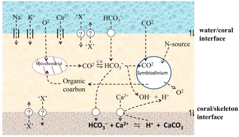
(20) exchange of materials only over short distances (e.g. over about 1 mm for oxygen exchange). The main physiological activities of corals, including respiration, digestion and elimination rely largely on diffusion. This diffusion based physiology, restricts corals to water and is facilitated by the large surface area provided by polyps with long thin tentacles. In scleractinian corals including A. millepora, the two main interfaces through which the material based diffusion (Zoccola et al., 1999; Irigaray et al., 1996) can occur within the water/coral /skeleton sandwich (Fig. 1.4.) are: (1) the oral ectoderm/water or endoderm/water interface, or (2) the aboral ectoderm/skeleton interface. Moreover, diffusion based physiological activities occur at the intracellular level, between the endodermal cells of the coral and the symbiotic dinoflagellates (Symbiodinium spp), which supply the host cells the main nutrition and energy (Muscatine and Porter, 1977; Papina et al., 2003).. Biomineralisation. Complex physiological metabolisms are involved in the biomineralisation process and the underlying molecular mechanisms are still largely unknown (reviewed by Allemand et al., 2004). In order to build the skeleton, which is composed of calcium carbonate (CaCO3) crystallized largely as aragonite (orthorhombic system), scleractinian corals have not only to supply calcium and inorganic carbon from ambient seawater (through the coral/sea water interface, Fig 1.4.) to the calcification site (through coral/coral skeleton interface, Fig. 1.4.), but also to eliminate the protons (through coral/coral skeleton interface, Fig. 1.4.) that result from the mineralising process:. Ca2+ + HCO3–. CaCO3 +H+. This process requires the movement of charged Ca2+ across the coral/sea water interface, presumably via calcium channels (Bénazet-Tambutté1996; Zoccola et al., 1999) and its transport across the coral/skeleton interface against a chemical gradient (Fig. 1.4), presumably requiring a Ca2+-ATPase (Zoccola et al., 2004). The 6.
(21) predominant form of dissolved inorganic carbon is as HCO3– which is present in sea water as well as in coral tissues at much higher concentration than are CO32– and CO2. The availability of HCO3– in coral tissue is ensured by the presence of the enzyme carbonic anhydrase (Allemand et al., 2004), which facilitates the following equilibrium:. CO2 +H2O. HCO3– + H+. The rate of biomineralisation may also modified by the photosynthetic activities of the symbiotic dinoflagellates. Photosynthetic activities in the symbiont can consume inorganic carbon, thus favoring carbonate precipitation. In addition, the liberation of OH- during photosynthesis can effectively neutralize protons arising from CaCO3 precipitation, thus facilitating calcification (Fig. 1.4; reviewed by Allemand et al., 2004).. Fig. 1.4 The diffusion based physiology of A. millepora. Materials can move within the water/coral /skeleton sandwich through diffusion interfaces (water/coral interface, coral/skeleton interface, coral/ Symbiodinium interface). X: other diffused materials like Se, Sr, Ba etc, and their distribution/movement in corals remain largely unclear. CA: carbonic anhydrase. The figure is adapted from Allemand et al., 2004.. The application of isotope flux kinetics and X-ray microanalysis indicates that the 7.
(22) distribution of many elements (Na, Mg, P, S, Cl, K, Ca, Sr, Ba) in coral tissues, symbiotic dinoflagellates and skeletons and the kinetics of movement of these elements between different compartments are under biological control (reviewed by Marshall and McCulloch, 2002). However, the biological roles of these elements and the significance of their distribution/movement in scleractinian corals remain largely unclear. 1.1.3 Bleaching and oxidative theory Because of their diffusion-based physiology, corals are particularly susceptible to physical (Lesser et al., 1990), chemical (Piniak 2007) and biological (reviewed by Rosenberg et al., 2007) stresses imposed from the living environments. For example, under elevated sea water temperatures, the symbiotic relationship between corals and their endosymbiotic dinoflagellates can breakdown, leading to the bleaching (loss of endosymbiotic dinoflagellates ) in a variety of scleractinian coral species (Downs et al., 2002; Hughes et al., 2003; Abrego et al., 2008) including Acropora millepora (Fig. 1.5). Over the last two decades, temperature-induced bleaching events have increased in both frequency and severity (Coles & Brown 2003; Hughes et al., 2003).. Fig 1.5. Bleached A. millepora. Photograph: by Dr Ray Berkelmans (Australian Institute of Marine Science). The 8.
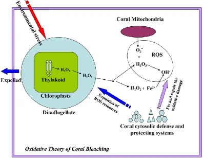
(23) symbiotic relationship between corals and their endosymbiotic dinoflagellates can breakdown, leading to the bleaching in a variety of scleractinian coral species including Acropora millepora.. The physiological mechanisms underlying coral bleaching remain unclear despite extensive investigation over the last few years. A number of studies (Downs et al., 2002; Abrego et al., 2008) suggest that oxidative stresses imposed on the coral by the symbiotic dinoflagellates play important roles in the process of sea-surface temperature-induced coral bleaching. The basic idea is that heat stress combined with intense ultraviolet irradiation destabilizes the Photosystem II-catalyzed electron transfer, resulting in increased production of reactive oxygen species (ROS) such as H2O2 (Giardi et al., 2001). It is proposed that H2O2 arising in this way in the dinoflagellate can diffuse into the coral cytoplasm (Fig. 1.6, Downs et al., 2002), where it may overload antioxidant buffering systems and potentially cause extensive tissue damage. To prevent this occurring, the theory goes, corals sense oxidative damage and move to eradicate the dominant source of ROS production by expelling their endosymbiotic dinoflagellates. Thus, the bleaching may be a last ditch attempt by the coral (Fig. 1.6, ―Oxidative Theory of Coral Bleaching” Downs et al., 2002) to deal with environmental stress. Several studies are consistent with this theory; for example, more temperature tolerant dinoflagellate strains (Symbiodinium type D) impose less ‗oxidative damage‘ on their coral hosts during acute temperature stress, and may thus facilitate adaptation of corals to warmer environments (Van Oppen et al., 2005; Abrego et al., 2008).. 9.
(24) Fig. 1.6. The oxidative theory of coral bleaching (adapted from Downs et al., 2002). Environmental stresses like. heat and UV accelerate the production of hydrogen peroxide (H2O2) in the chloroplasts of the algal symbionts either by damaging the thylakoid membrane or disrupting the Calvin cycle. H2O2 arising in the dinoflagellate can diffuse into the coral cytoplasm, and some of the ROS products like hydrogen peroxide can be accuamulated in the host cell, where they activate a cellular protecting response, which results in expulsion of symbionts and leads to the coral bleeching.. 1.2 Selenium (Se) biochemistry 1.2.1 The trace element selenium has similar chemical properties to sulfur The trace element selenium was discovered and named after Selene, the Greek goddess of the moon, in 1817 by Swedish chemist Jons Jacob Berzelius. In the periodic table, selenium (Se34) is in the same group (16# group) as sulfur (S16) and shares with them a number of chemical properties (Rosenberg et al., 1966). Because compounds of selenium and sulfur can act as reversible and specific oxidation agents to a variety of organic chemicals, they are important in biological redox regulation (Driscoll and Copeland, 2003). Replacement of S for Se tends to make the species 10.
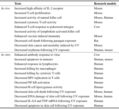
(25) more reducing under physiological conditions (Stadtman 1996), thus many enzymes whose active sites contain selenium catalyze oxidation/reduction reactions in vivo (Stadtman 2000). 1.2.2 The physiological roles of selenium Due to its chemical properties, selenium was historically regarded as a toxic agent, but is now known to be an essential trace element with a number of important physiological roles (reviewed by Rayman, 2000). The biological activities of selenium as a nutrient or a toxicant depend not only on the dose, but also on its chemical form (Ip et al., 1991). The most obvious biological role of selenium is as an antioxidant, as many. selenium-containing. enzymes. including. glutathione. peroxidases. and. thioredoxin reductases, are antioxidants (Rotruck et al., 1973).. The recommended dietary intake (RDI) for Se in the UK is 75 mg/day for adult males and 60 mg/day for adult females (reviewed by Mckenzie et al., 1998), and there is a considerable body of evidence linking Se-deficiency with a variety of disorders. These include Keshan disease, which occurs in areas of China with low Se soil (Chen et al., 1980), cardiovascular disease (Clark et al., 1996), cancer (Ip et al., 1991; Clark et al., 1996), rheumatoid arthritis and cataracts (Reviewed by Lockitch, 1989). The importance of Se in the mammalian immune system has been described at both the cellular (Table 1.1, reviewed by Mckenzie et al., 1998) and molecular levels (reviewed by Arthur et al., 2003). Moreover, Se can protect human keratinocytes against the cytotoxic effects of ultraviolet (UV) irradiation and hydrogen peroxide treatment (Shisler et al., 1998).. 11.
(26) Table 1.1 Effects of selenium supplementation on the mammalian immune system. In vivo. In vitro. Tests. Research models. Increased high-affinity of IL-2 receptor Increased T-cell proliferation Increased activity of natural killer cell Increased cytotoxic T-cell activity Enhanced T-cell response to pokeweed mitogen Increased activity of lymphokine activated killer cell Enhanced vaccine induced immunity Decreased cell death following paraquat exposure Decreased skin cancer and mortality induced by UV Decreased erythema following UV exposure. Mouse Mouse Mouse, Human Mouse Cow. Enhanced antibody response to virus Increased apoptosis in tumours Enhanced response in lymphocytes Increased killing by macrophages Increased killing by cytotoxic T cells Decreased HIV replication in T cells Decreased NF-kB activation Decreased B cell lipoxygenase activity Decreased skin cell death following UV exposure Decreased DNA damage of skin cell following UV exposure Decreased IL-6,8 and TNF mRNA following UV exposure Decreased apoptosis in skin cell following UV exposure. Cow Human, mouse Human Human Human Human Human Human Mouse, human Mouse, human Human Human. Mouse Rat Mouse Human, mouse. 1.2.3 Se geographical distribution Terrestrial distribution of Se. The geographical distribution of Se in soils is a function not only of the parent materials (Ure and Berrow, 1982) but also other soil properties including the loss on ignition (LOI) value and C concentration (Shand et al., 2010). Thus the terrestrial distribution of Se is highly variable. In general, organic-rich soils have higher Se concentrations. The variability of Se levels in soils leads to significant geographical differences in Se levels in crops; for example, American-grown wheat grain generally has a higher Se content than UK-grown grain (Adams et al., 2002).. Se contamination of aquatic ecosystems. Because inorganic selenium salts and 12.
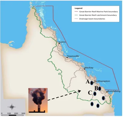
(27) compounds are soluble, mobile in the water column and tend to accumulate in organic-rich sediments, over long time periods environmental input of Se can result in local contamination of aquatic ecosystems. One major source of Se contamination of aquatic systems is agricultural irrigation drainage containing organic wastes, which is known to have caused severe teratogenesis in wild populations of aquatic birds in central California (Heinz et al., 1987). The other main contributors of aquatic Se contamination are coal-fired power plants, which often pollute nearby water bodies with Se-rich fly ash. This kind of pollution has been reported in lakes of central Alberta, Canada (Donahue et al., 2006), in Belews Lake, North California (Lemly 2002) and in Lake Macquarie, NSW, Australia (Kirby et al., 2001), frequently causing teratogenesis in fish and other aquatic organisms. To protect aquatic organisms, a water quality criterion of <2 μg/l for selenium has been recommended based on extensive review of the toxicology literature (reviewed by Hamilton and Lemly, 1999).. Potential Se pollution risks to the GBR. In Queensland, Se toxicity has been reported in some regions as a result of livestock feeding on Se accumulative plant species like Neptunia amplexicaulis (Peterson and Butler 1967). In addition, anthropogenic activities such as disposal of fly ash, raising of economic crops and mining operations have the potentiality to contribute substantially to the redistribution and cycling of Se in Queensland (reviewed by Tinngi, 2003). The issue of Se levels and distribution in the GBR (Great Barrier Reef) marine protected area is of urgent concern because there have been no base line studies and extensive risks exist in the forms of both agricultural run-off associated with sugarcane production and as the presence of large coal-fired power stations (Fig. 1.7). A priority should be widespread surveying and monitoring of Se levels across the whole GBR, but of particular concern are areas proximal to power stations such as that at Gladstone (Fig. 1.7 B).. 13.
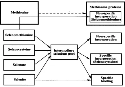
(28) Fig 1.7. The large coal fired-power stations (production ability more than 1000 megawatts) may affect the Se. distribution in the GBR protecting area. A: Stanwell power station (1440 megawatts); B: Gladstone power station (1650 megawatts); C: Callide power station (1700 megawatts). Information is adapted from Department of Mines and Energy, Queensland Government, Australia.. 1.3 Selenium containing proteins: general points 1.3.1 The forms of Se in vivo After ingestion of normal levels of selenite, selenate, or selenocysteine, nearly all of the element is metabolized via an intermediary pool and incorporated into specific Se-containing proteins (reviewed by Behne et al., 1991). The known Se-containing proteins (reviewed by Behne and Kyriakopoulos, 2001) can be divided into three groups: (1) proteins into which the element is incorporated nonspecifically, (2) 14.
(29) proteins that specifically bind selenium, and (3) proteins that contain selenium in the form of selenocysteine (Sec); this latter category are defined as selenoproteins, and in this case the Sec is encoded by a UGA codon. The incorporation of dietary selenium into the different types of selenium containing proteins is summarized in Figure 1.8.. Fig 1.8. The forms and kinetics of selenium in vivo (reviewed by Behne and Kyriakopoulos, 2001).. 1.3.2 Selenoproteins The mechanism of Sec incorporation.. The unique feature in the incorporation of. selenocysteine is the use of the UGA codon, which normally serves as a termination signal. The Sec codon of UGA needs specific stemloop structures located in the untranslated region of the mRNAs termed selenocysteine insertion (SECIS) elements, and trans-acting factors that associate with the SECIS elements (reviewed by Squires and Berry, 2008) which include the Sec elongation factor (EFSec) and the SECIS binding protein 2 (SBP2). Several models to describe the mechanism of Sec incorporation have been proposed in the past few years (reviewed by Papp et al., 2007), however, a clear and detailed picture is still lacking. In the commonly 15.
(30) recognized model, SBP2 stimulates Sec incorporation by associating with SECIS elements and recruiting the selenocysteyl-tRNA complexes to the ribosome. A simplified diagram illustrating the proposed complexes and their subcellular distribution is presented in Fig. 1.9.. Fig 1.9. The hypothetical mechanism of selenocysteine biosynthesis and incorporation into selenoproteins. (reviewed by Squires and Berry, 2008). In the process, a selenocysteyl-tRNA complex was synthesized and its insertion to the ribosome was bridged through the SECIS elements and the trans-acting factors.. Selenoproteins with known enzymatic functions. Most of the well characterised selenoproteins are enzymes, with the selenocysteine residue responsible for catalytic functions. The known selenoenzymes are listed in Table 1.2. Amongst the most widely distributed and best characterised selenoproteins are the glutathione peroxidases (GPx‘s), iodothyronine deiodinases, and the thioredoxin reductases (TR‘s). These selenoenzymes are catalytically active in redox processes as electron donors. Although their enzymatic functions have been established (Flohéet al., 1973; Tamura and Stadtman, 1996) for most of them the information on metabolic role and biological significance is far from complete.. 16.
(31) Non-enzymatic selenoproteins. In addition to the selenoenzymes, a large number of other selenoproteins have been identified on the basis of. 75. Se labeling, including. selenoprotein P, selenoprotein W, 15KD selenoprotein and selenophosphate synthetase 2 (SPS2; Low and Berry, 1996; Behne et al., 1988; Behne et al., 2000). Recently, computer programs have been developed that allow the identification of genes encoding selenoproteins by scanning the nucleotide sequence databases for the selenocysteine insertion sequence elements necessary for decoding UGA as selenocysteine. Novel selenoproteins discovered in this way include SelH, SelI, SelK, SelM, SelN, SelO, SelR, SelS SelT SelV SelW and 18 KD Selenoprotein (see Table 1.2; Kryukov et al., 1999; Kyriakopoulos et al., 1996; Kryukov 2003; Saijoh et al., 1995). For many of these selenoproteins, the function is unknown. In the cases of selenoprotein P (Hill et al., 1991), selenoprotein W (Vendeland et al., 1993) and the 15KD selenoprotein (Kalcklosch et al., 1995; Gladyshev et al., 1998), antioxidant functions have been suggested, but not confirmed, and mechanistic details are lacking.. Table 1.2 Selenoenzymes and non-enzymatic selenoproteins Selenoprotein. Abbreviations used. Significant studies. Glutathione peroxidases Cytosolic or classical GPx Gastrointestinal GPX, Plasma GPx Phospholipid hydroperoxide GPx Sperm nuclei GPx. GPxs cGPx, GPx1 GI-GPx, GPx2 pGPx, GPx3 PHGPx, GPx4 snGPx. Flohe et al., 1973 Chu et al., 1993 Takahashi et al., 1987 Ursini et al., 1985 Pfeifer et al., 2001. Thioredoxin reductases Thioredoxin reductase 1 Thioredoxin reductase 2 Thioredoxin reductase 3. TRs TR1 TR2 TR3. Tamura and Stadtman, 1996 Gasdaska et al., 1999 Sun 1999. Iodothyronine deiodinases Type 1 deiodinase Type 2 deiodinase Type 3 deiodinase. D1, 5‘DI D2, 5‘DII D3, 5‘DIII. Behne et al., 1990 Davey et al., 1995 Croteau et al., 1995. Difulfide bond formation protein A Methionine sulfoxide reductase 15KD selenoprotein 18KD selenoprotein Selenoprotein H. DsbA MsrA Sel15 Sel18 SelH. Jiang et al., 2010 Castellano et al., 2005 Gladyshev et al., 1998 Kyriakopoulos et al., 1996 Mendelev et al., 2010 17.
(32) Selenoprotein I Selenoprotein J Selenoprotein K Selenoprotein L Selenoprotein M Selenoprotein N Selenoprotein O Selenoprotein P Selenoprotein R Selenoprotein S Selenoprotein T Selenoprotein U Selenoprotein V Selenoprotein W Selenoprotein Z. SelI SelJ SelK SelL SelM SelN SelO SelP SelR SelS SelT SelU SelV SelW SelZ. Kryukov et al., 2003 Castellano et al., 2005 Kryukov et al., 2003 Shchedrina et al., 2003 Kryukov et al., 2003 Kryukov et al., 2003 Kryukov et al., 2003 Hill et al., 1991 Kryukov et al., 2003 Kryukov et al., 2003 Kryukov et al., 2003 Castellano et al., 2004 Kryukov et al., 2003 Vendeland et al., 1993 Lescure et al., 1999. 1.3.3 Selenium binding proteins Selenium binding proteins (SeBP) do not contain selenocysteine but selectively and specifically bind selenium (Bansal et al., 1989a). Two major families of selenium binding proteins are distinguished based on molecular mass, the 14 kDa SeBP first identified in mouse liver as a fatty acid-binding protein which was mainly distributed in mammalian research models (Bansal et al., 1989b); the 56 kDa SeBP type that is both highly conserved and widely distributed (Song et al., 2006; Bevan et al., 1998). It was found that the chemopreventive effects of selenium could be mediated by selenium-binding proteins other than glutathione peroxidase (Bansal 1989a). The chemical form of selenium present in SeBPs is not known, but the absence of selenocysteine implies that the association is non-covalent.. The 14 kDa SeBP. The mouse 14 kDa SeBP specifically binds selenite both in vitro or in vivo (Sani et al., 1988). Although its function is not known, it has been suggested to be active in the intracellular Se transport (Bansal et al., 1989a). Another suggestion is that it may act as a growth regulatory molecule and that by modulating its function selenium may inhibit cell growth (Bansal et al., 1989b).. 18.
(33) The 56 kDa SeBP. Levels of the 56 kDa SeBP in rat liver are significantly increased following. administration. of. aryl. hydrocarbon. (Ah)-receptor. ligands,. pentachlorobiphenyl (PCB126) and 3-methylcholanthrene (MC) (Ishii et al., 1996; Chang et al., 1997). There have been suggestions that the 56 kDa SeBP could also be induced by oxidative stress (Song et al., 2006). Involvement in anticarcinogenic growth regulation (Morrison et al., 1988; Yang and Sytkowski, 1998), reduction/oxidation modulation (Jamba et al., 1997), detoxification (Ishii et al., 1996a), and intra-Golgi protein transport (Porat et al., 2000) has been suggested, but the physiological functions, mode of induction and mechanism are still largely unclear. The known structure of the Sulfolobus tokodaii SeBP indicate that the 56 kDa SeBPs are typically monomeric (Fig. 1.9, Yamada et al., PDB: 2ECE, unpublished). It has recently been suggested (Jeong et al., 2009) that selenite can be specifically bound to the 56 kDa SeBP, but binding constant data were not provided. Whether or not the 56 kDa SeBP binds selenite through Cys(S)-Se bonds remains to be seen.. Fig 1.10. X-ray structure of monomeric 56 kDa SeBP from Sulfolobus tokodaii (Yamada et al., 2ECE in protein. data bank, unpublished).. 19.
(34) 1.4 Project aims As discussed above, the trace element Se and many of the proteins in which it occurs play crucial roles in many physiological processes. Unfortunately, most of what we know about selenium biology is from studies based on Bilateria models, and many aspects of the Se biochemistry of non-Bilateria animals need further research. Significant issues here are that many selenoprotein genes are incorrectly annotated, and there have been very few systematic analyses of non-enzymatic selenoproteins and selenium binding proteins. As a representative anthozoan cnidarian, characterization of the selenium protein repertoire of A. millepora may help clarify nature of the ancestral gene set as well as the importance of Se-proteins for the biology of reef animals. My project aims to carry out systematic research towards three key terms: Se, Se containing proteins and A. millepora (Fig. 1.11).. Fig 1.11 Project aims: systematic research towards Se mechanism in the coral A. millepora.. To achieve these goals: (Aim 1, in Chapter 2) bioinformatics tools were used to survey the repertoires of selenium-containing proteins in cnidarians and other model 20.
(35) organisms; (Aim 2, in Chapter 3) the expression of non-enzymatic selenoproteins and SeBP in A. millepora under oxidative stress was studied; (Aim 3, in Chapter 4) the 56 kDa A. millepora SeBP was characterized; and (Aim 4, in Chapter 5) the Se-binding mechanism of the A. millepora 56 kDa SeBP was investigated.. 21.
(36) Chapter 2 Evolutionary insights into ancestral selenium components 2.1 Introduction Selenium (Se) has similar redox properties to sulfur (S), while under physiological conditions it is of higher biochemical reactivity than sulfur. In a similar manner to the utilization of cysteine as the redox catalytic center in thiol (R-SH) proteins (Winterbourn and Hampton, 2008), selenium-containing proteins (R-SeH) often use selenocysteine or selenium to catalyze oxidation/reduction reactions in vivo. For example, the glutathione peroxidase (GPx) family of selenoproteins catalyze the reduction of peroxides (Rotruck et al., 1973). The specific selenium containing proteins known so far can be divided into two groups: the (specific) selenium binding proteins (SeBPs), and the selenoproteins, the latter of which contain selenium in the form of selenocysteine encoded by an in-frame UGA codon (Behne and Kyriakopoulos, 2001). Both selenium binding proteins and selenoproteins play important roles in a variety of physiological processes.. Based on the application of traditional 75Se labeling methods (Low and Berry, 1996; Behne et al., 1988; Behne et al., 2000) and bioinformatics-based approaches (Kryukov et al., 2003), large numbers of selenium-containing proteins have recently been reported from a wide range of organisms (Refer to Chapter 1 – Table 1.1). Among these, mammalian members of the GPx and TR (thioredoxin reductase) families have been highly studied in terms of their enzymatic properties, and their catalysis in redox processes. GPx proteins catalyze the reduction of hydrogen peroxide and organic hydroperoxides and thus protect cells from oxidative damage. To date, eight mammalian members of the GPx family have been identified, five of 22.
(37) which are selenoproteins (i.e. contain selenocysteine) and the remainder cysteine based thio-proteins. GPx1, which is also known as cytosolic GPx (cGPx), was the first selenoprotein to be identified (Flohe et al., 1973; Rotruck et al., 1973) and contributes to antioxidant defense against reactive molecules and free radicals. GPx2, originally known as gastrointestinal GPx (GI-GPx), is a tissue-specific selenoenzyme that was found in rats only in the GI tract and in humans only in the GI tract and liver (Chu et al., 1993); because of its tissue specificity, GI-GPx may be an important component of the defense system against ingested lipid hydroperoxides (Esworthy et al., 1998) and is thus of interest in the context of the prevention of colon cancer (Chu et al., 1997). GPx3, also known as Plasma GPx (pGPx), was identified as a secreted selenoprotein (Takahashi et al., 1987). GPx4, known as Peroxidase Phospholipid hydroperoxide GPx (PHGPx), has activity specifically on phospholipid/cholesterol hydroperoxides (Ursini et al., 1985; Thomas et al., 1990) and functions in mammalian spermatogenesis (Behne et al., 1982), has thus been considered the primary selenoprotein component of the system protecting biomembranes against oxidative damage (Roveri et al., 1994). The subcellular localization of GPx4 is dependent on specific promoters (Pushpa-Rekha et al., 1995; Arai et al., 1996), three different transcripts encoding cytosolic, mitochondrial (Calvin et al., 1981) and nuclear forms of the protein (Ursini et al., 1999). GPx-6, the fifth mammalian selenoprotein, is specifically expressed in the olfactory epithelium and was previously known as olfactory-metabolizing protein (OMP) (Dear et al., 1991). The other three members of the mammalian GPx superfamily, GPx-5 (Berry et al., 1997), GPx-7, GPx-8 (Reviwed by Toppo et al., 2008) are cysteine-based thio-proteins other than selenocysteine-based selenoproteins.. The thioredoxin reductase (TR) selenoprotein family was named for their ability to catalyze the NADPH-dependent reduction of oxidized thioredoxin. Thioredoxin reductase 1 (TR1), purified from 75Se-labeled human lung cancer cells, was the first mammalian selenocysteine-containing thioredoxin reductase to be identified (Tamura and Stadtman, 1996). A second such protein, the mitochondrial thioredoxin 23.
(38) reductase 2 (TR2), was described by four groups in 1999; TR2 cDNAs were cloned from human prostate and liver (Gasdaska et al., 1999), human adrenal (Miranda-Vizuete et al., 1999), rat liver (Li et al., 1999), and the amino acid sequence of bovine TR2 determined after purification of the protein from adrenal cortex (Watabe et al., 1999). The biological role of TR2 in mitochondria is unknown, but it is likely to be involved in protection against mitochondrial-mediated oxidative stress. A third Sec-containing thioredoxin reductase, known here as thioredoxin reductase 3 (TR3), was purified from. 75. Se labeled mouse testis, where it is. preferentially expressed (Sun et al., 1999). The deduced sequence of the human enzyme shows 70% identity to that of TR1.. Whereas selenoproteins contain selenocysteine, the chemical form of selenium present in selenium binding proteins (SeBPs) is not known, but the absence of selenocysteine codons (TGA) in the coding sequences and the independence of levels of the two proteins on dietary selenium supply, imply that the element is strongly but non-covalently bound to the proteins. The physiological function of the 56 kDa SeBP has been intensively researched. Levels of the protein in rat liver significantly increased following administration of various xenobiotics (Ishii et al., 1996; Ishida et al., 1998; Chang et al., 1997; Rushmore et al., 1991). These and other experiments led to the realization that the chemo-protective effects of selenium could be due at least in part to selenium-binding proteins other than glutathione peroxidase (Bansal et al., 1989a). A number of reports indicate that the expression of the 56 kDa SeBP is induced by oxidative stress (Song et al., 2006; Hassan et al., 1983). Roles have been proposed for SeBP in anti-oncogenic growth regulation (Morrison et al., 1988; Yang and Sytkowski, 1998), reduction/oxidation modulation (Jamba et al., 1997), detoxification (Ishii et al., 1996), and intra-Golgi protein transport (Porat et al., 2000), but its physiological functions in vivo and molecular mechanisms are still largely unknown.. By comparison with mammals, few studies have focused on selenium-containing 24.
(39) proteins from early-diverging animals, but data are accumulating at a rapid rate. The 56 kDa SeBP is the most highly conserved of known selenium-containing proteins, invertebrate, plant and microbial (Bansal et al., 1989b; Bevan et al., 1998) members of this family having high levels of similarity with their mammalian counterparts. Selenoproteins are also known from a wide range of organisms, including bacteria (Bock, 1994; Bock, 2000; Zhang et al., 2005). Both selenoproteins and the Sec insertion machinery are present in green algae but have been entirely lost in higher plants and fungi (Lobanov et al., 2007). In actinopterygian fishes and early-diverging chordates, a number of selenoproteins have been identified which include some restrictedly distributed selenoproteins like selenoprotein J (Castellano et al., 2005), selenoprotein L (Shchedrina et al., 2007) and disulfide bond formation protein A (DsbA, Jiang et al., 2010). In the insects, a Sec-containing TR is present in Caenorhabditis elegans (Gladyshev et al., 1999), and Drosophila melanogaster selenoprotein K (which was named by G-rich selenoprotein) and selenoprotein H (which was named by BthD) both contain Sec (Martin-Romero et al., 2001). The literature on cnidarian selenoproteins is very limited: a selenocysteine-containing protein most like GPx4 is known from Hydra (Dash et al., 2006).. An interesting aspect of selenoprotein evolution is that most of these proteins have homologs in which the selenocysteine catalytic center is replaced by cysteine. Examples include the GPx6 proteins, where the Sec present in (for example) the human sequence is replaced by Cys in the case of the rodent GPx6 genes (Kryukov et al., 2003), and the TR proteins, where the Sec present in the human protein is replaced by Cys in both D. melanogaster (Kanzok et al., 2001) and C. elegans (Lacey and Hondal, 2006).. The research summarized above indicates that selenium-containing proteins are widespread throughout the living world, but there have been few attempts to systematically survey their distribution and evolution. The recent availability of whole genome sequences and large EST datasets for a range of animals now permits 25.
(40) a systematic survey of the selenoprotein and selenium-binding protein complements of representative metazoans, and provides new perspectives on the evolution of the animal selenoproteome. In this chapter, we specifically address the evolution of the selenium-containing proteins GPx, TR and the 56 kDa SeBP, focusing particularly on the Sec/Cys switch in selenoproteins. For comparative purposes, the cephalochordate B. floridae, the ecdysozoans D. melanogaster and C. elegans, the cnidarians N. vectensis, H. magnipapillata and A. millepora, the poriferan Amphimedon queenslandica (sponge) were selected, as in every case except the last, whole genome sequences were available.. 2.2 Materials and Methods 2.2.1 Sequence Datasets B. floridae and N. vectensis genome and protein datasets were downloaded from the Joint Genome Institute website (http://genome.jgi-psf.org/). The H. magnipapillata and A. millepora datasets were obtained from the COMPAGEN platform (http://compagen.zoologie.uni-kiel.de/), in the latter case supplemented by 454/Illumina transcriptome assemblies generated locally by Sylvain Foret. Other sequences were obtained from the public database at NCBI. The SelenoDB 1.0 tool (Castellano et al., 2008) was used to manipulate some D. melanogaster and C. elegans datasets. 2.2.2 Sequence analyses and phylogeny construction The local Blast platform (http://compagen.zoologie.uni-kiel.de) and the public NCBI Blast platform were used for Blast analyses. Matches of selenoproteins identified in the original datasets were scrutinized for the presence of potential Sec encoding in-frame UGA codons. This process led to the identification of several mis-annotated selenoproteins, as indicated by asterisks against sequence identifiers in Table 2.1. Protein sequences were aligned using ClustalW version 2 (Larkin et al., 2007) and 26.
(41) maximum likelihood phylogenetic analyses were performed in SeaView version 4 package (Gouy et al., 2010) with PhyML version 3 (Guindon and Gascuel, 2003) using the LG substitution matrix (Le and Gascuel, 2008). The SH-like values calculated by PhyML were used as branch-support values in the constructed phylogenetic trees. 2.2.3 Secondary structure analysis Sec insertion sequence (SECIS) elements were analyzed using the SECISearch 2.19 program (Kryukov et al., 2003), and graphics of the stem-loop structures in the corresponding mRNAs also generated in this program.. 2.3 Results 2.3.1 The GPx family In mammals, some GPxs are expressed ubiquitously and have general roles, whereas the expression of others is restricted to specific tissues and associated with particular physiological processes. Based on phylogenetic analysis of these diversified GPxs, mammalian GPx sequences generally fall into four groups: GPx1, 2 as group A; GPx3, 5, 6 as Group B; GPx7, GPx8 as Group D; GPx4 as Group C (Toppo et al., 2008). By comparison with mammals, the animals included in this study have much simpler morphologies, however, a range of GPx isoforms were identified corresponding to each of the mammalian GPx groups (Fig. 2.1).. Based on the genome sequence database available via the JGI and NCBI website, fifteen predicted GPx sequences were identified in the cephalochordate Branchiostoma floridae (Table 2.1). Phylogenetic analysis grouped three of the Branchiostoma sequences with mammalian GPx1 and 2 counterparts, seven Branchiostoma sequences grouped with mammalian GPx3, 5 and 6, one grouped with mammalian GPx4 counterparts and the remaining four lancet sequences were 27.
(42) grouped with mammalian GPx7 and 8 (Fig 2.1). One particularly interesting aspect of the Brachiostoma data is that most of the predicted lancet GPx sequences contain cysteine rather than selencysteine, despite the fact that the Brachiostoma GPxs cluster with the mammalian selenocysteine-containing proteins.. The GPx repertoire of ecdysozoans varied considerably – whereas the genome of C. elegans encodes eight predicted GPx sequences, that of D. melanogaster encodes only one(Table 2.1). In phylogenic analysis, three of the C. elegans sequences were grouped with the mammalian GPx3/5/6 type and the other five others most resemble the mammalian GPx7/8 type. The sequence from D. melanogaster appeared to be highly diverged and did not cluster with any of the mammalian GPx groups (Fig 2.1). As in Brachiostoma, all of the predicted GPx sequences from C. elegans and D. melanogaster contained cysteine as opposed to selenocysteine.. Despite the absence of any selenocysteine-containing GPx forms in the model ecdysozoans, a GPx4 has previously been reported in the cnidarian Hydra vulgaris (Dash et al., 2006), suggesting that cnidarians might be informative with respect to the ancestral GPx repertoire. Scanning the available data for N. vectensis, H. magnipapillata and A. millepora allowed the identification of surprisingly complex GPx inventories that included both seleocysteine- and cysteine-containing predicted proteins (Table 2.1). Phylogenetic analysis grouped two selenocysteine-containing Nematostella GPx like sequences (JGI:Nemvel|90698 and Nemvel|140021) with mammalian GPx 1 and 2 with high bootstrap support (Fig 2.1). One selenocysteine-containing sequence (Nemvel|63846) and two cysteine-containing sequences (Nemvel|81508 and Nemvel|238222) grouped with the mammalian GPx3/5/6-type. Two cysteine-containing Nematostella GPx like sequence grouped with the mammalian GPx 7/ 8-type and the remaining (selenocysteine-containing) sequence fell into the clade defined by mammalian GPx4 (Fig 2.1). Of the six GPx sequences identified in H. magnipapillata, only two contained selenocysteine. Two of the Hydra GPx sequences clustered together with the GPx4 type, but illustrate the 28.
(43) lability of the cysteine/selenocysteine character – the phylogeny implies that these are products of independent duplication events but only one of them contains selenocysteine. One Hydra sequence (DN246918) clustered with sequences from Nematostella (jgi|Nemvel93209) and Acropora (DY579918) within the larger GPx4 clade, suggesting that these might represent orthologs of a cnidarian GPx-type. One Hydra sequence (DT619601) containing seleocysteine fell into the GPx3/5/6 clade and the remaining two sequences from this organism group together, diverging close to the base of the clade comprising both group A and group B sequences.. Moreover, two GPx like sequences were found in the sponge A. queenslandica, one clustered with cnidarian GPx-type in Group C, the other fell into GPx7/8 clade (Fig 2.1). No sequences of GPx1/2 and GPx3/5/6 are present in the A. queenslandica genome.. The broad pattern of distribution of GPx like sequences suggests an early emergence of the GPx4 type despite the fact that no sequences of this type are present in ecdysozoans. Cnidarian and sponge A. queenslandica genomes encode members of both the GPx4 and GPx7/8 types; cnidarians also contain members of the larger clade that comprises mammalian GPx1/2 and GPx3/5/6.. 29.
(44) Fig. 2.1 Phylogenetic analysis of GPx Family. The maximum likelihood (ML) tree shown is the result of analysis of a ClustalW alignment of the GPx homologues from the cephalochordate ‗BF‘ (B. floridae), two ecdysozoans ‗DM‘ (D. melanogaster) and ‗CE‘ (C. elegans), three cnidarians ‗NV‘ (N.vectensis), ‗HM‘ (H. magnipapillata), ‗AM‘ (A. millepora), and the poriferan ‗AQ‘ (A. queenslandica, sponge). The SH-like values were calculated to support the branches of the PhyML constructed ML tree. Four groups were resolved by this analysis, corresponding to the mammalian GPx1/2 (Group A), GPx3/5/6 (Group B), GPx4 (Group C) and GPx7/8 (Group D) types. ‗SEC‘ indicates the presence of selenocysteine; ‗NS‘ indicates the absence of selenocysteine. 30.
(45) 2.3.2 The TR like genes The amino acid sequence Gly-Cys-Sec-GlyOH (GCUG) located at the COOH termini of mammalian TRs functions as a redox centre (Kanzok et al., 2001). Due to Sec being encoded by an in-frame UGA, mis-annotation is common in the case of TR like sequences. To clarify the evolution of the TR protein family, candidate sequences were reevaluated in this study, leading to the finding that some sequences had been mis-annotated with respect to the UGA code (indicated in Table 2.1). After revaluation of the available sequence data, Sec-containing TR like sequences were identified in each of the cnidarians H. magnipapillata, N. vectensis, and A. millepora.. A second. TR like sequence was identified in N. vectensis, but this lacks Sec (Table 2.1). The sponge A. queenslandica also have two mis-annotated TR like sequences (GW169861 and XM_003389659), they were identified as Sec-containing TRs in this study (Fig 2.2).. Phylogenic analysis resolved two major groups of animal TRs, one of which is defined by mammalian TR3 (Fig 2.2), but it is interesting to note that most of the invertebrate members of this clade lack selenocysteine (except the sponge TR3 XM_003389659).. In contrast, those invertebrate TRs containing selenocysteine. grouped with the mammalian TR1/TR2 type in phylogenetic analysis (Fig 2.2). Although the (selenocysteine-containing) TR1/TR2 type appears to have been lost in D. melanogaster, members of this clade are present in C. elegans, in sponge A. queenslandica as well as each of the three cnidarians, H. magnipapillata, A. millepora, and N. vectensis (Fig 2.2).. Comparison of the C-terminal sequences indicates that the invertebrate TR3-type sequences differ from their mammalian counterparts in that the GCUG (Gly-Cys-Sec-GlyOH) motif present in the vertebrate proteins is replaced by GC--, GCCG or SCCS. The invertebrate TR1/TR2 type proteins, on the other hand have the conserved GCUG at the C-terminus as in their mammalian counterparts. 31.
(46) Fig 2.2 Phylogeny and C-terminal sequences of TR like proteins from representative animals. A: The maximum likelihood (ML) tree shown is the result of analysis of a ClustalW alignment of all of the TR homologues listed in Table 2.1 from a cephalochordate ‗Bf‘ (B. floridae), two ecdysozoans ‗DM‘ (D. melanogaster) and ‗CE‘ (C. elegans), three cnidarians ‗NV‘ (N.vectensis), ‗HM‘ (H. magnipapillata), ‗AM‘ (A. millepora), and the poriferan ‗AQ‘ (A. queenslandica, sponge). The SH-like values were calculated to support the branches of the PhyML constructed ML tree. ‗SEC‘ indicates the presence of selenocysteine; ‗NS‘ indicate the absence of selenocysteine. B: The alignment of C-terminal sequences shows the conservation of the Gly-Cys-Sec-GlyOH sequence (marked GCUG) in the invertebrate TR1/TR2 homologues. 32.
(47) 2.3.3 Other selenoproteins and related factors In addition to GPxs and TRs, other selenoprotein-like sequences were also surveyed in the available cnidarian sequence data. In some animals, many of the proteins are represented but have undergone loss of the selenocysteine residue. The most extreme example of this is C. elegans, in which a total of 20 selenoprotein-like sequences are present (this figure includes GPxs and TRs), but most of these lack Sec, only a single Sec-containing protein (a TR) was identified. Of the 14 selenoprotein-like sequences in D. melanogaster, only three contain Sec, and of the 40 selenoprotein-like sequences in B. floridae, only small portion of these proteins contain Sec (Table 2.1, 2.2). By comparison with these models, Cnidarians have maintained more selenoproteins than have the ecdysozoans, and a higher portion of these contain selenocysteine compared to the other invertebrates surveyed; 13 of 28 in N. vectensis, 21 of 27 in A. millepora and 12 of 21 in H. magnipapillata) selenoproteins identified contained Sec.. Patterns of retention and loss varied considerably across the range of invertebrates studied, which supports the mosaic evolution pattern of selenium elements in other research models (Castellano et al., 2005). Members of the 15 KD selenoprotein and Selenoprotein T families showed a conserved evolution pattern, which were present in each of the animals studied (Table 2.1). Selenoproteins P, S, V and W were present in amphioxus and cnidarians but appear to have been lost from the ecdysozoans studied. Conversely, selenoprotein K homologues were identified in both Drosophila and Caenorhabditis but were not found in either amphioxus or cnidarians. Homologues of selenoprotein N was found in cnidarians but were absent from both amphioxus and the ecdysozoans. More interestingly, some rare selenoproteins like selenoprotein L, J, U, DsbA (difulfide bond formation protein A), MsrA (methionine sulfoxide reductase) which are absent or only have Cys homologues in mammalian models were found in both amphioxus and cnidarians, indicated these selenoproteins 33.
(48) have diversified before the bilaterian-cnidarian split.. After sequence analysis using the program SECISearch 2.19 (Kryukov et al., 2003), Sec insertion sequence (SECIS) elements were found in the 3‘-untranslated regions (3‘-UTRs) of the cnidarian Sec-containing selenoproteins W and T (Fig. 2.3). These sequences each contain the ATGA_AA_GA pattern and are predicted to fold into a stem-loop structure in the corresponding mRNA.. Other related factors required for Sec biosynthesis (Kryukov et al., 2003), including Sec-specific elongation factor (eEFSec), SECIS binding protein and selenophosphate synthetase (SPS) were identified in N. vectensis, B. floridae and D. melanogaster, (Table 2.1), but in the case of C. elegans and H. magnipapillata the SECIS binding protein could not be identified.. Fig 2.3 SECIS elements in cnidarian selenoprotein W and T genes. (A) The primary sequence of SECIS elements in ‗NV‘ (N.vectensis), ‗HM‘ (H. magnipapillata) and ‗AM‘ (A. millepora) selenoprotein W, T genes; alignment of these SECIS elements shows a conserved ATGA_AA_GA pattern. (B) The predicted secondary stem-loop structure of these SECIS elements. 34.
Figure


+7


Related documents
Table 8 - Attrition and Survival Rates By Module of Study After Year 1 of University: Social
Based on the conceptualizations of learner empowerment by Frymier, Shulman and House (1996) and situational interest researchers in the field of accounting education, three
A CLINICAL STUDY OF MATERNAL AND PERINATAL OUTCOME IN HEART DISEASE COMPLICATING PREGNANCY.. Dissertation submitted in partial fulfilment
From the result of the study it was concluded that after 6weeks of treatment Hip abductors and lateral rotators strengthening with core stabilization (Hip protocol)
For example the CMVH Think Tank is now working on innovation in mental health services delivery based on the future framework developed during the process described in this paper,
Table 5: Area-wise effectiveness of visual package with mean, SD, mean percentage and µW¶ value of pre-test post-test knowledge scores of
(B) Influenza virus was treated with 20 mM methyl-  -cyclodextrin (c and d) or was left untreated (a and b) and was bound to the surfaces of MDBK cells on ice (binding) at a high
Some scholars report rapid accumulation In combination with low total factor productivity growth in Asia (Kim and Lau. And we w ill ana lyze patterns of TFPG among
