IN GP REMOVAL”
A dissertation submitted
in partial fulfillment of the requirements for the degree of
MASTER OF DENTAL SURGERY BRANCH IV
CONSERVATIVE DENTISTRY AND ENDODONTICS
THE TAMILNADU DR.M.G.R. MEDICAL UNIVERSITY
DECLARATION BY THE CANDIDATE
I hereby declare that this Dissertation titled ‘‘IN VITRO EVALUATION OF FOUR DIFFERENT TECHNIQUES IN GP REMOVAL” is a bonafide and genuine research
work done by me under the guidance of Dr.P.Hemalatha, M.D.S., Professor and Head of the Department, Department of Conservative Dentistry and Endodontics, Best Dental Science College, Madurai -625104.
“ Knowledge is in the end based on acknowledgement”
Immeasurable appreciation and deepest gratitude for the help and support are extended to
the following persons, who in one way or the another have contributed in making this
dissertation possible.
I take this opportunity to convey my immense thanks and sincere gratitude to our beloved
Chairman and managing trustee of Ultra Trust, Prof.K.R.Arumugam, M.Pharm., and our
Vice-Chairman, Prof.Dr.A.BabuThandapani, M.Pharm., for their care and support throughout my
course period.
It is my extreme pleasure to extend my gratitude to our respected Principal
Dr.K.Vijayalakshmi,M.D.S, and our beloved Vice- Principal Dr.K.S.Premkumar, M.D.S,
who has always been a great member, philosopher and pillar of moral support during my course
period.
My Sincere thanks to my Guide Dr.P.HEMALATHA, M.D.S,Professor and Head of
the Department, Department of Conservative Dentistry and Endodontics, Best Dental science
college, for their extreme patience to correct all my mistakes in dissertation works and being
supportive throughout every moments to complete my dissertation work.
Also extend my gratitude to Dr.M.ROBERT JUSTIN, M.D.S,Professor,Department of
Conservative Dentistry and Endodontics, Best Dental science college,for its kind guidance and
My heartfelt thanks to Dr. M.MUTHALAGU, M.D.S,Senior Lecturer, Department of
Conservative Dentistry and Endodontics, Best Dental Science College, who have been
supporting and encouraging me and were hand in hand with me to complete my work and for
there constant guidance and support.
My Sincere thanks to Dr. I. PORKODI,M.D.S, Reader and Dr. V. PRIYANKA, M.D.S, Senior Lecturer, Department of Conservative Dentistry and Endodontics, Best Dental
Science College, for their kind guidance and support.
I also express my heartfelt gratitude to Mr.Asaithambi, M.Sc., Biostatistician, for
helping me for statistical analysis.
I extend my thanks to Mr. Shankar, M.L.I.Sc., Librarian, Best Dental Science college,
for his support in searching articles.
I am grateful to my colleague and juniors for their extreme help and support. I also want
to thank all non-teaching staffs of Department of Conservative Dentistry and Endodontics, Best
Dental Science College.
I wish to thank all who has helped me directly and indirectly during the course of this
study.
ABOVE ALL I THANK GOD ALMIGHTY FOR THEIR BLESSINGS AND GRACE.
Urkund Analysis Result
Analysed Document: INTRODUCTIO1 rewritted (1).docx (D34501514)
Submitted: 1/9/2018 5:24:00 PM
Submitted By: [email protected]
Significance: 6 %
Sources included in the report:
http://www.jcd.org.in/article.asp?
issn=0972-0707;year=2016;volume=19;issue=2;spage=184;epage=188;aulast=Kocak http://djas.co.in/html/volume-VIII-16/DJAS-3-152.html
https://www.ncbi.nlm.nih.gov/pmc/articles/PMC4815551/
https://www.sciencedirect.com/science/article/pii/S0099239909006451
http://onlinelibrary.wiley.com/doi/10.1111/j.1365-2591.2012.02014.x/abstract http://eecampinas.com.br/publicacoes/publicacao_4.pdf
https://www.researchgate.net/
publication/38021710_In_Vitro_Evaluation_of_the_Effectiveness_of_ProTaper_Universal_Rotary_ Retreatment_System_for_Gutta-Percha_Removal_with_or_without_a_Solvent
https://www.researchgate.net/publication/221765562_Efficacy_of_D-RaCe_and_ProTaper_Universal_Retreatment_NiTi_instruments_and_hand_files_in_removing_gut ta-percha_from_curved_root_canals-A_micro-computed_tomography_study
http://a.umed.pl/endodoncja/prelekcje/6.Root%20canal%20retreatment.pdf
Instances where selected sources appear:
60
COPYRIGHT
DECLARATION BY THE CANDIDATE
I hereby declare that the Tamilnadu Dr.M.G.R. Medical University, Tamilnadu shall
have the rights to preserve, use and disseminate this research in print or electronic
format for academic/research purpose.
Signature of the candidate (Dr.I.MOHAMED ISMAIL) Date:
Place: MADURAI
This agreement herein after the “Agreement” is entered into on this day Dec 2017, between the Best Dental Science College represented by its Principal having address at Best Dental Science
College, Madurai–625104, (hereafter referred to as,‘the College’)
And
MISS.Dr. HEMALATHA.P aged 39years working as Professor in Department of Conservative
Dentistry&Endodontics at the College, having residence address at4/66, SappaniKoil Lane,
North MasiSreet, Madurai-625001 (herein after referred to as the‘Principal Investigator’)
And
Dr.MOHAMEDE ISMAIL.I aged 29years currently studying as Post Graduate student in
Department of Conservative Dentistry & Endodontics, Best Dental Science College,
Madurai-625104 (herein after referred to as the‘PG/Research student and Co-Investigator’)
Whereas the PG/Research student as part of her curriculum undertakes to research on‘‘IN
VITRO EVALUATION OF FOUR DIFFERENT TECHNIQUES IN GP REMOVAL’’
For which purpose PG/Principal Investigator shall act as Principal Investigator and the college
shall provide the requisite infrastructure based on availability and also provide facility to the
PG/Research student as to the extent possible as a Co-investigator.
Whereas the parties, by this agreement have mutually agreed to the various issues including in
Now this agreement witnessed as follows
1. The parties agree that all the Research material and ownership therein shall become the
vested right of the college, including in particular all the copyright in the literature
including the study, research and all other related papers.
2. To the extent that the college has legal right to do so, shall grant to license or assign the
copyright so vested with it for medical and/or commercial usage of interested
persons/entities subject to a reasonable terms/conditions including royalty as deemed by
the College.
3. The royalty so received by the college shall be shared equally by all the parties.
4. The PG Student and Principal Investigator shall under no circumstances deal with the
copyright, Confidential information and know – how generated during the course of research / study in any manner whatsoever, while shall sole vest with the college.
5. All expenses pertaining to the research shall be decided upon by the Principal
Investigator/Co-investigator or borne solely by the PG/Research Student,
(co-investigator).
6. The college shall provide all infrastructure and access facilities within and in other
institutes to the extent possible. This includes patient interactions, introductory letters,
recommendation letters and such other acts required in this regard.
7. The Principal Investigator shall suitably guide the Student Research right from selection
of the Research Topic and Area till its completion. However the selection and conduct of
research, topic and area of research by the student researcher under guidance from the
LIST OF ABBREVIATION USED
( IN ALPHABETICAL ORDER)
ABBREVATION
WORD EXPLANATION
AAE American association of Endodontists
ANOVA Analysis of variance
CT Computed tomography
GP Guttapercha
H-files Hedstrom - files
NiTi Nickel-titanium
NSRCT Non-surgical retreatment
Naocl Sodium hypochloride
PUI Passive ultrasonic irrigation
PTUS ProTaper universal Retreatment System
S.D Standard deviation
SS Stainless steel
S.NO. FIGURES PAGE NO.
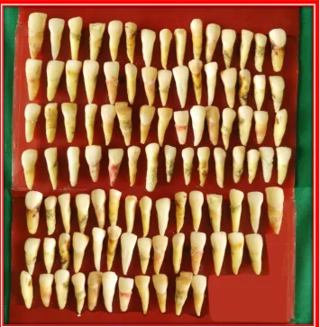
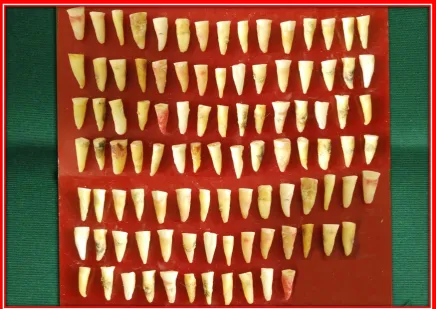
1 Extracted human maxillary anteriors 33
2 Armamentarium 34
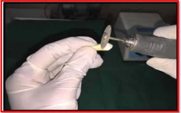
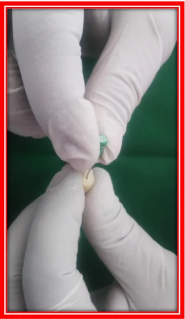
3 Decoronation of the teeth near CEJ 34
4 Decoranated maxillary anteriors 35
5 Cleaning and shaping by using k- file 36
6 Irrigation done by using 2ml of 3% Naocl and post-operative radiograph
36
7 Retreatment done with H-file 37
8 Retreatment done with PTUS 37
9 Retreatment done with D-race file 37
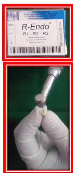
10 Retreatment done with R-endo file 37
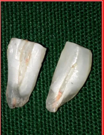
11 Vertically splitted tooth 38
12 Coronal portion 39
[image:15.595.70.537.132.750.2]14 Apical portion 39
TABLE NO.
DESCRIPTION
PAGE NO.
1
Presence of remaining obturating material and debris on
the dentin surface after retreatment in coronal area
41
2
Presence of remaining obturating material and debris on
the dentin surface after retreatment in middle area
42
3
Presence of remaining obturating material and debris
on the dentin surface after retreatment in apical area
43
LIST OF GRAPHS
S.NO.
GRAPHS
PAGE NO.
1.
.
Presence of remaining obturating material and debris on the dentin surface after retreatment in coronal area
45
2. Comparison of grading of four groups within the coronal area 46
3.. Presence of remaining obturating material and debris on the dentin surface after retreatment in middle area
47
4. Comparison of grading of four groups within the middle area 48
5. Presence of remaining obturating material and debris on the dentin surface after retreatment in apical area
49
6. Comparison of grading of four groups within the apical area 50
7.
S.NO
TITLE
PAGE NUMBER
1.
INTRODUCTION
1
2.
AIMS AND OBJECTIVES
5
3.
REVIEW OF LITERATURE
6
4.
MATERIALS AND METHODS
26
5.
RESULTS
41
6.
DISCUSSION
54
7.
SUMMARY
61
8.
CONCLUSION
64
9.
BIBLIOGRAPHY
1
The success of rootcanal therapy depends upon proper working length determination,
complete chemo mechanical debridement and three dimensional obturation. The Washington
study by Ingle in 1951, evaluated a total population of 3678 patients for the success and failure
of endodontic treatment, on a recall basis at 6 months, 1year, 2 years and 5 years. The
statistically significant two year recall analysis showed that there was a better success rate for
non surgical treatment which was assessed on the basis of improvement of the periradicular
health. The overall success rate for primary endodontic treatment is 94%1
Myriad of factors have been implicated in the failure of endodontic treatment. The usual
factors which can be attributed to endodontic failure are: persistence of microorganisms in root
canals, persistence of pulp tissue and remnants, deficiency in obturation, overextension of
obturating materials, improper coronal seal, untreated canals, iatrogenic procedural errors such as
poor access cavity design, complications of instrumentation like ledges, perforations, or
separated instruments.2
Endodontic retreatment is a procedure performed on a tooth that has received prior
attempted definitive treatment resulting in a condition requiring further endodontic treatment to
achieve successful results. As defined by the American association of endodontists (AAE),
retreatment is the removal of rootcanal filling materials from the tooth, followed by cleaning,
shaping and obturating the canals.3
Non-surgical retreatment should be considered as the first treatment option even in cases
of persistant periapical lesions, as it has been reported to have the success rate of 65% to 80%.4It
is also mandatory to assure the restorability of the tooth to facilitate its function.
When the choice is non surgical endodontic retreatment, the goal is to access the pulp
INTRODUCTION
repair the defects that are pathologic or iatrogenic in origin. In cases with obstructions within the
root canal system that make nonsurgical retreatment impossible, then surgical retreatment can be
the option that is preferred/recommended.5
According to Ingle, “Non-surgical retreatment (NSRCT) are disassembly and corrective procedures which are performed to potentially enable the clinician to properly clean, shape and
seal the root canal system”.5
The major goal of nonsurgical retreatment is to re-establish healthy periapical tissues
following ineffective root canal treatment and reinfection.6Root canal retreatment requires
complete removal of root filling material, inorder to uncover remaining necrotic tissues or
bacteria that may be responsible for periapical inflammation and post treatment disease.7,8
Removing of sealer and gutta-percha from inadequately prepared and obturated root
canal systems is critical in order to uncover remnants of necrotic tissue or bacteria that may be
responsible for periapical inflammation and failure.. GP removal will be settled by endodontic
hand files, heat-carrying instruments, ultrasonics, rotary instruments with or while not the help of
solvents.9
Many techniques are projected to get rid of remove filling materials from root canal
system, together with the utilization of endodontic hand files, Gates Glidden burs, heated
instrument, ultrasonic instruments, Nickel Titanium rotary instruments, laser and use of
adjunctive solvents like chloroform, halothane, eucalyptus oil, xylene, orange oil, turpentine oil
and whitepine oil.10
Conventionally, the removal of gutta-percha using manual files with or without solvent
can be a tedious, time-consuming process, especially when the root filling material is well
3
materials from root canal walls & varied studies reported their efficaciousness, cleansing ability
and safety.11
Hand files are employed in the retreatment procedures. Initially H-files with the single
helix, tear drop shaped cross sectional design and positive rake angle were the instruments
designed for the removal of filling materials. Unal et al. found K-files and H-files to be more
effective in removing filling material than ProTaper and R-Endo instruments in curved canals.12
Later rotary NiTi systems have been introduced for retreatment which resulted in
reduction in the treatment time when compared with hand instrumentation. Softening of
gutta-percha during rotary instrumentation caused by frictional heat results in easier penetration and
removal of the filling material is the main reason for this observation.
The cleaning ability of Protaper universal retreatment files depend on the
characteristics of the convex triangular cross sectional design of the instruments. The negative
cutting angle and the absence of radial lands permit a cutting action rather than a planning action.
Protaper-R file D1 incorporates a cutting tip to facilitate initial penetration into the filling
material. D2 and D3 each have non cutting tips and are used to take away material from the
middle and apical thirds.
RaCe NiTi instruments has a triangular cross section, variable and alternated helical
angle with non active tip. The alternate cutting edges and electro polished smooth surface
attributes to its effective removal of GP.
R-Endo retreatment instruments have a triangular cross section with three equally
spaced cutting edges; the instruments have neither radial land nor an active tip. This system
comprises of a stainless steel Rm hand file used to break the hard layer of root filling material
INTRODUCTION
the three rootcanal areas ( R1,R2,R3). Hence H-file, Protaper universal retreatment file, D-Race
file and R-Endo file have been taken for this study to evaluate the efficacy of removal of GP and
sealer.
Variety of chemical solvents like chloroform, xylol, eucalyptol, orange oil, halothane
are used for solubilization of gutta-percha and sealer inorder to facilitate easy removal of filling
material without damage to the tooth. Solvents acts as adjunct in the removal of the gutta-percha.
Hand or rotary files must be used to complete the removal of the entire filling material.
With the benefit of education and experience, one should be able to choose the proper
cases for endodontic retreatment and reject those that will obviously fail, but it is not always
possible. The reported success of retreatment is 87.9% .The lower success rate of secondary
treatment may be due to the incomplete elimination of certain microorganisms which are known
to be common in such cases, for example, E. faecalis, the elimination of this microbe could be
difficult because of its resistance to some disinfectants used during the treatment, particularly
calcium hydroxide. It has been postulated that E. faecalis may be able to invade the dentinal
tubules and adhere to collagen in the presence of human serum.14
The aim of this study was to compare the efficiency of different retreatment
instrumentation techniques to remove the filling material from the root canal walls, during
retreatment procedure and also to assess the percentage of remaining filling materials on root
AIMS AND OBJECTIVES
AIM:
The aim of this study was to compare the efficiency of different retreatment
instrumentation techniques to remove the filling material from the root canal walls, during
retreatment procedure and also to evaluate the percentage of remaining filling materials on root
canal walls using Stereomicroscope.
OBJECTIVES:
1) To compare the Efficiency of different files during retreatment instrumentation
techniques in removal of the filling material from the root canal walls during retreatment,
under stereo microscope.
2) To evaluate the Percentage of remaining filling material in coronal, middle and apical
REVIEW OF LITERATURE
Abramovitz .I et al (2012)1checked the efficacy of a two- stage retreatment
method in which the self-adjusting file (SAF) is used to remove root canal filling residue left
in the canal after using protaper universal retreatment files. The study concluded no system
removed the root filling material entirely. After rotary instrumentation using protaper universal
retreatment files followed by use of SAF resulted in a significant reduction in the amount of
filling residue in curved canals.
Akhavan et al (2012)2compared the ability concerning Mtwo and D-RaCe
retreatment systems to put off residual gutta-percha and sealer in the root canal after retreatment.
Group 1 become retreated with Mtwo and Group 2 with D-RaCe. Both groups have been then
divided into two subgroups retreated either without or with solvent. Teeth were then vertically
sectioned for evaluation of residual filling materials on the canal walls. In this study he
concluded a negative effect of solvent on removal of gutta-percha and sealer in both the
Mtwo and D-RaCe systems.
Asheibi et al (2014)3compared the effectiveness of ProTaper rotary files with
ProTaper-R and K-files in the removal of Resilon or guttapercha (GP) from canals filled
either by cold lateral condensation or thermal obturation using micro-CT. Group-1 was filled
with GP/AH-Plus and Group-2 with Resilon/RealSeal using cold lateral condensation. Group-3
was filled with GP/AH-Plus and Group-4 with Resilon/RealSeal using System Band Obtura
II. The roots were scanned by micro-CT. Each group was divided into two subgroups (n=12):
A, retreated using ProTaper files and B, using ProTaper-R and K-files. He concluded that
obturation using thermal technique resulted in significantly less remaining material than
7
Barletta et al (2007)5compared the capacity of a reciprocating system
(Endo-Gripper) and a rotary system (Profile .04) for mechanical removal of root-filling material
from curved root canals. Eighty canals (40 mesiobuccal and 40 mesiolingual) from
mandibular first molars were instrumented and filled. After 6 months, the volume of
root-filling mass was measured by computed tomography (CT). Root root-fillings were removed by
either the reciprocating system with K-type files or the rotary system with NiTi files. The
volume of filling debris remaining after the removal procedures was assessed by CT. He
concluded there were no significant differences between the reciprocating and rotary systems
with regard to the volume of filling material left inside the canals after mechanical
instrumentation.
Betti .L .V et al (2001)6study was to compare Quantec SC rotary instruments
and hand files for removal of gutta-percha during retreatment. The time for root filling
removal was significantly less when Quantec SC was used. Although Quantec SC
instruments took less time, hand instruments and solvent cleaned canals more effectively.
Bernardes RA et al (2015)7used micro-CT to quantitatively measure the number of
residual filling material after using numerous techniques to get rid of root filling with and
without ultrasonic activation and to analyze the cleanliness of the root canal walls and
dentine tubules with scanning electron microscopy (SEM).The method used for removing the
root filling: G1-Reciproc (using only instrument R50), G2-Protaper Universal Retreatment
System, and G3-Manual (Hand Files and Gates Glidden burs). None of the methods were
able to completely remove root filling material. Ultrasonic activation stepped forward the
REVIEW OF LITERATURE
Bodrumlu E et al (2008)8compared the retreatment of a root canal within the case of
infection needs get rid off previous rootcanal filling material. This study analyse the
efficaciousness of three techniques in removing laterally compacted Resilon/Epiphany and
GP/AH Plus from straight and curved canals throughout retreatment.
Clovis Monteiro Bramante et al (2010)9 ex vivo study evaluated the warmth
release, time needed, and clean up effectivity of MTwo (VDW, Munich, Germany) and
ProTaper Universal Retreatment systems (Dentsply/Maillefer, Ballaigues, Switzerland) and
hand instrumentation in the removal of filling material. ProTaper UR and MTwo R
caused the greatest and lowest temperature increase on root surface, respectively;
regardless of the type of instrument, more heat was released in the cervical third. Pro Taper
UR needed less time to remove fillings than MTwo R. All techniques left filling debris in the
root canals.
Carlos Eduardo da Silveira Bueno et al (2006)10 assessed in vitro the efficacy of
nickel-titanium K3 rotary files and hand files for removal of guttapercha and sealer from
obturated root canals using either chloroform or chlorhexidine as solvents Group I: size 3
Gates-Glidden drills plus size 30 hand K-files and Hedstrom files and chloroform; Group II:
K3 NiTi rotary files and chloroform; and Group III: K3 NiTi rotary files and 2%
chlorhexidine gel. The findings of this study showed that, despite the technique used for
removal of filling material, none of the retreated canals were completely free of gutta-percha
and sealer remnants. The use of stainless steel hand files resulted in a lesser amount of
9
Rodrigo Sanches Cunha et al (2007)11 evaluated the filling material removal and
reinstrumentation operating time of canals which was filled with Resilon/Real Seal in
comparison with canals filled with gutta-percha/AH Plus. When compared with GP cones and
the AH plus cement ,Resilon/Real Seal system was removed in higher quantities from the canal
walls. Time was not a big issue. Under scanning electron microscopy analysis, the teeth given
material remnants in the 3 analyzed thirds. Resilon was higher off from the canal than the
gutta-percha cones and the AH Plus.
De carvalhomaciel .A.C et al (2006)14 compared automated and manual
instrumentation techniques for removing filling material from root canal walls during root
canal retreatment. He concluded that a photo micrographic method by epiluminescence
was more effective than the radiographic method to evaluate filling debris. There was no
significant difference between the filling materials in terms of their removal. K3 and
ProTaper were more efficient than manual instrumentation.
De Mello Junior JE et al (2009)15compared the efficacy of guttapercha/sealer
removal from endodontically treated extracted human teeth with and without the aid of a clinical
operating microscope/ultrasonic instruments. Teeth were divided into 2 groups: group I,
re-treated using a conventional technique with burs and solvent; and group II, re-re-treated using a
conventional technique with burs and solvent plus clinical operating microscope/ultrasonic tips.
The use of the dental operating microscope and ultrasonic tips removed the filling material from
root canal walls higher, however all examined teeth, in each groups, had remaining filling
REVIEW OF LITERATURE
Duarte et al (2010)17evaluate the effectiveness of manual and rotary instrumentation
techniques for removing root fillings after different storage times. 24 canals from palatal
roots of human maxillary molars were instrumented and filled with gutta-percha and zinc-oxide
eugenol-based sealer (Endofill), and were hold on in saline for 6 years. Non-aged control
specimens were treated in the same manner and hold on for 1 week. All canals were
retreated using hand files or ProTaper Universal NiTi rotary system. The roots were vertically
split, the halves were examined with a clinical microscope and the obtained images were
digitized. There was no statistically significant difference between the manual and rotary
techniques for filling material removal regardless the ageing effect on endodontic sealers. He
concluded that all canals presented residual filling material after endodontic retreatment
procedures.
Ersev .H et al (2012)18 evaluated residual root filling material following
removal of three newly developed root canal sealers used with a matched-taper single
cone root filling technique and to compare the efficiency of protaper universal rotary
retreatment instruments with conventional manual technique. When using a gross radiographic
criteria, the Activ GP used to be greater efficaciously eliminated from root canals than AH plus
together with hand instrumentation. Hybrid root seal, Endosequence BC sealer and AH plus
has been eliminated to a comparable extent. Protaper universal retreatment instruments and
hand instruments were same as safe and effective in reaching the working length.
Fenoul .G et al (2010)20evaluated the effectiveness of the R-Endo rotary nickel
titanium instrumentation system and hand instrumentation to take away gutta-percha or
11
for removal of filling were lower with R-Endo than with Hedstrom files. Both
instrumentation techniques left filling material inside the root canal and mainly in the apical
third. There was no difference between the instrumentation techniques.
Gergi et al (2007)21evaluated ex vivo the effectiveness of hand files, ProTaper and
R-Endo rotary instruments when removing gutta-percha from curved root canals. All
instruments left filling material inside the root canal. ProTaper and R-Endo rotary instruments
were inadequate for the complete removal of filling material from the root canal system.
Valentina Giuliani et al (2008)22 study was to judge the efficaciousness of the
ProTaper Universal System rotary retreatment system and of Profile 0.06 and hand instruments
(K-file) in the removal of root filling materials. The ProTaper-R files and ProFile rotary
instruments worked considerably quicker than the K-file. The ProTaper-R files left cleaner root
canal walls than the hand instruments K-fies and the ProFile Rotary instruments, although
none of the devices used guaranteed to get rid off filling materials. The rotary NiTi system
proved to be faster than hand instruments in removing root filling materials.
Gu L.S et al (2008)25evaluated the efficiency of the protaper-R system for GP
removal from root canals. In Group A GP removal completed with the protaper universal rotary
retreatment system and with additional canal repreparation accomplished with protaper
universal rotary instruments, Group B –GP removal was completed using Gates Glidden drills and Hedstrom files with chloroform as a solvent, followed with additional canal repreparation
with protaper universal rotary instruments, Group C identical as group B for GP removal with
further canal preparation with stainless steel K flex files. He concluded that all techniques left
REVIEW OF LITERATURE
GP and sealer from maxillary anterior teeth is by the protaper universal rotary retreatment
system.
Haapasallo M et al (2012)26used micro-computed tomography to evaluate the
amount of remaining root filling material in oval canals filled by using 2 obturation techniques
after retreatment with the ProTaper Universal Retreatment with or without solvent. He
concluded that none of the retreatment techniques were able to completely remove all
gutta-percha/sealer from the oval canals. More root filling material was left in the root canals
filled by using the continuous wave condensation technique than those filled by using the cold
lateral condensation technique after retreatment. In the non solvent groups, less time was
needed to achieve satisfactory gutta-percha removal and root canal refinement than in the solvent
groups.
Hammad et al (2008)27study was to compare the percentage of 3D volume of
remaining filling materials in canals filled with realseal, endorez, guttaflow and gutta-percha
after retreatment with endodontic manual files and retreatment with endodontic manual files and
retreatment protaper rotary files by using micro computed tomography. The study showed that,
by using hand and rotary files were not completely removed all tested filling materials during
retreatment. Guttapercha was more efficiently removed by using hand K files. In future,a
combination of a pair of techniques, hand files and protaper-R files, might result in more
efficient removal of material.
Hassanloo et al (2007)29assessed the efficaciousness of retreatment of canals filled
with the epiphany system with and without solvent, with particular reference to the extent of
13
and without chloroform, with lesser efficacy than guttapercha and AH plus sealer. The major
amount of residues were present in the apical segments of canals were removed before
retreatment which is enhanced by apical enlargement beyond the diameter of the canal .
Horvath D et al (2009)29determined the influence of solvents on gutta-percha
and sealer remaining on root canal walls and in dentinal tubules Removal of root fillings was
undertaken after 2 weeks using Gates Glidden burs and hand files without solvent (group
2), with eucalyptol (60 microL; group 3) and with chloroform (60 microL; group 4) to size 50
Solvents led to more gutta-percha and sealer remnants on root canal walls and inside dentinal
tubules.
Hulsmann .M et al (2004)31evaluated the efficacy, cleaning ability and safety
of three different rotary nickel-titanium instruments with and without a solvent (eucalyptol)
versus hand files in the removal of gutta-percha root fillings. GP removal was performed with
the following devices and techniques: FlexMaster, GTRotary, ProTaper and Hedstrom files.
Flex-Master and ProTaper NiTi instruments proved to be efficient and timesaving devices for
the removal of gutta-percha. The use of eucalyptol as a solvent shortened the time to
reach the working length and to remove the gutta-percha, but this was not significant.
Imura. N et al (2000)32study was to quantify the quantity of remaining
gutta-percha/sealer on the walls of root canals once two engine-driven instruments (Quantec
and ProFile) and two hand instruments (K-file and Hedstrom file) were used to remove
these materials. The number of apically extruded debris and the time needed for treatment were
REVIEW OF LITERATURE
material inside the root canal. Throughout the retreatment procedure there is a risk for
instrument breakage, particularly rotary instruments.
EmreIriboz et al (2014)33evaluated the effectiveness of the protaper and Mtwo
retreatment systems for removal of resin based obturation techniques during retreatment.
In the present study, in teeth obturated using the Resilon-Epiphany technique, less
remnant filling material was observed with the other techniques. Teeth prepared with
Mtwo instruments contained significantly more remaining filling material than those prepared
with protaper. The time required to remove gutta-percha+AHplus was significantly less than
that required for the other obturation techniques The retreatment time of the teeth prepared with
protaper was significantly reduced compared to that of those prepared with Mtwo.
Jamie Ring et al (2009)34compared the effectiveness and working time of
two rotary instrumentation file systems with two solvents for the removal of gutta-percha
(GP) (ProTaper Universal, Dentsply Tulsa Dental,Tulsa, Okla.) or resin-based composite
(RBC) (RealSeal 1 Bonded Obturator, SybronEndo, Orange, Calif.) endodontic obturation
material Re-treatment with EndoSequence rotary files was quicker than re-treatment with
ProTaper Universal re-treatment files (Dentsply Tulsa Dental). However, in this study, the file
systems were similarly effective in removing GP and RBC. Orange solvent was as effective
as chloroform in removing obturation materials, but its use is less time-consuming.
Kefah .M et al (2002)38compared the cleanliness of the root canal walls
after retreatment using nickel titanium (NiTi) rotary and stainless steel (SS) files. Also
compared were time of retreatment and canal deviation. Results showed that the mean
15
was 15.2% for the NiTi group. There was no statistically significant difference (p = 0.361). No
severe canal deviation occurred with either retreatment method. Mean retreatment time for
the SS group and NiTi roup was 6.3 min and 7.9 min ; the difference was statistically important
(‘t’ test p < 0.001). Lastly, NiTi rotary and SS hand files were similar in removing filling material remaining after retreatment, however SS hand files was a small amount quicker
Fir .A .K et al (2012)39compared the ability over five techniques for the
elimination of root filling material yet to test the hypothesis that radiographs fail to represent
the real extent of remaining material on canal walls. As a whole, 11–26% of the canal wall remained covered with filling material; no significant variation was found among the
groups. The mechanized methods were faster than manual removal of filling material. The
use of solvent did not speed up the mechanized procedures. Radiographic evaluation did not
thoroughly and reliably detect the quantity of filling material remaining on the canal walls,
which was later determined by microscopic analysis. He concluded that all methods left root
canal filling material on the canal walls. Radiographic evaluation failed to detect the extent
of remaining root filling material, which could only be detected using microscopy of
mandibular molars.
Khalilak Z et al (2013)40assess the effectiveness of H-File and ProTaper-R
with or without chloroform in the elimination of filling material GP during retreatment of
mandibular premolars. Removal of GP was performed with H-File and ProTaper. All
techniques were used with or without chloroform. The treated teeth were split
longitudinally and the area of remaining gutta-percha/sealer on the root canal wall was
explored under stereomicroscope. In all groups, no significant difference was found in
REVIEW OF LITERATURE
reduced the time of retreatment. ProTaper left significantly less remaining filling materials than
H-File in canals with no or slight curvature. Retreatment time was significantly different
between the studied groups. ProTaper Ni-Ti instruments proved to be more efficient and
time-saving devices for removal of gutta-percha compared to H-File in canals with no or slight
curvature.
Kok D et al (2014)41assessed the penetrability of two sealers ( MTA
Fillapex and AH plus) into dentinal tubules, which was submitted to rootcanal treatment and
sooner or later to rootcanal retreatment. There was no significant difference was observed
among the two experimental groups (P > 0.05). On the other hand, the sealers in the control
group were not capable to penetrate into dentinal tubules after endodontic treatment (P >
0.05). In retreatment cases, no sealers would be able to penetrate into dentin tubules . It can
be concluded that sealer penetrability is high in rootcanal treatment. But, MTA Fillapex and
AH Plus do no longer penetrate into dentinal tubules after rootcanal retreatment.
Kosti .E et al (2006)42compared the efficacy of ProFile rotary Nickel–Titanium (Ni–Ti) instruments and Hedstrom - files (H-files) combined with Gates-Glidden (GG) drills during removal of gutta-percha root fillings used in combination with one of the four
representative sealers. Sealer remnants were observed with both techniques mainly in the
middle and apical third of the root canal. The ProFile system and the H-files were
associated with similar amounts of remaining filling material. In the cervical third of the root
canal all sealer remnants were removed with both techniques. In the middle and apical third
AH26 was associated with a statistically significant greater quantity of remnants on the root
17
the removal of root fillings was totally effective, especially in the apical third of the root
canal.
Marciano. M et al (2010)44reported that Confocal laser scanning microscopy
will be employed in laboratory research to get a series of optical XY images through the
thickness of the dentin of endodontically treated teeth. With this information the study of
the resin/dentin interface of root canal fillings is attainable. By comparing SEM and Confocal
microscopy, confocal microscopy has the advantage of providing accurate information and
one of the easy method to see the variation and distribution of sealers within dentinal tubules in
non-dehydrated samples through the use of Rhodamine-marked sealers. 3D reconstructions
can also be generated with the digital data. In this work, some examples which includes epoxy
and methacrylate resin sealers are given in relation to the sealer/dentin interface for different
endodontic materials .
Marcus Vinícius Reis Só et al (2008)45evaluated the efficacy of ProTaper
-R system and hand files for removal of filling material during retreatment and the
influence of sealer type on the presence of filling debris in the reinstrumented canals.
G1, EndoFill/hand files; G2, AH Plus/hand files; G3, EndoFill/ProTaper; G4, AH
Plus/ProTaper. He concluded that all techniques were similar in the apical third. All groups
presented filling debris in the 3 canal thirds after reinstrumentation.
Marcus Vinícius Reis SO (2012)46 evaluated the effectiveness of ProTaper
Universal rotary retreatment system and also the influence of sealer type on the presence
REVIEW OF LITERATURE
Remnants was left in all canal thirds, irrespective of the retreatment technique. The greatest
differences between techniques and sealers were found within the cervical third.
Marfisi .K et al (2010)47evaluated the efficaciousness of protaper-R files, Mtwo
retreatment files and twisted files for removal of guttapercha and resilon in straight root canals.
He concluded that none of the system entirely removed the root filling material . Mtwo
retreatment files needed less time to get rid of root filling material than the alternative
instruments. Resilon was removed considerably higher from the canal walls than guttapercha,
irrespective of the rotary instruments used.
Marques da silva B.et al (2012)48assessed the efficacy of different
retreatment rotary files in removing guttapercha and endodontic sealer from canals. Group
I – protaper universal retreatment group, Group II – protaper universal retreatment group+protaper F4,Group III- D-Race retreatment group, Group IV- D-Race retreatment
group+Race size 40..04 taper, Group V-Mtwo retreatment group, Group VI – Mtwo retreatment group +Mtwo size 40..04 taper. The study concluded that none of the systems
completely removed filling material from the root canal. Amongst groups in which
additional instrumentation was sued, the PTUR was the most effective system, especially when
compared with D-Race. There were no differences in outcomes between groups with the use of
additional instrumentation.
Martos .J et al (2011)49evaluated the solubility of five root canal sealers
in orange oil, eucalyptol, xylol and chloroform solvents. The study concluded that xylol
was the most effective solvent followed by the chloroform and the essential oils. Orange
19
Masiero .A.V (2005)50evaluated the effectiveness of various techniques for
removing filling material from root canals in vitro. The apical third had the most remaining
material, whilst the cervical and middle thirds were significantly cleaner. Comparison of
the techniques revealed that teeth instrumented with K3 rotary instruments had a lower ratio of
remaining filling material in the apical third. In the apical third, K3 rotary instruments
were more efficient in removing gutta-percha filling material than the other techniques,
which were equally effective for the other thirds.
Mollo .A et al (2012)51compared the effectiveness of two NiTi systems
and hand files for removing guttapercha and sealer from root canals. He concluded None of
the techniques was able to remove guttapercha and sealer completely from the root canal.
NiTi engine driven rotary instruments were significantly faster and more effective in removing
guttapercha than hand files.
Müller GG et al (2013)52investigated whether a final rinse with Endosolv
R solvent and ultrasound resulted in cleaner root canal walls during endodontic retreatment. A
total of 56 extracted premolar teeth were manually instrumented using a step-back flare
technique and filled with gutta-percha and AH Plus sealer. After 9 months, the canals were
retreated by using ProTaper Universal Retreatment rotary instrument up to an F5 file to remove
gutta percha and sealer . As a final step, the teeth were randomly divided in 4 groups (n=14)
and were subjected to passive ultrasonic irrigation (PUI) with either Endosolv R or distilled
water. In the control groups, the irrigants were left undisturbed. All groups presented
filling debris in the three root canal thirds after retreatment. There were no significant
REVIEW OF LITERATURE
concluded PUI with Endosolv R was not effective in the removal of filling debris from root canal
walls.
Ronald Ordinola - Zapata et al (2009)53compared the percentage and depth of
penetration of sealer into dentinal tubules during rootcanal obturation using Guttaflow, sealer 26
or seal apex in root canals filled with the lateral compaction technique using confocal
microscopy. The study concluded that depth of penetration of root canal sealers into dentinal
tubules using the lateral compaction technique is influenced by the type of sealer and by the root
canal level, with penetration decreasing apically. Sealapex showed deeper penetration into
the dentinal tubules. Neverthless, the percentage of sealer adaptation into the root canal walls
was similar in the 3 sealers.
Pirani C et al (2009)55 evaluated the root canal wall morphology below
scanning electron microscopy magnification when removal of 2 forms of root canal fillings by
using ultrasonic tips, nickel-titanium (NiTi) rotary instruments, and hand K-files.
Retreatment was completed by using K-files , M-Two NiTi rotary instruments , or ESI
ultrasonic tips (group1,2,or3) in 12 roots each respectively. He concluded that all
retreatment techniques showed similar performances in terms of smear layer morphology,
debris, and surface profile. None of the above instruments completely removed filling material
debris from dentinal tubules of apical third.
Rached-junior F.J.A et al (2014)57evaluated the bond strength of a resin based
sealer (AH plus) to root canal dentine after the elimination of a zinc oxide-eugenol based sealer
(Endofill), using distinct retreatment techniques. He concluded that operating microscope was
21
oxide eugenol based sealer negatively affected the bond strength of AH plus to root
canal walls, irrespective of the retreatment technique
Robert W. Ladley et al (1991)59compared halothane and chloroform used with
hand or ultrasonic instrumentation to remove gutta-percha and sealer from root canals,
apically extruded debris, residual debris, time for filling removal and amount of solvent
used were determined. The extruded apical debris and radiographically visible residual debris
were not significant. Ultrasonic instrumentation required significantly less time to remove the
root canal filling than did hand instrumentation. The only significant difference in the amount of
solvent used occurred when the ultrasonic-chloroform group compared with the hand
instrumentation-chloroform group. Halothane was found to be an acceptable alternative to
chloroform for removing gutta-percha and sealer from obturated root canal.
Rodig .T et al (2012)60 compared the efficacy of two rotary NiTi
retreatment systems and Hedstrom files in removing filling material from curved root
canals. The root fillings were removed with D-RaCe instruments, ProTaper Universal
Retreatment instruments or Hedstrom files. He concluded that D-RaCe instruments were
associated with significantly less residual filling material as compared to ProTaper
Universal Retreatment instruments and hand files. Hedstrom files removed significantly less
dentine than both rotary NiTi systems. Retreatment with rotary NiTi systems resulted in a high
incidence of procedural errors.
Roggendorf .M.J et al (2010)62 evaluated the efficiency of removing Activ
GP or GuttaFlow from root canals using nickel titanium instruments. He concluded that both
REVIEW OF LITERATURE
enlargement up to two sizes beyond the pre-retreatment size was necessary to minimize the
amount of seale Roggendorf .M.J et al (2010)62 evaluated the efficiency of removing
Activ GP or GuttaFlow from root canals using nickel titanium instruments. He concluded that
both obturations with ActivGP and GuttaFlow were removed with NiTi instruments. Canal
enlargement up to two sizes beyond the pre-retreatment size was necessary to minimize the
amount of sealer remaining.
Varawan Sae – Lim et al (2000)63 investigated the retreatment effectiveness of 0.04 taper nickel titanium rotary profiles. Retreatment for group A done using profile
alone, group B using profile and chloroform ,and group C using hand files with chloroform.
The results showed that the mean scores in group A and B were typically less than group C
.Mean scores of apical thirds tended to be higher than the middle and the cervical third,
except group A .Profile with or without chloroform appeared to be a viable various retreatment
methodology.
Schirrmeister .J. F. et al (2006)65also compared the detectability of residual
Epiphany and gutta-percha after root canal retreatment using a dental operating microscope and
radiographic examination with the residual area measured after rendering the roots
transparent. He concluded that especially for remaining gutta-percha, the operating
microscopes provided better detection of residual root filling material in retreated maxillary
incisor teeth.
Schirrmeister .J .F et al (2006)66assess the effecaciousness of hand and rotary
instrumentation for removal of vertically compacted Epiphany and GP during retreatment. He
23
Sealant was removed more effectively than gutta-percha and AH Plus sealer. Hedstrom
files were more rapid than RaCe rotary instruments.
Silva et al (2015)68compared the effectiveness of reciprocating and rotary
techniques to get rid off gutta-percha and sealer from root canals. Two experimental
retreatment groups: ProTaper Retreatment System (PTRS) and WaveOne System (WS).
The percentage of residual material was calculated .No system completely removed the root
filling material from the root canal. No significant differences were observed between the
systems, in terms of residual filling material in any tested third. WS was faster in removing
filling material than PTRS. He concluded that although no differences were observed in the
efficacy of PTRS and WS for removing root filling material, WS was faster than PTRS
Francesco Somma et al (2008)69compared the effectiveness of the Mtwo
R (Sweden & Martina, Padova, Italy), ProTaper- R files (Dentsply-Maillefer, Ballaigues,
Switzerland), and a H files manual technique in the removal of three different filling
materials (gutta-percha, Resilon [Resilon Research LLC, Madison, CT], and EndoRez
[UltradentProductsInc, South Jordan, UT]) during retreatment. In conclusion, all instruments
left residues of filling material and debris on the root canal walls irrespective of the root
filling material used. Both the engine-driven NiTi rotary systems proved to be safe and
fast devices for the removal of endodontic filling material.
Takahashi et al (2009)70evaluated the efficacy of a NiTi rotary instrument
system with or while not a solvent versus SS hand files for GP removal. They were divided
into 4 groups: GatesGlidden and K-files, Gates-Glidden and K-files with chloroform,
REVIEW OF LITERATURE
system with chloroform. He ended that all techniques proved useful for the removal filling
material, and that they were similar in material remaining after retreatment, however the
ProTaper-R without chloroform was quicker.
Tesdemir T et al (2008)71 studied the ability of three rotary NiTi
instruments and hand instruments to remove GP and sealer. The ProTaper group had less filling
material within the root canals than the other groups, however a major distinction was found
between solely the ProTaper and Mtwo groups. The retreatment time for Mtwo and ProTaper
was considerably shorter when compared with R-Endo and manual instrumentation Hedstrom
files. R-Endo was considerably quicker than manual instrumentation. Under the experimental
conditions, ProTaper left considerably less GP and sealer than Mtwo instruments. Complete
removal of GP and sealer did not occur with any of the instrument systems.
CelikUnal G et al (2009)72compared the efficaciousness of standard and new
retreatment instruments once removing gutta-percha root fillings in curved root canals.
Within the bucco-lingual direction, the remaining filling material was considerably less
following manual instrumentation than R-Endo and ProTaper instrumentation. Within the
proximal view, it had been considerably less following manual and ProFile instrumentation
than R-Endo. Complete removal of filling material occurred solely in three specimens
(with manual instruments). Manual instruments were significantly faster than R-Endo and
ProFile. More procedural errors (five fractured instruments and two perforations) were
noted when using ProTaper. The laboratory study in curved molar roots shows that ProTaper
-R and -R-Endo instruments were less effective in removing filling material from canal walls
25
Lisa R. Wilcox et al (1987)73examined the appearance of root canal walls
after retreatment. Four techniques were used to remove gutta-percha and sealer: method 1-heat
and files; method 2-heat, files, and Cavi-Endo; method 3-chloroform and files; and method
4-chloroform, files, and Cavi-Endo. The results showed that no technique removed all
debris. When AH26 was the sealer, method 4 was significantly less effective. When
Roth's 801 was the sealer, method 1 was significantly less effective. Teeth obturated using
Roth's 801 sealer were significantly cleaner after reinstrumentation.
Lisa R. Wilcox et al (1989)74studied forty extracted teeth were
instrumented using a step-back flare technique and obturated with guttapercha and either
AH26 or ROTH’S 801 sealer. After 3 months the canals were retreated by removing
gutta-percha and sealer with hot instruments followed by chloroform and files. As a final
step, the teeth were instrumented using ultrasonics with either chloroform or with NaOcl. Most
teeth were well cleaned. No significant differences were found between sealer groups or
between the two irrigants as to the ability to remove gutta-percha/sealer.
Li lixu et al (2012)76aimed to assess variation in the incidence and depth of
residual filling material in dentinal tubules after gutta-percha removal with H files, the protaper
universal system and Sybron endo K3 system. He concluded that the protaper universal
system and Sybron endo K3 system left filling material in a greater proportion of dentinal
26
MATERIALS
S.no
Material used
Brand name/
Manufacturer details
1. Human maxillary Anteriors n=100
2. Round diamond burs Mani, Japan
3. Airotor highspeed handpiece NSK, Japan
4. X Smart Endomotor Dentsply Maillefer
5. Safe side Diamond disc Mani Inc, Japan
6. Sodium hypochloride 3% Prime Dental Products PVT
7. 17% EDTA solution Desmer
8. 2.5 ml disposable syringes Hindustan syringes & Medical
Services Ltd
9. K files 15 size–21 mm Mani, Japan
10. Protaper universal rotary retreatment files Dentsply Maillefer
11. R-Endo rotary files VDW.Germany
12. D-Race rotary files FKG
13. H-Files Mani, Japan
MATERIALS AND METHODS
15. Spreaders-21mm Mani, Japan
16. 2% taper GP points Dentsply Maillfiller
17. Normal saline Claris Otsuka Private Ltd, India
18. Micro motor handpiece NSK pana-max plus,
Nakanishi International, Tokyo
19. Stereo microscope Chongqing optec Instrument
28
METHODS
SAMPLE SELECTION:
This study was approved by Institutional Ethical Committee / Institutional Review
Board. A total of 100 extracted maxillary anteriors were selected. Teeth with fully formed
apices, absence of calcifications and straight root canal were collected, cleaned and stored in
saline with 0.1% thymol and washed under running water.
SAMPLE PREPARATION:
Access cavities were prepared and 15 size K-file was placed into the canal until it
was visible at the apical foramen and the working length was established 1 mm short of this
length. The crowns were removed at the CEJ using a diamond-coated high-speed bur with
air-water spray coolants. The roots were ground coronally to establish a uniform 16-mm
working length for all teeth. Instrumentation of all rootcanals was performed by a single
operator. The coronal thirds of all root canals were enlarged with size 1, 2 and 3
Gates-Glidden drills . The apical two-thirds were enlarged to working length using K-files up to
size50 with a balanced-force technique (Roane et al. 1985). At each instrument change, 2 ml
of 2.5% NaOCl was used for irrigation. The root canals were dried with paper points and
filled using laterally compacted gutta-percha, spreaders and zinc oxide eugenol. After
completion of the procedure, a heated plugger was used to remove the excess gutta-percha to
a level 2 mm short of the canal orifice followed by vertical compaction with a cold plugger.
The coronal orifice of each canal was sealed with a temporary filling material , and the teeth
were stored at 370C in 100% humidity for 1 week to allow complete setting of the sealer.
Teeth were radiographed in B-L and M-D directions to confirm the radiographic adequacy of
root filling, using the following criteria: reaching working length, uniform radiopacity and no
MATERIALS AND METHODS
Retreatment procedures
The temporary fillings were removed, and a 5-mm coronal portion of each root canal
filling was removed using number 2 and 3 Gates -Glidden drills. All specimens were then
coded and randomly assigned to four groups of 25 specimens each.
Retreatment procedures
Group I-H files
The obturating material was removed with H-files of sizes 20, 25 and 30 in a circumferential
quarter turn push and pull motion until working length was achieved.
Group II-Protaper Universal Retreatment File
The D1 file was used to remove sealer and gutta-percha from the coronal third of the
root canal. The D2 file was used in the coronal two thirds of the root canal. The D3 file was
used with mild apical pressure in the WL. The instruments were used at 500 rpm.
Group III- D-RaCe
DR1 was used to remove sealer and gutta-percha from the coronal third of the root
canal at 1000 rpm. DR2 was used at 600 rpm to remove sealer and gutta-percha from the
middle and apical third of the root canal.
GROUP A H-file (n=25) GROUP B Protaper universal retreatment file (n= 25) GROUP C D-race ( n=-25)
30
Group IV- R-Endo
RM file was used to locate the canal orifice and to create a pathway. Then RE file was
used to a depth of 1-3 mm towards to the apex with circumferential filing. Following this the
R1 instrument was used for the coronal third, R2 was used for the middle and R3 was used
for the apical third. All instruments were used to remove filling material in a brushing
circumferential movement. The instruments were used at a speed of 300 rpm
.
The preparation was complete when no more obturation material was observed
sticking to the retreatment instruments. After the retreatment procedure, all roots were
radiographed in B-L orientation to verify the removal of filling material by various removal
technique . Root canals were irrigated in between all instrument changes using 2ml of 3%
NaOCl and a final irrigation with 2ml of 17% EDTA and 5ml of distilled water.
After instrumentation, the roots were split vertically on the buccal and lingual
surfaces, using a water‑cooled diamond disk and taking care to avoid touching the root canal.
They were then split into halves longitudinally with a chisel and mallet and each half then
split into coronal, middle and apical third. The cleanliness of the canal wall was evaluated
through an optical stereomicroscope with 20x magnification and photographs were taken by a
digital Camera.
One blinded reviewer categorized Stereomicroscope images according to the
following criteria by Somma et al. A 4‑point grading system was used with respect to
residual obturation material and debris at the coronal, middle and apical third of each canal.
Variation in the scoring occurred rarely, and when discrepancy was observed, an average of
MATERIALS AND METHODS
• 0: 0%–25% of the dentin surface covered with obturation debris.
• 1:<50% of the dentin surface covered with obturation debris
• 2: 50%–75% of the dentin surface covered with obturation debris
32
Flowchart 1
SAMPLE SIZE n = 100
Sample preparation
Fully formed teeth(apex)
Absence of calcification stored in saline with 0.1% thymol Straight root canal
Decoronated with diamond disc to standardize Root length = 16mm
Working length = 1 mm short of this length
Retreatment procedure done by
GROUP A H-file(n=25) GROUP B Protaper universal retreatment file (n=25) GROUP C D-race(n=25) GROUP D R-endo(n=25)
STEREOMICROSCOPE IMAGE ANALYSIS
carried out in the coronal, middle and apical 3rdsamples of all the four groups.Cleaning and shaping done uniformly till 50 size k-file
Obturation done by lateral condensation method
SECTIONING
MATERIALS AND METHODS
34
Fig.2: Armamentarium
[image:57.595.119.487.487.717.2]MATERIALS AND METHODS
36
Fig.5: Cleaning and shaping by using k-files
[image:59.595.113.494.514.701.2]MATERIALS AND METHODS
[image:60.595.347.464.469.746.2] [image:60.595.121.225.475.748.2]Fig.7: Retreatment done with H- File Fig 8: Retreatment done with protaper universal retreatment file
38
MATERIALS AND METHODS
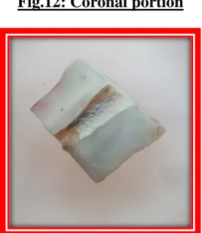
Fig.12: Coronal portion
[image:62.595.228.368.564.731.2]Fig.13: Middle portion
40
41
TABLE 1 : PRESENCE OF REMAINING OBTURATING MATERIAL AND DEBRIS
ON THE DENTIN SURFACE AFTER RETREATMENT IN CORONAL AREA
Presence of obturating material
and debris
Number of cases in Group A H-file(n=25) Group B Protaper(n=25) Group C D-race(n=25) Group D R-endo(n=25)
No. % No. % No. % No. %
None to slight presence 12 48.0 10 40.0 16 64.0 9 36.0
Some presence 10 40.0 13 52.0 9 36.0 10 40.0
Moderate presence 3 12.0 2 8.0 0 0 6 24.0
Heavy presence 0 0 0 0 0 0 0 0
Mean Score 0.64 0.68 0.36 0.88
S.D. 0.7 0.63 0.49 0.78
‘p’ value between
Groups A,B,C & D 0.053 Not significant
Groups A & B 0.4 Not Significant
Groups A & C 0.108 Not significant
Groups A & D 0.258 Not significant
Groups B & C 0.05 Not significant
Groups B & D 0.323 Not significant
Groups C & D 0.007 Significant
RESULTS
TABLE 2 : PRESENCE OF REMAINING OBTURATING MATERIAL AND DEBRIS
ON THE DENTIN SURFACE AFTER RETREATMENT IN MIDDLE AREA
Presence of obturating material
and debris
Number of cases in Group A H-file(n=25) Group B Protaper(n=25) Group C D-race(n=25) Group D R-endo(n=25)
No. % No. % No. % No. %
None to slight presence 13 52.0 10 40.0 14 56.0 7 28.0
Some presence 8 32.0