0022-538X/97/$04.0010
Copyrightq1997, American Society for Microbiology
Functional Interactions between Papillomavirus E1 and
E2 Proteins
MIKAEL BERGANDARNE STENLUND*
Cold Spring Harbor Laboratory, Cold Spring Harbor, New York 11724
Received 10 October 1996/Accepted 14 February 1997
DNA replication of papillomaviruses requires the viral E1 and E2 proteins. These proteins bind coopera-tively to the viral origin of replication (ori), which contains binding sites for both proteins, forming an E1-E2-ori complex which is essential for initiation of DNA replication. To map the domains in E2 that are involved in the interaction with E1, we have used chimeric bovine papillomavirus (BPV)/human papillomavirus type 11 (HPV-11) E2 proteins. The results from this study show that both the DNA binding domain and the transactivation domain from BPV E2 independently can interact with BPV E1. However, the roles of these two interactions are different: the interaction between E1 and the activation domain of E2 is necessary and sufficient for cooperativity in binding and for DNA replication; the interaction between E1 and the DNA binding domain of E2 is required only when the binding sites for E1 and E2 are adjacent to each other, and the function of this interaction appears to be to facilitate the interaction between E1 and the transactivation domain of E2. These results indicate that the cooperative binding of E1 and E2 to the BPV ori takes place via a novel two-stage mechanism where one interaction serves as a trigger for the formation of the second, productive, interaction between the two proteins.
DNA replication in mammalian cells has been studied mainly by using DNA viruses as model systems, and simian virus 40 has been of particular importance (8, 49). From stud-ies of DNA replication of many different replicons, it is now evident that transcription factors play a role in DNA replica-tion in virtually all eukaryotic replicareplica-tion systems (for a review, see reference 21). Frequently, binding sites for transcription factors flank the binding sites for the initiator protein. For example, the simian virus 40 and polyomavirus origins of rep-lication (oris) are flanked by nearby enhancer elements which stimulate viral DNA replication in vivo (for a review, see ref-erence 13). Transcription factors other than the ones that naturally bind to the enhancer can in some cases substitute for this stimulatory function, provided that binding sites for these factors are present in the ori (3, 10, 18). These results demon-strate that the requirement for transcription factors in replica-tion in most cases show only a limited degree of specificity. The function of these transcription factors in DNA replication is unclear; it has been suggested however, that the bound tran-scription factors can function to reverse nucleosomal repres-sion (9, 10, 30). It has also been demonstrated that the activa-tion domains of certain transcripactiva-tion factors can interact directly with the single-stranded DNA binding protein replica-tion protein A (RPA) and possibly serve to recruit RPA to the ori (20, 29). A different function for transcription factors in replication, which has attracted less attention, has been dem-onstrated for adenovirus DNA replication. Binding of the ad-enovirus preterminal protein/DNA polymerase to the viral or-igin of replication is facilitated through a specific interaction with the cellular transcription factor NF-1, which binds to an adjacent site. This interaction results in cooperative binding of the two proteins to the viral DNA (5, 36, 37).
The minimal ori from bovine papillomavirus (BPV) consists of an approximately 60-bp-long sequence that contains an
A/T-rich region, a binding site for the E1 protein, and a binding site for the virus-encoded transcription factor E2 (15, 23, 24, 45, 56, 60). The E1 protein has several activities that are normally associated with viral initiator proteins, including ori-specific DNA binding activity, DNA-dependent ATPase activity, heli-case activity, and unwinding activity (15, 32, 44, 52, 56, 57, 60, 62). The E2 protein is a sequence-specific transcriptional ac-tivator that serves to regulate viral gene expression (1, 19, 33, 46). In contrast to the relaxed requirement for a transcriptional activator observed for other viral replicons, E2 cannot be re-placed by other transcription factors. For example, a hybrid activator, VP16-E2, which contains the activation domain from the herpes simplex virus protein VP16 fused to the DNA bind-ing domain of E2, fails to support replication (56).
It has been well established that interactions between the E1 and E2 proteins can be observed by coimmunoprecipitation assays and two-hybrid analysis as well as by cooperative bind-ing of the two proteins to the ori (4, 6, 15, 31, 35, 45, 47, 60). This interaction between the E1 and E2 proteins results in the formation of a specific complex (E1-E2-ori complex) on the ori (41, 42). We have recently demonstrated by genetic means that the formation of this complex is required for DNA replication activity. The interaction between the E1 and E2 proteins has several consequences, one of which is that the specificity of binding of the E1 protein to the ori is substantially increased. Furthermore, in the presence of E2, E1 binds to the ori in a form that in the absence of E2 lacks, or has very low, DNA binding activity, demonstrating that the interaction with E2 alters the DNA binding activity of the E1 protein (42).
A number of studies have been performed to determine what regions in E2 are required for replication activity as well as for interactions with the E1 protein (4, 7, 14, 17, 22, 35, 38, 39, 50, 56, 58). The conclusion from these studies using a variety of methods all indicate that mutations in the N-terminal activation domain of E2 are deleterious for replication activity. However, the interpretations of these studies are complicated by the limited structural information about the E2 protein. Essentially all deletions in the activation domain of E2 are inactive both for DNA replication and for transcriptional
ac-* Corresponding author. Mailing address: Cold Spring Harbor Lab-oratory, 1 Bungtown Rd., Cold Spring Harbor, NY 11724. Phone: (516) 367-8407. E-mail: [email protected].
3853
on November 9, 2019 by guest
http://jvi.asm.org/
tivation and are likely to result from misfolded protein (56, 58). Recently, also point mutations in the activation domain have been shown to have structural effects (14). One approach that has been used to control for defects resulting from improper folding, or faulty structure, of E2 mutants has been to measure transcriptional activation by E2 as a positive control. Whether a proportional relationship exists between the requirement for E2 in replication and transcription is unclear; the two assays show very different kinetics and differ also in their dependence on the cell cycle. In a effort to circumvent this issue, we have chosen instead to use an approach that utilizes the DNA rep-lication activity of E2 as a positive control. This can be accom-plished by using chimeric E2 proteins with differing specifici-ties.
It has previously been demonstrated that E1 and E2 proteins can function in mixed and matched combinations. For exam-ple, BPV E1 can function together with human papillomavirus type 11 (HPV-11) E2 to replicate a HPV ori plasmid, indicat-ing a conserved function of these proteins in the initiation of replication (11, 12). However, some exceptions exist: the BPV E1 and HPV-11 E2 combination does not function for repli-cation of a BPV minimal ori, in spite of a high degree of conservation between different E2 proteins (41). In this study, we have taken advantage of this observation and have gener-ated chimeric BPV-HPV E2 proteins to map the domains of E2 that are required for the interaction with E1 on the ori. Our results demonstrate that two specific interactions can take place between BPV E1 and BPV E2. One of these interactions, consistent with previous mutational analyses (see above), oc-curs between E1 and the activation domain of E2. This inter-action is conserved and occurs equally well between either BPV E2 or HPV-11 E2. We demonstrate here that this inter-action is sufficient for replication activity. A second interinter-action takes place between E1 and the DNA binding domain of BPV E2. This interaction is not conserved between BPV E2 and HPV-11 E2 and is responsible for the specific requirement for BPV E2 that we observe for the interaction on the minimal BPV ori. However, we also show that the requirement for an interaction between the E1 and the DNA binding domain of E2 can be bypassed by changing the distance between the E1 and E2 binding sites, indicating that this particular interaction serves to alleviate steric hindrance.
MATERIALS AND METHODS
Plasmid constructs. (i) E2 chimeras.Plasmids used for expression of E2 proteins were pET11C for bacterial expression (51) and pCG for eukaryotic expression (53, 55). The coding sequences for BPV and HPV-11 E2 were am-plified by PCR using a 59primer containing an NdeI site and a 39 primer containing a BamHI site, and the PCR product was cloned between NdeI and
BamHI in pET11C. The chimeric E2 proteins were generated by first designing
swap points between the transactivation domain and the hinge and between the hinge and the DNA binding domain. Point mutations were generated in BPV E2 and HPV-11 E2 by oligonucleotide-directed mutagenesis to generate unique restriction sites at equivalent positions in the two E2 sequences without changing the coding sequence. These sites were at positions 3213 (TaqI) and 3576 (BstUI) in BPV E2 and at nucleotides (nt) 3324 (TaqI) and 3568 (BstUI) in HPV-11 E2. Simple subcloning was subsequently used to generate the chimeric E2 proteins. For transient replication assays, the chimeric E2s were transferred into the pCG vector as an XbaI-BamHI fragment.
(ii) Truncated E2s.The short versions of E2 were cloned in pET11C and expressed in Escherichia coli. These constructs were generated by PCR amplifi-cation of the respective segment, using one internal primer including an initiator AUG codon with an NdeI restriction site and one primer corresponding to the sequence downstream of the cloning site in pET11C. The PCR fragments were digested with NdeI and BamHI and ligated into pET11C digested with NdeI and
BamHI. In this way, the BPV DNA binding domain (--B, amino acids 323 to
410), the HPV-11 DNA binding domain (--H, amino acids 280 to 366), and E2C (amino acids 249 to 410) were constructed. Some of these constructs were transferred into the eukaryotic expression vector pCG as described for the full-length E2s.
(iii) E2-GCN4 chimeras.The E2 proteins with the GCN4 DNA binding and dimerization domain were constructed by fusing the activation domain of BPV E2 (amino acids 1 to 202) or the activation domain and the hinge (amino acids 1 to 319) to the DNA binding domain of GCN4 (amino acids 218 to 282) in pET11C. The GCN4 DNA binding domain fragment was generated by PCR using primers from the GCN4 coding sequence using the template pAB 100 (2). These E2-GCN4 chimeras in pET11C were transferred into pCG as described for full-length E2s.
(iv) Ori plasmids.The starting material for all ori constructs was either (i) the minimal BPV ori (nt 7914 to 27) (54) with the wild-type (wt) E2 binding site replaced with the E2 binding site from the HPV-11 ori (ACCGAAAACGGT) or (ii) the HPV-11 ori (nt 7900 to 94) cloned into the pUC19 polylinker between the
XbaI and HindIII sites. The chimeric oris B/H ori and H/B ori were constructed
by PCR using internal ori primers containing a restriction site for the enzyme
BsaI, in combination with the universal primers flanking the polylinker sequence.
Digestion of the PCR products with BsaI removes the BsaI recognition sequence and generates a unique 59overhang. In this way, fragments with unique com-plementary overhangs can be generated at any given position in the ori se-quences. By combining fragments generated in this manner from the BPV and HPV oris, chimeric oris with switch points at any position in the ori sequence can be generated and cloned into the pUC19 polylinker. The sequences at the switch points in the B/H ori were GTTGTTAACAATAATcttagtta and in the H/B ori were ttatacttaataacaatCACACCATCACCGTT, with the sequences from BPV shown uppercase and the sequences from HPV in lowercase.
The122 ori construct was generated from the ori construct110 (54), which has an insertion of 10 bp between the E1 and E2 binding sites. This insertion was extended by annealing a primer complementary to the 10-bp extension, which adds 12 bp between the E1 and E2 binding sites and contains a high-affinity E2 binding site, followed by PCR amplification using an upstream universal primer. The sequence of the 22-bp insertion is CGCACCACACCGTATACCAATG. Replacement of the E2 binding site with a binding site for GCN4 was accom-plished by PCR using a primer complementary to the 22-bp extension and containing the GCN4 binding site (ATGACTCAT).
Protein expression and purification.For in vitro studies, the E1 and E2 proteins were expressed by using the pET system (51). The description of the purification of E1 will be presented elsewhere (43). The E2 proteins in this study were purified using DNA affinity chromatography as described by Kadonaga (27), using high-affinity E2 binding sites. E2 proteins were expressed in E. coli BL21(DE3) and induced at 188C overnight. Bacterial pellets were resuspended in a lysis buffer (50 mM Tris [pH 7.5], 0.1 M NaCl, 5 mM EDTA, 5 mM dithiothreitol [DTT], 20% sucrose, 0.1% Nonidet P-40 [NP-40]) and lysed in a French press. The lysate was then sonicated to shear the DNA, cleared by centrifugation, and directly applied onto the DNA affinity column. After exten-sive washing, the bound E2 protein was eluted with 1 M NaCl. Peak fractions were pooled, aliquoted, and frozen in liquid nitrogen. The E2 proteins were quantitated both by using a Bradford assay and by sodium dodecyl sulfate (SDS)-polyacrylamide gel electrophoresis (PAGE). DNA binding activity of each of the different E2 proteins was measured by an electrophoretic mobility shift assay. The E2 proteins were diluted in buffer P and incubated with a probe containing a high-affinity E2 binding site. After 20 min, the samples were ana-lyzed on a 6% polyacrylamide gel in 0.53Tris-borate-EDTA. For glutathione
S-transferase (GST) pull-down experiments, E2 proteins were expressed by using
the TNT coupled transcription-translation system as specified by the manufac-turer (Promega). The pET11C constructs were used as templates for the tran-scription reactions.35S-labeled in vitro-translated proteins were quantitated with a Fuji BAS 1000 after SDS-PAGE.
Probes.For all in vitro studies, specific PCR fragments were used as probes. These probes were generated by using labeled universal sequencing primers (USP and RSP) and the same ori plasmids that were used for in vivo replication assays. The probes were purified by PAGE and eluted.
Electrophoretic mobility shift assays.The assay have been described earlier (41). Briefly, E1 and E2 were mixed together in buffer P (20 mM KPO4[pH 7.5], 0.1 M NaCl, 10% glycerol, 5 mM EDTA, 2.5 mM DTT, 0.7 mg of bovine serum albumin per ml, 0.1% NP-40) containing probe (5,000 cpm/sample). After 30 min of incubation in room temperature, glutaraldehyde (0.02%, final concentration) was added, and the mixture was incubated for another 30 min. The complexes were then analysed on 0.8% Tris-acetate-EDTA agarose gels.
McKay assay.Five nanograms of E1 and 1 ng of E2 were added to 10ml of a mixture containing 5,000 cpm of each probe and 100 ng of competitor DNA [poly(dA-dT)] in buffer P (20 mM KPO4[pH 7.5], 0.1 M NaCl, 10% glycerol, 5 mM EDTA, 2.5 mM DTT, 0.7 mg of bovine serum albumin per ml, 0.1% NP-40) and incubated for 20 min at room temperature. Then 2.5ml of glutathione-agarose beads was added, and the sample was diluted to 50ml in buffer P and incubated on a rotating wheel for another 20 min at room temperature. After a brief spin, the beads were washed three times with 200ml of buffer P. A stop buffer (1% SDS, 50 mM EDTA, 0.1 M NaCl, 50mg of tRNA per ml) was added, and after phenol extraction and ethanol precipitation, the recovered DNA was analyzed by PAGE. All quantitations were performed with a Fuji BAS 1000.
Transient replication assays.Transient replication assays were performed essentially as described previously (55). Briefly, expression vectors for E1 (1mg), E2 (0.2mg), and ori-containing plasmid (0.5mg) were transfected into CHO cells by electroporation. At 36, 60, and 84 h posttransfection, low-molecular-weight
on November 9, 2019 by guest
http://jvi.asm.org/
DNA was recovered by a modified alkaline lysis method. The DNA was treated with proteinase K, phenol extracted, precipitated, and digested with DpnI and
HindIII to linearize the replicated plasmids and digest input DNAs. The
prod-ucts were run on an agarose gel, blotted onto nitrocellulose, and hybridized with an ori-specific probe. Products were quantitated with a Fuji BAS 1000.
GST pull-down experiment.Radiolabeled, in vitro-translated proteins were quantitated with a Fuji imager, and normalized quantities of the E2 fragments were incubated with 100 ng of purified GST-E1 protein in buffer P. After incubation at room temperature for 20 min, glutathione-agarose beads were added, and this mixture was incubated on a rotating wheel for 1 h at room temperature. The beads were washed four times with buffer P, Laemmli loading dye was added, and the proteins were subjected to SDS-PAGE. Due to the low molecular mass of the DNA binding domain fragments (10 kDa), a Tris-Tricine buffer system was used instead of the customary Tris-glycine buffer (40).
RESULTS
The C-terminal half of the BPV E2 protein is required for
complex formation with BPV E1 and for replication.
Papillo-mavirus E2 proteins have been divided into three separate functional domains based on structure predictions (16) and biochemical analysis (for a review, see reference 33). The N-terminal domain consisting of approximately 200 amino acids is required for transactivation. This domain is followed by a hinge region of variable length and composition and a C-terminal DNA binding and dimerization domain of about 85 amino acids. Because of the substantial homology between E2 proteins from different papillomaviruses and the conservation of the sequence specificity for DNA binding, we decided to test E2 proteins other than BPV E2 for the ability to support replication from the BPV ori. The E2 protein from HPV-11 was inactive for replication in vivo as well as for cooperative DNA binding with BPV E1 in a DNase I footprint assay (41). This was the case irrespective of whether an E2 binding site from HPV or BPV was used, demonstrating that differences in DNA binding specificities were not responsible for the lack of interaction. These results suggested that by using chimeric BPV–HPV-11 E2 proteins, we might be able to map the re-gions of E2 that are specifically required for interactions with E1 and for replication activity. This approach would have sev-eral advantages compared to more traditional mutational anal-yses, since it would allow us (i) to perform all experiments in the context of the intact protein, (ii) to perform in vivo repli-cation assays with a different ori to determine whether negative results were due to inactive or defective protein, and (iii) to measure activities of the different chimeras in stringent in vivo replication assays.
We first generated two chimeric E2 proteins by generating a swap point between the transactivation domain and the hinge and switching the two halves of the protein, such that the transactivation domain of HPV-11 E2 was transferred to the hinge and DNA binding domain of BPV E2 and vice versa. The resulting chimeras, HBB and BHH (Fig. 1), were tested in transient replication assays and also for interaction with BPV E1 in an in vitro interaction assay as shown in Fig. 2. The two chimeric E2 proteins were compared to BPV E2 and HPV-11 E2 for the ability to support replication from the BPV ori. To rule out that intrinsic differences in DNA binding specificity of the two DNA binding domains would affect the result, we replaced the BPV E2 binding site with a high-affinity E2 bind-ing site from HPV-11 which binds both E2s with high affinity. This ori was used throughout this study as a BPV wt ori.
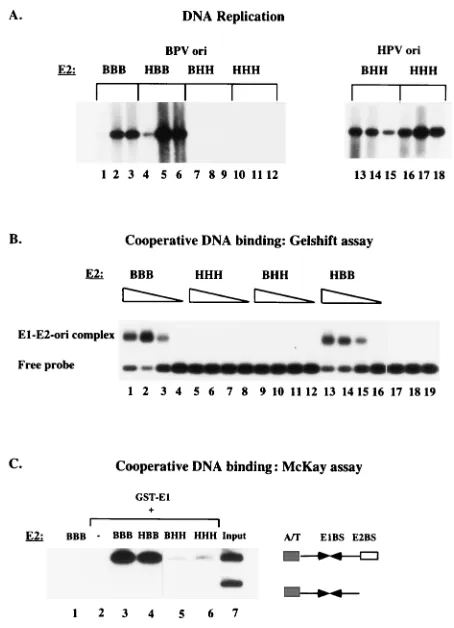
As expected, BPV E2 (Fig. 2A, lanes 1 to 3) supported replication of the BPV ori efficiently. The chimera containing the N-terminal half of HPV E2 and the C-terminal half of BPV (HBB) was equally active (lanes 4 to 6). However, the chimera containing the N-terminal half of BPV E2 and the C-terminal half from HPV E2 (BHH) failed to support replication at a detectable level and was as inactive as HPV-11 E2 (lanes 7 to
12). To ensure that BHH and HPV-11 E2 were expressed, replication assays were performed with the HPV-11 ori (lanes 13 to 18). Both BHH and HPV-11 E2 were capable of sup-porting replication of this origin, demonstrating that the pro-teins were well expressed and active.
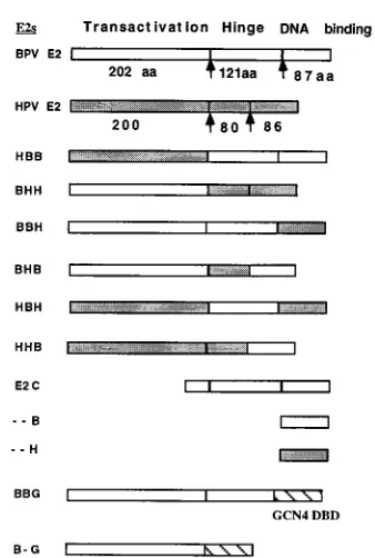
[image:3.612.350.519.84.335.2]We have previously demonstrated that the ability to form an E1-E2-ori complex is required for replication in vivo (41). This complex forms through cooperative binding of E1 and E2 to the ori, and the formation can be monitored by using a gel shift assay. At low concentrations of E1, where binding of E1 alone cannot be observed, addition of E2 gives rise to a complex containing both E1 and E2 (Fig. 1B). The E1-E2-ori complex forms at 20- to 50-fold-lower concentrations of E1 than is required for binding of E1 alone and requires an interaction between E1 and E2 (32, 41). E2 alone under these conditions does not give rise to detectable complex. To determine whether the chimeric E2 proteins could function for coopera-tive binding with E1, we overexpressed the different chimeric E2 proteins in E. coli and purified them by DNA affinity chro-matography. The different E2s were determined to be active for DNA binding by gel shift assays using an E2 binding site probe (data not shown). The different E2 proteins were then tested for cooperative binding with BPV E1. In this experi-ment, the E1 concentration was kept constant, and comparable 10-fold dilutions of the different purified E2 proteins were added. As we have previously demonstrated, BPV E2 readily formed the E1-E2-ori complex (Fig. 2B, lanes 1 to 3); likewise, the chimera HBB formed the E1-E2-ori complex, while both HPV-11 E2 (HHH) and the chimera BHH were inactive. FIG. 1. Schematic representation of the chimeric E2 proteins used in this study. The three domains of BPV and HPV-11 E2 are indicated as well as the switch points that were used to generate chimeric E2 proteins. The open boxes represent sequences derived from BPV E2, and the shaded boxes represent sequences originating from HPV-11 E2. The chimeric E2s were named by as-signing a letter (B or H) to each of the three different segments denoting whether the segment originated from BPV E2 or HPV-11 E2; for example, the chimera BHH contains the activation domain from BPV E2 and the hinge and DNA
binding domain (DBD) from HPV-11 E2. aa, amino acids.
on November 9, 2019 by guest
http://jvi.asm.org/
These results demonstrated that a correlation existed between the ability to form the E1-E2-ori complex and replication ac-tivity.
We wanted to use a different assay where we could use an internal standard for more accurate quantitation of in vitro association of E1 and E2. We therefore designed a precipita-tion assay that we refer to as a McKay assay (34). This assay was performed by incubating purified GST-E1 fusion protein with two labeled probes in the absence or presence of E2. The two probes are different in length, but both contain sequences from the BPV ori. The longer probe contains both the E1 binding site and the E2 binding site, while the shorter probe lacks the E2 binding site. The GST-E1 protein can be
recov-ered by using glutathione-agarose beads, and probes bound to GST-E1 can be analyzed by PAGE. Since the E1-E2-ori com-plex can form only on the probe containing both the E1 and the E2 binding sites whereas an E1-ori complex can form on both probes, the preferential recovery of the larger fragment con-taining both the E1 binding site and the E2 binding site gives a direct measure of the degree of cooperative binding. Figure 2C shows results of a McKay assay using the same E2 proteins that were used in the gel mobility shift assay in Fig. 2B. As expected, no DNA could be recovered with BPV E2 alone (Fig. 2C, lane 1). In the presence of GST-E1 but in the absence of added E2, the two probes could be recovered equally well but at a very low level (lane 2). Addition of BPV E2 resulted in an approximately 50-fold stimulation of the recovery of the larger probe that contains both the E1 and the E2 binding sites compared to the probe lacking the E2 binding site (lane 3), demonstrating cooperativity between E1 and E2. The chimera HBB cooperated for binding with E1 as well as BPV E2, while the chimera BHH (lane 5) and HPV-11 E2 (lane 6) failed to show significant cooperativity. These results were virtually identical to the results obtained in the gel shift assay above, demonstrating that the McKay assay could replace the gel shift assay. In the experiments described below, we have exclusively used the McKay assay to measure cooperative binding between E1 and E2.
The DNA binding/dimerization domain of BPV E2 is re-quired for complex formation with BPV E1 and for replication.
The results of these experiments indicated that the N-terminal transactivation domain from BPV E2 and HPV E2 had equiv-alent function and that the specificity that we could observe resided in the hinge and/or DNA binding domain. We there-fore generated further chimeras to identify the sequences im-portant for activity. These four new chimeras, BBH, BHB, HBH, and HHB (Fig. 1), were tested for replication and co-operative binding together with E1 as shown in Fig. 3. Two of FIG. 2. (A) Replication of the BPV minimal ori specifically requires the
C-terminal half of BPV E2. The chimeric E2 proteins BHH and HBB in addition to BPV E2 and HPV-11 E2 were tested for the ability to support replication in a transient replication assay together with BPV E1, using either the BPV ori (left panel) or the HPV 11 ori (right panel). Low-molecular-weight DNA was ex-tracted at 36, 60, and 84 h posttransfection, digested with HindIII and DpnI, and analyzed by Southern analysis using an ori probe. Replicated DNA migrates as a linear molecule of 2.8 kb. (B) Formation of the E1-E2-ori complex in vitro requires the C-terminal half of BPV E2. A gel mobility shift assay was used to determine the abilities of the different chimeric E2s to bind cooperatively with the BPV E1 protein. Equal quantities of the different E2s (and 10-fold dilutions) were incubated together with a constant amount of BPV E1 and a labeled BPV ori probe followed by cross-linking with glutaraldehyde and agarose gel electro-phoresis. Lanes 17 to 19 are controls; lane 17 contains 1 ng of E1 alone, lane 18 contains 1 ng of E2 alone, and lane 19 contains probe alone. (C) Cooperative binding of E1 and E2 to the BPV ori was measured in a McKay assay. GST-E1 in the absence or presence of different E2 proteins was incubated together with two BPV ori probes, one containing the E2 binding site and one lacking the E2 binding site, as illustrated schematically to the right. After incubation with glutathione-agarose beads and washes, probes bound to the glutathione-agarose beads were recovered and analyzed by PAGE. In the absence of GST-E1, no DNA was recovered (lane 1); in the presence of GST E1 alone, very low levels of probes were recovered (lane 2). The input ratio of the two probes is shown in lane 7.
FIG. 3. (A) The DNA binding domain of BPV is necessary for efficient replication of the BPV ori. BPV E2, HPV-11 E2, and four chimeric E2s (BBH, BHB, HBH, and HHB) were tested for the ability to replicate the BPV ori or the HPV ori. At 36, 60, and 84 h posttransfection, low-molecular-weight DNA was recovered and analyzed as described for Fig. 2A. (B) The DNA binding domain of BPV E2 is required for cooperative interaction with BPV E1 on the BPV ori. BPV E2, HPV-11 E2, and the four chimeric E2s (BBH, BHB, HBH, and HHB) were tested for cooperative DNA binding with BPV E1, using a McKay assay as described in the legend to Fig. 2C. The input ratio of the two probes is shown in lane 8. With no E2 added (lane 1), low but equal levels of the two probes were recovered.
on November 9, 2019 by guest
http://jvi.asm.org/
[image:4.612.64.291.67.381.2] [image:4.612.321.548.68.264.2]these chimeric E2 proteins, BHB and HHB (Fig. 3A, lanes 7 to 9 and 13 to 15), were capable of supporting replication from the BPV ori at roughly the same levels as the BPV E2 (lanes 1 to 3). The other two chimeras, BBH and HBH (lanes 4 to 6 and 10 to 12), were inactive. When we tested these chimeras for replication with the HPV ori (lanes 19 to 24), both supported replication of the HPV ori, demonstrating that these chimeric E2 proteins were expressed in active form.
The four chimeric E2 proteins, BBH, BHB, HBH, and HHB, were also expressed and purified from E. coli and used for in vitro binding assays (McKay assays) together with GST-E1 as described above (Fig. 3B). The same chimeras that were active for DNA replication, BHB and HHB, functioned as well as BPV E2 for cooperative binding (lanes 4 and 6), while the two chimeras that were inactive for replication, BBH and HBH, showed poor cooperative binding (lanes 3 and 5). These results demonstrated a very good correlation between the ability of a given E2 molecule to bind cooperatively with E1 and the ability to function in replication. Also, these results mapped very clearly the domain specifically required for rep-lication and cooperative binding on the BPV ori: all E2s that were active in these assay contained the DNA binding domain from BPV E2, demonstrating that the requirement for BPV E2 for replication from the BPV ori resides in the DNA binding domain of E2.
The DNA binding domain of BPV E2 protein can bind
co-operatively with BPV E1. The results obtained above clearly
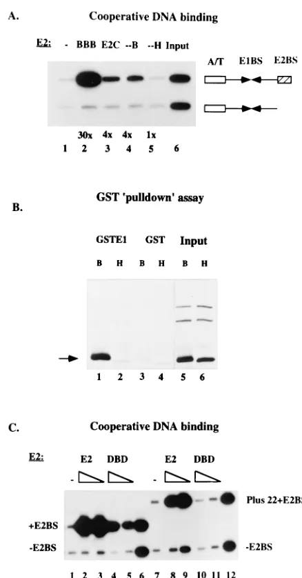
indicated that the DNA binding domain was required for the ability of BPV E2 to interact with BPV E1 in the cooperative binding assays as well as for activity in the replication assays. We therefore wanted to determine whether an interaction could be detected between the DNA binding domain of E2 and E1. We expressed in E. coli and purified three different trun-cated forms of E2 that all contain the DNA binding domain and tested these in McKay assays (Fig. 4A). These three forms are all inactive for support of replication in vivo (data not shown). We used E2C (amino acids 162 to 410), which is a naturally occurring form of BPV E2 that lacks the 161 N-terminal amino acids of the full-length protein (26, 28). We also expressed and purified an 85-amino-acid-long C-terminal fragment from both BPV E2 and HPV-11 E2 which constitutes the minimal DNA binding domain for these proteins. After quantitation of the different proteins by gel shift assays, we added equal quantities of DNA binding activity of the different E2s and compared the abilities of these different forms of E2 to bind cooperatively with E1, using the standard probes. As shown in Fig. 4A, full-length E2 stimulated binding of E1 to the E2 binding site-containing probe approximately 30-fold (lane 2). E2C and the DNA binding domain from BPV E2 (--B) stimulated binding of E1 less well but still significantly (fourfold) (lanes 3 and 4), while the DNA binding domain from HPV-11 E2 (--H) failed to stimulate binding of BPV E1 (lane 5). These results demonstrated that a specific interaction takes place between E1 and the DNA binding domain of BPV E2 which results in cooperative binding to the ori.
The DNA binding domain of BPV E2 interacts with GST-E1
in the absence of DNA.To determine if the interactions that we
observed in the cooperative DNA binding assays could be detected in the absence of DNA, we performed GST pull-down assays using GST-E1 and radiolabeled fragments of HPV E2 and BPV E2 generated by using a coupled in vitro transcription-translation system (Fig. 4B). We used two differ-ent E2 constructs encoding either the 85-amino-acid DNA binding domain from BPV E2 or the corresponding DNA binding domain fragment from HPV-11 E2 (lanes 5 and 6). The 85-amino-acid fragment containing the BPV E2 DNA
domain could be specifically recovered in this assay (lane 1), while the corresponding DNA binding domain fragment from HPV-11 E2 failed to associate at detectable levels with GST-E1 (lane 2). Neither fragment associated at appreciable FIG. 4. (A) The DNA binding domain alone can bind cooperatively with E1. A McKay assay was used to measure cooperative binding of four different E2 proteins to the BPV ori. The four E2 proteins were BPV E2 (BBB), E2C, which is a naturally occurring form of BPV E2 that lacks the N-terminal 161 amino acids, an 85-amino-acid C-terminal fragment that contains the DNA binding domain of BPV E2 (--B), and the corresponding fragment from HPV 11 E2 (--H). With no E2 added (lane 1), low levels of both probes were recovered. The input ratio of probes is shown in lane 6. (B) The DNA binding domain of BPV E2 can interact directly with BPV E1. Two different E2 fragments were trans-lated in vitro, using a coupled in vitro transcription-translation system. The fragments were the 85-amino-acid DNA binding domain from BPV E2 (B) and the corresponding fragment containing the DNA binding domain from HPV 11 E2 (H). These samples were incubated with either GST or GST-E1 protein and glutathione-agarose beads. After several washes, the labeled proteins recovered with the glutathione beads were analyzed by SDS-PAGE. The arrow indicates the position of the DNA binding domain translation products. (C) The BPV DNA binding domain contributes to interaction with E1 only when the binding sites for the two proteins are proximal. The full-length BPV E2 protein and the DNA binding domain from BPV E2 (DBD) were tested for the ability to bind cooperatively with E1, using a probe where the E2 binding site is proximal to the E1 binding site (1E2BS) and a probe where the E2 binding site is distal to the E1 binding site (Plus 221E2BS). In lanes 1 and 7, no E2 was added, and lanes 6 and 12 show the input probe mixtures.
on November 9, 2019 by guest
http://jvi.asm.org/
[image:5.612.323.542.64.483.2]levels with GST alone (lanes 3 and 4). These results are con-sistent with the notion that the cooperative DNA binding that we observe between BPV E1 and the DNA binding domain of BPV E2 is caused by a physical interaction between these two proteins and that the failure of the DNA binding domain from HPV-11 E2 to bind cooperatively with E1 is due to the lack of this physical interaction.
We next examined whether we could detect a qualitative difference between the interaction that requires the activation domain of E2 and the weaker interaction that only requires the DNA binding domain of E2. We compared full-length BPV E2 and the BPV DNA binding domain for the ability to bind cooperatively with BPV E1, using probes with different spacing between the E1 and E2 binding sites (Fig. 4C). In these exper-iments, we used three different probes. One probe which is present in all samples was generated from the BPV ori lacking the E2 binding site. The second probe was generated from the BPV ori and contains an E2 binding site in its natural proximal position (lanes 1 to 6). The third probe is identical to the second probe except that 22 bp (two turns of the helix) were inserted between the E1 and E2 binding sites (lanes 7 to 12). As we had observed previously, the full-length E2 stimulated binding of E1 well to both of the E2 binding site-containing probes (60- and 35-fold); compare lanes 2 and 3 to lanes 8 and 9). Interestingly, the DNA binding domain behaved differently: although substantial stimulatory activity could be observed in assays using the probe with the proximal E2 binding site (10-fold; lanes 4 and 5), the E2 DNA binding domain failed to show a significant effect on E1 binding in assays using the probe with the distal E2 binding site (lanes 10 and 11). These experiments indicated that while the DNA binding domain of BPV E2 appears to contribute to the interaction between E1 and E2 in the proximal position, the E2 DNA binding domain does not contribute to the interaction between E1 and E2 when the E2 binding site is in a distal position.
Increased distance between the E1 binding site and the E2 binding site alleviates the specific requirement for the BPV E2
DNA binding domain.The results described above indicated
that an important difference existed between the BPV and HPV oris and that this difference was responsible for the dif-fering requirements for E2. We knew that these differences were not due to the E2 binding site per se, since we could replace the low-affinity E2 binding site normally present in the BPV ori with a high-affinity E2 binding site from HPV-11 and obtain the same results as for the wt BPV ori. To determine what aspect of the two oris resulted in the observed differences, we constructed hybrid oris as shown in Fig. 5. We generated two chimeric origins with a switch point between the BPV E1 and E2 binding sites. The resulting chimeric origins, B/H ori and H/B ori, were both tested for replication with either BPV E2 or HPV-11 E2. The B/H ori was active with both BPV and HPV E2s, while the H/B ori was inactive with both E2s (data not shown). The B/H ori is identical to the BPV ori with two exceptions: the distance between the E1 and E2 binding is increased by 22 bp in the B/H ori, and the B/H ori also contains two E2 binding sites. This observation indicated that one or both of these differences were responsible for the ability of the B/H ori to function with HPV E2. Based on these results, we generated two artificial BPV oris that contained an insertion of 22 bp of random sequence between the E1 and E2 binding sites. One of these (Plus 22E2x2 ori) contains a duplicated E2 binding site, while the other (Plus 22E2x1 ori) contains a single E2 binding site. These artificial oris were tested for replication with all the different chimeric E2 proteins.
[image:6.612.65.291.70.275.2]As demonstrated in Fig. 6A, HPV E2 as well as all the chimeric E2s that were inactive for replication of the BPV ori could be restored to activity by using the Plus 22E2x2 ori. BBH (lanes 4 to 6), HBH (lanes 13 to 15), BHH (lanes 19 to 21), and HHH (lanes 22 to 24), which were inactive for replication of the BPV ori (Fig. 2A and 3A), all showed significant activity. Similar results were obtained using the Plus 22E2x1 ori (data FIG. 5. Schematic representation of hybrid oris, illustrating the organization
of the BPV and HPV-11 oris as well as two chimeric oris, B/H ori and H/B ori, and two artificial oris (Plus 22 E2x2 ori and Plus 22 E2x1 ori) that were gener-ated. The solid arrows represent the BPV E1 binding site; the open arrows repre-sents the HPV-11 E1 binding site. The hatched lines reprerepre-sents random sequence that was inserted as a spacer into the artificial oris. (See text for further details.)
FIG. 6. (A) The six chimeric E2 proteins as well as BPV E2 (BBB) and HPV 11 E2 were tested for the ability to support replication of the artificial ori Plus 22E2x2 ori as described for Fig. 2. (B) The same E2 proteins that were tested for replication in panel A were tested for cooperative DNA binding in a McKay assay. In this experiment, in addition to the two probes used previously, a larger probe corresponding to the Plus 22E2x2 ori with two E2 binding sites in the distal position was added. In the absence of added E2 added (lane 1), low levels of all three probes were recovered. The input ratio of probes is shown in lane 10.
on November 9, 2019 by guest
http://jvi.asm.org/
[image:6.612.323.545.72.297.2]not shown). These results demonstrated that the requirement for the BPV E2 DNA binding domain that we observed for replication of the BPV ori could be circumvented by increasing the distance between the E1 and E2 binding sites by 22 bp, providing an explanation for the different activities of the chi-meric E2s on the BPV and HPV oris. Interestingly, however, one of the chimeric E2s that was fully active for replication of the BPV ori (HHB) showed little or no activity for replication of this artificial ori. At present, we have no simple explanation for this result; we believe that in certain contexts, from a distal E2 binding site, the hinge region from BPV E2 may contribute to interactions with E1 (see also Fig. 7). Similarly, the chimera BHH was significantly less active than HHH on the Plus 22E2x2 ori than on the HPV ori, where these two E2s had similar activities (Fig. 2A). This result indicates that other properties of the ori, in addition to the distance between the E1 and E2 binding sites, play some role in determining the activity of a given E2 protein.
We also tested the behavior of the artificial ori in the McKay assay in combination with the different chimeric E2 proteins (Fig. 6B). In this assay we added, in addition to the standard probes, a larger probe corresponding to the artificial ori (Plus 22E2x2) for a direct comparison. The preference of the differ-ent chimeric E2s can be clearly seen: BPV E2 (BBB) and the chimeras BHB, HBB, and HHB all preferentially stimulated binding to the probe with the proximal site. HPV E2 (HHH), and the chimeras BBH, HBH, and BHH preferentially stimu-lated binding to the probe with the distal E2 binding sites.
When the results from the replication assay in Fig. 2A, 3A, and 6A were compared to the results from the McKay assays, the following relations emerged (Table 1): BPV E2 (BBB) stimulated E1 binding to both probes and was highly active for replication of both the proximal and distal site oris. BBH stimulated binding poorly to the proximal site probe but well to the probe with the distal E2 binding site and was active for replication with the distal site ori only. BHB stimulated binding well to the proximal site probe and poorly to the distal site probe and was highly active for replication of the proximal site ori, with low activity for replication of the distal site ori. HBB stimulated binding to the proximal site probe but not to the distal site probe and was active for replication for both the proximal and distal site oris. HBH stimulated binding poorly to the proximal site probe but showed significant stimulation to the distal site probe and was active for replication only with the distal site ori. HHB stimulated binding from the proximal site only and was active only for replication of the proximal site ori. BHH stimulated binding only from the distal site and was active only for replication of the distal site ori. Finally, HHH stimulated binding poorly to the proximal site probe but func-tioned well for the distal site probe and was active for
replica-tion only for the distal site ori. Taken together, these results demonstrated a very good correlation between the ability of a given E2 protein to function for replication and to bind coop-eratively with E1. Furthermore, these results also supported the notion that increasing the spacing between the E1 and E2 binding sites relaxes the requirement for a specific E2: only a single chimeric E2 protein failed to support replication from the artificial ori with the distal E2 binding sites.
The activation domain of BPV E2 is sufficient for interaction
with E1 and for replication.The results presented above
[image:7.612.362.512.68.280.2]indi-cated that the activation domain of E2 alone might suffice for the interaction with E1 provided that the E2 binding site was in the distal position. To address this question, we generated two fusion proteins where we replaced either the DNA binding domain only (BBG) or the hinge and the DNA binding domain (B-G) in BPV E2 with the DNA binding domain from the yeast transcription factor GCN4 (25) (Fig. 1). We also constructed an ori where we replaced the E2 binding site in the distal position (Plus 22E2x1 ori) with a binding site for GCN4. It has previously been demonstrated that an in vitro translated E2-GAL4 fusion protein can function for cooperative binding with E1 (59). However, our previous experience indicated that GAL4-E2 fusions resulted in poorly expressed proteins with low activity when expressed in mammalian cells. Since in vivo replication assays were our primary objective, we instead chose the GCN4 protein as a fusion partner. We decided to use the GCN4 protein because the overall architecture is similar to that of E2. The DNA binding domain is located at the extreme C terminus of the protein, and the DNA binding and dimer-ization domain is similar in size to that in E2. As shown in Fig. 7, the two fusion proteins, BBG and B-G were both capable of FIG. 7. (A) The activation domain of E2 is sufficient for cooperative binding and DNA replication. Two hybrid E2 proteins, BBG, which contains the activa-tion domain and hinge from BPV E2 linked to the DNA binding domain of the yeast protein GCN4, and B-G, which has the activation domain of BPV E2 linked directly to the GCN4 DNA binding domain, were tested for the ability to support replication in the transient replication assay. Expression vectors for these two proteins were transfected into CHO cells together with an ori plasmid containing either an E2 binding site or a GCN4 binding site in the distal position (122) and tested for replication as described for Fig. 2. (B) The hybrid E2 protein B-G (see above) was tested in the cooperative binding assay for its ability to bind coop-eratively with E1, using a probe that contains a GCN4 binding site in the distal position (122). As a control for the specificity of the probe, BPV E2 was added (lanes 4 and 5). In lane 1, no E2 or GCN 4 fusion was added. The input ratio of probes is shown in lane 6.
TABLE 1. Position of the E2 binding sitea
Construct Proximal Distal
Binding Replication Binding Replication
BBB 36 Y 14 Y
BBH 3 N 13 Y
BHB 23 Y 2 (Y)
HBB 16 Y 4 Y
HBH 4 N 7 Y
HHB 12 Y 2 N
BHH 2.5 N 10 (Y)
HHH 2 N 12 Y
aReplication is indicated as follows: Y, yes; N, no; (Y), low-level replication.
on November 9, 2019 by guest
http://jvi.asm.org/
[image:7.612.59.301.80.190.2]supporting replication of the origin containing the GCN4 bind-ing site (Fig. 7A, lanes 4 to 6 and 10 to 12) but not of an origin containing E2 binding sites (lanes 1 to 3 and 7 to 9). These results demonstrated that the interaction between the activa-tion domain of E2 and E1 is sufficient to support replicaactiva-tion activity and provide further evidence that tethering of E2 to the DNA is required for activity, since these fusion proteins were inactive in the absence of a GCN4 binding site. We also tested these fusion proteins for the ability to interact with E1 in vitro by using the McKay assay. As shown in Fig. 7B, the B-G fusion protein containing the activation domain of E2 linked to the GCN 4 DNA binding domain showed strong cooperative binding with the E1 protein on a probe containing a distal GCN4 binding site (lanes 2 and 3). As expected, E2 had no effect on E1 binding on this probe (lanes 4 and 5). We also tested BBG in this assay and obtained results similar to those observed with B-G (data not shown).
DISCUSSION
The results presented above provide a great deal of infor-mation both about E2 and about the interaction between E1 and E2. All chimeric E2 proteins that we have tested, without exception, are active for replication under some conditions, providing good support for the idea that E2 is a modular protein, as has been suggested based on structure predictions. The results also indicate that the domains in the different E2s have activities that are structurally and functionally compati-ble. Furthermore, our results constitute very strong evidence that the interaction between the E1 and E2 proteins, as de-tected by cooperative DNA binding, is a requirement for rep-lication in vivo. We observe a very good correlation between replication activity in vivo and cooperative binding in vitro for a large number of ori constructs and chimeric E2s. In addition, the simultaneous gain, and loss, of both replication activity and cooperative binding that we observe when we replace the HPV DNA binding domain with the BPV DNA binding domain, or vice versa, argues strongly that the cooperative binding be-tween E1 and E2 is a crucial activity that is required for DNA replication. While this finding does not rule out that E2 has other activities, such as interaction with RPA, or acts to pre-vent nucleosome formation, it certainly indicates that the crit-ical activity of E2 for DNA replication is its interaction with E1. A reasonable possibility is that the interaction with E1 is the only essential activity that is provided by E2 for DNA replication. We have recently demonstrated that the interac-tion between E1 and E2 results in substantially altered DNA binding properties of E1 (41) and, furthermore, that E1 in the presence of E2 binds to the origin of replication in a form that otherwise appears to lack DNA binding activity (42). Both of these findings are consistent with a role for E2 primarily af-fecting the DNA binding activity of E1, indicating similarities with the role of NF-1 for adenovirus replication (5, 36, 37).
Cooperative binding of E1 and E2 to an ori thus is a very good predictor of replication activity, at least on the BPV ori. In contrast, because of the dependence on the position of the E2 binding site, interaction between E1 and E2 in the absence of DNA is not a good predictor for activity in DNA replication. HPV-11 E2 has been shown to interact with BPV E1 in im-munoprecipitation assays (61) but is completely inactive for cooperative binding and for replication together with BPV E1 in assays using the BPV ori. However, HPV-11 E2 is active for cooperative binding and replication together with BPV E1 on an ori with a distal E2 binding site. Thus, interaction between E1 and E2 in the absence of DNA reveals a potential for a productive interaction, but the binding site arrangement
de-termines whether this potential is realized such that the two proteins can interact productively for cooperative DNA bind-ing, which in turn appears to be a requirement for DNA rep-lication.
Our results present the first positive evidence that the part of E2 specifically required for its activity in replication is the N-terminal activation domain. This 200-amino-acid fragment, which previously has been shown to be necessary and sufficient for cooperative binding with E1 in vitro (59), is also necessary and sufficient for replication in vivo when tethered to DNA through an unrelated DNA binding domain. The activation domain shows no apparent specificity, both the activation do-mains from HPV-11 E2 and BPV E2 interact with BPV E1 and support replication at comparable levels. In contrast, the hinge and the DNA binding domain of E2 are not specifically re-quired for replication. A different DNA binding/dimerization domain can function very well, and a mutant that lacks the hinge is still replication competent, albeit at reduced levels (Fig. 7). However, the DNA binding domain is solely respon-sible for the differential abilities of BPV and HPV-11 E2 to function for replication of the BPV ori. This study also very clearly reaffirms our previous finding that tethering of E2 to the ori is a requirement for replication (54). The E2-GCN4 fusion protein is inactive in the absence of the cognate binding site (Fig. 7). Furthermore, the crucial importance of the posi-tion of the E2 binding sites relative to the E1 binding site strongly supports a requirement for tethering of E2 to DNA. Our objective in initiating this study was to utilize the dif-ferential activities of HPV-11 E2 and BPV E2 to map, with the use of chimeric E2 proteins, the regions of interaction between BPV E1 and BPV E2. We expected to be able to determine what sequences in the activation domain of E2 were involved in interactions with E1. This expectation was based on results from several laboratories using various assays that indicated that the N-terminal portion of E2 was capable of interaction with E1 (4, 7, 22, 35, 38, 39, 59). Our data agree with the existence of an interaction domain in the N terminus of E2. Furthermore, we demonstrate that this same part of E2 is necessary and sufficient for E2 activity in replication. We have found, however, that the interaction between the E1 and E2 proteins is more complex than we had anticipated; in fact, we have determined that the basis for the differential specificities of BPV E2 and HPV-11 E2 does not reside in the activation domain. On the contrary, the HPV and BPV E2 activation domains are equally capable of interacting with BPV E1. The differential specificities of these different types of E2 instead originate in the DNA binding domains of the two proteins. The DNA binding domain of BPV E2 is specifically required for cooperative binding of E1 and E2 to the BPV ori and can direct the interaction between E1 and either the BPV E2 or HPV E2 activation domains. The BPV E2 DNA binding do-main, contrary to the conclusion reached by, for example, Winokur et al. (59), is also independently capable of interact-ing with BPV E1 in the presence and absence of DNA, while the DNA binding domain of HPV E2 fails to interact in either assay. Our results thus demonstrate that BPV E2 interacts with E1 via two separate and different interactions involving differ-ent domains of E2. As will be discussed below, these two separate interactions may have different functions.
An important factor in the interaction between E1 and E2 is the relative positions of the binding sites for the two proteins. Our experiments show that the HPV ori arrangement, where the E1 and E2 binding sites are separated by 22 bp, is the arrangement with the lowest stringency; all E2s can function on this type of ori, and when placed in this position as a GCN4 fusion protein, the activation domain alone can support
on November 9, 2019 by guest
http://jvi.asm.org/
cation and interaction with E1. The arrangement found in the BPV ori, where the E1 and E2 binding sites are proximal, is more restrictive: exclusively E2 proteins with the DNA binding domain from BPV E2 are functional with this ori. The proxi-mal position of the E2 binding site is also required for coop-erative binding between E1 and the DNA binding domain of BPV E2.
Our results show that under certain conditions, the minimal requirement for E2 for replication as well as for cooperative binding can be provided by a fusion protein containing only the activation domain of E2 fused to the DNA binding domain from GCN4 (Fig. 7). This result demonstrates that the activa-tion domain of E2 contains all of the sequences that are re-quired for a productive in vivo interaction with E1. Under these same conditions, the activation domains of BPV E2 and HPV-11 E2 (compare, for example, BBB and HBB in Fig. 2) appear to have equivalent activities, indicating that the se-quences in the two activation domains that take part in the interaction with E1 are conserved. Thus, the explanation for the failure of HPV-11 E2 to function for replication of the minimal BPV ori and to interact with E1 must be that although the activation domain of HPV-11 E2 is nominally capable of interaction with E1, this interaction does not take place. One way to overcome this inhibition is by increasing the distance between the E1 and E2 binding sites to 22 bp (two turns of the helix). The increased distance allows interaction between E1 and the activation domain of HPV-11 E2 as well as replication. Thus, a likely explanation for the inability of HPV-11 E2 to interact with E1 on the BPV ori is that sterical constraints prevent the activation domain of HPV-11 E2 from contacting E1 when the binding sites for the two proteins are proximal. Most likely, the increased flexibility that results from the in-creased distance between the sites alleviates this sterical prob-lem and allows interaction between the E2 activation domain and E1.
A second way to overcome this inhibition is by transferring the DNA binding domain from BPV E2 to HPV-11 E2. The resulting protein (HHB) is fully capable of interaction with E1 when the binding sites are proximal (BPV ori), while the re-ciprocal chimera (BBH) becomes inactive (see Fig. 3). It is reasonable to suggest that the BPV E2 DNA binding domain functions similarly to the increased distance, i.e., to alleviate steric hindrance in the interaction between E1 and E2. There are several possible mechanisms through which the E2 DNA binding domain could exert this effect. The simplest possibility is that the close proximity of the E1 and E2 binding sites in the BPV ori affects cooperative binding by preventing simulta-neous binding of the two proteins to DNA. If, for example, the size or shape of the DNA binding domain from HPV-11 E2 is different from that of BPV E2, such that simultaneous binding of E1 and HPV-11 E2 to the proximal sites is prevented, this would explain the inability of HPV E2 to bind cooperatively with E1. Unfortunately, because E1 fails to bind DNA in mo-nomeric form in the absence of an interaction with E2, it is difficult to test this possibility biochemically. However, the difference in size between the two DNA binding domains is unlikely to be the whole explanation; an increased distance between the E1 and E2 binding sites by 10 bp still does not allow cooperative binding between E1 and HPV-11 E2 but functions well for cooperative binding between E1 and BPV E2 (48). Thus, we believe that the interactions that we can observe between E1 and the BPV DNA binding domain, but not the HPV E2 DNA binding domain, in both cooperative DNA binding assays (McKay assays) and GST pull-down as-says are highly significant. This interaction, which is weaker than the interaction between E1 and the E2 activation domain
and does not allow replication by itself, might serve to alter the conformation of E1, of E2, or of both proteins to allow a productive interaction between E1 and the E2 activation do-main. In this scenario, the interaction between E1 and the DNA binding domain of E2 could act as a trigger or switch to facilitate the interaction between E1 and the activation domain of E2.
Based on the results we have described here, we propose a simple model for how the E1 and E2 proteins interact with each other in different combinations and on different oris. This model (Fig. 8) accommodates and explains the majority of our experimental findings. In Fig. 8A, which represents the situa-tion on the BPV ori (a proximal E2 binding site) and BPV E1 and E2 proteins, we envision that the E1 and E2 proteins can bind to their respective sites. The interaction between E1 and the DNA binding domain of E2 results in a conformational change in E1 and/or E2 which allows the interaction between E1 and the activation domain of E2, resulting in strong coop-erative binding and a stable complex. In Fig. 8B, when BPV E2 is replaced by HPV-11 E2, the interaction between E1 and the DNA binding domain of E2 cannot take place, the required conformational change does not occur, and the activation do-main fails to interact with E1. In Fig. 8C, however, when the E2 binding site is moved to the distal position (HPV-type ori), the steric hindrance that is observed when the E1 and E2 binding sites are proximal is circumvented and the flexibility of the intervening DNA allows positioning of the E2 activation do-main relative to E1 such that interaction between the two proteins can take place without requiring a conformational change.
Our approach to study the interactions between the E1 and E2 proteins and the relationship between these interactions and DNA replication has been to use chimeric E2 proteins in combination with chimeric origins of replication. This ap-proach has allowed us to identify some of the constraints af-FIG. 8. A model for the interactions between E1 and E2. See text for de-tails.
on November 9, 2019 by guest
http://jvi.asm.org/
[image:9.612.322.546.67.328.2]fecting the interactions between the E1 and E2 proteins such that we with few exceptions, can predict the behavior of E1 and E2 in the context of a given ori. We believe that this relation-ship between E2 structure and ori structure will be of impor-tance in further defining the interactions between the E1 and E2 proteins and to determine what molecular consequences result from these interactions.
ACKNOWLEDGMENTS
We thank J. Sedman and C. Sanders for critical reading of the manuscript.
This work was supported by National Institutes of Health grant CA 13106 to A.S.
REFERENCES
1. Androphy, E. J., D. R. Lowy, and J. T. Schiller. 1987. Bovine papillomavirus E2 trans-activating gene product binds to specific sites in papillomavirus DNA. Nature (London) 325:70–73.
2. Arndt, K., and G. R. Fink. 1986. GCN 4 protein, a positive transcription factor in yeast, binds general control promoters at all 59TGACTC 39 se-quences. Proc. Natl. Acad. Sci. USA 83:8516–8520.
3. Bennet-Cook, E. R., and J. A. Hassel. 1991. Activation of polyomavirus DNA replication by yeast Gal 4 is dependent on its transcriptional activation domains. EMBO J. 10:959–969.
4. Benson, J. D., and P. M. Howley. 1995. Amino-terminal domains of the bovine papillomavirus type 1 E1 and E2 proteins participate in complex formation. J. Virol. 69:4364–4372.
5. Bosher, J., E. C. Robinson, and R. T. Hay. 1990. Interactions between the adenovirus type 2 DNA polymerase and the DNA binding domains of nu-clear factor I. New Biol. 2:1083–1090.
6. Blitz, I., and L. Laiminis. 1991. The 68-kilodalton E1 protein of bovine papillomavirus is a DNA binding phosphoprotein which associates with the E2 transcriptional activator in vitro. J. Virol. 65:649–656.
7. Brokaw, J. L., M. Blanco, and A. A. McBride. 1996. Amino acids critical for the functions of the bovine papillomavirus type 1 E2 transactivator. J. Virol. 70:23–39.
8. Challberg, M., and T. Kelly. 1989. Animal virus DNA replication. Annu. Rev. Biochem. 58:671–718.
9. Cheng, I., and T. Kelly. 1989. Transcriptional activator nuclear factor I stimulates the replication of SV40 minichromosomes in vivo and in vitro. Cell 59:541–551.
10. Cheng, I., J. I. Workman, R. E. Kingston, and T. J. Kelly. 1992. Regulation of DNA replication in vitro by the transcriptional activation domain of GAL4-VP16. Proc. Natl. Acad. Sci. USA 89:589–593.
11. Chiang, C.-M., M. Ustav, A. Stenlund, T. F. Ho, T. R. Broker, and L. T. Chow.1992. Viral E1 and E2 proteins support replication of homologous and heterologous papillomaviral origins. Proc. Natl. Acad. Sci. USA 89: 5799–5803.
12. Del Vecchio, A. M., H. Romanczuk, P. M. Howley, and C. C. Baker. 1992. Transient replication of human papillomavirus DNAs. J. Virol. 66:5949– 5958.
13. DePamphilis, M. L. 1993. Eukaryotic DNA replication: anatomy of an ori-gin. Annu. Rev. Biochem. 62:29–63.
14. Ferguson, M. K., and M. Botchan. 1996. Genetic analysis of the activation domain of bovine papillomavirus protein E2: its role in transcription and replication. J. Virol. 70:4193–4199.
15. Gillette, T. G., M. Lusky, and J. A. Borowiec. 1994. Induction of structural changes in the bovine papillomavirus type 1 origin of replication by the viral E1 and E2 proteins. Proc. Natl. Acad. Sci. USA 91:8846–8850.
16. Giri, I., and M. Yaniv. 1988. Structural and mutational analysis of E2 trans-activating proteins of papillomaviruses reveals three distinct functional do-mains. EMBO J. 7:2823–2829.
17. Grossel, M. J., F. Sverdrup, D. E. Breiding, and E. J. Androphy. 1996. Transcriptional activation function is not required for stimulation of DNA replication by bovine papillomavirus type 1 E2. J. Virol. 70:7264–7269. 18. Guo, Z.-S., and M. L. DePamphilis. 1992. Specific transcription factors
stimulate simian virus 40 and polyomavirus origins of DNA replication. Mol. Cell. Biol. 12:2514–2524.
19. Hawley-Nelson, P., E. J. Androphy, D. R. Lowy, and L. T. Schiller. 1988. The specific DNA recognition sequence of the bovine papillomavirus E2 protein is an E2-dependent enhancer. EMBO J. 7:525–531.
20. He, Z., B. T. Brinton, J. Greenblatt, J. A. Hassel, and C. L. Ingles. 1993. The transactivator proteins VP16 and Gal 4 bind replication factor A. Cell 73: 1223–1232.
21. Heintz, N. H. 1992. Transcription factors and the control of DNA replica-tion. Curr. Opin. Cell Biol. 4:459–567.
22. Hibma, M., K. Raj, S. J. Ely, M. Stanley, and L. Crawford. 1995. The interaction between human papillomavirus type 16 E1 and E2 proteins is
blocked by an antibody to the N-terminal region of E2. Eur. J. Biochem. 229:517–525.
23. Holt, S. E., G. Schuller, and V. G. Wilson. 1993. DNA binding specificity of the bovine papillomavirus E1 protein is determined by the sequences con-tained within an 18-base-pair inverted repeat element at the origin of rep-lication. J. Virol. 68:1094–1102.
24. Holt, S. E., and V. G. Wilson. 1995. Mutational analysis of the 18-basepair inverted repeat element at the bovine papillomavirus origin of replication: identification of critical sequences for E1 binding and in vivo replication. J. Virol. 69:6525–6532.
25. Hope, I. A., and K. Struhl. 1986. Functional dissection of a eukaryotic transcriptional activator protein, GCN4 of yeast. Cell 46:885–894. 26. Hubbert, N. L., J. T. Schiller, D. R. Lowy, and E. J. Androphy. 1988. Bovine
papilloma virus-transformed cells contain multiple E2 proteins. Proc. Natl. Acad. Sci. USA 85:5864–5868.
27. Kadonaga, J. T. 1991. Purification of sequence-specific binding proteins by DNA affinity chromatography. Methods Enzymol. 208:10–23.
28. Lambert, P. F., N. L. Hubbert, P. M. Howley, and J. T. Schiller. 1989. Genetic assignment of multiple E2 gene products in bovine papillomavirus-transformed cells. J. Virol. 63:3151–3154.
29. Li, R., and M. Botchan. 1993. The acidic transcriptional activation domains of VP16 and p53 bind the cellular replication protein A and stimulate in vitro BPV-1 DNA replication. Cell 73:1207–1221.
30. Li, R., and M. Botchan. 1994. Acidic transcription factors alleviate nucleo-some-mediated repression of DNA replication of bovine papillomavirus type 1. Proc. Natl. Acad. Sci. USA 91:7051–7055.
31. Lusky, M., and E. Fontane. 1991. Formation of the complex of bovine papillomavirus E1 and E2 proteins is modulated by E2 phosphorylation and depends upon sequences within the carboxyl terminus of E1. Proc. Natl. Acad. Sci. USA 88:6363–6367.
32. Lusky, M., J. Hurwitz, and Y.-S. Seo. 1994. The bovine papillomavirus E2 protein modulates the assembly but is not stably maintained in a replication-competent multimeric E1-replication origin complex. Proc. Natl. Acad. Sci. USA 91:8895–8899.
33. McBride, A. A., H. R. Romanczuk, and P. M. Howley. 1991. The papilloma-virus E2 regulatory proteins. J. Biol. Chem. 266:18411–18414.
34. McKay, R. D. G. 1981. An immuno assay for the interaction between an SV40 T antigen related protein and DNA. J. Mol. Biol. 145:471–482. 35. Mohr, I. J., R. Clark, S. Sun, E. J. Androphy, P. MacPherson, and M. R.
Botchan.1990. Targeting the E1 replication protein to the papillomavirus origin of replication by complex formation with the E2 transactivator. Sci-ence 250:1694–1699.
36. Mul, Y. M., C. P. Verrijzer, and P. C. van der Vliet. 1990. Transcription factors NF I and NF III/oct-1 function independently, employing different mechanisms to enhance adenovirus DNA replication. J. Virol. 64:5510–5518. 37. Mul, Y. M., and P. C. van der Vliet. 1992. Nuclear factor I enhances ade-novirus DNA replication by increasing the stability of a preinitiation com-plex. EMBO J. 11:751–760.
38. Piccini, A., A. Storey, P. Massimi, and L. Banks. 1995. Mutations in the human papillomavirus type 16 E2 protein identify multiple regions of the protein involved in binding to E1. J. Gen. Virol. 76:2909–2913.
39. Sakai, H., T. Yasugi, J. D. Benson, J. J. Dowhanick, and P. M. Howley. 1996. Targeted mutagenesis of the human papillomavirus type 16 E2 transactiva-tion domain reveals separable transcriptransactiva-tional activatransactiva-tion and DNA replica-tion funcreplica-tions. J. Virol. 70:1602–1611.
40. Schagger, H., and G. von Jagow. 1987. Tricine-sodium dodecyl sulfate-polyacrylamide gel electrophoresis for the separation of proteins in the range from 1 to 100 kDa. Anal. Biochem. 166:368–379.
41. Sedman, J., and A. Stenlund. 1995. Co-operative interaction between the initiator E1 and the transcriptional activator E2 is required for replication of bovine papillomavirus in vivo and in vitro. EMBO J. 14:6218–6228. 42. Sedman, J., and A. Stenlund. 1996. The initiator protein E1 binds to the
bovine papillomavirus origin of replication as a trimeric ring-like structure. EMBO J. 15:5085–5092.
43. Sedman, T., J. Sedman, and A. Stenlund. 1997. Binding of the E1 and E2 proteins to the origin of replication of bovine papillomavirus. J. Virol. 71: 2887–2896.
44. Seo, Y.-S., F. Muller, M. Lusky, and J. Hurwitz. 1993. Bovine papillomavirus (BPV)-encoded E1 protein contains multiple activities required for BPV DNA replication. Proc. Natl. Acad. Sci. USA 90:702–706.
45. Seo, Y.-S., F. Muller, M. Lusky, E. Gibbs, H.-Y. Kim, B. Phillips, and J. Hurwitz.1993. Bovine papilloma virus (BPV)-encoded E2 protein enhances binding of E1 protein to the BPV replication origin. Proc. Natl. Acad. Sci. USA 90:2865–2869.
46. Spalholz, B. A., Y. C. Yang, and P. M. Howley. 1985. Transactivation of a bovine papillomavirus transcriptional regulatory element by the E2 gene product. Cell 42:183–191.
47. Spalholz, B. A., A. A. McBride, T. Sarafi, and J. Quintero. 1993. Binding of bovine papillomavirus E1 to the origin is not sufficient for DNA replication. Virology 193:201–212.
48. Stenlund, A., and M. Berg. Unpublished observation.
on November 9, 2019 by guest
http://jvi.asm.org/
49. Stillman, B. 1989. Animal virus DNA replication. Annu. Rev. Cell Biol. 5:197–245.
50. Storey, A., A. Piccini, P. Massimi, V. Bouvard, and L. Banks. 1995. Muta-tions in the human papillomavirus type 16 identify a region of the protein involved in binding to E1 protein. J. Gen. Virol. 76:819–826.
51. Studier, F., and B. Moffatt. 1986. Use of the bacteriophage T7 RNA poly-merase to direct selective high-level expression of cloned genes. J. Mol. Biol. 189:113–130.
52. Thorner, L., D. Lim, and M. Botchan. 1993. DNA binding domain of bovine papillomavirus type 1 E1 helicase: structural and functional aspects. J. Virol. 67:6000–6014.
53. Tanaka, M., and W. Herr. 1990. Differential transcriptional activation by Oct-1 and Oct-2: interdependent activation domains induce Oct-2 phosphor-ylation. Cell 60:375–386.
54. Ustav, E., M. Ustav, P. Szymanski, and A. Stenlund. 1993. The bovine papillomavirus origin of replication requires a binding site for the E2 tran-scriptional activator. Proc. Natl. Acad. Sci. USA 90:898–902.
55. Ustav, M., and A. Stenlund. 1991. Transient replication of BPV-1 requires two viral polypeptides encoded by the E1 and E2 open reading frames. EMBO J. 10:449–457.
56. Ustav, M., E. Ustav, P. Szymanski, and A. Stenlund. 1991. Identification of
the origin of replication of bovine papillomavirus and characterization of the viral origin recognition factor E1. EMBO J. 10:4321–4329.
57. Wilson, V. G., and J. Ludes-Meyers. 1991. A bovine papillomavirus E1-related protein binds specifically to bovine papillomavirus DNA. J. Virol. 65:5314–5322.
58. Winokur, P. L., and A. A. McBride. 1992. Separation of the transcriptional activation and replication functions of the bovine papillomavirus-1 E2 pro-tein. EMBO J. 11:4111–4118.
59. Winokur, P. L., and A. A. McBride. 1996. The transactivation an DNA binding domain of the BPV-1 E2 protein have different roles in cooperative origin binding with the E1 protein. Virology 221:44–53.
60. Yang, L., R. Li, I. Mohr, R. Clark, and M. Botchan. 1991. Activation of BPV-1 replication in vitro by the transcription factor E2. Nature (London) 353:628–633.
61. Yang, L., I. Mohr, R. Li, T. Nottoli, S. Sun, and M. Botchan. 1991. Tran-scription factor E2 regulates BPV-1 DNA replication in vitro by direct protein-protein interaction. Cold Spring Harbor Symp. Quant. Biol. 56:335– 346.
62. Yang, L., I. Mohr, E. Fouts, D. A. Lim, M. Nohaile, and M. Botchan. 1993. The E1 protein of the papillomavirus BPV-1 is an ATP dependent DNA helicase. Proc. Natl. Acad. Sci. USA 90:5086–5090.