Evaluation of dried blood spots as a feasible alternative to plasma
for detection, quantification and genotyping of hepatitis C virus:
A pilot study
Dissertation submitted in partial fulfilment of the rules and
regulations for the M.D. (Branch-IV Microbiology) examination
ACKNOWLEDGEMENT
Firstly, I would like to thank God for his blessings and providing me the strength to
endure the difficult times.
I extend my sincere gratitude to my guide, Dr. Priya Abraham, Professor, Department
of Clinical Virology, for guiding me through the course of my dissertation. She has been
a source of constant encouragement. This study would not have been possible without
her support and motivation. Thank you for inspiring me to strive harder in times of
difficulty. The dedication, passion for your field of work and the immaculate attention
to detail that you possess are certain qualities that I would like to inculcate in myself.
Thank you for providing me the opportunity to learn from you and for shepherding me
through the duration of my study.
I would like to thank Dr. Rajesh Kannangai, Professor and Head of Department of
Clinical Virology for giving me permission to undertake my study in his department
and for the support and encouragement provided.
I am grateful to Dr. V. Balaji, Professor and Head of Department of Clinical
Microbiology for the motivation, concern and compassion to complete this study.
I am thankful to Dr. John G. Fletcher, for his valuable suggestions and insight for the
betterment of the work.
I am obliged to Mr. Raghavendran A and Dr. Visalakshi J. for their enormous help and
support in various stages of my study.
I am indebted to Mrs. Kalaivani, Mr. Suresh, Mr. Deenadhayalan and Mr. John Calvin
a patient that could be recruited in my study, for teaching me the details of the molecular
techniques and for helping me in setting up the experiments.
Lastly, I thank my parents and brother for being with me in the times of need and being
the constant source of strength. It is only because of them that I was able to do what I
have done.
0
Contents
INTRODUCTION ... 1
AIMS AND OBJECTIVES ... 4
REVIEW OF LITERATURE ... 5
MATERIALS AND METHODS ... 43
RESULTS ... 73
DISCUSSION ... 92
CONCLUSION ... 100
1 INTRODUCTION
Hepatitis C virus (HCV) is an enveloped, positive sense RNA virus of 9.6 kb belonging
to family Flaviviridae and genus Hepacivirus (1,2). It has a single open reading frame
(ORF), which encodes 3 structural proteins required for formation of the virus and 5
non-structural proteins which serve as enzymes required for viral replication (3).
Global prevalence of HCV is about 1%, with highest prevalence of 2.3% in East
Mediterranean region (4). India has a prevalence of 0.09-2.02%, with about 6-11 million
people infected with HCV (5,6).
There are 7 confirmed genotypes of HCV and they are further classified into various
subtypes (7). Genotype 1 predominates globally (8), while genotype 3 infects the
majority in the Indian subcontinent (9).
Being a blood borne virus, hepatitis C is transmitted mainly by sharing of contaminated
needles among IV drug abusers (10) and transfusion of blood or blood products.
Perinatal, occupational and sexual are other modes of transmission, though less frequent
(11).
It is a major cause of morbidity and mortality, with only about 15-25% of acute
infections leading to resolution and the rest progressing to chronicity (12). It can
multiply and form a niche for itself in the body and remain asymptomatic for years,
with the symptoms appearing only when histological damage has occurred. Chronic
liver disease, hepatic cirrhosis and hepatocellular carcinoma are the long term
complications of this disease (13,14). Annually, hepatic cirrhosis accounts for more
number of deaths (720,000 deaths) followed by hepatocellular carcinoma (470,000
2
Hepatitis C virus infection can be diagnosed by Rapid Diagnostic Tests (RDTs) and
serological tests such as Enzyme-Linked Immunosorbent Assays (ELISA) (15) and
Chemiluminescent Microparticle Immunoassay (CMIA), which mainly detect the
antibody response to such an infection. Serological tests, particularly RDTs are
performed in low resource settings because of their wider availability. Nevertheless,
these tests are not good predictors of current (active) infection as the antibodies begin
to rise only later in the course of infection and persist for a long time.
Molecular tests that detect and quantify HCV RNA are the gold standard, which if
positive imply an active infection. They also help in monitoring response to therapy as
the viral RNA decreases with effective therapy (16).
Recently, HCV core antigen detection has also been employed as an alternative to HCV
RNA quantification as it rises early in the infection and correlates well with HCV RNA
levels, giving a better idea of level of replication of the virus or its decline (17,18).
Genotyping of HCV is required, as currently the treatment and duration of therapy
varies with different genotypes (19).
PEG-IFN-α and ribavirin combination therapy were the preferred treatments for HCV
a few years ago. With the advent of directly acting antivirals (DAAs), there is a
possibility of cure. DAAs have shown an efficacy that is almost double to that of the
previous regimens (20). Now, pangenotypic drugs such as sofosbuvir and velpatasvir
have been licensed for use (21).
Many people living in remote areas are still not aware that they have HCV infection and
there is a dire need for better sampling and transportation of samples from such distant
3
Globally, only about 1 million of the total 80 million population affected with HCV
have access to treatment (22). DAAs are cost-effective, efficacious medications with
less side effects which should become available to all individuals with demonstrable
HCV viraemia even in the remote, less accessible areas of the country.
Detection of HCV RNA and infecting genotype are required to start the treatment and
assess the response. Collection of plasma and its appropriate transport to referral
laboratories from distant resource-limited settings is challenging and may negatively
impact on the accuracy of results. The challenges are due to duration of shipment and
extremes of temperature that the clinical samples can be subjected to.
Dried blood spots (DBS) have recently been evaluated as an alternative to plasma for
detecting HCV RNA and HCV genotyping and it has been recommended in the recent
WHO guidelines (23).
There are not many studies evaluating DBS as a sample for HCV RNA detection in our
country. Also the influence of extreme temperatures of the Indian subcontinent on such
samples has not been addressed to. Our study aims at filling the lacunae in knowledge
about feasibility of DBS as a means of collection, storage and transport for diagnosing
4 AIMS AND OBJECTIVES
AIM
To assess the feasibility of dried blood spots as an alternative specimen to plasma for
hepatitis C virus RNA detection and quantification, HCV genotyping and estimation of
hepatitis C virus core antigen.
OBJECTIVES
1. To compare dried blood spots (DBS) with plasma as an alternative specimen source
to detect/quantify HCV RNA, hepatitis C core antigen and to determine HCV genotype.
2. To analyse the effect of different temperatures (4°C and ≥37°C for 15 days) on storage
and transportation of DBS samples, prior to detection and quantification of HCV RNA,
5 REVIEW OF LITERATURE
With the identification of non-A non-B viral hepatitis in the 1970s as a causative factor
for transfusion related hepatitis, concerted investigations were undertaken and after
employing molecular techniques, a novel infectious agent was identified in 1989;
termed as hepatitis C virus (24,25). It was perceived to be a cause of hepatitis in blood
donors (26), post transfusion patients, and intra-venous drug users (27). It can lead to
acute infection or progress to chronicity in majority of patients (28) and can also be
attributed to cirrhosis and hepatocellular carcinoma (29) after several years.
1. Epidemiology
1.1. Global Epidemiology
The global prevalence of hepatitis C virus infection was 2.8% with >184 million people
infected in 2005 (30). According to Global Hepatitis Report 2017 (WHO), its
prevalence is about 1%, with about 71 million people living with chronic HCV infection
as per estimates. Only 20% (14 million) of the population infected with HCV are
diagnosed while the majority are still unaware of the infection (4). Highest prevalence
(2.3%) is seen in Eastern Mediterranean region, followed by European region (1.5%)
and African region (1%) (4). China with about 29.8 million people has the highest
number of individuals infected with HCV, due to its largest population. It is followed
by India (18.2 million), Egypt (11.8 million) and Pakistan and Indonesia with 9.4
million each (31). Globally, it is known to be implicated in 26% of hepatocellular
carcinoma and 28% of cirrhosis cases (32). Mortality due to chronic viral hepatitis is a
matter of great concern. In 2015, 1.34 million deaths were attributed to complications
6 1.2. Indian Scenario
Hepatitis C virus prevalence in India has been shown to vary from 0.09%-2.02% (6)
and 6-11 million people are thought to be infected with HCV in India (5).
Studies done in the northern part of India reported its prevalence as 5.2% in 2012 and
3.2% in 2016 (33). Community based studies done in West Bengal found the
seroprevalence of HCV to be 0.87% (34) and 1.5% (35). While, in the Western part of
India the seroprevalence was 0.12% (36). Southern India shows a prevalence of 1.4%
and 2.02% (37,38). S.A. Naik et al.,in their study from one of the southern states of
India found the prevalence to be 2.4% (39). In Vellore, a prevalence of 0.22% was
observed by V. Gowri and colleagues, in a tertiary care centre (40).
The various risk factors that have been ascribed for causation of hepatitis C virus
infection are: (i) intravenous drug use: seroprevalance in such group of patients is about
92%, (ii) multiple transfusions: 23.9%, 16.7 to 21% and 9.93% seroprevalence in
haemophilia patients, thalassemia patients and haemodialysis patients, respectively
(41). Thus, it is imperative to follow safe injection practices to prevent the spread of
HCV in India.
2. Hepatitis C virus
2.1. Taxonomy
Hepatitis C virus is a positive sense RNA virus with an open reading frame which
encodes for a polyprotein precursor of 3010 amino acids and belongs to Flaviviridae
family because of sequence similarity to other Flaviviruses (1) and genus Hepacivirus
7 2.2. Structure of virus
HCV is roughly spherical, 55-65 nm in size, enveloped, 9.6 kilobase pairs long positive
sense single stranded RNA virus. The RNA is enclosed in a nucleocapsid composed of
core protein. Envelope has E1 and E2 glycoproteins.
It contains a single large open reading frame encoding 3010 amino acids which forms
various viral proteins after several modifications. The open reading frame is flanked by
highly conserved 5' and 3' untranslated regions of 341 and 230 nucleotides, respectively
(3).
2.2.1. Untranslated regions
The 5' untranslated region (UTR) is highly conserved region composed of 341
nucleotides. It is here that an internal ribosomal entry site (IRES) is present which helps
in initiating polyprotein translation by binding to 40S subunit of ribosome (3).
The 3' UTR region consists of a 40 nucleotide long variable region, a poly U/UC tract
which varies in its length from 30 to 80 nucleotides (42) and an X tail composed of 98
nucleotides which are highly conserved (43). This region is important for viral
replication and synthesis of negative strand RNA and it also forms a "kissing loop" with
NS5b region, which is essential for viral replication (42).
2.2.2. Polyproteins
The single open reading frame (ORF) has 9024 to 9111 nucleotides, with the number
of nucleotides varying with genotype. This ORF encodes a polyprotein with 3010 amino
8
processing by the viral and cellular encoded proteases (44). The various regions of the
ORF can be divided as follows:
- Amino terminal end, encoding the structural proteins (Core, E1 and E2
glycoproteins)
- Central region encoding p7 and NS2 proteins, which are essential for virus
production
- Carboxy terminal end which encode the non-structural proteins such as NS3,
NS4A, NS4B, NS5A and NS5B.
The various proteins are designated based on their molecular weight, as depicted in the
following figure. Core antigen has a molecular weight of 22 kilo daltons and expressed
as p22. NS1 protein is referred as p7 widely in the literature (45).
Figure 1: Open reading frame of HCV genome with the proteins produced. Adapted from Ashfaq et al., 2011 (44)
2.2.2.1. Structural proteins
i. Core protein: It is a highly conserved basic protein which forms the
9
three domains i.e. domain 1 (amino acid 1-117), which is formed mainly of basic
residues, domain 2 (amino acid 118-174) and domain 3 which is highly
hydrophobic (175-191 amino acids) (44). Core antigen (p22) is released into the
plasma during viral replication (46), which is used in diagnosis of HCV infection.
ii. E1 & E2 glycoprotein: These are highly glycosylated envelope proteins, which
help in entry of the virus into the cell. E1 helps in fusion of the virus to the cell
while E2 serves as receptor binding subunit (44). There is more genetic diversity
in these proteins with respect to the different genotypes of HCV (47).
In addition, E2 envelope glycoprotein has a hypervariable region, which helps in
evasion of neutralisation by antibodies (47).
2.2.2.2. p7 and NS2 proteins
These two proteins are not essential for replication but are important for release of
virions. The p7 protein acts as an ion channel and is supposed to be involved in calcium
ion transport (48). It has also been shown to confer infectivity to hepatitis C virus (49).
NS2 is a non-structural protein which acts in tandem with amino terminal of NS3 to
form a NS2-3 protease (50,51). It loses protease activity after being dissociated from
NS3 and is degraded by protein kinase casein kinase II (52).
2.2.2.3. Non-structural proteins
NS3, NS4A, NS4B, NS5A, NS5B constitute the remaining non-structural proteins of
HCV. NS3 protein has a helicase as well as a serine protease domain and it acts along
10
NS4B/NS5A and NS5A/NS5B junctions. NS4B causes changes in the morphology of
endoplasmic reticulum and serves an important function in viral replication (53). NS5A
protein has transcription activation functions (54) and has even been shown to be a
cause of interferon resistance (55). NS5B serves as an RNA dependent RNA
polymerase and is a target for many new antivirals against HCV (56).
2.3. Replication
HCV gets attached to the host cell with the help of E1 and E2 glycoproteins. CD81,
scavenger receptor class B type 1, CLDN 1, 6 and 9, mannose binding lectins such as
DC-SIGN and L-SIGN have been identified as potential receptors for hepatitis C virus
on the host cell surface (44).
Virus is then internalised into the host cell, RNA genome is released in the cytoplasm
(57) and the positive sense RNA serves as mRNA and initiates translation of polyprotein
after binding of IRES to the 40S ribosomal subunit. This translation occurs at the
endoplasmic reticulum membrane and a "membranous web" is formed as a result (58).
HCV also recruits certain host cell proteins such as phosphatidylinositol 4-kinase III
(PI4KIII) for its replication (59). RNA dependent RNA polymerase synthesises
negative stranded RNA, which forms the positive strand RNA again and a "replication
complex" is formed near the nuclear membrane. Encapsidation of genome occurs in
endoplasmic reticulum and the virus is enveloped in the golgi apparatus of the host cell
and released by exocytosis after the process of maturation and assembly is complete
11 2.4. Genetic diversity:
2.4.1. Quasispecies
NS5B RNA polymerase lacks proof reading activity and thus HCV replication is
susceptible to errors and mutations. This results in accumulation of a host of closely
related but still distinct HCV variants in the infected individual, called quasispecies
(61). This might result in selection of treatment resistant mutants, resulting in reduced
efficacy of new anti-viral therapies (62).
2.4.2. HCV genotypes
There exists enormous heterogeneity among sequences of HCV isolated from different
individuals, which is over and above the quasispecies found in an individual infected
with hepatitis C. There are 7 confirmed genotypes of HCV. These genotypes are further
classified into 67 subtypes, 20 provisionally assigned subtypes and 20 subtypes which
are still unassigned (7). Each of the genotypes differ from each other by about 30-35%
of nucleotides, while related subtypes vary by about 20% in sequence homology and
the variation is 10% in the different isolates of same genotype (63). Hepatitis C virus
genotyping is carried out by sequencing specific regions of the virus, such as 5' UTR,
Core, E1 and NS5b (64,65). The different genotypes differ in their susceptibility to
various drugs and antiviral agents and thus the therapy needs to be modified according
to the genotype (66).
2.4.2.1 HCV genotypes: Global distribution
Genotype 1 is the most common (44%) prevalent genotype worldwide. It is followed
12
(8). Genotype 1 is seen mostly in high income countries such as USA and northern
Europe. Genotype 2 is seen mostly in Japan, Europe and North America. Genotype 3,
even though worldwide in distribution, is detected predominantly in South Asia and the
Indian subcontinent. Genotype 5 is mainly limited to South Africa and genotype 6 is
predominantly seen in Australia and the far east (64,67). Genotype 7 was first identified
in Canada from an African patient and has since been identified in a handful of patients
from Democratic Republic of Congo (68).
2.4.2.2. HCV genotypes: Indian scenario
As is aforementioned, genotype 3 is most prevalent in the Indian subcontinent.
Amarapurkar D et al., in 2001 showed the prevalence of genotype 3, genotype 2 and
genotype 1 to be 54%, 25% and 21%, respectively (9).
The prevalence was shown to be 63.38% for genotype 3, 30.98% for genotype 1 and
5.63% for genotype 2 in the study done by Anita Chakravarti and colleagues in 2011.
In the study spanning over a decade and a sample size of over 450 patients undertaken
by Christdas et al., the prevalence of genotype 3 was 63.85%. The prevalence of
genotype 1, genotype 4 and genotype 6 was 25.72%, 7.5% and 2.7%, respectively (69).
Genotype 3 was predominantly seen in Eastern and Northeastern India while South
India had preponderance of genotype 1, followed by genotypes 3 and 4. Genotype 4
was seen mostly in patients from South India while genotype 6 was prevalent in patients
from North Eastern states of India (70,71). Genotype 2 was seen only in a single patient
13
were observed. A recombinant strain of HCV genotype 1b and 2k was also seen in 2
patients during the duration of study (69).
In a study from Kolkata by Chaudhuri et al., prevalence of genotype 3 was 79.8% while
genotype 1 was 10.2% and genotype 2 was 3.8% (72). Similarly, study from Northern
India showed highest prevalence of genotype 3 i.e. 80.2%, followed by genotype 1,
genotype 4 and genotype 2 with 13.1%, 3% and 2.5%, respectively (73).
3. Transmission of hepatitis C virus infection
Hepatitis C virus is one of the most important blood borne viruses to cause a chronic
infection, leading to death in a certain subset of patients (74). The most common route
of HCV transmission is through sharing of contaminated needles by intra-venous drug
users with 15-30% of such people acquiring hepatitis C virus infections (10). Global
prevalence among people who inject drugs is around 67% (75) . It is followed closely
by cases of transfusion of blood or blood products mainly in people requiring multiple
transfusions; such as patients with haematological disorders and patients on dialysis
with such patients accounting upto 40% of the global burden of HCV (76). Other routes
of transmission of HCV are occupational exposure (risk of about 0.2-10% after needle
stick injury), perinatal (in 3-10% cases) (77) and unsafe sexual practices (11).
4. Clinical course of HCV infection
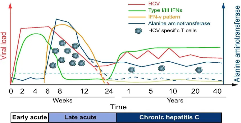
i) Acute infection
Hepatitis C virus has an incubation period ranging from 2 months to 6 months. Acute
14
RNA is detectable for the first 2 weeks. Later on, an elevation in the liver enzymes is
seen from 2-8 weeks. Symptoms of hepatitis namely malaise, fatigue, right upper
quadrant pain, nausea, dark urine and jaundice can be observed in about 25-30% of
patients within 3-12 weeks of acquiring HCV. Anti-HCV also appears in the patients
by about 7-8 weeks after infection, with delayed seroconversion seen in a group of
patients (78).
Spontaneous resolution which is defined as undetectable HCV RNA in blood is seen in
about 15-25% patients, while the rest progress to chronicity (12).
ii) Chronic infection
Chronic HCV infection is defined as persistence of HCV RNA in blood of the patient
for 6 months or more. The various factors that influence the progress to chronicity are
age at the exposure, male sex, race, co-infections and host factor such as the immune
status of the patient (12). Patients with chronic hepatitis C infection are predominantly
asymptomatic but have raised serum transaminases, which can fluctuate over time. Only
6% of patients might have symptoms. Fatigue, nausea, abdominal pain and decreased
appetite are the common symptoms. The disease can remain clinically silent for decades
while hepatic inflammation and fibrosis continue in the liver. The factors influencing
hepatic fibrosis are alcohol consumption, age at the time of infection, male gender,
immunosuppression, obesity and insulin resistance (79,80). Hepatic cirrhosis occurs in
about 5-20% of patients after 20-30 years with 10 year survival rate for such patients
15
Cirrhotic patients are at a higher risk of developing decompensated liver disease and
hepatocellular carcinoma (HCC), with about 1-4% of such patients progressing to HCC
every year (13,14).
5. Extra hepatic manifestations of HCV
HCV infection can also result in certain extrahepatic manifestations in about 40-74%
of patients. Mixed cryoglobinemia is the most common among them.
Other manifestations are non-Hodgkin lymphoma and porphyria cutanea tarda.
Metabolic disorders such as insulin resistance and steatosis can also be seen in a certain
group of patients (13).
6. Immune response to hepatitis C virus infection
Pathogen associated molecular patterns (PAMPs) are the moieties present on the
infective virions which are recognized by their corresponding receptors on the host cell,
resulting in initiation of an immune response. The pathways triggered by viral infection
are either toll-like receptor dependent pathway or the cytosolic pathway involving
[image:24.612.216.413.153.339.2]RIG-1 (retinoic acid inducible gene-RIG-1). This results in activation of NF-kB and interferon
16
regulatory factors which ensues in production of interferons by activation of interferon
stimulator genes, thus resulting in initiation of antiviral response of the body (82).
HCV is known to evade immune response of the host and to survive for a longer
duration resulting in disease progression.
6.1. Innate immune response
First line of defence to any infection is innate immunity. Interferons act as mainstay for
innate immunity. It induces an antiviral state and activates the natural killer cells. Cells
infected with viruses give rise to type I and type II interferons, while NK cells produce
interferon-γ.
Hepatitis C virus infection leads to initiation of TLR and RIG-1 pathways which
culminate in activation of IRF3 (Interferon regulatory factor 3). This IRF3 along with
NF-kB initiates transcription of interferons which bring about an antiviral state in
nearby cells. Type I and type III INFs are produced in the early acute phase of hepatitis
C infection and are capable of controlling viral replication upto a certain extent but not
of eliminating it (82). Interferons bind to their specific receptor and cause activation of
Janus kinase/Signal transducer and activator of transcription (Jak-STAT) pathway. As
a result, interferon-stimulated genes (ISGs) are induced which have antiviral properties.
RIG -1 and interferon regulatory factor 7 (IRF-7) also function as ISGs and promote
interferon production (82).
Natural killer cells also play a major role in acute HCV infection. NK cells mediate
17
ligand (TRAIL) or perforin mediated (direct cytolytic) and IFN-γ mediated
(non-cytolytic) pathways (82).
Ubiquitin molecules also play a role in controlling HCV infection. IFN type I induces
E3 ubiquitin ligase tripartite-motif 22 (TRIM22), which is thought to be associated with
[image:26.612.96.519.207.423.2]response to treatment by peg-IFN-α-2a/RBV therapy (83).
Figure 3. Course of HCV infection. Adapted from Heim and Thimme, 2014 (82)
6.2. Attenuation of innate immunity of host by hepatitis C virus
The various mechanisms by which HCV attenuates the host's innate immunity are (84):
a) Protease activity of HCV NS3/4A results in cleavage of toll-IL-1 receptor
domain containing adapter inducing IFN-β (TRIF) and IFN-β, thus blocking
RIG-1 signalling and ablating TLR3 signalling, culminating in decreased IRF3
18
b) HCV core protein can increase the level of suppressor of cytokine signalling
protein 3 (SOCS-3), which in turn act as inhibitors of Jak-STAT pathway and
form a negative feedback loop decreasing IFN-α/β receptor signalling.
c) NS5A protein of HCV stimulates IL-8, which causes attenuation of ISG
expression and antagonisation of type I IFN signalling.
Figure 4. Evasion of Innate immune response by HCV. Adapted from Barbara Rehermann (2009) (85)
6.3. Adaptive immune response
6.3.1. Humoral immunity
Antibodies to hepatitis C virus are detectable in the blood by about 7-8 weeks after
infection. These are specific for an isolate and exert a selection pressure leading to
evolution of various quasispecies. Although, there is no clear role of humoral response
in controlling the infection that has been elucidated; it has been proven that antibodies
19
phase of infection has even shown to be associated with spontaneous clearance of
hepatitis C virus from the body (86).
It is mainly the hypervariable region 1 (HVR1) of E2 that has been shown to lead to
persistence of infection. Antibodies which target a more conserved region, other than
HVR 1 have been shown to result in virus clearance (87).
A lack of robust immune response and antibodies during the early phase of infection
can lead to chronicity. Increased severity, rapid progression of disease and chronic
illness is seen in patients with less antibodies, thus suggesting that these antibodies play
a crucial role in HCV disease progression (88).
6.3.2. Cell mediated response
CD8+ and CD4+ T-cell response specific to HCV are of critical importance in
suppression and clearance of HCV infection. Patients who spontaneously recover and
clear the infection are shown to have increased levels of IL-2 and IFN-γ, which are
formed by the HCV specific CD4+ cell. In patients who develop chronic infection, the
HCV specific CD4+ cell responses are weak or absent (85).
CD8+ cells specific to HCV are detected in blood of patients in acute phase of HCV
infection. These cells produce cytokines such as TNF-α and IFN-γ and even result in
cytolysis of HCV infected cells. IFN-γ secreting CD8+ cells are seen predominantly in
people who recover from hepatitis C virus infection (89). Impaired CD4+ cell function
leads to exhaustion of CD8+ cells. There is loss of cytolytic activity of the CD8+ cells,
as well as a substantial decrease in TNF-α and INF-γ production. As a consequence
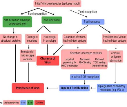
20 6.3.3. Evasion of humoral and cellular immune response by hepatitis C virus
HCV causes a chronic infection in majority of the patients. It does so by evading the
immune response of the body by following mechanisms (84,90,91):
a) Mutation in epitopes of hepatitis C virus
NS5b protein of HCV which acts as RNA dependent RNA polymerase has a
lesser proof reading capability and is prone to errors. Such errors result in
formation of mutational variants which have certain differences in their epitopes
enabling them to evade the cellular immune response.
b) Anergy of CD8+ cells specific to HCV
CD8+ cells loose there specificity for HCV overtime, which might help the virus
in evading the immune response.
c) Populations of regulatory T-cells
CD8+ T cells present in the liver of an individual infected with HCV are known
to produce interleukin-10. IL-10 downregulates effector T cells and hampers the
production of IFN-α. Moreover, CD4+CD25+ regulatory T cells are also
observed in chronically infected patients.
d) T cell inhibitory receptors
1 (Programmed death-1) is a T cell inhibitory receptor. Levels of surface
PD-1 are found to be significantly higher in people who develop chronic HCV
infection (90). Thus, this can also be considered as a mechanism of evasion by
21
The various mechanisms of evasion of adaptive immune response by HCV is depicted
[image:30.612.101.539.136.503.2]as follows:
Figure 5. Mechanisms of evasion of adaptive immune response by HCV. Adapted from Burke and Cox (90)
7. Diagnosis of HCV infection
Hepatitis C virus infection testing is advised in symptomatic patients with a suspicion
of underlying liver disease, in individuals belonging to high risk groups and as a
mandatory screening procedure for blood donors in blood banks (since 2002). WHO
22
testing in adults and adolescents from population with high HCV burden and in all
individuals with a high clinical suspicion of viral hepatitis (23). In addition, focused or
targeted testing in specific high risk groups such as IV drug users, prisoners, MSM and
sex workers, HIV infected individuals, family and close contact of already infected
cases and health care workers should be undertaken (23,92).
The various tests that are available for diagnosing hepatitis C virus can be grouped into:
i. Rapid diagnostic tests (RDTs): Based on detection of antibodies to HCV by
immunofiltration or "flow through technology" (93).
ii. Serologic tests: Antibodies to HCV are detected by using recombinant antigens
by ELISA and CMIA methodologies. Recently, HCV core antigen has also been
added in certain CMIA and ELISA formats.
iii. Molecular tests: Nucleic acid amplification tests are employed for detection and
quantification of HCV RNA and genotyping of HCV.
iv. Other tests: Liver function tests, fibroscan, acoustic radiation force impulse
(ARFI), AST to platelet ratio index (APRI) and liver biopsy.
7.1. RDTs (Rapid diagnostic tests)
Rapid diagnostic tests can be used as point of care tests and may be employed as a
preliminary diagnostic test in low resource settings, as no elaborate procedures are
required and results are available within minutes.
These tests are based on immunochromatographic, immunofiltration and "flow through
technology". Highly purified HCV antigens from core, NS3, NS4, NS5 regions are
23
to those antigens will give a positive reaction. The sensitivity of such tests are lower
than that of ELISA (93).
7.2. Serological tests (ELISA and CMIA)
a) Detection of antibodies
Antibodies to hepatitis C virus can be detected in patients by 1-2 months of infection.
ELISA which employs the principle of enzymatic reaction to ascertain antibodies in a
patient's sample can be used. There are various generations of ELISAs that have evolved
over a period of time. First generation ELISA uses c100-3 part of NS4 genome of HCV
as the antigen. Lower sensitivity of first generation ELISA led to the development of
2nd and 3rd generation ELISAs. To add to the sensitivity of the assay, c22-3 and c200
proteins were used additionally in 2nd generation ELISA. NS5 protein were the addition
in third generation ELISA (15). Recently, combination ELISAs have come into the
market which have antibodies to core antigen coated on the microtitre wells, so
antibodies and core antigen can be detected resulting in higher specificity.
Chemiluminescence immunoassay, such as Abbott ARCHITECT platforms utilises
microparticles on which the recombinant antigens or antibodies are coated for detection
of corresponding elements in the patient's sample.
RIBA (Recombinant immunoblot assay) was another type of test to detect specific
antibodies in the patient's sample. In this, recombinant antigens of HCV (namely,
recombinant c33c and NS5 antigens and synthetic 5-1-1, c100 and c22 peptides) were
coated on a nitrocellulose membrane and a conjugate and substrate were added after
24
(SIA) where no bands meant a negative result, one band indicated indeterminate result
and a minimum of two bands were taken as positive (95).
b) Detection and quantification of HCV core antigen
Hepatitis C virus core antigen is a highly conserved structural protein of 191 amino
acids in length. It can be detected in blood during viral replication. The advantage of
estimating core antigen is that it is positive even during the pre-seroconversion window
period, in immunosuppressed individuals and in neonates where the antibodies to HCV
are negative (17). A positive result confirms active infection. There are various
combination ELISAs available for detection of HCV core antigen along with antibodies
in the patient's sample (96). Chemiluminescence assays with microparticles coated with
monoclonal antibody to HCV core antigen can also be employed for its detection.
Sensitivity ranges from 80-99%, while specificity is about 96-100% (18,97). It is a
simple and cost-effective method to establish active HCV infection in settings where
the facilities for molecular tests are absent. The only limitation with core antigen
detection is that for core antigen to be detectable in plasma, HCV RNA value should be
1000 IU/mL.
7.3. Molecular tests
7.3.1. HCV RNA detection
Detection of viral nucleic acid is essential to establish active infection. Apart from being
the confirmatory test of hepatitis C virus infection, HCV RNA detection also serves as
a mainstay of diagnosis in neonates and immunosuppressed patients. Branched-DNA
assay, transcription mediated amplification (TMA), reverse transcriptase polymerase
25
quantify HCV RNA (98,99). With the discovery of antiviral agents, there has been an
immediate need to quantify HCV RNA for monitoring therapy and response to
treatment. Viral RNA is measured in "International Units" (IU) instead of viral copy
numbers, according to the WHO. The viral copies/mL= viral load in IU/mL X 2.7 (for
Roche COBAS TaqMan). Newer assays have lower limit of detection ranging from
12-50 IU/ml and specificity of 98-99% (16).
7.3.2. Genotyping
7.3.2.1. HCV genotyping
Treatment of HCV depends on the genotype of the virus infecting the individual.
Certain genotypes such as genotype 1, require a prolonged duration of treatment. In the
recent past, pegylated-IFNα and ribavirin were in use for treatment and now directly
acting antivirals (DAAs) are the mainstay of treatment. Different treatment regimens
and duration are advised for each genotype, thus necessitating the role of HCV
genotyping (100,101).
Genotyping can be performed by sequencing core, 5'UTR, NS3 or NS5b regions of
hepatitis C virus (19). Line probe assay i.e. reverse hybridisation of amplified products
on probes specific for genotypes, PCR-restriction fragment length polymorphism
(RFLP) in which amplicons are digested by help of restriction enzymes to yield
fragments specific to different genotype are other methods used to genotype HCV.
Recently, real-time PCR with specific primers and probes for different genotypes have
26 7.3.2.2. IL-28B genotyping
Interleukin-28B gene (rs12979860 and rs8099917) polymorphisms predict sustained
virological response (SVR) in patients. Thus, detection of such single nucleotide
polymorphism is important for ascertaining the prognosis after treatment (103).
Genotyping of IL-28B region which is present on chromosome 19 is performed by
RFLP or sequencing.
7.4. Other tests
Liver function tests, fibroscan, acoustic radiation force impulse (ARFI) test can also be
of use to detect any pathology in the liver. Aspartate transaminase to platelet ratio
(APRI) is a new score to assess the degree of liver fibrosis (104). Liver biopsy is
recommended to stage the degree of hepatic necrosis, fibrosis and inflammation but is
generally not preferred, as it is an invasive procedure (105).
8. Treatment
8.1. Rationale for treatment
HCV infection is a significant cause of morbidity and mortality. It results in chronic
infection in about 80% of patients. Most of these infections are asymptomatic and
slowly progress towards liver cirrhosis and hepatocellular carcinoma. Even in absence
of significant hepatic symptoms, severe extra hepatic symptoms can predominate. Thus,
treatment of hepatitis C virus becomes a necessity. With the introduction of DAAs in
the past few years, there is a possibility of cure. Antiviral treatment is helpful in
27
extra-hepatic manifestations of HCV. Virus clearance takes upto 24 weeks after starting
effective therapy. Sustained virological response (SVR) refers to absence of virus (HCV
RNA) in the patient 12 or 24 weeks after completion of therapy and it is considered as
a measure of effectiveness of the drug regimen (106). Timely and effective treatment
can reduce the chances of complications of chronic HCV infection and reduce the
liver-related mortality (107).
8.2. Anti HCV drugs
1) Interferon and ribavirin combination therapy
Interferon-α was the mainstay of treatment of chronic hepatitis C for more than two
decades. Even though it was not very effective, it resulted in viral clearance in about
10-25% of patients (108). INF-α and ribavirin combinations were in use since the late
1990s. This combination resulted in almost two fold increase in virological response.
Pegylated interferons were introduced in early 2000s and because of less side effect
profile compared to interferons, its combination with ribavirin has been in use since
then. The duration of therapy varies with the genotype of hepatitis C virus infecting the
patient. Genotypes 2 & 3 require a shorter duration i.e. 24 weeks, while genotypes 1, 4,
5 and 6 infections require longer duration of therapy (109).
2) Directly acting antivirals (DAAs)
With the sustained virological response being only about 40% even after 48 weeks of
therapy with peg-INF-α and ribavirin combination therapy for patients infected with
HCV genotype 1, finding suitable and more effective drugs for treatment was the need
28
of HCV have been developed in the recent years. The first drugs to be approved were
boceprevir and teleprevir, which act on NS3/4A protease and bring about its inhibition
(20). The use of these drugs in the combination with the existing therapy showed a
marked increase in the virological response but did not help in the management of
people who had failed a previous interferon therapy. Adverse effects such as severe
anaemia and hypersensitivity reactions were also observed with these drugs and
consequently these were removed from the market (110). Since then, other new DAAs
such as sofosbuvir, ledipasvir, daclatasvir etc. have been introduced in HCV treatment
regimens as these have better tolerability, better safety profile and a SVR of more than
95% after the completion of regimen (111).
8.3. Interferon and ribavirin combination therapy
There have been constant revisions in the treatment guidelines of HCV over the past
few decades. Newer drugs have been added and duration of therapy has been revised.
The various regimens and drug combinations in use for the treatment of HCV in the
past few decades have been outlined below:
i. Interferon-α monotherapy
Interferon-α has been shown to be effective in patients with HCV infection since
early 1990s (108,112). Interferons stimulate ISGs through various cascades and
have an immunomodulatory effect resulting in increased cytokine production
and activation of natural killer cells which leads to antiviral effect. It was the
29
However, with severe adverse effects such as neutropenia, myalgia,
neuropsychiatric manifestations, fatigue (113) etc. and with sustained virological
response ranging from 10-25% in patients, this was used in combination with
ribavirin.
ii. Interferon-α and ribavirin combination
Ribavirin is a guanosine analogue, synthesised in 1970 and was used in treating
respiratory syncytial virus infection in children. It had a broad antiviral spectrum
and thus was introduced as a combination therapy along with interferon-α for
treatment of chronic hepatitis C. The use of ribavirin as a combination drug lead
to almost doubling of rates of sustained viral response (114). The various
mechanisms of action postulated for ribavirin are (115):
a) Inhibition of replication of HCV: Ribavirin is a guanosine analogue and can
be incorporated in the viral RNA in its phosphorylated state. This brings about
chain termination and inhibition of HCV replication.
b) Inosine-monophosphate-dehydrogenase inhibition: Ribavirin competitively
inhibits inosine-monophosphate-dehydrogenase which leads to GTP depletion.
GTP is vital for viral RNA synthesis and this inhibits HCV replication.
c) Mutagenesis: According to Crotty S. et al., ribavirin can act as a viral mutagen
and result in formation of virions with decreased infectivity (116,117).
iii. Pegylated interferon-α
Pegylated interferon-α is formed by covalent conjugation of monomethoxy
30
(100). PEG-INF-α has a better pharmacological profile compared to INF-α and
results in a higher SVR rate reaching upto 82% (118,119). Recent studies have
shown higher incidence of adverse effects and mortality on use of
PEG-interferon-α and ribavirin combination regimen (120).
8.4.1. Directly acting antivirals (DAAs)
Combination therapy with pegylated interferon-α and ribavirin were not effective with
SVR rates hovering around 40% in certain cases. This necessitated the search for newer
compounds which were better tolerated and had better SVR rates. Ciluprevir
(BILN-2061) was the first drug to be tested but was withdrawn after severe side effects were
observed in animals (121). Serine protease inhibitor therapy trial 2 (SPRINT-2) was
undertaken to assess boceprevir, a newer drug targeting the NS3 region of HCV. Triple
regimen therapy with PEG-IFN-α, ribavirin and boceprevir resulted in a SVR rate of
68% after 24 weeks of therapy, which was significantly higher than the existing dual
drug combination therapy of PEG-IFN-α and ribavirin (122); thus heralding a new era
of directly acting antivirals.
In early 2010s new oral drugs were approved for the treatment of chronic hepatitis C
infection. The first such drugs were the protease inhibitors (boceprevir and teleprevir),
which target the NS3/4A region of HCV genome (123). Since then, several other drugs
targeting other regions such as NS5A and NS5B of hepatitis C virus have been
31
Figure 6. Timeline of development of DAAs. Adapted from Akihiro Tamori et al.
(124)
The classification of directly acting antiviral drugs for HCV with the profile and
mechanism of action for each drug are elucidated as following (125):
a. Protease Inhibitors: NS3 protein of HCV has protease function and NS4
protein acts as a cofactor to aid in the protease and helicase function of NS3/4A.
NS3 exhibits protease activity at the amino terminal and helicase activity at the
carboxy terminal. Substrate attaches to a shallow cleft present between the two
terminals. Teleprevir and boceprevir are the prototype drugs of this class.
Simeprevir, grazoprevir, paritaprevir are other drugs belonging to this class
(126). Asunaprevir, voxileprevir and glecaprevir are currently under clinical
32
Antiviral property is because of direct inhibition of HCV replication by
inhibition of protease function and indirect effect by inhibition of cleavage of
TRIF and mitochondrial antiviral signalling protein (MAVS) adaptor molecules
by NS3/4A in TLR3 and DDX58 pathways resulting in increased INF-β(128).
Adverse effects such as rash and hematopoietic suppression is common with this
class of drugs, owing to which boceprevir and teleprevir were withdrawn from
the market by the end of 2015 (129).
b. NS5B inhibitors: This class of compounds bind to NS5B protein of HCV which
serves as a RNA dependent RNA polymerase. This NS5B polymerase is
composed of about 590 amino acids and has a right hand topology with palm,
thumb and finger regions. NS5B inhibitor drugs bind at the substrate site of the
polymerase and bring about inhibition of viral replication.
These NS5B inhibitors can be further classified as nucleotide and non-nucleotide
inhibitors (130). Non-nucleotide inhibitors (NNIs) such as dasabuvir and
beclabuvir bind to allosteric site on NS5B and induce a confirmational change
in the RNA dependent RNA polymerase with a resultant blockage of enzymatic
activity.
Sofosbuvir is the prototype drug belonging to nucleotide inhibitor class. It is a
uridine analogue, administered in the prodrug form and needs to be changed to
its active triphosphate form to bind to NS5B protein. It competes with natural
nucleoside triphosphate and prevents the replication of RNA (125). Sofosbuvir
33
Nausea, diarrhoea, flatulence, headache and rash are the common side effects of
this class of drugs (131).
c. NS5A inhibitors: NS5A is a phosphoprotein of 44 amino acids with three
domains. It is essential for replication of HCV and for morphogenesis of virions,
with its specific role not yet known (125).
NS5A inhibitors are proposed to inhibit membranous web formation, which is
essential for viral replication. In addition, these inhibitors may form complexes
with host factors such as TIP47 (Tail interacting protein of 47 kD) or PI4KIIIα
and adversely affect viral replication and assembly (132). Daclatasvir was the
first NS5A inhibitor produced. Ledipasvir, ombitasvir and elbasvir are newer
compounds of this class. Velpatasvir is a widely used pangenotypic drug
34 Figure 7. Site of action of directly acting antivirals. Adapted from Gotte and Feld
(125)
8.4.2. DAAs: treatment regimens
Direct acting antivirals have a high tolerability and compliance due to once daily oral
dosage, less severe side effects and lead to a SVR reaching upto 95% (134). These
DAAs are generally advised as a combination of two drugs to achieve a high sustained
virological response (SVR). Ribavirin can also be included in the combination therapy.
The dosage of various drugs for the treatment are (135):
Sofosbuvir: 400 mg/day in 1 dose {Or in combination with ledipasvir & velpatasvir}
Ledipasvir: 90 mg/day in 1 dose
Daclatasvir: 60 mg/day in 1 dose
35
WHO recommended regimens for the treatment of hepatitis C virus infection for the
year 2016, adapted from “American Association for the Study of Liver Diseases
(AASLD) and European Association for the Study of the Liver (EASL)" (136):
Figure 8. Recommended regimens for HCV treatment. Adapted from WHO guidelines, 2016 (136).
8.4.3. Resistance to DAAs
Resistance to directly acting antivirals is a relatively new phenomenon observed in
individuals who don't achieve SVR even after completion of the full duration of therapy
(137). The various mechanisms suspected to be involved in resistance to DAAs are
(138):
a) Quasispecies: Presence of a large number of viral variants in the individual because
of high error rate and lack of proof reading ability of NS5B protein of HCV. This can
result in selection of variants with mutations favouring survival of the virus.
b) Nucleotide polymorphisms and mutations: S282T, L159F and V321A are certain
36
didn't achieve SVR with teleprevir were found to have V36A/M, T54A/S, R155K/T,
A156S/T and D168N mutations (139).
8.4.4. Monitoring of viral response in the era of DAAs
Only about 1 million of the estimated total 80 million people infected with hepatitis C
virus have access to HCV treatment. With the use of DAAs for treatment of HCV
infection, there is a possibility of cure. Within 3 months of treatment, 95% of patients
attain cure, especially because of efficacy of drugs, increased compliance, ease of
treatment and low cost. Continuous monitoring of HCV RNA levels is required to assess
the response to therapy and to modify the drug regimen, if needed.
9. Limitations for adequate management of hepatitis C virus infection
Management of HCV infection involves estimation of HCV RNA and detection of the
infecting HCV genotype. Initiation of therapy depends on pre-treatment viral load and
HCV genotype. Duration of therapy and the current treatment strategies are influenced
by HCV RNA quantification (viral load estimation) and HCV genotype (140). These
assays are unfortunately available in very few laboratories in India, leaving low resource
settings at a disadvantage in optimally managing HCV infected individuals (141).
HCV quantification is most reliably done by freezing plasma/serum within 6-8 hours
after blood draw. Detection of HCV RNA in resource-limited settings therefore is
difficult due to the challenges of collection, storage and transport to larger reference
laboratories. Plasma/serum samples need to be centrifuged and separated within 6 hours
37
reference laboratories for these tests requires dry ice shipments, which is also logistically
difficult.
Thus, newer and simpler methods for collection and transport of sample from distant
places to higher centres are required.
10. Dried blood spots
Dried blood spots (DBS) have recently been studied as an alternative specimen to
plasma for detection of antibodies to HCV as well as for detection and quantification of
HCV RNA (142). DBS has already been evaluated as a method of collection and storage
of blood for HIV and HBV infections. It is a cost effective and easier method for
collection of blood samples (143,144). Collection of DBS is a less cumbersome and
minimally invasive method requiring only a finger prick and spotting of whole blood
onto filter paper cards (145). Thus, DBS collection in resource-limited settings and then
its transportation at ambient temperature to the reference laboratories for HCV RNA
estimation, is one of the means by which current HCV infection can be detected and
followed-up, in remote areas (146).
The idea of collecting blood on a paper card and subsequently using these dried blood
spots (DBS) for diagnostic purposes originated a century ago, with Ivar Christian Bang
using DBS eluates to determine blood glucose (147). Since then, DBS testing for
decades focused on infectious diseases diagnosis (148) especially in low resource
settings and for the inherited metabolic disorders screening in newborns (149).
Recently, DBS has been used for a variety of tests and applications.
38
a. Lesser volume of blood, compared to conventional venipuncture method is
required.
b. It provides a cost-effective, minimally invasive and simple alternative for
collection of blood for testing.
c. There is minimal risk of haemolysis of blood or contamination by other
micro-organisms such as bacteria.
d. Less deterioration of analytes overtime and easy storage and transport for
prolonged durations.
There has been an upsurge in the usage of DBS as a preferred method of collecting
samples from remote areas because of its aforementioned advantages. In the past
decade, it has been evaluated as a sample for testing of HIV, HBV and HCV infections
(151,152). With the advent of DAAs for therapy of hepatitis C virus infection,
monitoring of HCV RNA has gained even more importance for ascertaining response
to therapy.
39 10.1. Evaluation of DBS for virological studies
To evaluate DBS as a method for sample collection, storage and transport to referral
centres, many studies have been undertaken.
In a study by Johannessen et al., in rural Tanzania, 98 plasma and DBS pairs were
compared and HIV-1 RNA was detected in 100% of DBS samples when plasma viral
loads were >3000 copies/ml (153). Another study in Burkina Faso, demonstrated the
benefits of using DBS for the serological detection of HIV, HBV and HCV infections
in settings where the laboratory facilities for their detection were non-existent (151).
Marjorie Monleau et al., studied the effects of different storage conditions i.e. 20ᵒC &
37ᵒC on DBS and came to the conclusion that viral monitoring is still feasible from
DBS after storage for 3 months at 37ᵒC (154). A study done in Bangalore, by Neogi et
al. studied the effects of various storage conditions on DBS and its effect on HIV viral
load and came to the conclusion that DBS can be used as a collection and storage
method of blood samples for viral load monitoring in HIV (155).
Studies by Lee CE et al. (156), Ross RS et al. (143), Boa-Sorte et al. (157) and Mössner
BK et al. (158) in various parts of the world have further emphasised that dried blood
spots can be used as a means of sampling in remote areas where there are no facilities
for viral testing.
10.2. Evaluation of DBS for HCV testing
Various studies have evaluated DBS as a specimen for HCV testing. In one of the
earliest studies on DBS for HCV in 1997, anti-HCV antibodies were detected in all 168
40
correlated later in the study by McCarron B. et al. where detection of anti-HCV in DBS
samples showed to have very high sensitivity and specificity (160). In a study by Abe
K. et al., serum samples were spotted onto filter papers and stored at room temperature
for upto 4 weeks. PCR was done at the end of 1, 2, 3 and 4th week to check for stability
of HCV RNA, which was detectable at the end of 4th week, though showed a reduction
of almost 10 folds in amount (161). Solomone M. and colleagues in 2002, assessed the
stability of HCV RNA in DBS for over a period of 11 months and the nucleic acid was
preserved until the duration of the study (162).
Studies conducted in the recent past by Tullion E et al. in 2010 (146) and Dokubo EK
and colleagues in 2014 (142), concluded that DBS samples are sensitive and specific
for HCV antibodies and HCV RNA and can be used as an alternative to plasma.
Cloherty et al. concluded that DBS, being stable at room temperature and at 2-8ᵒC for
upto 10 weeks can allow for storage and shipment and is an effective method for HCV
screening (163). DBS has been shown to be stable at different conditions such as
22-26ᵒC, 2-8ᵒC and -20ᵒC (164). In a review, Greenman et al. have concluded that DBS
has potential for use in HCV RNA detection, quantification and genotyping. DBS may
be sufficient to detect and quantify HCV RNA for the purpose of HCV diagnosis. There
was significant variation among viral load endpoint detection limits reviewed in the
study. Lowest threshold of 150–250 IU/mL was found in highest-powered study
designed specifically to measure endpoint sensitivity (165).
The following table shows cumulative sensitivity and specificity of DBS for various
41
Tests Sensitivity Specificity References
HCV RNA 65.9%-98.1% 100% (165–168)
Anti-HCV 97.5%-100% 95.95%-100% (146,164,166,169)
HCV core antigen 64.1% 100% (167)
There are only a handful of studies from India evaluating the feasibility of DBS as a
sample. Most studies have evaluated DBS for HIV detection and quantification, with
high correlation between DBS and plasma values suggesting it can be used as a sample
in suspected HIV cases (155,170–172).
Nandagopal et al., in their study published in 2014, evaluated DBS samples for detection
of anti-HCV and it was found to be 100% sensitive and specific (169). In a study by
Lakshmi V. and colleagues, HCV was detected by loop mediated isothermal
amplification (LAMP) from DBS samples with the resulting sensitivity and specificity
of 100% (173). This further corroborates that DBS is comparable to plasma for
qualitative detection of hepatitis C virus.
11. Rationale for this study
There are no studies evaluating the effect of the extreme temperature conditions of the
Indian subcontinent on DBS for the detection of HCV RNA. Many studies from
developed countries have studied the role of room temperature on DBS for estimating
HCV viral loads. Room temperature in most parts of the developed world is mostly
42
temperatures in this country, which is often ≥37ᵒC, especially between April to
September of each year.
Our study aims at filling the lacunae in the knowledge about the feasibility and
effectiveness of DBS as a sample for collection, storage and transport of blood for HCV
RNA quantification, HCV core antigen detection and HCV genotyping at variable
temperatures (4ᵒC & ≥37ᵒC). The lower temperature of 4ᵒC is chosen because some
laboratories may be able to store DBS samples at this temperature and also ship them at
4ᵒC. Most laboratories will only be able to store and transport them at ambient
temperature which is highly variable and often exceeding 37ᵒC, thus justifying the
43 MATERIALS AND METHODS
1.1. Study Design
This is an observational study to assess the diagnostic accuracy, in which hepatitis C
infected, treatment naive patients who presented to the Department Of Clinical
Virology, Christian Medical College, Vellore for HCV viral load quantification and
genotyping were included. Dried blood spots from patients were collected, which were
later eluted and quantified using Abbott Real Time HCV assay (for HCV RNA) and
Abbott ARCHITECT HCV Ag assay (for HCV core antigen). This was then compared
with the routine plasma based assays.
Packed cell volume (PCV) values were estimated for each of the samples, to arrive at a
correction factor/normalisation coefficient. This was to adjust for difference in sample
volume and sample type between plasma and DBS samples.
1.2. Ethics Approval
This study was approved by Institutional Review Board, Christian Medical College and
Hospital, Vellore (IRB Min No: 9687 (DIAGNO) dated 20.10.2015).
1.3. Funding
The study was funded by Internal Fluid Research Grant and Virology special fund.
1.4. Study Duration
44 1.5. Study Sample
Samples from 40 consecutive treatment naive patients, with high index of suspicion of
having hepatitis C virus infection; previous HCV RNA positive test results and HCV
antibody levels ≥10 S/Co, referred from liver clinic were included in the study. This
was over and above the required sample size calculated prior to starting the study.
Written informed consent was obtained from the patients at the time of sample
collection.
1.5.1. Sample size calculation
The required sample size to show that there was an agreement of about 0.8 (anticipated)
in viral load between routine HCV RNA detection and whole blood spotted on to 2
strips of Whatman 903 filter paper was found to be 36 patients with a power of 90%
and at 5% level of significance.
Agreement - Single Group - Continuous outcome-ICC (Testing against Population value)
Sample reliability value 0.7 0.7 0.8 0.8 0.8 0.8
Population reliability value 0.3 0.3 0.4 0.4 0.5 0.5
Power (1- beta) % 80 90 80 90 80 90
Alpha error (%) 5 5 5 5 5 5
1 or 2 sided 2 2 2 2 2 2
Number of replicates 2 2 2 2 2 2
45 Formula for sample size calculation(174):
1.5.2. Inclusion Criteria
- Patients with hepatitis C virus infection
- Patients more than 18 years of age
- Treatment naive patients
- Patients who give consent for the study
1.5.3. Exclusion Criteria
- HCV RNA negative plasma samples
- Pregnancy
- Patients less than 18 years of age
46 1.6. Data Sources
Patient information such as age, sex, hospital no., address, AST, ALT, ALP, PT, APTT
and haematocrit (PCV) values were collected from the online patient clinical records
(Clinical Workstation). Samples were stored and tested in the Department of Clinical
Virology.
1.7. Study algorithm
47 Study algorithm
Treatment naive patient comes to Department of Clinical Virology for routine
molecular tests for HCV (detection/quantification/genotyping)
Informed consent taken
9ml of blood collected in EDTA tube
2 ml of blood sample taken in K2EDTA tubes and sent to
Department of Clinical Pathology for estimation of PCV, to arrive at a correction factor
Approximately 7ml of whole blood to generate plasma for routine HCV RNA detection using reference standard (HCV
RNA real-time PCR)
Whole blood (50µl on each circle, 5 spots on each strip) spotted onto two strips of Whatman 903 filter paper
One strip with desiccant placed at 4ᵒC for 15 days
One strip placed at ≥37ᵒC, with
desiccant for 15 days Negative for HCV RNA Positive for HCV RNA Excluded from the study
Stored at -20ᵒC until time of testing
DBS samples of HCV RNA positive patients alone eluted
Abbott m2000sp automated extraction system
RNA extract
Detection/quantification of HCV RNA
Abbott m2000rt HCV Real time PCR
HCV genotyping
NS5B region based sequencing using ABI PRISM 310 Genetic Analyzer, for a proportion of patients
HCV core antigen estimation
48 2.1. Processing of blood samples
9 ml blood collected in sterile vacutainer tubes containing potassium ethylene diamine
tetra acetic acid, after obtaining informed written consent. An additional 4 ml K2EDTA
tube taken and 2ml blood collected in it for the purpose of estimating packed cell
volume (PCV). Two Whatman 903 protein saver cards collected and labelled with
unique study identification number, date of collection and the temperature (4ᵒC or
≥37ᵒC), in which it will be kept.
The whole blood was spotted onto two Whatman 903 protein saver cards. Each
Whatman 903 card has 5 sample slots. 50 µl of whole blood spotted onto each of the
sample slots in the two cards (Totally 500 µl spotted, on two cards). The cards were left
to dry, at room temperature for 6 hours. Cards were then put into separate zip lock
pouches with desiccant. Afterwards, card labelled as A (4ᵒC) was placed in a box and
stored in refrigerator and card labelled as B (≥37ᵒC) was put in another box and kept in
walk in incubator with temperature ≥37ᵒC, for 15 days duration.
After 15 days, the cards were taken out of their respective boxes and kept at -20ᵒC, until
the time of testing.
3.1. Elution of DBS cards
3.1.1. Materials required
(i) Abbott mLysis Buffer
(ii) 15ml Tarson tubes
49
(iv) Clean forceps
(v) Sterile petri dishes
(vi) Ethanol
(vii) Calibrated pipettes: 10-1000µl with appropriate sterile tips
3.1.2. Procedure
HCV RNA elution was performed according to the protocol by David et al. (171)
Bio-safety hood cleaned with ethanol and ultraviolet light switched on for 15
minutes.
DBS samples, mLysis buffer, 15 ml Tarsons tubes (labelled), puncher, sterile
forceps, sterile petri dishes, pipettes and appropriate tips placed near the site.
1.7 ml of mLysis Buffer aliquoted into separate Tarsons tubes.
DBS cards selected, unique identification number and date of collection noted
and matched with that of the Tarsons tube.
3 punches from one sample slot, punched out using the puncher. Two sample
slots (6 punches) for each DBS strip.
The 6 punched out DBS samples put in the corresponding Tarsons tube and kept
at room temperature for 2 hours with intermittent agitation.
After 2 hours, Tarsons tube with the DBS punches centrifuged at 4500rpm for 2
minutes.