0095-1137/05/$08.00
⫹
0
doi:10.1128/JCM.43.8.3877–3883.2005
Copyright © 2005, American Society for Microbiology. All Rights Reserved.
Simultaneous Detection of Hepatitis C Virus (HCV) Core Antigen
and Anti-HCV Antibodies Improves the Early Detection of
HCV Infection
Syria Laperche,
1* Nadine Le Marrec,
1Annie Girault,
1Franc¸oise Bouchardeau,
1Annabelle Servant-Delmas,
1Miche
`le Maniez-Montreuil,
2Pierre Gallian,
3Thierry Levayer,
4Pascal Morel,
5and Nicole Simon
6National Reference Center for Hepatitis B and C in Transfusion, Institut National de la Transfusion Sanguine, Paris, France
1;
Etablissement Franc¸ais du Sang Nord-de-France, Lille, France
2; Etablissement Franc¸ais du Sang Alpes-Me´diterrane´e,
Marseille, France
3; Etablissement Franc¸ais du Sang Pyre´ne´es-Me´diterrane´e, Montpellier, France
4;
Etablissement Franc¸ais du Sang Bourgogne Franche-Comte´, Dijon, France
5; and Unite´
d’He´modialyse Association pour l’Utilisation du Rein Artificiel, Paris, France
6Received 28 December 2004/Returned for modification 9 March 2005/Accepted 9 May 2005
To evaluate whether a new enzyme immunoassay developed for the simultaneous detection of hepatitis C
virus (HCV) core antigen (Ag) and anti-HCV antibodies (anti-HCV Ab) (Monolisa HCV Ag/Ab ULTRA;
Bio-Rad) could improve the early detection of HCV infection, we compared its sensitivity to that of anti-HCV,
HCV core Ag, and HCV RNA assays. The populations studied included 12 blood donor samples positive for
HCV RNA and HCV core Ag but negative for anti-HCV antibodies and 23 hemodialysis patients who developed
anti-HCV Ab (seroconversion) during the follow-up. From these 23 individuals, 83 samples sequentially
collected prior to seroconversion and 108 samples collected after seroconversion were tested. Six of 12 blood
donations were positive by the HCV Ag/Ab assay. In the hemodialysis cohort, the 24 HCV RNA-negative
samples were negative by the HCV Ag/Ab assay and 23 of the 59 HCV RNA-positive samples (39%) were
positive. The HCV Ag/Ab assay detected HCV infection on average 21.6 days before the most sensitive antibody
assay. The HCV Ag/Ab assay did not detect HCV infection as early as the HCV RNA assay (mean delay, 30.3
days) or HCV Ag assay (mean delays, 27.9, and 16.3 days by the HCV core Ag quantification assay and the HCV
Ag blood screening assay, respectively). This new assay provides a notable improvement for the early detection
of HCV infection during the so-called window period compared with anti-HCV Ab assays and could be a useful
alternative to HCV RNA detection or HCV core Ag assays for diagnosis or blood screening when nucleic acid
technologies or HCV core Ag detection are not implemented.
Since the development of the first assay in 1989 (20), assays
for detection of hepatitis C virus (HCV) antibodies (Ab) have
allowed progress in the early detection of HCV infection (46).
This increased sensitivity of the last-generation assays has
dra-matically reduced the risk of HCV transmission by blood
com-ponents by reducing the window period from 82 days (5) to 66
days (3, 12). To further reduce the residual risk (2, 5, 16, 18, 36,
37, 41, 48), nucleic acid testing (NAT) for HCV RNA was
introduced in several high-income countries (2, 14, 15, 21, 30,
39). In some countries, an assay for the detection of HCV core
antigen (Ag) by use of the enzyme immunoassay (EIA)
tech-nology has been chosen as an alternative to NAT for the early
diagnosis of infection (1, 8, 25, 38). In addition, some authors
emphasized the clinical advantage of HCV core Ag
quantifi-cation as a direct marker of viral repliquantifi-cation in the chronic
phase of infection (4) and as a relevant marker for predicting
and monitoring the response to therapy (7, 29, 31). Indeed, the
HCV core Ag assays have sensitivities close to that of NAT,
with mean detection differences of 1 to 2 days in the window
period with the specific assay developed for blood screening
(11, 32, 35, 45) and 0.29 day with the immunoassay capable of
detecting and quantifying HCV core Ag (23). A recent study
reported that a prototype assay based on the simultaneous
detection of HCV core Ag and anti-HCV Ab significantly
closed the time gap between HCV RNA detection and the first
appearance of detectable anti-HCV Ab (42). However, this
assay is not yet available for routine use. More recently, a new
combination assay has been developed and licensed in Europe
(Monolisa HCV Ag/Ab ULTRA; Bio-Rad, Marnes la
Co-quette, France). To assess its sensitivity for the detection of
HCV infection during the window period or at the early phase
after seroconversion, we tested two panels and compared the
results with those obtained using the two available assays for
HCV Ag (HCV core Ag EIA blood screening assay and trak-C
assay) and HCV RNA. The overall objective was to determine
if this new test could constitute an alternative to NAT for the
diagnosis of HCV infection during the window period and
whether the sensitivity for antibody detection is preserved.
MATERIALS AND METHODS
Panels. (i) Panel 1: individual samples from volunteer blood donors.Panel 1 (Table 1) consisted of 12 blood donor samples which were negative for anti-HCV Ab (Ortho HCV 3.0 EIA test system Enhanced SAVe; Ortho Clinical Diagnos-tics, Raritan, NJ) but positive for HCV RNA. The plasma from each of these blood donations was immediately aliquoted and stored at⫺30°C until it was
* Corresponding author. Mailing address: National Reference
Cen-ter for Hepatitis B and C in Transfusion, Institut National de la
Transfusion Sanguine, 6 rue Alexandre-Cabanel, 75015 Paris, France.
Phone: (33) 1 44 49 30 52. Fax: (33) 1 44 49 30 59. E-mail: slaperche
@ints.fr.
3877
on May 15, 2020 by guest
http://jcm.asm.org/
used. All these samples were subsequently found to be positive for HCV core Ag by the trak-C assay (23), and 9 of the 11 samples tested were also positive by the HCV core Ag blood screening assay. Eight samples had been collected before the seroconversion (among these, five had been identified by the French Frac-tionation and Biotechnology Laboratory by detection of HCV RNA in plasma pools, and three had been identified by NAT in a pool of 8 or 24 donations after its implementation in blood donation centers), and three had been collected from three chronic immunosilent HCV carriers. Among the donors who pro-vided the latter three samples, the first one developed antibodies 2 years after
donation (13), and the second and the third ones were still negative for anti-HCV Ab 1 year and 6 months after donation, respectively (unpublished data). The last one was not explored. Viral loads ranged from 3.6⫻104
to 8⫻107 IU/ml (Amplicor HCV Monitor test v 2.0; Roche Diagnostics, Branchburg, NJ). Ge-notypes were determined by the InnoLipa test (Innogenetics, Zwijndrecht, Bel-gium).
(ii) Panel 2: sequential samples from hemodialysis patients.Panel 2 consisted of 191 samples collected from 23 hemodialysis patients who developed anti-HCV antibodies (seroconversion) during the follow-up. Each sample had been
imme-TABLE 1. Results of HCV Ag/Ab assay for the 12 HCV RNA-positive, anti-HCV Ab-negative blood donor samples included in panel 1
Case no. Clinical
contexta Genotype
HCV RNA level (IU/ml)b
HCV core Ag result by blood screening assay
(S/CO)c
trak-C assay result (Ag level [pg/ml])
Monolisa HCV Ag/Ab ULTRA result
(S/CO)c
1
WP
1a
1.6
⫻
10
7⫹
(20.6)
⫹
(183.5)
⫹
(5.95)
2
IS
1b
8
⫻
10
5⫹
(2.14)
⫹
(48.8)
⫺
(0.65)
3
WP
2a/2c
8
⫻
10
6⫹
(20.1)
⫹
(
⬎
217)
Gray zone (0.90)
4
WP
1b
6.5
⫻
10
5⫹
(3.05)
⫹
(51.7)
⫺
(0.40)
5
WP
3
3.6
⫻
10
4⫺
(0.27)
⫹
(2.8)
⫺
(0.10)
6
WP
1
8
⫻
10
7⫹
(
⬎
53.8)
⫹
(
⬎
217)
⫹
(
⬎
6.64)
7
WP
1a
4.7
⫻
10
7⫹
(49)
⫹
(
⬎
217)
Gray zone (0.93)
8
IS
1a
1.7
⫻
10
7NA
d⫹
(
⬎
217)
⫹
(1.20)
9
WP
3a
1.2
⫻
10
5⫺
(0.45)
⫹
(15.2)
⫺
(0.10)
10
IS
4a
⬎
5
⫻
10
5⫹
(9.1)
⫹
(
⬎
278)
⫹
(2.58)
11
?
3a
7
⫻
10
6⫹
(1.3)
⫹
(105)
⫺
(0.13)
12
WP
1b
1.8
⫻
10
7⫹
(38)
⫹
(
⬎
240)
⫺
(0.50)
aWP, window period; IS, immunosilent chronic carrier. bAmplicor HCV Monitor Roche v 2.0.
[image:2.585.46.542.81.227.2]cS/CO, OD for the sample/cutoff OD; S/COⱖ1⫽positive; 0.9ⱕS/CO⬍1⫽gray zone. dNA, not available.
TABLE 2. Number of positive samples among anti-HCV Ab-negative samples included in panel 2, according to the assay
Patient no. Genotypeb No. of samples
tested (n⫽83)
No. of samplesc:
RNA HCV positive
HCV core Ag blood screening assay
positive
trak-C assay positive
Monolisa HCV Ag/Ab ULTRA positivea
S9
1b
3
2
2
2
0
S10
1b
2
1
1
1
1
S11
nt
7
6
3
6
0
S12
1b
2
1
1
1
1
S13
3a
1
0
0
0
0
S14
1a
3
2
2
2
2
S16
1b
1
0
0
0
0
S17
1b
11
10
6/6
9/9
6
S18
1b
3
2
1
2
0
S19
1
5
5
3
4
0
S20
1b
1
0
0
0
0
S21
2a/2c
3
2
2
2
1
S23
1b
2
1
0
1
0
S24
3a
5
4
4
4
3
S29
1b
4
2
2
2
2
S31
4c/4d
2
1
1
1
0
R1
2a/2c
7
5
4
4
3
R2
2a/2c
6
5
0
3/3
0
R5
2a/2c
3
1
0
1
0
R6
2a/2c
4
4
3
3/3
2
R7
2a/2c
3
2
1/1
2
2
R8
2a/2c
1
0
0
0
0
R9
2a/2c
4
3
3
2/2
0
No. positive/no. of HCV
RNA-positive samples
tested (%)
59/59 (100)
39/54 (72.2)
52/54 (96.3)
23/59 (39)
aIncluding four samples in the gray zone. bnt, nontypeable.
cWhere not all samples were tested, values are numbers of positive samples/numbers tested.
on May 15, 2020 by guest
http://jcm.asm.org/
[image:2.585.40.548.406.703.2]diately aliquoted after collection and stored at⫺30°C until it was used. Samples were previously tested for (i) anti-HCV Ab by using third-generation assays (Ortho HCV 3.0 EIA test system Enhanced SAVe and RIBA 3.0; Ortho Clinical Diagnostics); (ii) HCV RNA (Amplicor HCV and Amplicor HCV Monitor test v 1.2 [Roche Diagnostics], which have detection limits of 50 IU/ml and 400 copies/ml, respectively); (iii) HCV core Ag, determined with the two available assays (HCV core Ag blood screening assay and trak-C assay; Ortho Clinical Diagnostics); and (iv) HCV genotype (InnoLipa test; Innogenetics) (11, 23). For each patient except the one who was positive only for HCV RNA in the first available sample, a negative sample for all above-mentioned HCV markers was obtained. This negative sample preceded the first available sample positive for at least one of these markers. Among the 191 samples included in panel 2, 83 (1 to 11 samples for each patient) were collected during the HCV window period (59 were positive and 24 were negative for HCV RNA) and 108 were collected after seroconversion, which was defined as a positive HCV Ab EIA result or a signal, even weak, for NS3 or core protein by a recombinant immunoblot assay (RIBA test).
Methods.Each sample was tested simultaneously by Monolisa anti-HCV Plus version 2 for anti-HCV Ab detection and by Monolisa HCV Ag/Ab ULTRA for the combined detection of HCV antigen and HCV antibodies, according to the manufacturer’s instructions.
Monolisa HCV Ag/Ab ULTRA is based on the combination of an indirect test for the antibodies and a sandwich test for Ag detection. The main steps of the procedure were as follows: 50l of each sample and controls was incubated at 37°C for 90 min with 100l of conjugate 1 (containing biotinylated anti-HCV core monoclonal antibody) in microwells coated with a monoclonal antibodies specific for HCV core protein, purified NS3 and NS4 recombinant HCV proteins (from genotypes 1 and 3), and a core Ag-specific peptide. After a cycle of
washings, 100l of conjugate 2 (containing peroxidase-labeled antibodies to human immunoglobulin G and streptavidin-peroxidase) was added to each well and the plate was incubated for 30 min at 37°C. After a second cycle of washing, the antigen-antibody complex was revealed by the addition of substrate, and the reaction was stopped after a 30-min incubation at room temperature by the addition of 100l of a 1 N sulfuric acid solution to each well. The absorbance was measured at 450 nm with a 620-nm reference wavelength. A sample was consid-ered positive when its optical density (OD) was greater than or equal to the cutoff value (determined by the mean of the positive control OD divided by 4). More-over, a 10% gray zone was recommended by the manufacturer, which advises consideration of such a sample as possibly positive.
RESULTS
Panel 1: individual blood donor samples.
Table 1
summa-rizes the results of HCV RNA testing, HCV core Ag blood
screening assay, trak-C assay, and Monolisa HCV Ag/Ab
UL-TRA for the 12 HCV RNA-positive and anti-HCV
Ab-nega-tive blood donor samples of panel 1. Four samples (from cases
1, 6, 8, and 10) were positive by the HCV Ag/Ab assay, and two
samples (from cases 3 and 7) were in the gray zone. Except for
the sample from case 12 (HCV RNA level, 1.8
⫻
10
7IU/ml),
[image:3.585.39.542.80.357.2]which was repeatedly negative by the HCV Ag/Ab assay (data
not shown in Table 1), the six HCV Ag/Ab-positive samples
TABLE 3. Delays in the detection of early HCV infection between assays for patients included in panel 2
Patient
Delay (days) between the two mentioned tests (no. of samples/total no. of samples tested)d
HCV RNA and Monolisa HCV Ag/Ab
HCV core Ag blood screening assay and Monolisa HCV Ag/Ab
trak-C assay and Monolisa HCV
Ag/Ab
Monolisa HCV Ag/Ab and the most sensitive
anti-HCV assaya
Monolisa HCV Ag/Ab and Monolisa HCV
S9
46 (2)
46 (2)
46 (2)
0
0
S10
0
0
0
29 (1)
44 (2)
S11
61 (6)
Not assessable
61 (6)
⫺
7 (1)
c0
S12
0
0
0
28 (1)
28 (1)
S13
0
⫺
28 (1)
b0
0
0
S14
0
0
0
47 (2)
47 (2)
S16
0
0
0
0
25 (4)
S17
354 (4)
Not assessable
Not assessable
287 (6)
287 (6)
S18
28 (2)
19 (1)
28 (2)
0
0
S19
ⱖ
103 (5)
48 (3)
76 (4)
0
16 (1)
S20
0
0
0
0
0
S21
25 (1)
25 (1)
25 (1)
30 (1)
30 (1)
S23
15 (1)
0
15 (1)
0
31 (1)
S24
28 (1)
28 (1)
28 (1)
28 (3)
35 (2)
S29
0
0
0
69 (2)
90 (3)
S31
28 (1)
28 (1)
28 (1)
0
0
R1
5 (2)
0
0
51 (4)
51 (4)
R2
133 (5)
Not assessable
133 (3)
0
12 (2)
R5
91 (1)
0
91 (1)
0
0
R6
28 (2)
19 (1)
19 (1)
93 (2)
93 (2)
R7
0
0
0
77 (2)
77 (2)
R8
0
0
0
0
0
R9
44 (3)
44 (3)
44 (2)
0
40 (2)
Mean delay
30.3 (36/59)
e16.3 (11/39)
f27.9 (29/52)
g21.6
h30.1
iaThe most sensitive assay is defined as the first assay which detected the appearance of anti-HCV Ab in each patient included in the panel. bMonolisa HCV Ag/Ab ULTRA positive before HCV core Ag positive by HCV core Ag blood screening assay.
cSignal in RIBA test before Monolisa HCV Ag/Ab ULTRA positive result.
dThe data for some patients were excluded from calculation of the mean due to the absence of available results of suitable delays between sampling. These patients
are indicated in footnotesetoi.
eS13, S17, S20, R5, R8. For patient S19 with a follow-up beginning with an HCV RNA positive sample with a low viral load (400 copies/ml), we considered that the
delay between the first HCV RNA-positive sample and the first Monolisa HCV Ag/Ab ULTRA was 104 days.
fS11, S17, S20, S29, R2, R5, and R8. gS13, S17, S20, R5, and R8. hS13, S17, S20, and R8. iS13, S17, S20, R7, and R8.
on May 15, 2020 by guest
http://jcm.asm.org/
corresponded to the highest viral loads (HCV RNA levels,
from 8
⫻
10
6IU/ml to 8
⫻
10
7IU/ml).
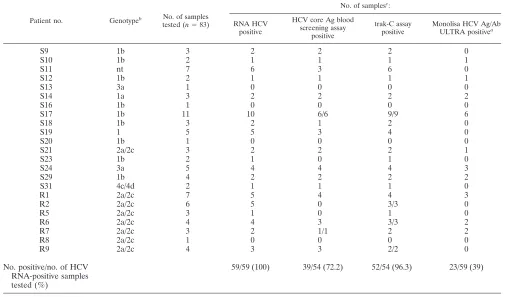
Panel 2: hemodialysis patient samples.
Of the 83 anti-HCV
Ab-negative samples collected before the seroconversion
(Ta-ble 2), the 24 HCV RNA- and HCV core Ag assay-negative
samples were also negative by the Monolisa HCV Ag/Ab
UL-TRA assay. Among the 59 anti-HCV Ab-negative, HCV
RNA-positive samples (39 of 54 were HCV core Ag blood screening
assay positive and 52 of 54 were positive by the trak-C assay),
23 (39%) were positive by the HCV Ag/Ab assay. No
differ-ence was observed according to genotype. All 23 samples were
also positive by both HCV core Ag assays. Table 3 shows the
mean delays for the early detection of HCV infection between
assays. The mean delays between positive results by HCV
RNA testing and positive results by the Monolisa HCV Ag/Ab,
between positive results by the trak-C assay and Monolisa
HCV Ag/Ab ULTRA assay, and between positive results by
the HCV core Ag blood screening assay and the Monolisa
HCV Ag/Ab ULTRA assay were 30.3 days (36 of 59
⫽
61% of
discrepant results), 27.9 days (29 of 52
⫽
55.8% of discrepant
results), and 16.3 days (11 of 39
⫽
28% of discrepant results),
respectively. In addition, the Monolisa HCV Ag/Ab ULTRA
could detect HCV infection before the most sensitive HCV Ab
assay (defined as the first HCV Ab assay which detected the
appearance of anti-HCV Ab for each patient) and Monolisa
anti-HCV Plus version 2 could, with mean delays of 21.6 days
and 30.1 days, respectively.
Among the 52 trak-C assay-positive samples that were
col-lected during the window phase, 22 of 26 samples with an HCV
core Ag concentration greater than 260 pg/ml were Monolisa
HCV Ag/Ab ULTRA positive, whereas only 1 of 26 samples
with a core Ag level below 260 pg/ml was Monolisa HCV
Ag/Ab ULTRA positive (
P
⬍
10
⫺3).
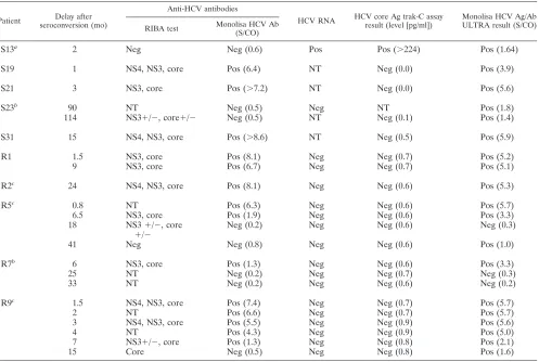
Of the 108 samples collected during or after seroconversion,
4 were Monolisa HCV Ag/Ab ULTRA negative. One sample
(patient S11, Table 3) corresponded to an early seroconversion
(RIBA test c33
⫹
/
⫺
) and was positive for HCV RNA but
negative by the Monolisa HCV Ab (the trak-C assay was not
performed). The three other Monolisa HCV Ag/Ab
ULTRA-negative samples (which were also ULTRA-negative for all other HCV
markers) (Table 4) were collected from two patients who
re-covered from infection (patient R5, one sample collected 17
months after interferon treatment; patient R7, two samples
collected 25 and 33 months after seroconversion and
corre-TABLE 4. Characteristics of the 22 samples collected after HCV seroconversion from 10 patients who became negative for anti-HCV
antibody (Monolisa HCV Ab assay) (
n
⫽
8) and/or HCV core Ag (trak-C assay) (
n
⫽
20) during the follow-up
dPatient Delay after seroconversion (mo)
Anti-HCV antibodies
HCV RNA HCV core Ag trak-C assay result (level [pg/ml])
Monolisa HCV Ag/Ab ULTRA result (S/CO) RIBA test Monolisa HCV Ab
(S/CO)
S13
a2
Neg
Neg (0.6)
Pos
Pos (
⬎
224)
Pos (1.64)
S19
1
NS4, NS3, core
Pos (6.4)
NT
Neg (0.0)
Pos (3.9)
S21
3
NS3, core
Pos (
⬎
7.2)
NT
Neg (0.0)
Pos (5.6)
S23
b90
NT
Neg (0.5)
Neg
NT
Pos (1.8)
114
NS3
⫹
/
⫺
, core
⫹
/
⫺
Neg (0.5)
NT
Neg (0.1)
Pos (1.4)
S31
15
NS4, NS3, core
Pos (
⬎
8.6)
NT
Neg (0.5)
Pos (5.9)
R1
1.5
NS3, core
Pos (8.1)
Neg
Neg (0.7)
Pos (5.2)
9
NS3, core
Pos (6.7)
Neg
Neg (0.7)
Pos (5.1)
R2
c24
NS4, NS3, core
Pos (8.1)
Neg
Neg (0.6)
Pos (5.3)
R5
c0.8
NT
Pos (6.3)
Neg
Neg (0.6)
Pos (5.7)
6.5
NS3, core
Pos (1.9)
Neg
Neg (0.6)
Pos (3.3)
18
NS3
⫹
/
⫺
, core
⫹
/
⫺
Neg (0.2)
Neg
Neg (0.6)
Neg (0.3)
41
Neg
Neg (0.8)
Neg
Neg (0.6)
Pos (1.0)
R7
b6
NS3, core
Pos (1.3)
Neg
Neg (0.6)
Pos (3.3)
25
NT
Neg (0.2)
Neg
Neg (0.7)
Neg (0.3)
33
NT
Neg (0.2)
Neg
Neg (0.6)
Neg (0.2)
R9
c1.5
NS4, NS3, core
Pos (7.4)
Neg
Neg (0.7)
Pos (5.7)
2
NT
Pos (6.6)
Neg
Neg (0.7)
Pos (5.7)
3
NS4, NS3, core
Pos (5.5)
Neg
Neg (0.9)
Pos (5.6)
4
NT
Pos (4.3)
Neg
Neg (0.9)
Pos (5.0)
7
NS3
⫹
/
⫺
, core
Pos (1.3)
Neg
Neg (0.8)
Pos (2.1)
15
Core
Neg (0.5)
Neg
Neg (0.8)
Pos (1.6)
aIntermediate antibody-negative result. bSpontaneous resolved infection.
cInterferon treatment (R2, 15 months after seroconversion; R5, 1 month after seroconversion; R9, 1 month after seroconversion). dAbbreviations: NT, not tested; Neg, negative; Pos, positive; S/CO, sample OD/cutoff OD.
on May 15, 2020 by guest
http://jcm.asm.org/
[image:4.585.46.543.89.423.2]sponding to a spontaneous resolved infection). Of the 104
Monolisa HCV Ag/Ab ULTRA-positive samples, 17 were
trak-C assay negative (14 were anti-HCV Ab positive and 3
were anti-HCV Ab negative by the Monolisa HCV Ab assay)
and 8 were Monolisa HCV Ab assay negative (3 were trak-C
assay negative). All these 17 samples (Table 4) (14 of which
were HCV RNA negative and 3 of which were not tested) were
collected from patients who recovered from infection
sponta-neously or after antiviral treatment.
DISCUSSION
The diagnosis of HCV infection is currently based on the
detection of anti-HCV Ab by EIAs, and it is confirmed by a
positive result obtained by an immunoblot assay or by the
presence of HCV RNA (27, 34). However, when an early
diagnosis of HCV infection is required, such as in emergency
situations (health care worker exposures) (47), in blood
screening to reduce the residual risk of transmission through
blood (5) or grafts (9, 24), or as a pretreatment screening for
at-risk populations, two diagnostic approaches based on the
detection of the HCV RNA or HCV core Ag are currently
available. The prevention of viral transmission by blood,
thanks to the implementation of these methods for routine
blood screening, has been reported (10, 14, 25, 39, 40, 44).
However, since nucleic acid testing could not totally prevent
HCV transmission, anti-HCV serologic screening must be
con-tinued (6, 22, 33) and viral screening approaches can be
im-plemented only as supplemental tests. Thus, a new assay that
combines the detection of HCV antigen and antibodies could
preclude the need for additional assays in routine screening
and thereby minimize the cost of the improvement of blood
safety, especially with regard to NAT procedures (17, 28, 43).
Concerning the early diagnosis of HCV infection in
sero-negative individuals, our study indicates that the Monolisa
HCV Ag/Ab ULTRA assay was able to detect the infection in
40.8% of HCV-RNA positive samples. The performance in
terms of sensitivity was not as high as that observed by HCV
core Ag assays (73.8% and 96.9% of positive samples for HCV
core Ag blood screening assay and trak-C assay, respectively)
(Table 5). These differences were due to the lower sensitivity of
the Monolisa HCV Ag/Ab ULTRA assay for the detection of
HCV core proteins than those of specific HCV core Ag assays.
However, this HCV Ag/Ab combined assay significantly
re-duces the window period to 21.6 days and 30.1 days compared
with that of the most sensitive assay and Monolisa HCV Ab
assay, respectively. Moreover, 50% of the blood donation
sam-ples in panel 1 that were positive only by NAT were also
detected by the Monolisa HCV Ag/Ab ULTRA assay,
indicat-ing a better sensitivity compared to those of the antibody-only
assays. When the present seronegative window period of 66
days is considered (2), the yield of this HCV Ag/Ab assay
would correspond to a 33 to 46% reduction in the HCV
trans-fusion-transmitted residual risk. Thus, we can assume that the
implementation of this assay in countries where only the
anti-HCV antibody screening is performed would be an efficient
measure that could be used to improve blood safety.
After seroconversion, HCV RNA, as well as HCV antigen,
become undetectable (especially if the blood screening HCV
Ag core assay is used). This is the consequence of a progressive
decline in viremia after seroconversion (5), and it is also
prob-ably due to the fact that HCV antigens are in the form of
immune complexes. In such cases, NAT and HCV Ag-specific
assays could be inefficient for the detection of HCV infection
(19). Monolisa HCV Ag/Ab ULTRA also gave negative
re-sults, but only after spontaneous or treatment-induced
resolu-tion of infecresolu-tions, when, evidently, all markers of HCV
infec-tion are undetectable. Contrary to what is observed in rare
circumstances of early infection with human immunodeficiency
virus (HIV) with some HIV Ag/Ab combined assays (26), we
did not evidence a gap between Ag and immunoglobulin G
detection, despite the fact that the HCV combined assay uses
an indirect EIA for anti-HCV antibody detection. Thus, the
improved sensitivity of the Ag detection test did not impair
antibody detection.
[image:5.585.43.543.83.161.2]Although this new assay is not as sensitive as HCV
Ag-specific assays, it presents undeniable performance in terms of
sensitivity for the detection of HCV infection, especially in the
early phase, when antibodies are undetectable. Its represents a
notable improvement for the detection of HCV infection when
it is compared with the anti-HCV Ab detection assays. The
simultaneous detection of HCV antigen and antibodies
pro-vides a valuable alternative to methods that directly detect
viremia, such as NAT or an HCV Ag-specific assay, especially
with regards to cost, organization, emergency, and logistic
dif-ficulties. It could be implemented for blood screening in
de-veloping countries where NAT or HCV Ag assays are not
affordable or not feasible. In addition, the requirement for a
fast screening of organ transplant donors makes the HCV
Ag/Ab combined assay an attractive option, since a high HCV
prevalence has been observed in this population (9). Finally, it
could contribute to decision making when preventive antiviral
therapy has to be implemented in the case of occupational
exposure. As observed for HIV Ag/Ab combined assays (26),
the improvement of the HCV antigen detection of HCV
TABLE 5. Sensitivity of Monolisa HCV Ag/Ab ULTRA for HCV RNA-positive but anti-HCV Ab-negative samples
Sample
Total no. of HCV RNA-positive
samples
No. of samples positive by individual assay/total no. tested (%):
HCV core Ag blood
screening assay trak-C assay
Monolisa HCV Ag/Ab ULTRA assay
Blood donors
12
9/11 (81.8)
12 (100)
6
a(50)
Hemodialyzed patients
59
39/54 (72.2)
52/54 (96.3)
23/59
b(39)
Total
71
48/65 (73.8)
64/66 (96.9)
29/71 (40.8)
a
Two of six samples were in the gray zone.
b
Four of 23 samples were in the gray zone.
on May 15, 2020 by guest
http://jcm.asm.org/
Ag/Ab assays would allow the benefit provided by these new
tools to be increased.
ACKNOWLEDGMENT
We thank Camille Sureau for helpful advice and comments.
REFERENCES
1.Alvarez, M., D. Planelles, E. Vila, J. Montoro, and E. Franco.2004. Pro-longed hepatitis C virus seroconversion in a blood donor, detected by HCV antigen test in parallel with HCV RNA. Vox Sang.86:266–267.
2.Assal, A., J. Coste, V. Barlet, S. Laperche, C. Cornillot, W. Smilovici, J. Pillonel, and G. Andreu.2003. Application of molecular biology to blood transfusion safety: the nucleic acid testing. Transfus. Clin. Biol10:217–226. 3.Barrera, J. M., B. Francis, G. Ercilla, M. Nelles, D. Achord, J. Darner, and S. R. Lee.1995. Improved detection of anti-HCV in post-transfusion hepa-titis by a third-generation ELISA. Vox Sang.68:15–18.
4.Bouvier-Alias, M., K. Patel, H. Dahari, S. Beaucourt, P. Larderie, L. Blatt, C. Hezode, G. Picchio, D. Dhumeaux, A. U. Neumann, J. G. McHutchison, and J. M. Pawlotsky.2002. Clinical utility of total HCV core antigen quan-tification: a new indirect marker of HCV replication. Hepatology36:211– 218.
5.Busch, M.2001. Closing the windows on viral transmission by blood trans-fusion, p. 33–54.InS. L. Stramer (ed.), Blood safety in the new millenium. American Association of Blood Banks, Bethesda, Md.
6.Busch, M. P., L. H. Tobler, W. H. Gerlich, S. Schaefer, C. Giachetti, and R. Smith.2003. Very low level viremia in HCV infectious unit missed by NAT. Transfusion43:1173–1174.
7.Buti, M., C. Mendez, M. Schaper, S. Sauleda, A. Valdes, F. Rodriguez-Frias, R. Jardi, and R. Esteban.2004. Hepatitis C virus core antigen as a predictor of non-response in genotype 1 chronic hepatitis C patients treated with peginterferon alpha-2b plus ribavirin. J. Hepatol.40:527–532.
8.Cano, H., M. J. Candela, M. L. Lozano, and V. Vicente.2003. Application of a new enzyme-linked immunosorbent assay for detection of total hepatitis C virus core antigen in blood donors. Transfus. Med.13:259–266.
9.Challine, D., B. Pellegrin, M. Bouvier-Alias, P. Rigot, L. Laperche, and J. M. Pawlotsky.2004. HIV and hepatitis C virus RNA in seronegative organ and tissue donors. Lancet364:1611–1612.
10.Coste, J., T. Levayer, J. Izopet, S. Laperche, P. Parneix, H. Rech, J. L. Payen, and J. F. Schved.2003. Acute HCV infection detected in a blood donor by NAT. Transfusion43:116–118.
11.Courouce, A. M., N. Le Marrec, F. Bouchardeau, A. Razer, M. Maniez, S. Laperche, and N. Simon. 2000. Efficacy of HCV core antigen detection during the preseroconversion period. Transfusion40:1198–1202. 12.Courouce, A. M., J. Pillonel, et al.1996. Transfusion-transmitted viral
infec-tions. N. Engl. J. Med.335:1609–1610.
13.Durand, F., A. Beauplet, and P. Marcellin.2000. Evidence of hepatitis C virus viremia without detectable antibody to hepatitis C virus in a blood donor. Ann. Intern. Med.133:74–75.
14.Eiras, A., S. Sauleda, D. Planelles, M. Sedeno, A. Ibarra, M. A. Vesga, E. Franco, J. A. Montoro, J. M. Hernandez, and R. G. Villaescusa.2003. HCV screening in blood donations using RT-PCR in mini-pool: the experience in Spain after routine use for 2 years. Transfusion43:713–720.
15.Fang, C. T., S. P. Field, M. P. Busch, and P. Heyns Adu.2003. Human immunodeficiency virus-1 and hepatitis C virus RNA among South African blood donors: estimation of residual transfusion risk and yield of nucleic acid testing. Vox Sang.85:9–19.
16.Glynn, S. A., S. H. Kleinman, D. J. Wright, and M. P. Busch.2002. Inter-national application of the incidence rate/window period model. Transfusion 42:966–972.
17.Jackson, B. R., M. P. Busch, S. L. Stramer, and J. P. AuBuchon.2003. The cost-effectiveness of NAT for HIV, HCV, and HBV in whole-blood dona-tions. Transfusion43:721–729.
18.Kolk, D. P., J. Dockter, J. Linnen, M. Ho-Sing-Loy, K. Gillotte-Taylor, S. H. McDonough, L. Mimms, and C. Giachetti.2002. Significant closure of the human immunodeficiency virus type 1 and hepatitis C virus preseroconver-sion detection windows with a transcription-mediated-amplification-driven assay. J. Clin. Microbiol.40:1761–1766.
19.Krajden, M., R. Shivji, K. Gunadasa, A. Mak, G. McNabb, M. Friesenhahn, D. Hendricks, and L. Comanor.2004. Evaluation of the core antigen assay as a second-line supplemental test for diagnosis of active hepatitis C virus infection. J. Clin. Microbiol.42:4054–4059.
20.Kuo, G., Q. L. Choo, H. J. Alter, G. L. Gitnick, A. G. Redeker, R. H. Purcell, T. Miyamura, J. L. Dienstag, M. J. Alter, C. E. Stevens, et al.1989. An assay for circulating antibodies to a major etiologic virus of human non-A, non-B hepatitis. Science244:362–364.
21.Laperche, S.2005. Blood safety and nucleic acid testing in Europe. Euro Surveill.10:3–4.
22.Laperche, S., F. Bouchardeau, M. Maniez, M. Beolet, M. H. Elghouzzi, and J. J. Lefrere.2004. Nucleic acid testing in blood donations reactive to
hep-atitis C virus antibody, but with an extremely low viral load. Vox Sang. 86:198.
23.Laperche, S., N. Le Marrec, N. Simon, F. Bouchardeau, C. Defer, M. Maniez-Montreuil, T. Levayer, J. P. Zappitelli, and J. J. Lefrere.2003. A new HCV core antigen assay based on disassociation of immune complexes: an alternative to molecular biology in the diagnosis of early HCV infection. Transfusion43:958–962.
24.Lefrere, J. 1998. Se´curite´ virale et transplantation d’organes. Virologie 2:227–232.
25.Letowska, M., E. Brojer, M. Mikulska, A. Gronowska, and A. Rosiek.2004. Hepatitis C core antigen in Polish blood donors. Transfusion44:1067–1071. 26.Ly, T. D., S. Laperche, C. Brennan, A. Vallari, A. Ebel, J. Hunt, L. Martin, D. Daghfal, G. Schochetman, and S. Devare.2004. Evaluation of the sensi-tivity and specificity of six HIV combined p24 antigen and antibody assays. J. Virol. Methods122:185–194.
27.Maniez-Montreuil, M., and F. Dubois.2000. Interpretation of hepatitis C virus serology: immunoblot and genome amplification. Transfus. Clin. Biol. 7(Suppl. 1):25s–30s.
28.Marshall, D. A., S. H. Kleinman, J. B. Wong, J. P. AuBuchon, D. T. Grima, N. A. Kulin, and M. C. Weinstein.2004. Cost-effectiveness of nucleic acid test screening of volunteer blood donations for hepatitis B, hepatitis C and human immunodeficiency virus in the United States. Vox Sang.86:28–40. 29.Maynard, M., P. Pradat, P. Berthillon, G. Picchio, N. Voirin, M. Martinot,
P. Marcellin, and C. Trepo.2003. Clinical relevance of total HCV core antigen testing for hepatitis C monitoring and for predicting patients’ re-sponse to therapy. J. Viral Hepat.10:318–323.
30.Mine, H., H. Emura, M. Miyamoto, T. Tomono, K. Minegishi, H. Murokawa, R. Yamanaka, A. Yoshikawa, and K. Nishioka. 2003. High throughput screening of 16 million serologically negative blood donors for hepatitis B virus, hepatitis C virus and human immunodeficiency virus type-1 by nucleic acid amplification testing with specific and sensitive multiplex reagent in Japan. J. Virol. Methods112:145–151.
31.Netski, D. M., X. H. Wang, S. H. Mehta, K. Nelson, D. Celentano, S. Thongsawat, N. Maneekarn, V. Suriyanon, J. Jittiwutikorn, D. L. Thomas, and J. R. Ticehurst.2004. Hepatitis C virus (HCV) core antigen assay to detect ongoing HCV infection in Thai injection drug users. J. Clin. Micro-biol.42:1631–1636.
32.Nubling, C. M., G. Unger, M. Chudy, S. Raia, and J. Lower.2002. Sensitivity of HCV core antigen and HCV RNA detection in the early infection phase. Transfusion42:1037–1045.
33.Operskalski, E. A., J. W. Mosley, L. H. Tobler, E. W. Fiebig, M. J. Nowicki, L. T. Mimms, J. Gallarda, B. H. Phelps, and M. P. Busch.2003. HCV viral load in anti-HCV-reactive donors and infectivity for their recipients. Trans-fusion43:1433–1441.
34.Pawlotsky, J. M.2002. Use and interpretation of virological tests for hepa-titis C. Hepatology36:S65–S73.
35.Peterson, J., G. Green, K. Iida, B. Caldwell, P. Kerrison, S. Bernich, K. Aoyagi, and S. R. Lee.2000. Detection of hepatitis C core antigen in the antibody negative ‘window’ phase of hepatitis C infection. Vox Sang.78:80– 85.
36.Pillonel, J., and S. Laperche.2005. Trends in risk of transfusion-transmitted viral infections (HIV, HCV, HBV) in France between 1992 and 2003 and impact of nucleic acid testing (NAT). Euro Surveill.10:5–8.
37.Pillonel, J., S. Laperche, C. Saura, J. C. Desenclos, and A. M. Courouce. 2002. Trends in residual risk of transfusion-transmitted viral infections in France between 1992 and 2000. Transfusion42:980–988.
38.Raker, C. A., E. Tabor, A. Okayama, M. Y. Yu, M. Kohara, N. E. Mueller, H. Tsubouchi, and S. O. Stuver.2004. HCV core antigen as an alternative to NAT to detect HCV viremia. Transfusion44:307–308.
39.Roth, W. K., M. Weber, S. Buhr, C. Drosten, W. Weichert, W. Sireis, D. Hedges, and E. Seifried.2002. Yield of HCV and HIV-1 NAT after screen-ing of 3.6 million blood donations in central Europe. Transfusion42:862– 868.
40.Sanz, C., D. Tassies, J. Costa, C. Freire, and A. Pereira.2002. The first case of HCV infection detected before seroconversion in blood donors tested by HCV core antigen ELISA. Transfusion42:505–506.
41.Seed, C. R., A. Cheng, S. L. Ismay, W. V. Bolton, P. Kiely, T. J. Cobain, and A. J. Keller.2002. Assessing the accuracy of three viral risk models in predicting the outcome of implementing HIV and HCV NAT donor screen-ing in Australia and the implications for future HBV NAT. Transfusion 42:1365–1372.
42.Shah, D. O., C. D. Chang, L. X. Jiang, K. Y. Cheng, A. S. Muerhoff, R. A. Gutierrez, T. P. Leary, S. M. Desai, I. V. Batac-Herman, V. A. Salbilla, A. S. Haller, J. L. Stewart, and G. J. Dawson.2003. Combination HCV core antigen and antibody assay on a fully automated chemiluminescence ana-lyzer. Transfusion43:1067–1074.
43.Simmonds, P., J. Kurtz, and R. Tedder.2002. The UK blood transfusion service: over a (patent) barrel. Lancet359:1713–1714.
44.Stramer, S. L., S. A. Glynn, S. H. Kleinman, D. M. Strong, C. Sally, D. J. Wright, R. Y. Dodd, and M. P. Busch.2004. Detection of HIV-1 and HCV infections among antibody-negative blood donors by nucleic acid-amplifica-tion testing. N. Engl. J. Med.351:760–768.
on May 15, 2020 by guest
http://jcm.asm.org/
45.Tanaka, E., C. Ohue, K. Aoyagi, K. Yamaguchi, S. Yagi, K. Kiyosawa, and H. J. Alter.2000. Evaluation of a new enzyme immunoassay for hepatitis C virus (HCV) core antigen with clinical sensitivity approximating that of genomic amplification of HCV RNA. Hepatology32:388–393.
46.Tobler, L. H., S. L. Stramer, S. R. Lee, B. L. Masecar, J. E. Peterson, E. A. Davis, W. E. Andrews, J. P. Brodsky, S. H. Kleinman, B. H. Phelps, and M. P. Busch.2003. Impact of HCV 3.0 EIA relative to HCV 2.0 EIA on blood-donor screening. Transfusion43:1452–1459.
47.Toyoda, H., H. Sakamoto, T. Mizuno, Y. Horiguchi, and H. Nakano.2000. Eradication of hepatitis C virus 1b by interferon in a health care worker with acute hepatitis following needlestick transmission from a patient with chronic hepatitis C unresponsive to interferon. Scand. J. Gastroenterol. 35:1117–1120.
48.Velati, C., L. Romano, L. Baruffi, M. Pappalettera, V. Carreri, and A. R. Zanetti.2002. Residual risk of transfusion-transmitted HCV and HIV infec-tions by antibody-screened blood in Italy. Transfusion42:989–993.