Nonstructural Protein NS5B and Fatty Acid Synthase
Jing-Tang Huang,a,bChing-Ping Tseng,cMei-Huei Liao,aShao-Chun Lu,dWei-Zhou Yeh,aNaoya Sakamoto,eChuan-Mu Chen,b Ju-Chien Chenga
Department of Medical Laboratory Science and Biotechnology, China Medical University, Taichung, Taiwana
; Department of Life Sciences, National Chung Hsing University, Taichung, Taiwanb
; Department of Medical Biotechnology and Laboratory Science, Chang Gung University, Taoyuan, Taiwanc
; Graduate Institute of Biochemistry and Molecular Biology, National Taiwan University, Taipei, Taiwand
; Department of Gastroenterology and Hepatology, Tokyo Medical and Dental University, Tokyo, Japane
Hepatitis C virus (HCV) nonstructural protein 5B (NS5B) is an RNA-dependent RNA polymerase (RdRp) that acts as a key
player in the HCV replication complex. Understanding the interplay between the viral and cellular components of the HCV
rep-lication complex could provide new insight for prevention of the progression of HCV-associated hepatocellular carcinoma
(HCC). In this study, the NS5B protein was used as the bait in a pulldown assay to screen for NS5B-interacting proteins that are
present in Huh7 hepatoma cell lysates. After mass spectrophotometric analysis, fatty acid synthase (FASN) was found to interact
with NS5B. Coimmunoprecipitation and double staining assays further confirmed the direct binding between NS5B and FASN.
The domain of NS5B that interacts with FASN was also determined. Moreover, FASN was associated with detergent-resistant
lipid rafts and colocalized with NS5B in active HCV replication complexes. In addition, overexpression of FASN enhanced HCV
expression in Huh7/Rep-Feo cells, while transfection of FASN small interfering RNA (siRNA) or treatment with FASN-specific
inhibitors decreased HCV replication and viral production. Notably, FASN directly increased HCV NS5B RdRp activity
in vitro
.
These results together indicate that FASN interacts with NS5B and modulates HCV replication through a direct increase of NS5B
RdRp activity. FASN may thereby serve as a target for the treatment of HCV infection and the prevention of HCV-associated
HCC progression.
H
epatitis C virus (HCV) is a positive-stranded RNA virus
clas-sified in the
Hepacivirus
genus in the family
Flaviviridae
. The
9.6-kb viral RNA genome encodes a precursor polyprotein that is
processed to generate at least 10 viral proteins, including
struc-tural proteins (core, E1, E2, and p7) and nonstrucstruc-tural proteins
(NS2, NS3, NS4A, NS4B, NS5A, and NS5B) (
1
). The NS5B RNA
polymerase, together with other nonstructural viral proteins
(NS3, NS4A, NS4B, and NS5A) and host factors, constitutes the
active replication complexes (RC) for viral RNA replication.
These proteins are directly or indirectly associated with the
endo-plasmic reticulum (ER)-derived structure called the
“membra-nous web,” where replication occurs (
2
,
3
). However, the exact
host factors and detailed interactions within the RC remain to be
determined.
HCV infection usually causes chronic hepatitis and frequently
leads to cirrhosis and hepatocellular carcinoma (HCC). Besides,
HCV-induced end-stage liver disease is an important indication
for liver transplantation in most of the Western countries (
4
,
5
). At
present, no effective vaccine to prevent HCV infection is available.
The current treatment strategies for HCV infection are valid only
for individuals with a particular single nucleotide polymorphism
(SNP) in the interleukin-28B gene or for infections caused by
certain viral genotypes (
6
). Accordingly, the prevalence of hepatic
steatosis in HCV-infected patients is much higher than that in the
general population or in hepatitis B virus (HBV)-infected patients
(
7
). Hepatic steatosis has also been reported to be associated with
an increased rate of HCC in chronic hepatitis C patients (
8
).
Lipid metabolic pathways are essential for the entry, secretion,
and replication of HCV. For example, apolipoprotein E (apoE) is
essential for the production of HCVcc (HCV produced in cell
culture) and for viral entry (
9
,
10
). Downregulation of apoA-I
decreases levels of HCV replication and viral particle production
in cell culture (
11
). HCV coopts the secretory pathway of very low
density lipoprotein (VLDL) for its own secretion (
12
,
13
).
More-over, HCV replication is regulated through induction of lipogenic
gene expression in HCV replicon cells (
14
) or geranylgeranylation
of host proteins required for HCV RNA replication (
15
,
16
). Fatty
acid synthesis is also required for HCV RNA replication (
14
).
In-hibition of fatty acid synthase (FASN) by cerulenin (
17
) or C75
(
18
) reduces the replication of subgenomic HCV replicons as well
as JFH-1-based HCVcc virion production. Although the
underly-ing mechanisms are not yet completely understood, these studies
imply that FASN is essential for HCV replication.
In this study, HCV NS5B was used as the bait to screen for
NS5B-interacting proteins that are present in Huh7 hepatoma cell
lysates. After mass spectrophotometric analysis, FASN was found
to interact with NS5B, and this interaction was further confirmed
in vitro
and
in vivo
. Our data indicate that FASN interacts with
NS5B to enhance NS5B RNA-dependent RNA polymerase
(RdRp) activity and subsequently facilitate HCV replication.
Taken together, these data suggest a critical role for FASN-NS5B
Received15 September 2012Accepted7 February 2013
Published ahead of print20 February 2013
Address correspondence to Ju-Chien Cheng, [email protected], or Chuan-Mu Chen, [email protected].
Supplemental material for this article may be found athttp://dx.doi.org/10.1128 /JVI.02526-12.
Copyright © 2013, American Society for Microbiology. All Rights Reserved.
doi:10.1128/JVI.02526-12
on November 7, 2019 by guest
http://jvi.asm.org/
interaction in the modulation of HCV replication. The roles of
FASN in the regulation of HCV pathogenesis are also discussed.
MATERIALS AND METHODS
Cells and materials.The HCV subgenomic replicon cell line Sg-PC2 (19) was a gift from Jing-Hsiung Ou (University of Southern California, Los Angeles, CA). The HCV subgenomic replicon cell line Huh7/Rep-Feo (genotype 1b) expressing a luciferase construct was obtained from Naoya Sakamoto (Tokyo Medical and Dental University, Tokyo, Japan). The Huh7.5.1 cells and plasmid JC1-Luc2A, which replaces the Rluc gene of pJ6/JFH(p7-Rlu2A) with the firefly luciferase (Luc) gene, were kindly pro-vided by Robert T. Schooley (University of California, San Diego). A rabbit polyclonal antibody against the HCV NS5B protein was kindly provided by Takaji Wakita (National Institute of Infectious Diseases, To-kyo, Japan). Plasmid pCMV-SPORT6-FASN, the transfection reagent Li-pofectamine 2000 (LF2000), and the Alexa Fluor 568-conjugated goat anti-rabbit IgG were purchased from Invitrogen (Carlsbad, CA). The syn-thetic small interfering RNA (siRNA) for FASN (siFASN; 5=-AACCCTG AGATCCCAGCGCTG-3=) and siCONTROL nontargeting siRNA-2 (siC) were from Dharmacon (Lafayette, CO). The siRNA for green fluo-rescent protein (siGFP) was purchased from Ambion (Austin, TX). The pLKO.1 vector carrying short hairpin RNA (shRNA) for GFP (pLKO.1-shGFP; clone identification [ID], TRCN0000072197), pLKO.1-shFASN (clone IDs, TRCN000003127 and TRCN000003128), pCMVdR, and pMD2.G were purchased from the National RNAi Core Facility (Aca-demia Sinica, Taipei, Taiwan). The transfection reagent Arrest-In was obtained from Open Biosystems (Lafayette, CO). The FASN inhibitor C75 and the anti-caveolin-2, anti-calnexin, and mouse monoclonal anti-HCV NS5B antibodies were purchased from Enzo Life Sciences (Farmingdale, NY). S7 nuclease and the FuGENE HD transfection reagent were pur-chased from Roche (Mannheim, Germany). Glutathione Sepharose 4B and protein A Sepharose were purchased from GE Healthcare (Piscat-away, NJ). The 3-(4,5-dimethylthiazol-2-yl)-5-(3-carboxymethoxyphe-nyl)-2-(4-sulfophenyl)-2H-tetrazolium (MTS) reduction assay and Bright-Glo luciferase assay reagents were purchased from Promega (Mad-ison, WI). The anti-HCV NS5A antibody was purchased from BioDesign (Carmel, NY). The anti-HCV NS3 antibody was purchased from Novo-castra (Newcastle, United Kingdom). The anti-HCV core antibody was purchased from Thermo Scientific (Rockford, IL). Proteinase K, pyruvate kinase, the anti--actin antibody, and the anti-Flag M2 affinity gel were purchased from Sigma (St. Louis, MO). The anti-FASN and anti-GRP78 antibodies and normal rabbit IgG were purchased from Santa Cruz Bio-technology (Santa Cruz, CA). HiLyte Fluor 488-conjugated goat anti-mouse IgG was purchased from AnaSpec (San Jose, CA). The NS5B in-hibitor 2=-C-methyladenosine (2=CMA) was purchased from Carbosynth (Berkshire, United Kingdom). Phosphoenolpyruvate was purchased from Alfa Aesar (Ward Hill, MA). EasyBlocker and EasyBlot anti-rabbit IgG were purchased from GeneTex (Irvine, CA). [␣-32P]CTP was purchased from Perkin-Elmer (Wellesley, MA).
Plasmid construction.To generate pGEX-2T-NS5Bd21, the NS5B frag-ment with a deletion of the C-terminal 21 amino acids (NS5Bd21) (20) was amplified by PCR using the forward primer NS5bGF (5=-CGCGGATCCAC CATGTCCTACACATGG-3=) and the reverse primer NS5bER (5=-GGC TACTTAAGTCAGCGGGGTCGGGCACGAG-3=). The PCR product was then subcloned into the pGEM-T Easy vector (Promega, Madison) to generate pGEN-T-NS5B. The BamHI-EcoRI fragment of pGEM-T-NS5B, containing the NS5B coding sequences, was subcloned into pGEX-2T (GE Healthcare, Sweden). The NS5Bd21 fragment from pGEM-T-NS5B was further subcloned into pFlag-CMV2 to generate pCMV-Flag-NS5B or into pET32a to generate pET32a-NS5Bd21.
For NS5B deletion mutants, the deletion fragments were generated by PCR using pCMV-Flag-NS5B as the template. The primer sets are listed in Table 1. The PCR product was ligated into the pTOPO vector and was further subcloned into pFlag-CMV2 to generate pCMV-Flag-NS5B/1-180, pCMV-Flag-NS5B/1-335, and pCMV-Flag-NS5B/179-570.
To generate pcDNA3.1/HisC-FASN, an EcoRI-XhoI DNA fragment of pCMV-SPORT6-FASN containing FASN cDNA sequences was cloned into pCMV-Tag3 (Stratagene). Then an EcoRI-XhoI DNA fragment con-taining FASN sequences was subcloned into pcDNA3.1/HisC (Invitro-gen) to generate pcDNA3.1/HisC-FASN.
GST pulldown assay.The glutathioneS-transferase (GST) pulldown assay was performed as described by the manufacturer (GE Healthcare). Briefly, plasmids pGEX-2T-NS5Bd21 and pGEX-2T were transformed into theEscherichia colistrain BL21(DE3)pLysS, respectively. The bacteria were then induced with 1 mM isopropyl-D-1-thiogalactopyranoside
(IPTG) at 22°C overnight and were subsequently collected by centrifuga-tion at 6,000 rpm for 20 min. Cell pellets were resuspended in lysis buffer (1⫻phosphate-buffered saline [PBS] containing 1% Triton X-100 and 1 mM dithiothreitol [DTT]) and were sonicated for 4 min (10-s sonication and 10-s pause). Ten milligrams of bacterial lysates was incubated with 66 l glutathione Sepharose 4B for 1 h at 4°C. After three washes with lysis buffer, the GST- and GST fusion protein-binding beads were mixed with 1.5 mg Huh7 cell lysates suspended in GST pulldown buffer {10 mM Tris-HCl, 140 mM NaCl, 0.5 mM calcium chloride, 0.5 mM magnesium chloride, and freshly added 1% 3-[(3-cholamidopropyl)-dimethylammo-nio]-1-propanesulfonate (CHAPS)} at 4°C overnight. The beads were then washed four times with GST pulldown buffer and were separated by 10% sodium dodecyl sulfate-polyacrylamide gel electrophoresis (SDS-PAGE) for further analyses.
Silver staining.After protein separation by SDS-PAGE, the polyacryl-amide gel was fixed in buffer A (50% methanol and 25% acetic acid) for 2 h, followed by incubation with buffer B (30% methanol) for 15 min. After three rinses in distilled water, the gel was incubated in buffer C (0.8 mM sodium thiosulfate) for 2 min and was rinsed three times in distilled water. The gel was then incubated with buffer D (0.2% silver nitrate) for 25 min and was rinsed twice with distilled water. The gel was subsequently devel-oped with buffer E (0.28 M sodium carbonate, 0.85% formaldehyde, and 16M sodium thiosulfate) until protein bands were visible. The reaction was stopped by rinsing the gel briefly in distilled water, and the gel was then incubated in buffer F (42 mM EDTA).
In-gel digestion and MALDI-TOF mass spectrometric analysis.The band of interest displayed by silver staining was subjected to excision. Gel digestion was performed as described previously (21). Briefly, the gel was washed with wash buffer (25 mM NH4HCO3 and 50% acetonitrile [ACN]) and was destained in a solution with 1% potassium ferricyanide and 1.5% sodium thiosulfate. The protein was reduced in 10 mM dithio-threitol in 25 mM NH4HCO3for 1 h at 56°C and was alkylated with 55 mM iodoacetamide in 25 mM NH4HCO3at room temperature for 30 min in the dark. The protein was dehydrated with ACN and was then digested with trypsin at 37°C overnight, followed by extraction with 0.5% trichlo-roacetic acid (TCA). Matrix-assisted laser desorption ionization–time of flight mass spectrometric (MALDI-TOF MS) analysis was then performed using the Ultraflex MALDI-TOF mass spectrometer (Bruker-Daltonics).
TABLE 1Primer sequences for plasmid constructs
Primer name Primer sequencea
Length (mer)
CNS5BYF 5=-CACGCGTCGACGTCGATGTCCTACACATGG-3= 30 CNS5BtYR 5=-GGACTAGTCCGCGGGGTCGGGCACGAG-3= 27 NS5BGF 5=-CGCGGATCCACCATGTCCTACACATGG-3= 27 NS5BER 5=-GGCTAGAATTCTCAGCGGGGTCGGGCACGAG-3= 31 NS5B 1-180 F 5=-TTCGATGTCCTACACATGGACA-3= 22 NS5B 1-180 R 5=-TCAGGAGACCACATCGTAAAGG-3= 22 NS5B 179-570 F 5=-GCATCCCAAGCTTGTCTCCACCCTTCCTCAAG-3= 32 NS5B 179-570 R 5=-CGGAATTCTCAGCGGGGTCGGGCACGAGA-3= 29 NS5B 1-335 F 5=-CCCAAGCTTTCGATGTCCTACACATGGACAG-3= 31 NS5B 1-335 R 5=-CGGAATTCTCAGCTCGCCGCGTCCTCCTGG-3= 30
a
Underlining indicates the SalI site for CNS5BYF, the SpeI site for CNS5BtYR, the BamHI site for NS5BGF, the EcoRI site for NS5BER, the HindIII site for NS5B 179-570 F and NS5B 1-335 F, and the EcoRI site for NS5B 179-570 R and NS5B 1-335 R.
on November 7, 2019 by guest
http://jvi.asm.org/
The peptide sequence data were searched against the MASCOT search database (Matrix Science, London, United Kingdom).
Transfection.HEK293T or Huh7 cells were seeded into 60-mm or 35-mm culture dishes for 24 h. Three micrograms of pCMV3B-FASN and 3g of pCMV-Flag-NS5B or pcDNA-NS5A were cotransfected into 293T cells by use of the Arrest-In transfection reagent (Thermo Scientific) or into Huh7 cells by use of the Fugene HD transfection reagent (Roche). At 48 h posttransfection, either cell lysates were prepared for immunopre-cipitation or the cells were fixed for immunofluorescence staining. To evaluate the effects of FASN expression on HCV replication, Huh7/Rep-Feo cells were seeded at a density of 1⫻104/well. Then 2g of pCMV3B-FASN or the pCMV3B vector control plasmid was transfected into the cells by use of LF2000. At 48 h after transfection, the luciferase activity was quantified using the Bright-Glo luciferase assay reagent. For lentivirus-mediated knockdown of FASN, HEK293 cells were seeded at a density of 2⫻106/60-mm culture dish for 24 h. Then 4g of the indicated pLKO.1-shRNA (Fig. 5) and 4g of pCMVdR and pMD2.G were transfected into HEK293 cells by use of the Arrest-In transfection reagent. At 4 h after transfection, the supernatant was removed and was replaced by fresh me-dium. The virus-containing supernatants collected at 24 and 36 h after transfection were combined and clarified by low-speed centrifugation, passed through a filter (pore size, 0.45m), and stocked at⫺80°C for further use.
Immunoprecipitation assay.The cells were dissolved in EBC lysis buffer (125 mM NaCl, 50 mM Tris-HCl [pH 7.4], 0.5 mM EDTA, 0.25% Nonidet P-40, 10g/ml aprotinin, 10g/ml leupeptin, 1 mM phenyl-methylsulfonyl fluoride [PMSF], 200M sodium orthovanadate, 10 mM sodium fluoride, and 100M EGTA) and were incubated on ice for 30 min. Subsequently, the lysates were clarified by centrifugation at 13,000 rpm for 2 min. The protein extracts were then incubated with 2g of the indicated antibody or control IgG at 4°C for 2 h. Protein A Sepharose beads were then added and were incubated at 4°C for 4 h. The immuno-precipitated complexes were washed three times with radioimmunopre-cipitation assay (RIPA) buffer (0.15 M NaCl, 0.01 M sodium phosphate [pH 7.2], 50 mM sodium fluoride, 0.2 mM sodium vanadate, 1% sodium deoxycholate, 0.1% SDS, 1% NP-40, 2 mM EDTA, and 100 U/ml apro-tinin). The protein complexes were then separated by 8% SDS-PAGE for Western blot analysis using the enhanced chemiluminescence (ECL) kit as described previously (20).
Immunofluorescence staining.The cells were washed twice with 1⫻ PBS and fixed with 4% paraformaldehyde at room temperature for 20 min. After permeabilization by 0.2% Triton X-100 on ice for 20 min, the permeable cells were incubated with 4% bovine serum albumin (BSA) at 37°C for 1 h. The cells were then incubated with the indicated antibody for 1 h at room temperature and were subsequently detected by an Alexa 488-conjugated anti-mouse secondary antibody or a rhodamine-conju-gated anti-rabbit secondary antibody for 1 h. The fluorescence-labeled proteins were observed with a Leica TCS SP2 confocal microscope and were analyzed in thex-zandy-zsections. The colocalization coefficient for the cell images (n⫽10) were calculated with MetaMorph software (ver-sion 7.0; Molecular Devices) (20).
RNA isolation and real-time quantitative RT-PCR.Total RNA was isolated by REzol C & T RNA extraction reagent as described by the man-ufacturer (Protech, Taipei, Taiwan). For the quantification of HCV RNA expression, total cellular RNA (100 ng) was subjected to one-step reverse transcription-PCR (RT-PCR) in a 25-l reaction mixture containing 2⫻ SYBR green PCR Master Mix with the primer set for HCV (HCV-F, 5=-T GCGGAACCGGTGAGTACA-3=; HCV-R, 5=-CTTAAGGTTTAGGATT CGTGCTCAT-3=) or the primer set for FASN (FASN-F, 5=-GAAACTGC AGGAGCTGTCC-3=; FASN-R, 5=-CACGGAGTTGAGCCGCAT-3=). The reaction conditions were as follows: 1 cycle of 48°C for 30 min, 1 cycle of 95°C for 10 min, and 40 cycles of 95°C for 15 s and 60°C for 1 min, by use of the ABI Prism 7000 sequence detection system.
The method for detecting the minus strand of HCV RNA was modi-fied from that in a previous report (22). In brief, the cDNA was generated
with a Taq-149 primer (5=-ACATGCGCGGCATCTAGATGCGGAACC GGTGAGTACA-3=) using Moloney murine leukemia virus (M-MLV) re-verse transcriptase and was subsequently quantified by real-time PCR with a 25-l reaction mixture containing 2⫻SYBR green PCR Master Mix and a primer set consisting of Taq (5= -ACATGCGCGGCATCTAGA-3=) and HCV-R. The reaction conditions were as follows: 1 cycle of 95°C for 10 min, followed by 40 cycles of 95°C for 15 s and 60°C for 1 min, by use of the ABI Prism 7000 sequence detection system. The expression of -actin was used as a normalization control. HCV RNA expression was quantified using the⌬⌬CTmethod, whereCTrepresents the threshold
cycle.
Infectious HCV particle production and infection inhibition assay.
Infectious HCV particles (HCVcc) were produced as described previously (23,24). Briefly,in vitro-transcribed genomic JC1-Luc2A RNA was deliv-ered into Huh7.5.1 cells by electroporation. The JC1-Luc2A HCV re-porter virus was recovered from cell culture medium. The virus-contain-ing supernatant was clarified by low-speed centrifugation, passed through a 0.45-m-pore-size filter, and concentrated by ultracentrifugation. For the infection inhibition assay, Huh7.5.1 cells were seeded in 24-well plates at a density of 5⫻104/well. Twenty-four hours later, the cells were in-fected with a lentivirus carrying shFASN in the presence of Polybrene (8 g/ml) for 24 h. The virus-containing supernatant was then removed, and fresh medium with puromycin was added for an additional 48 h. The FASN knockdown cells were then seeded in 96-well plates at a density of 1⫻104/well. At 24 h after plating, the HCV reporter virus (multiplicity of infection [MOI], 0.1) was added to each well for 24 h. The virus-contain-ing supernatant was then removed and was replaced with fresh medium for an additional 96 h. The cell lysates were then collected for further analyses as indicated.
Membrane flotation assay.The membrane flotation assay was per-formed as described previously (25). Briefly, Huh7/Rep-Feo cells were harvested in 100l of hypotonic buffer (10 mM Tris-HCl [pH 7.5], 10 mM KCl, and 5 mM MgCl2) and were incubated for 30 min on ice. The cell lysates were passed through a 26-gauge needle, followed by centrifu-gation at 1,000⫻gfor 5 min at 4°C. One milligram of the cell lysate was either left untreated or treated with 1% NP-40 on ice for 1 h before the application of a 10%-to-72% sucrose gradient at 38,000 rpm for 14 h at 4°C (Hitachi S55S rotor). Two hundred microliters of each fraction was collected from the top to the bottom, and the fractions were subsequently separated by 10% SDS-PAGE.
Preparation of CRC.Crude replication complexes (CRC) were pre-pared as described previously (26). Briefly, the cells were suspended in hypotonic buffer (10 mM Tris-HCl [pH 7.5], 10 mM KCl, 1.5 mM MgCl2, 0.5 mM PMSF, and 2g/ml aprotinin) and were passed through a 26-gauge needle. Cell debris was removed by centrifugation at 1,000⫻gfor 10 min, and the supernatant (S1) was collected. The S1 supernatant was further centrifuged at 69,000⫻gfor 1 h at 4°C (Hitachi S55S rotor). The supernatant (S2) and pelleted CRC were collected. To understand if FASN is located in the CRC with HCV NS5B, CRC were treated with 1 mg/ml proteinase K (total volume, 100l) at 37°C for 5 min. The proteinase K-treated CRC were concentrated by adding 25l of TCA (containing 1 mM PMSF), followed by centrifugation at 13,000 rpm for 10 min. The pellet was washed with cold acetone twice at 13,000 rpm for 10 min each time and was then dried at 95°C for 10 min, followed by protein fraction-ation using 10% SDS-PAGE. In parallel, an equal amount of CRC was incubated with 2 U/l of S7 nuclease at 37°C for 15 min, and the reaction was stopped with 10 mM EGTA. The reaction activity was confirmed by the detection of 28S RNA, and the HCV RNA level was quantified by real-time RT-PCR and anin vitroreplicase activity assay as described previously (26). Briefly, the reaction mixture, containing 20 mM Tris-HCl (pH 7.5), 10 mM MgCl2, 5 mM dithiothreitol, 5 mM KCl, 40g/ml of actinomycin D, 20Ci of [␣-32P]CTP, 10M CTP, 1 mM (each) ATP and UTP, 5 mM GTP, 2.5 mM phosphoenolpyruvate, 1 U of RNasin, and 1 U of pyruvate kinase in a total volume of 20l, was incubated at 35°C for 60 min. The reaction products were purified by phenol-chloroform
on November 7, 2019 by guest
http://jvi.asm.org/
traction and isopropanol precipitation, followed by denaturing agarose gel electrophoresis. The radioactive signal was detected and analyzed by phosphorimaging using the Typhoon Trio 9410 instrument (GE Health-care).
Protein purification.Histidine-tagged proteins were purified with Ni-nitrilotriacetic acid (NTA) His·Bind resin (Novagen, Darmstadt, Ger-many). For His-FASN, pcDNA3.1 HisC-FASN was transfected into HEK293 cells, and 1 mg of the collected cell lysates was incubated with His·Bind resin at 4°C for 1.5 h. The protein was then eluted with 200 mM imidazole. For the production of recombinant NS5B, the pET32a-NS5Bd21 plasmid was transformed intoE. coliBL21(Lys), followed by induction with 1 mM IPTG at 20°C for 24 h. Subsequently, the bacterial suspension was centrifuged (13,000 rpm) at 4°C for 1 h. The His-NS5B proteins that were present in the supernatant were eluted with 200 mM imidazole and were concentrated using an Amicon Ultra-4 50K centrifu-gal filter unit (Millipore, Bedford, MA).
RdRp activity assay.The RNA-dependent RNA polymerase (RdRp) activity assay was performed as described previously with minor modifi-cations (27). Briefly, the reaction reagents were added to a 96-well plate with a final volume of 100l/well in the following order. At first, reaction buffer A [0.5g/ml oligo(G12) and 5g/ml poly(C)] was preincubated at room temperature for 10 min. In parallel, reaction buffer B (20 mM HEPES [pH 7.3], 7.5 mM DTT, 20 U/ml RNAsin, 1M GTP, 10 mM MgCl2, 5 mM NaCl, and 100g/ml BSA) was prepared and was mixed with buffer A. Then reaction buffer C (0.03 U/ml ATP sulfurylase, 0.5 mM coenzyme A, 310MD-luciferin, and 5M adenosine 5=
-phosphosul-fate) was added to the reaction mixtures. Finally, the enzyme mixture (250 nM NS5B and 1 nM luciferase) was added to the well, and RdRp activity was detected by a SpectraMax microplate reader (Molecular Devices).
RESULTS
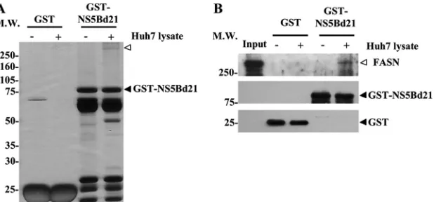
FASN is identified as an NS5B-interacting protein by proteomic
and MALDI-TOF analysis.
To elucidate the mechanisms
under-lying HCV replication and to explore the interplay between viral
and host factors, we attempted to identify proteins interacting
with HCV RNA polymerase NS5B in the lysates of Huh7 cells, a
cell line well known for its ability to facilitate HCV RC formation
and viral production. A GST fusion protein, GST-NS5Bd21,
lack-ing the C-terminal 21 amino acids of NS5B, was used as the bait to
pull down NS5B-interacting proteins. After protein fractionation
by SDS-PAGE and silver staining, a candidate protein with a
mo-lecular size of approximately 250 kDa was present in the pulldown
lysates of GST-NS5Bd21 but not in those of the control protein
GST (
Fig. 1A
). The identity of the candidate protein was unveiled
by spectrum analysis of the peptide profiles generated by
MALDI-TOF MS. With 18% sequence coverage, the protein was identified
as the lipogenic enzyme FASN (see Table S1 in the supplemental
material). The calculated molecular mass of FASN is 276 kDa,
which is consistent with the location of the candidate protein on
SDS-PAGE gels. Western blot analysis of the GST-NS5Bd21
pull-down lysates using an anti-FASN antibody further confirmed
FASN as the NS5B-interacting protein (
Fig. 1B
).
NS5B-FASN interaction is mediated by the N terminus of
NS5B.
To determine whether FASN interacts directly with NS5B
in the absence of other viral proteins, FASN and NS5B expression
plasmids were cotransfected into 293T cells, followed by
immu-noprecipitation with an anti-FASN antibody. Western blot
anal-ysis using an anti-NS5B antibody revealed that NS5B was present
in protein complexes immunoprecipitated by the anti-FASN
an-tibody but not by the IgG control anan-tibody (
Fig. 2A
), implying
that other viral proteins do not contribute to the interaction of
NS5B and FASN.
To define the region of NS5B that mediates its interaction with
FASN, serial NS5B deletion mutants (
Fig. 2B
) were generated and
were coexpressed with FASN in 293T cells. After
immunoprecipi-tation with an anti-FASN antibody, Western blot analyses were
performed to determine whether NS5B mutant proteins were
present
in
FASN-immunoprecipitated
protein
complexes
(
Fig. 2C
). Like NS5B/1-570, both NS5B/1-180 and NS5B/1-335
were able to bind FASN. In contrast, the FASN binding activity of
NS5B was abrogated by deleting amino acids 1 to 178
(NS5B/179-570). In agreement with these findings, FASN can be detected in
the immunoprecipitated protein complexes of full-length NS5B
but not in those of NS5B/179-570 (
Fig. 2D
). These data suggest
that the first 178 amino acids, where the finger domain of NS5B is
located (
28
), play a pivotal role in mediating the binding of NS5B
to FASN.
FASN interacts with NS5B in HCV subgenomic replicon cells
and HCVcc-infected cells.
To confirm the interaction between
endogenous FASN and NS5B, HCV subgenomic replicon cells
were subjected to coimmunoprecipitation and
immunofluo-rescence staining assays. The subgenomic replicon cell lysates
were immunoprecipitated with an anti-FASN antibody,
fol-FIG 1FASN interacts with NS5B. (A) GST and GST-NS5B fusion proteins (GST-NS5Bd21) were purified with glutathione Sepharose 4B beads for a pulldown assay in the absence (⫺) or presence (⫹) of Huh7 cell lysates. Equal volumes of purified fusion proteins and pulldown proteins were separated by 10% SDS-PAGE, followed by silver staining. The open arrowhead indicates the candidate protein that specifically bound to GST-NS5Bd21. MW, molecular weight (in thousands). (B) Pulled down proteins were analyzed by Western blotting using anti-FASN (top), anti-NS5B (center), and anti-GST (bottom) antibodies. Accordingly, FASN was confirmed to interact with NS5B. Input, 1/30 of the Huh7 cell lysates used for GST pulldown.on November 7, 2019 by guest
http://jvi.asm.org/
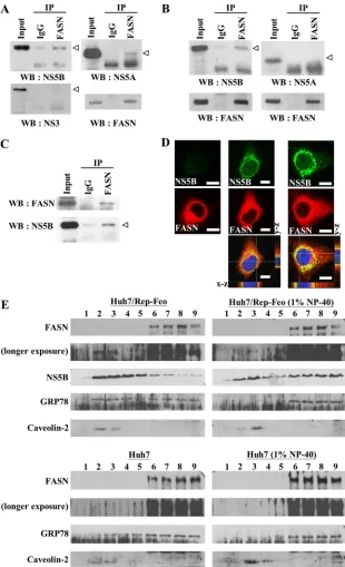
[image:4.585.140.459.66.210.2]lowed by Western blotting of the HCV proteins. Our data
re-vealed that NS5B and NS5A, but not NS3, were present in the
immunocomplexes of FASN (
Fig. 3A
). The interaction
be-tween NS5B and FASN still occurred when
immunoprecipita-tion was performed using lysates from Huh7 cells coexpressing
FASN and NS5B expression plasmids. However, NS5A was not
detectable in the immunocomplexes of FASN when FASN and
NS5A were coexpressed in Huh7 cells (
Fig. 3B
). In addition, a
coimmunoprecipitation assay of the HCVcc-infected Huh7.5.1
cell lysates using an anti-FASN antibody further confirmed the
interactions of NS5B and FASN (
Fig. 3C
). These data not only
define the interactions between FASN and NS5B but also
sug-gest that FASN, likely through NS5B, interacts with other HCV
proteins, such as NS5A, and forms large protein complexes
with them.
To further demonstrate the interaction between FASN and
NS5B, Huh7/Rep-Feo subgenomic replicon cells and
HCVcc-in-fected Huh7.5.1 cells were subjected to immunofluorescence
staining using a rabbit anti-NS5B or mouse anti-FASN antibody.
NS5B was found to be distributed mostly near the perinuclear
region, whereas FASN was found in the cytoplasm. The merged
images at the
x
-
z
and
y
-
z
sections revealed that FASN and NS5B
were colocalized in the perinuclear region, with a colocalization
coefficient of 80% (
Fig. 3D
), supporting the notion that FASN is
an NS5B-interacting protein.
NS5B protein is usually associated with the detergent-resistant
lipid raft membrane fraction (
29
). To determine whether FASN
and NS5B colocalize in the lipid rafts of Huh7/Rep-Feo cells, the
lysates were extracted in the absence or presence of 1% NP-40 and
were fractionated by a 10-to-72% sucrose gradient. Each fraction
of the gradient was collected for Western blot analysis. Fractions 2
and 3 were considered lipid rafts based on the presence in these
fractions of caveolin-2, a lipid raft membrane protein resistant to
1% NP-40 solubilization (
25
), regardless of whether the cell
ly-sates were extracted in the presence or absence of NP-40. On the
other hand, GRP78, an ER marker not associated with lipid rafts
and sensitive to 1% NP-40 solubilization (
25
), was detectable in
the bottom of the gradient and was not detected in the
detergent-resistant membrane fractions. In agreement with previous studies
(
29
), the viral protein NS5B was associated with lipid raft fractions
in the sucrose gradient (
Fig. 3E
, top). Portions of FASN protein
were codistributed with NS5B in lipid rafts (
Fig. 3E
, top). Notably,
the distribution of FASN in lipid rafts was not observed in Huh7
cells that did not express any HCV proteins (
Fig. 3E
, bottom).
These results suggest that FASN was recruited by HCV proteins,
most likely through the interaction with NS5B, into lipid rafts.
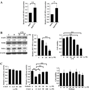
Inhibition of FASN activity decreases HCV protein and RNA
expression levels.
The interaction of FASN with HCV NS5B may
be involved in the regulation of HCV proteins and RNA
expres-sion. To elucidate whether there is a correlation between FASN
expression and the infectivity of HCV, FASN expression in
HCV-permissive Huh7.5.1 cells was compared with that in the parental
Huh7 cells. Real-time RT-PCR analysis revealed that FASN
mRNA expression was higher in HCV-permissive Huh7.5.1 cells
than in Huh7 cells (
Fig. 4A
, left) (
P
⬍
0.01). In agreement with
these observations, FASN expression was also increased in
HCVcc-infected (MOI, 3) Huh7.5.1 cells (
Fig. 4A
, right) (
P
⬍
0.05). These data suggest that FASN expression likely facilitates
HCV infection and replication.
The lipid raft is the major cellular compartment for the
assem-bly of HCV RC. The colocalization of FASN and NS5B in the lipid
raft led us to delineate the role of FASN in HCV replication. First,
subgenomic replicon cells were treated with C75, an inhibitor of
the

-ketoacyl synthase domain of FASN (
30
), to inhibit cellular
FASN activity. Western blot analysis revealed that C75 had no
effect on FASN expression but caused a decrease in the expression
of NS5A and NS5B from that for the untreated control or cells
treated with the solvent dimethyl sulfoxide (DMSO) (
Fig. 4B
, left).
In agreement with these findings, HCV RNA expression was also
inhibited by C75 in a dose-dependent manner, with 34% and 68%
inhibition at the noncytotoxic concentrations of 20 and 40
M,
respectively (
Fig. 4B
, center and right).
The subgenomic replicon cells were also treated with orlistat,
an inhibitor of the thioesterase domain of FASN and an
FDA-approved drug used for treating obesity (
31
), to elucidate FASN
function in viral replication. The expression levels of HCV RNA,
as reflected by luciferase activity in Huh7/Rep-Feo cells, were
de-termined. At the dosage of the 50% effective concentration (EC
50)
(50
M), which is noncytotoxic, orlistat caused a 45% decrease in
FIG 2FASN binds to the N terminus of NS5B. (A) The NS5B and FASNexpression plasmids were cotransfected into HEK293T cells, followed by im-munoprecipitation (IP) with an anti-FASN antibody. The immunoprecipi-tated proteins were detected by Western blotting (WB) using an anti-FASN or anti-NS5B antibody. (B) Schematic representation of the NS5B deletion mu-tants. The six motifs essential for the RNA-dependent polymerase are denoted as A to F. (C) Expression plasmids for the indicated NS5B deletion mutants were cotransfected with the FASN expression plasmid into HEK293T cells. At 48 h after transfection, immunoprecipitation was performed using an FASN antibody, and NS5B was detected by Western blotting using an anti-NS5B or anti-Flag tag antibody. Asterisks indicate the binding of anti-NS5B to FASN. MW, molecular weight (in thousands). (D) The FASN expression plas-mid was cotransfected with the indicated NS5B expression plasplas-mids into HEK293T cells. At 48 h after transfection, immunoprecipitation was per-formed using an anti-Flag tag antibody, and FASN was detected by Western blotting using an anti-FASN antibody. Open arrowheads indicate the NS5B deletion mutant proteins. The asterisk indicates the binding of FASN to NS5B.
on November 7, 2019 by guest
http://jvi.asm.org/
[image:5.585.45.284.66.331.2]FIG 3NS5B interacts and colocalizes with FASN in HCV replicon cells and HCVcc-infected cells. (A) Immunoprecipitation (IP) of subgenomic replicon cell lysates was performed with an anti-FASN antibody. The immunoprecipitated proteins were analyzed by Western blotting (WB) with an anti-NS5B, anti-NS5A, anti-NS3, or anti-FASN antibody. (B) Huh7 cells were cotransfected with plasmids expressing FASN and NS5B (left) or FASN and NS5A (right) for 48 h. The transfected cell lysates were then immunoprecipitated using an anti-FASN antibody. NS5B or NS5A was subsequently detected by Western blotting using an anti-NS5B or anti-NS5A antibody, respectively. (C) Huh7.5.1 cells were infected with JC1-Luc2A HCVcc (MOI, 3). After infection for 72 h, the infected-cell lysates were collected for immunoprecipitation with an anti-FASN antibody. The immunoprecipitated proteins were analyzed by Western blotting using an anti-NS5B or anti-FASN antibody. (D) Immunofluorescence staining of Huh7 cells (left), subgenomic replicon cells (center), and Huh7.5.1 cells infected with JC1-Luc2A HCVcc (MOI, 3) (right) was performed using anti-FASN and anti-NS5B antibodies. The rhodamine- or Alexa 488-conjugated secondary antibody was then used to visualize the FASN or NS5B protein, respectively. The cell images were captured using a Leica SP2 confocal spectral microscope. The images of thex-zsection and they-zsection were acquired from the dashed lines from the centerx-ypanel of the bottom merged image. The yellow area indicates the colocalization of NS5B with FASN. Bars, 10m. (E) Huh7/Rep-Feo and Huh7 cell lysates were harvested in parallel with a hypotonic buffer and were either left untreated or mixed with 1% NP-40 for 1 h on ice. The lysate was fractionated by a 10-to-72% sucrose gradient at 38,000 rpm for 14 h. After centrifugation, the sample was separated into nine fractions, which were analyzed by Western blotting with the indicated antibodies.
on November 7, 2019 by guest
http://jvi.asm.org/
[image:6.585.136.446.65.574.2]luciferase activity (
Fig. 4C
, center and right). Although palmitate
(C16:0), the principal product of FASN, had no effect on HCV
expression alone (
Fig. 4C
, left), the suppressive effect of orlistat on
HCV RNA expression could be abrogated in a dose-dependent
manner by cotreatment with palmitate (C16:0)
(
Fig. 4C
, middle
panel
)
. These data suggest that FASN activity is important for
HCV replication.
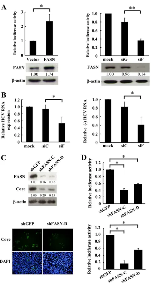
Abrogation of FASN expression diminishes HCV expression
and replication.
To further delineate the role of FASN in HCV
expression and replication, FASN was overexpressed in Huh7/
Rep-Feo cells, and the luciferase activity, corresponding to the
level of HCV RNA expression, was analyzed (
Fig. 5A
, left). In
FASN-overexpressing cells, luciferase activity was increased
2.4-fold over that in control vector-transfected cells (
n
⫽
3;
P
⬍
0.05).
In agreement with these findings, knockdown of FASN in the
rep-licon cells by small interfering RNA (siF) caused a 44% decrease in
luciferase activity from that in cells transfected with nontargeting
control small interfering RNA (siG) (
Fig. 5A
, right). Moreover,
the expression levels for total and negative-strand HCV RNA were
decreased by 41% and 42%, respectively, in siF-transfected
sub-genomic replicon cells (
Fig. 5B
).
To elucidate whether FASN has any functional impact on
HCVcc production, Huh7.5.1 cells were transfected with the
JC1-Luc2A-based HCV reporter virus (MOI, 0.1) and were infected
with a lentivirus encoding shFASN (shFASN-C or shFASN-D). As
indicated by Western blotting and immunofluorescence staining
analyses, Huh7.5.1 cells expressing either of the shFASN
con-structs exhibited a 65% decrease in the level of HCV core protein
(
Fig. 5C
). In agreement with these findings, the luciferase activity
reflecting the level of HCVcc production was decreased by
ap-proximately 60% (
Fig. 5D
, top). Moreover, when the supernatants
from shFASN-C- or shFASN-D-transduced HCVcc-infected cells
were collected to reinfect naïve Huh7.5.1 cells, the luciferase
ac-tivity of the reinfected cells was decreased by 83% or 43%,
respec-tively (
Fig. 5D
, bottom). These data suggest that FASN expression
modulates HCV replication and affects viral production.
FASN is a component of HCV replication complexes and
modulates NS5B RdRp activity.
Previous study has indicated that
FIG 4FASN activity is required for HCV replication. (A) Cellular RNAs from the indicated cells (left) or mock- or JC1-Luc2A HCVcc-infected Huh7.5.1 cells (right) were collected for real-time RT-PCR to analyze FASN mRNA expression. (B) (Left) HCV subgenomic replicon cells were treated with the FASN inhibitor C75 for 48 h. The cell lysate was harvested, and Western blot analysis was performed using the indicated antibodies. The ratios for the relative band intensities of each indicated protein normalized by-actin are shown. C, control untreated cells; D, DMSO-treated cells. (Center) In parallel, cellular RNA was collected, and viral RNA was detected by real-time RT-PCR. The expression of glyceraldehyde-3-phosphate dehydrogenase was used as a control for normalization. The expression level for untreated cells was arbitrarily set at 1. Data are means⫾standard deviations (n⫽3; **,P⬍0.01). (Right) The viability of HCV subgenomic replicon cells treated with the indicated concentrations of C75 was measured by an MTS assay. Data are means⫾standard deviations (n⫽3; **,P⬍0.01). (C) (Left and center) Huh7/Rep-Feo cells were treated with the indicated concentrations of palmitate (C16:0) (left) or the FASN inhibitor orlistat combined with the
indicated dose of palmitate for 72 h (center). Luciferase activities were determined, and the relative luciferase activity is shown. The luciferase activity for the negative control was arbitrarily set at 1. Data are means⫾standard deviations (n⫽3; *,P⬍0.05; **,P⬍0.01). (Right) The viability of HCV Huh7/Rep-Feo cells treated with the indicated concentrations of orlistat was measured by an MTS assay. Data are means⫾standard deviations (n⫽3).
on November 7, 2019 by guest
http://jvi.asm.org/
[image:7.585.135.448.64.380.2]NS5B is present in HCV RC that are formed by invaginations of
cholesterol- and sphingomyelin-rich membranes during
replica-tion. Due to the vesicular membrane structures, the complexes are
resistant to protease and nuclease treatment (
26
). Considering the
functional role of FASN in viral replication and the interplays
between FASN and NS5B, we first investigated whether FASN is
present in RC and then employed siF- and siC-transfected
rep-licon cells to delineate the effects of FASN knockdown on the
activity of RC. The total-cell lysate, supernatant (S1), CRC, and
proteinase K-treated CRC (RC) were prepared from both siF- and
siC-transfected cells. Western blot analyses revealed that FASN
was present in both CRC and RC and that, together with NS5B, it
FIG 5FASN expression is required for HCV replication. (A) Huh7/Rep-Feocells were transfected with an FASN expression plasmid (left) or a siFASN oligonucleotide (siF) (right) for 48 h. Mock-transfected or siGFP-transfected (siG) cells were used as controls for the FASN knockdown experiment. The luciferase activities were determined, and Western blot analysis was performed in parallel to determine the level of FASN expression. The ratios for the relative band intensity of FASN after normalization with-actin are shown. The ratio for the vector control or for mock transfection was arbitrarily set at 1. Lucif-erase activities are means⫾standard deviations for three independent exper-iments (*,P⬍0.05; **,P⬍0.01). (B) The expression levels of total and minus-strand (⫺) HCV RNA for Huh7/Rep-Feo cells with FASN knockdown were determined by real-time RT-PCR. The expression of glyceraldehyde-3-phosphate dehydrogenase was used as a control for normalization. The expres-sion level for mock-transfected cells was arbitrarily set at 1. siC is a nontarget-ing siRNA control. Data are means ⫾ standard deviations for three independent experiments (*,P⬍0.05). (C) Huh7.5.1 cells were seeded in 24-well plates at a density of 5⫻104/well for 24 h and were infected with a
lentivirus carrying shGFP or shFASN (shFASN-C and shFASN-D). The cells were then seeded in a 96-well plate at a density of 1⫻104/well, followed by
infection with the JC1-Luc2A HCV reporter virus (MOI, 0.1). The infected cells were fixed and stained with an anti-HCV core antibody. In parallel, the cell lysates were collected to determine the expression of FASN and HCV core
proteins by Western blotting. (D) (Top) The HCV infectivity of the cells was measured by a luciferase activity assay. (Bottom) The viruses in the infected-cell supernatants were collected and used to reinfect naïve Huh7.5.1 infected-cells. The reinfected cell lysates were collected, and the luciferase activities were deter-mined. Data are means⫾standard deviations for three independent experi-ments (*,P⬍0.05).
FIG 6Fatty acid synthase is a component of the HCV replication complex. (A) Subgenomic replicon cells were transfected with a control (siC) or siFASN (siF) oligonucleotide. CRC were isolated from the cell lysates as described in Materials and Methods. The presence of the indicated proteins in the total-cell lysate (TC), supernatant (S1), CRC, and proteinase K-treated CRC (RC) was analyzed by Western blotting. (B) (Left) The RNA from the CRC fractions of the indicated transfected cells, either left untreated or treated with S7 nuclease, was analyzed by agarose gel electrophoresis for the expression levels of 28S RNA and by real-time RT-PCR for HCV RNA expression. The expression level for cells transfected with the nontargeting siRNA control (siC) without S7 nuclease treatment was arbitrarily set at 1. Data are means⫾standard devia-tions for three independent experiments (**,P⬍0.01). (Right) Thein vitro replicase activity of S7 nuclease-resistant RC for the indicated cell lines was analyzed, and the reaction products were analyzed by denaturing agarose gel electrophoresis, followed by phosphorimaging of the dried gel. The major reaction product of thein vitroreplicase assay is indicated by an arrow.
on November 7, 2019 by guest
http://jvi.asm.org/
[image:8.585.316.522.65.341.2] [image:8.585.44.285.69.521.2]is among the protein components of RC in siC-transfected cells
(
Fig. 6A
). Knockdown of FASN by small interfering RNA (siF) did
not decrease the amount of NS5B in CRC but resulted in an 80%
decrease of the amount of NS5B in RC. The efficiency of
protei-nase K digestion was monitored by the cleavage of the ER
mem-brane protein calnexin; the C terminus of calnexin resides at the
cytoplasmic side of the ER membrane and is susceptible to digest
by proteinase K. Western blot analysis using an antibody that
rec-ognizes the N terminus of calnexin revealed the absence of
full-length calnexin, concomitant with the presence of a calnexin
cleavage product, indicating complete and effective proteinase K
digestion (
Fig. 6A
). A large portion of the NS5B and FASN
pro-teins in CRC was digested, while a small part of these two propro-teins
remained resistant in RC. These data indicate that FASN
colocal-izes with NS5B and plays a role in retaining NS5B in active RC.
To further unveil the effect of FASN on HCV RNA expression,
total RNA in CRC of control and FASN knockdown cells was
isolated with or without treatment by S7 nuclease. The relative
amount of HCV RNA was determined by real-time RT-PCR,
whereas 28S RNA was analyzed by gel electrophoresis. In
accor-dance with a previous study (
26
), HCV, but not 28S RNA, can be
detected in the S7 nuclease-treated active RC. HCV RNA
expres-sion in the S7 nuclease resistant RC was decreased by 56% in FASN
knockdown cells (
Fig. 6B
). Analysis of the replicase activity in S7
nuclease-resistant RC further confirmed the decrease in the level
of newly synthesized HCV RNA by FASN knockdown (
Fig. 6B
).
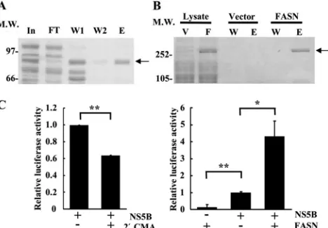
To elucidate whether FASN is directly involved in the
regula-tion of RdRp activity during HCV replicaregula-tion,
in vitro
NS5B RdRp
activity was compared in the presence or absence of recombinant
FASN protein. The NS5B and FASN recombinant proteins were
purified by a His tag affinity column (
Fig. 7A
and
B
), and the
expression of these two proteins was confirmed by Western
blot-ting (data not shown). The NS5B-specific inhibitor 2
=
CMA at the
dosage of the 50% inhibitory concentration (IC
50) (
32
) inhibited
37% of the RdRp activity, implying that the activity assay is NS5B
specific (
Fig. 7C
, left). The presence of FASN in the reaction
mix-ture containing NS5B protein increased the RdRp activity 4-fold,
while FASN alone elicited minimal residual RdRp activity
(
Fig. 7C
, right). These data indicate that by interacting with NS5B,
FASN increases the
in vitro
RdRp activity of NS5B.
DISCUSSION
In addition to viral factors, host proteins play a pivotal role in
many steps of RNA virus replication (
33
,
34
). Nevertheless, the
components of HCV RC are still not completely understood.
Some of the host factors involved in HCV replication were found
to be tightly linked to lipid metabolism (
17
,
35
). In this study,
FASN is revealed to interact with NS5B, with two major functional
implications. The interaction with NS5B causes a portion of FASN
proteins to shift from the cytoplasm to lipid rafts. On the other
hand, FASN enhances NS5B-dependent RdRp activity and causes
an increase in HCV replication and viral production. This study
thereby demonstrates for the first time that FASN regulates HCV
replication and viral production through a direct interplay with
NS5B.
The main function of FASN is to catalyze the synthesis of
palmitate from acetyl coenzyme A (acetyl-CoA) and
malonyl-CoA, subsequently forming long-chain saturated fatty acids in the
presence of NADPH (
36
). FASN expression is significantly
in-creased in HCV-infected livers (
37
), and serum FASN
concentra-tions are significantly increased in patients with chronic HCV
in-fection (
38
,
39
). FASN is also upregulated in HCV-infected Huh7
cells (
18
). Accordingly, a significantly higher level of free fatty acid
was detected in HCV-infected than in uninfected Huh 7.5 cells
(
40
). Consequently, more lipid droplets were formed for HCV
production (
41
). Hence, FASN plays roles in HCV infection.
However, the mechanism of FASN involvement in HCV
replica-tion is unclear. The data we present in this study indicate that both
the enzymatic activity of FASN and FASN-NS5B interactions are
important for the role of FASN in regulating HCV replication.
Accordingly, FASN knockdown and the inhibition of FASN
enzy-matic activity by C75 and orlistat result in decreases in the level of
HCV replication activity. Notably, we demonstrate for the first
time that FASN is a component of HCV RC and, by interacting
with NS5B, enhances NS5B-dependent RdRp activity. In this
mode of action, an allosteric effect of FASN, but not its enzymatic
activity, is likely to play a major role in modulating NS5B activity.
This notion is based on the observations that the inhibition of
FASN activity by C75 and orlistat or FASN knockdown alone does
not result in complete inhibition of HCV replication, suggesting
that both FASN activity and protein expression are involved in the
regulation of HCV expression. Moreover, FASN has been
re-ported to elicit palmitoylation activity, and the palmitoylation of
NS4B is important for HCV RC formation (
42
,
43
). Whether
FASN is recruited, by interacting with NS5B, into the
NS4B-asso-ciated membrane web and palmitoylates NS4B protein remains to
be investigated further.
FIG 7FASN enhances HCV NS5B RdRp activity. (A) The NS5B protein was purified as described in Materials and Methods and was then stained with Coomassie blue. The input (In) is the protein before purification, and FT, W, and E represent the flowthrough, wash, and elution fractions during NS5B purification, respectively. The arrow indicates the size of HCV NS5B. MW, molecular weight (in thousands). (B) The FASN protein (F) was purified from pcDNA-HisC-FASN-transfected cells, and the proteins were visualized by sil-ver staining. The pcDNA3.1-HisC vector (V)-transfected cell lysates were pu-rified in parallel as a control. W and E represent the wash and elution fractions during purification, respectively. (C) (Left) Purified NS5B was incubated alone or with the NS5B inhibitor 2=-C-methyladenosine (2=CMA) at 2M to evaluate the specificity of NS5B RdRp activity. (Right) Two picomoles of pu-rified His-FASN was incubated with 250 nM pupu-rified NS5B to evaluate the effect of FASN on RdRp activity. An equal volume of pcDNA3.1-HisC vector-transfected cell lysates purified in parallel was used as a background control. Luciferase activity was detected at 5 min after the initiation of the reaction. The relative RdRp activity after subtraction of background luciferase activity is shown. Data are means⫾standard deviations for three independent experi-ments (*,P⬍0.05; **,P⬍0.01).
on November 7, 2019 by guest
http://jvi.asm.org/
[image:9.585.305.539.68.231.2]The expression of FASN has been reported to be regulated by
HCV core protein (
44
), NS2 (
45
), and NS4B (
46
). These viral
proteins enhance sterol regulatory element-binding protein 1
ex-pression, followed by activation of the FASN promoter (
47
).
Ac-cordingly, FASN expression was increased in HCV-permissive
cells and HCVcc-infected Huh7.5.1 cells (
Fig. 4A
), indicating that
FASN plays a role in HCV replication. As in HCV, FASN
expres-sion can be induced by some other types of viral infection. The
hepatitis B virus large surface protein accumulated on the
endo-plasmic reticulum leads to the unfolding of proteins and promotes
FASN expression through the binding of the activated NF-Y
tran-scription factor to the FASN promoter (
48
). The Epstein-Barr
virus early-stage protein BRLF1 also activates FASN expression,
leading to the induction of Epstein-Barr virus Z transcription for
the lytic stage (
49
). On the other hand, the replication of two
enveloped viruses, human cytomegalovirus and influenza virus, is
inhibited by a fatty acid biosynthesis inhibitor that subsequently
modulates the membrane composition for virus budding (
50
).
Moreover, dengue virus NS3 interacts with FASN and recruits it to
the site of RC formation, leading to membrane expansion and an
increase in membrane fluidity (
51
). In agreement with our
find-ings, Yang et al. have reported that HCV replication and viral
entry are decreased by a FASN inhibitor and FASN RNAi (
18
).
HCV was thought to replicate in the NS4B-induced membrane
web and the viral particles were assembled on the outer surface of
lipid droplets (
41
,
52
). Hence, it was thought that HCV
infection-induced FASN is involved mainly in the production of
phospho-lipids which are participated in the formation of
detergent-resis-tant membranes (
18
). Our data offer a new insight: by interacting
with NS5B, FASN is recruited into HCV replication complexes
and directly enhances HCV RdRp activity. Thus, promising new
antiviral approaches to combat HCV infection based on the
inhi-bition of FASN activity are worthy of consideration.
Increased FASN expression and activity are found in several
cancers, including HCV-associated HCC (
53
,
54
). Our data
dem-onstrate that NS5B, FASN, and caveolin-1 (Cav-1) can interact
with each other in HCV replicon cells (data not shown). Cav-1 is a
palmitoylated lipid raft protein and has been found to interact
with FASN in prostate cancer cells to promote tumor growth and
survival (
55
). Recently, Cav-1 was also reported to promote the
motility and invasion of hepatocellular carcinoma (HCC) and
metastasis in a mouse model though an unknown mechanism (
56
,
57
). We therefore postulate that following HCV infection, FASN
interacts with NS5B and, together with Cav-1, is recruited to lipid
rafts, followed by the induction of signaling leading to
hepatocel-lular carcinoma.
In conclusion, we demonstrate for the first time that FASN
interacts with NS5B, leading to an increase in HCV RdRp activity.
Furthermore, inhibition of FASN reduces HCV replication and
viral production. This study thereby contributes to our
under-standing of the role of FASN in the control of HCV replication and
offers new insight for developing novel anti-HCV therapeutic
ap-proaches.
ACKNOWLEDGMENTS
We thank Jing-Hsiung Ou for providing HCV subgenome replicon cells, Robert T. Schooley for providing Huh7.5.1 cells and plasmid JC1-Luc2A, Charles Rice for providing plasmid J6/JFH(p7-Rlu2A), and Takaji Wakita for providing a polyclonal antibody against NS5B. We also thank Chien-Ling Huang, Yung-Ju Yeh, and Chia-Fan Lin for technical support.
This work was supported by grants NSC97-2320-B-039-026-MY3, NSC94-2320-B-039-008-, and NSC 93-2314-B-039-029- from the Na-tional Science Council (J.-C.C.).
REFERENCES
1.Lindenbach BD, Rice CM.2005. Unravelling hepatitis C virus replication from genome to function. Nature436:933–938.
2.Egger D, Wolk B, Gosert R, Bianchi L, Blum HE, Moradpour D, Bienz K.2002. Expression of hepatitis C virus proteins induces distinct mem-brane alterations including a candidate viral replication complex. J. Virol.
76:5974 –5984.
3.Moradpour D, Penin F, Rice CM.2007. Replication of hepatitis C virus. Nat. Rev. Microbiol.5:453– 463.
4.Samuel D, Feray C.2000. Recurrent hepatitis C after liver transplanta-tion: clinical and therapeutical issues. J. Viral Hepat.7:87–92.
5.Thomson BJ, Finch RG.2005. Hepatitis C virus infection. Clin. Micro-biol. Infect.11:86 –94.
6.Poenisch M, Bartenschlager R.2010. New insights into structure and replication of the hepatitis C virus and clinical implications. Semin. Liver Dis.30:333–347.
7.Hwang SJ, Lee SD.2011. Hepatic steatosis and hepatitis C: still unhappy bedfellows? J. Gastroenterol. Hepatol.26(Suppl. 1):96 –101.
8.Negro F.2010. Abnormalities of lipid metabolism in hepatitis C virus infection. Gut59:1279 –1287.
9.Jiang J, Luo G.2009. Apolipoprotein E but not B is required for the formation of infectious hepatitis C virus particles. J. Virol.83:12680 – 12691.
10. Liu S, McCormick KD, Zhao W, Zhao T, Fan D, Wang T.2012. Human apolipoprotein E peptides inhibit hepatitis C virus entry by blocking virus binding. Hepatology56:484 – 491.
11. Mancone C, Steindler C, Santangelo L, Simonte G, Vlassi C, Longo MA, D’Offizi G, Di Giacomo C, Pucillo LP, Amicone L, Tripodi M, Alonzi T.2011. Hepatitis C virus production requires apolipoprotein A-I and affects its association with nascent low-density lipoproteins. Gut60:378 – 386.
12. Gastaminza P, Cheng G, Wieland S, Zhong J, Liao W, Chisari FV.2008. Cellular determinants of hepatitis C virus assembly, maturation, degrada-tion, and secretion. J. Virol.82:2120 –2129.
13. Huang H, Sun F, Owen DM, Li W, Chen Y, Gale M, Jr., Ye J.2007. Hepatitis C virus production by human hepatocytes dependent on assem-bly and secretion of very low-density lipoproteins. Proc. Natl. Acad. Sci. U. S. A.104:5848 –5853.
14. Kapadia SB, Chisari FV.2005. Hepatitis C virus RNA replication is regulated by host geranylgeranylation and fatty acids. Proc. Natl. Acad. Sci. U. S. A.102:2561–2566.
15. Wang C, Gale M, Jr, Keller BC, Huang H, Brown MS, Goldstein JL, Ye J.2005. Identification of FBL2 as a geranylgeranylated cellular protein required for hepatitis C virus RNA replication. Mol. Cell18:425– 434. 16. Ye J, Wang C, Sumpter R, Jr, Brown MS, Goldstein JL, Gale M, Jr.2003.
Disruption of hepatitis C virus RNA replication through inhibition of host protein geranylgeranylation. Proc. Natl. Acad. Sci. U. S. A.100:15865– 15870.
17. Su AI, Pezacki JP, Wodicka L, Brideau AD, Supekova L, Thimme R, Wieland S, Bukh J, Purcell RH, Schultz PG, Chisari FV.2002. Genomic analysis of the host response to hepatitis C virus infection. Proc. Natl. Acad. Sci. U. S. A.99:15669 –15674.
18. Yang W, Hood BL, Chadwick SL, Liu S, Watkins SC, Luo G, Conrads TP, Wang T.2008. Fatty acid synthase is up-regulated during hepatitis C virus infection and regulates hepatitis C virus entry and production. Hepatology48:1396 –1403.
19. Choi J, Lee KJ, Zheng Y, Yamaga AK, Lai MM, Ou JH.2004. Reactive oxygen species suppress hepatitis C virus RNA replication in human hep-atoma cells. Hepatology39:81– 89.
20. Cheng JC, Chang MF, Chang SC.1999. Specific interaction between the hepatitis C virus NS5B RNA polymerase and the 3=end of the viral RNA. J. Virol.73:7044 –7049.
21. Weng LP, Wu CC, Hsu BL, Chi LM, Liang Y, Tseng CP, Hsieh LL, Yu JS.2008. Secretome-based identification of Mac-2 binding protein as a potential oral cancer marker involved in cell growth and motility. J. Pro-teome Res.7:3765–3775.
22. Craggs JK, Ball JK, Thomson BJ, Irving WL, Grabowska AM.2001.
on November 7, 2019 by guest
http://jvi.asm.org/
Development of a strand-specific RT-PCR based assay to detect the repli-cative form of hepatitis C virus RNA. J. Virol. Methods94:111–120. 23. Cheng JC, Yeh YJ, Tseng CP, Hsu SD, Chang YL, Sakamoto N, Huang
HD.2012. Let-7b is a novel regulator of hepatitis C virus replication. Cell. Mol. Life Sci.69:2621–2633.
24. Wakita T, Pietschmann T, Kato T, Date T, Miyamoto M, Zhao Z, Murthy K, Habermann A, Krausslich HG, Mizokami M, Bartenschlager R, Liang TJ.2005. Production of infectious hepatitis C virus in tissue culture from a cloned viral genome. Nat. Med.11:791–796.
25. Gao L, Aizaki H, He JW, Lai MM. 2004. Interactions between viral nonstructural proteins and host protein hVAP-33 mediate the formation of hepatitis C virus RNA replication complex on lipid raft. J. Virol.78: 3480 –3488.
26. Quinkert D, Bartenschlager R, Lohmann V.2005. Quantitative analysis of the hepatitis C virus replication complex. J. Virol.79:13594 –13605. 27. Lahser FC, Malcolm BA.2004. A continuous nonradioactive assay for
RNA-dependent RNA polymerase activity. Anal. Biochem.325:247–254. 28. Mosley RT, Edwards TE, Murakami E, Lam AM, Grice RL, Du J, Sofia MJ, Furman PA, Otto MJ.2012. Structure of hepatitis C virus polymerase in complex with primer-template RNA. J. Virol.86:6503– 6511. 29. Shi ST, Lee KJ, Aizaki H, Hwang SB, Lai MM.2003. Hepatitis C virus
RNA replication occurs on a detergent-resistant membrane that cofrac-tionates with caveolin-2. J. Virol.77:4160 – 4168.
30. Kuhajda FP, Pizer ES, Li JN, Mani NS, Frehywot GL, Townsend CA.
2000. Synthesis and antitumor activity of an inhibitor of fatty acid syn-thase. Proc. Natl. Acad. Sci. U. S. A.97:3450 –3454.
31. Kridel SJ, Axelrod F, Rozenkrantz N, Smith JW.2004. Orlistat is a novel inhibitor of fatty acid synthase with antitumor activity. Cancer Res.64: 2070 –2075.
32. Migliaccio G, Tomassini JE, Carroll SS, Tomei L, Altamura S, Bhat B, Bartholomew L, Bosserman MR, Ceccacci A, Colwell LF, Cortese R, De Francesco R, Eldrup AB, Getty KL, Hou XS, LaFemina RL, Ludmerer SW, MacCoss M, McMasters DR, Stahlhut MW, Olsen DB, Hazuda DJ, Flores OA.2003. Characterization of resistance to non-obligate chain-terminating ribonucleoside analogs that inhibit hepatitis C virus replica-tion in vitro. J. Biol. Chem.278:49164 – 49170.
33. Ahlquist P, Noueiry AO, Lee WM, Kushner DB, Dye BT.2003. Host factors in positive-strand RNA virus genome replication. J. Virol.77: 8181– 8186.
34. Moriishi K, Matsuura Y.2007. Host factors involved in the replication of hepatitis C virus. Rev. Med. Virol.17:343–354.
35. Alvisi G, Madan V, Bartenschlager R.2011. Hepatitis C virus and host cell lipids: an intimate connection. RNA Biol.8:258 –269.
36. Jayakumar A, Tai MH, Huang WY, al-Feel W, Hsu M, Abu-Elheiga L, Chirala SS, Wakil SJ.1995. Human fatty acid synthase: properties and molecular cloning. Proc. Natl. Acad. Sci. U. S. A.92:8695– 8699. 37. Fujino T, Nakamuta M, Yada R, Aoyagi Y, Yasutake K, Kohjima M,
Fukuizumi K, Yoshimoto T, Harada N, Yada M, Kato M, Kotoh K, Taketomi A, Maehara Y, Nakashima M, Enjoji M.2010. Expression profile of lipid metabolism-associated genes in hepatitis C virus-infected human liver. Hepatol. Res.40:923–929.
38. Aragones G, Alonso-Villaverde C, Oliveras-Ferraros C, Beltran-Debon R, Rull A, Rodriguez-Sanabria F, Camps J, Martin AV, Menendez JA, Joven J.2010. Infection with HIV and HCV enhances the release of fatty acid synthase into circulation: evidence for a novel indicator of viral in-fection. BMC Gastroenterol.10:92. doi:10.1186/1471-230X-10-92. 39. Joven J, Espinel E, Rull A, Beltran-Debon R, Aragones G,
Rodriguez-Gallego E, Camps J, Pedro-Botet J, Sans T, Menendez JA, Alonso-Villaverde C.2011. Serum fatty acid synthase concentration is increased in patients with hepatitis viral infection and may assist in the prediction of liver steatosis. J. Clin. Virol.51:199 –201.
40. Woodhouse SD, Narayan R, Latham S, Lee S, Antrobus R,
Gangadha-ran B, Luo S, Schroth GP, Klenerman P, Zitzmann N.2010. Transcrip-tome sequencing, microarray, and proteomic analyses reveal cellular and metabolic impact of hepatitis C virus infection in vitro. Hepatology52: 443– 453.
41. Miyanari Y, Atsuzawa K, Usuda N, Watashi K, Hishiki T, Zayas M, Bartenschlager R, Wakita T, Hijikata M, Shimotohno K.2007. The lipid droplet is an important organelle for hepatitis C virus production. Nat. Cell Biol.9:1089 –1097.
42. Ueno K.2000. Involvement of fatty acid synthase in axonal development in mouse embryos. Genes Cells5:859 – 869.
43. Yu GY, Lee KJ, Gao L, Lai MM.2006. Palmitoylation and polymerization of hepatitis C virus NS4B protein. J. Virol.80:6013– 6023.
44. Jackel-Cram C, Babiuk LA, Liu Q. 2007. Up-regulation of fatty acid synthase promoter by hepatitis C virus core protein: genotype-3a core has a stronger effect than genotype-1b core. J. Hepatol.46:999 –1008. 45. Oem JK, Jackel-Cram C, Li YP, Zhou Y, Zhong J, Shimano H, Babiuk
LA, Liu Q.2008. Activation of sterol regulatory element-binding protein 1c and fatty acid synthase transcription by hepatitis C virus non-structural protein 2. J. Gen. Virol.89:1225–1230.
46. Park CY, Jun HJ, Wakita T, Cheong JH, Hwang SB.2009. Hepatitis C virus nonstructural 4B protein modulates sterol regulatory element-binding protein signaling via the AKT pathway. J. Biol. Chem.284:9237– 9246.
47. Jackel-Cram C, Qiao L, Xiang Z, Brownlie R, Zhou Y, Babiuk L, Liu Q.
2010. Hepatitis C virus genotype-3a core protein enhances sterol regula-tory element-binding protein-1 activity through the phosphoinositide 3-kinase-Akt-2 pathway. J. Gen. Virol.91:1388 –1395.
48. Foo NC, Yen TS.2000. Activation of promoters for cellular lipogenic genes by hepatitis B virus large surface protein. Virology269:420 – 425. 49. Li Y, Webster-Cyriaque J, Tomlinson CC, Yohe M, Kenney S.2004.
Fatty acid synthase expression is induced by the Epstein-Barr virus imme-diate-early protein BRLF1 and is required for lytic viral gene expression. J. Virol.78:4197– 4206.
50. Munger J, Bennett BD, Parikh A, Feng XJ, McArdle J, Rabitz HA, Shenk T, Rabinowitz JD.2008. Systems-level metabolic flux profiling identifies fatty acid synthesis as a target for antiviral therapy. Nat. Biotechnol.26: 1179 –1186.
51. Heaton NS, Perera R, Berger KL, Khadka S, Lacount DJ, Kuhn RJ, Randall G.2010. Dengue virus nonstructural protein 3 redistributes fatty acid synthase to sites of viral replication and increases cellular fatty acid synthesis. Proc. Natl. Acad. Sci. U. S. A.107:17345–17350.
52. Miyanari Y, Hijikata M, Yamaji M, Hosaka M, Takahashi H, Shimo-tohno K.2003. Hepatitis C virus non-structural proteins in the probable membranous compartment function in viral genome replication. J. Biol. Chem.278:50301–50308.
53. Menendez JA, Lupu R.2007. Fatty acid synthase and the lipogenic phe-notype in cancer pathogenesis. Nat. Rev. Cancer7:763–777.
54. Wu JM, Skill NJ, Maluccio MA.2010. Evidence of aberrant lipid metab-olism in hepatitis C and hepatocellular carcinoma. HPB (Oxford)12:625– 636.
55. Di Vizio D, Adam RM, Kim J, Kim R, Sotgia F, Williams T, Demichelis F, Solomon KR, Loda M, Rubin MA, Lisanti MP, Freeman MR.2008. Caveolin-1 interacts with a lipid raft-associated population of fatty acid synthase. Cell Cycle7:2257–2267.
56. Cokakli M, Erdal E, Nart D, Yilmaz F, Sagol O, Kilic M, Karademir S, Atabey N.2009. Differential expression of Caveolin-1 in hepatocellular carcinoma: correlation with differentiation state, motility and invasion. BMC Cancer9:65. doi:10.1186/1471-2407-9-65.
57. Tse EY, Ko FC, Tung EK, Chan LK, Lee TK, Ngan ES, Man K, Wong AS, Ng IO, Yam JW.2012. Caveolin-1 overexpression is associated with hepatocellular carcinoma tumourigenesis and metastasis. J. Pathol.226: 645– 653.
on November 7, 2019 by guest
http://jvi.asm.org/