0022-538X/95/$04.0010
Copyrightq1995, American Society for Microbiology
Efficient Nuclear Localization and Immortalizing Ability, Two
Functions Dependent on the Adenovirus Type 5 (Ad5) E1A
Second Exon, Are Necessary for Cotransformation
with Ad5 E1B but Not with T24ras
JANET L. DOUGLAS†ANDMARGARET P. QUINLAN*
Department of Microbiology and Immunology, University of Tennessee Health Science Center, Memphis, Tennessee 38163
Received 10 May 1995/Accepted 22 August 1995
Expression of adenovirus type 5 E1A 12S is sufficient to immortalize primary baby rat kidney cells, but another viral or cellular oncogene, such as E1B or T24ras, is necessary for complete transformation. The regions of 12S sufficient for T24rascotransformation have been well characterized and are located in the first exon. The second exon is dispensable forrascotransformation, although it contains a region which appears to modulate the transforming phenotype. The same 12S first exon regions important inrastransformation are also necessary for E1B transformation. Analysis of an extensive series of second exon deletion and amino acid point mutations demonstrated that mutations affecting either the efficient nuclear localization and/or the immortalizing ability of the 12S protein also prevented cooperation with E1B. In general, the entire C-terminal half of 12S, including the nuclear localization signal, was necessary for efficient cotransformation with E1B. In addition to the differences between T24rasand E1B regarding 12S regions necessary for cotransformation, the characteristics of E1B-cotransformed foci differed from those of T24ras. The E1B foci took longer to appear and had a much slower growth rate. No hypertransformed foci were produced with E1B cotransfections, and established E1A-E1B lines exhibited minimal growth in soft agar compared with that of E1A-T24raslines.
At least 80% of all solid tumors are carcinomas and are therefore of epithelial origin. Adenovirus (Ad), a DNA tumor virus, is a good system for studying epithelial cell transforma-tion, because it can transform primary rodent kidney epithelial cells. The transforming genes of Ad are two of the early genes, E1A and E1B (for a review, see references 3 and 6). E1A encodes at least five transcripts, 13S, 12S, 11S, 10S, and 9S, through differential splicing. Two of these, 13S and 12S, en-code polypeptides important for both immortalization and transformation of primary cells. These proteins are identical except for an internal 46 amino acids in 13S that are known as the transactivating domain. This region is responsible for trans-activating many of the other early Ad genes as well as many cellular genes; however, it is dispensable for both immortaliza-tion and transformaimmortaliza-tion. E1A alone can immortalize primary cells (18, 27) but needs E1B for complete transformation (14, 16). E1B encodes at least 4 transcripts, 22S, 14.5S, 14S, and 13S, through differential splicing. The 22S mRNA encodes the two transforming oncoproteins of E1B, 19K and 55K, from different but overlapping reading frames. The 13S mRNA also encodes the 19K protein.
The roles of 19K and 55K in transformation are still being investigated. Whether or not 19K is even necessary for trans-formation appears to be a controversy (1, 2, 4, 8, 12, 36). The use of viral infections versus transfections and the use of many different cell types, both primary and established lines, have contributed to the conflicting results. Some of the speculations
for 19K’s transforming functions include its ability to disrupt the intermediate filament, vimentin, and the nuclear lamins (37). The 19K protein has also been identified as an antiapop-totic factor, like bcl-2 (30). Recent work has suggested that the ability of 19K and bcl-2 to block apoptosis might be due to their ability to relieve p53-mediated transcriptional repression (32), suggesting another possible transforming activity.
The major transforming role attributed to the 55K protein has been its ability to bind and cytoplasmically sequester the tumor suppressor protein p53 (43). However, several 55K mu-tants that retain p53 binding but are still defective for trans-formation have been identified (41). Also, the Ad type 12 (Ad12) 55K protein does not cytoplasmically sequester p53 (44) but is quite capable of causing transformation. Both Ad5 and Ad12 55K proteins are able to inhibit p53-mediated tran-scriptional activation (41), which could also explain how they mediate transformation. A recent report suggests that E1B contains a transcriptional repression domain that, through its interaction with p53, is targeted to p53-activated genes (42). Although the exact transforming functions of 19K and 55K are unclear, their dependence on E1A to manifest these functions is not. Neither 19K nor 55K, individually or in tandem, has any transforming effect on primary cells or normal cell lines.
Determining the E1A regions and functions necessary for transformation has received much attention and has, thus, been quite well defined, although questions still remain. The majority of studies have utilized an activated ras gene for cotransformation and, therefore, do not address the require-ments for E1B cotransformation. Several conserved regions located in the first exon of 12S are important for ras transfor-mation (20, 21, 24, 31, 33, 39). The functions of these regions are thought to be mediated through the binding of several cellular peptides (17, 40), including pRb (38), p130, p107, cyclins A and E (13, 26), and p300 (17, 40). The induction and
* Corresponding author. Mailing address: Department of Microbi-ology and ImmunMicrobi-ology, University of Tennessee, 858 Madison Ave., Memphis, TN 38163. Phone: (901) 448-8219. Fax: (901) 448-8462. Electronic mail address: [email protected].
† Present address: Department of Virology, St. Jude Children’s Re-search Hospital, Memphis, TN 38105.
8061
on November 9, 2019 by guest
http://jvi.asm.org/
transient maintenance of DNA synthesis and proliferation are encoded by these same regions (29, 31, 33) and are necessary for both immortalization and transformation. The second exon of 12S is dispensable for cotransformation with ras; however, we and others have shown that it can modulate the transfor-mation phenotype (9, 34). Mutations in certain second exon regions result in a hypertransformed phenotype, indicating a possible transformation-suppressive function for wild-type (WT) 12S. A polypeptide with a size of 48 kDa, CtBP, that binds to the region responsible for modulating transformation has been identified (7), and perhaps it mediates this function. The E1B and Ras proteins have very different subcellular localizations and functions (see references 6, 11, and 22 and references therein). Because the mechanisms of Ras and E1B transformation are probably quite different, it is likely that their need for E1A functions may also be. It appears that both T24ras and E1B (35) require the same 12S first-exon-encoded regions. However, E1B seems to require second exon functions as well, because large deletions of the C terminus of 12S prevent cooperation with E1B (35). We have analyzed a series of 12S second exon deletions and amino acid point mutants to further map the necessary E1B cotransformation regions and to determine what 12S functions are needed.
We have previously demonstrated that a region encompass-ing nucleotides 1437 to 1488 of the Ad5 12S second exon has the ability to modulate transformation with an activated ras gene (9). When coexpressed with T24ras, 12S genes with point mutations or deletions in this area result in hypertransforma-tion. Introduction of WT 12S into such cells suppresses this phenotype (15). This suggests the presence of a possible trans-formation suppressor function in WT 12S. To determine whether this phenomenon occurs with 12S-E1B cotransforma-tion and to investigate the structural and funccotransforma-tional contribu-tion of the 12S second exon to E1B cotransformacontribu-tion, a large panel of mutants, shown in Fig. 1, was analyzed in cotransfor-mation studies. The construction of these mutants has been described previously (9, 10).
E1A-E1B transformants are distinct from E1A-T24ras
transformants. An E1A-independent E1B expression vector was made by cloning the genomic E1B region (nucleotides 1680 to 3600) downstream of the cytomegalovirus promoter in pcDNAI/Amp to create pCMVE1B. Cotransfection of primary baby rat kidney (BRK) cells with pCMVE1B and either WT or mutated E1A 12S genes resulted in two categories of foci. The most abundant foci were well-delineated islands of epithelial-like cells resembling immortalized foci (Fig. 2A). The second type of focus was composed of slowly growing, densely packed fibroblasts with ill-defined borders (Fig. 2C). Both types of foci were quite adherent to the culture dish and took up to 6 weeks to appear. Cotransfections involving these same E1A genes and pT24ras also resulted in the production of these two types of foci; however, they appeared by 2 weeks posttransfection (Fig. 2B and D). In addition to these, a third type of focus was discovered (Fig. 2F). This type of focus was made up of very refractile, loosely attached cells of an unknown cell type. These cells had the ability to detach from the dish and reseed else-where, indicating their ability to grow in suspension. Their rapid rate of growth, as well as their ability to detach and reattach elsewhere, enabled these foci to completely cover the dish within 3 weeks posttransfection. They also had the ability to invade the slower growing epithelial- and fibroblast-like foci, as depicted in Fig. 2F. These hypertransformed foci, which have been extensively discussed in a previous study (9), were never observed in the E1B cotransfections, indicating that they are specific to E1A-T24ras transformation (Fig. 3 [mutant XS3]). Another difference between E1B and ras
cotransfec-tions was the need for higher plasmid levels for E1B cooper-ation to obtain reproducible foci formcooper-ation (10 mg of pCMVE1B and 5mg of E1A plasmid compared with 2mg of pT24ras and 2 mg of E1A plasmid). Neither pT24ras nor pCMVE1B, when transfected alone, gave rise to significant numbers of transformed foci (Fig. 3). All types of foci, from both E1B and ras cotransfections, could be cloned and pas-saged indefinitely in culture. Cell lines were established from these foci and were examined for their ability to grow in semi-solid media (Fig. 4). E1A-T24ras-transformed cells (104)
formed large, rapidly growing colonies when plated in soft agar. In contrast, E1A-E1B-transformed cells (106) formed
very small, slowly growing colonies. A higher number of
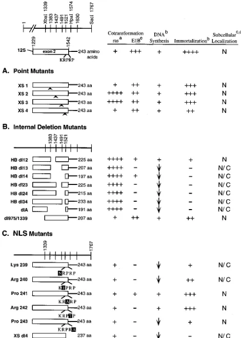
E1A-FIG. 1. Map of 12S second exon mutants and their functions. To the left are shown maps of the second exons of the WT and mutated 12S genes. All of these constructs have intact first exons. The top line, at the left, is a map indicating the salient restriction enzyme sites of the 12S second exon. The numbers above each line represent Ad nucleotide numbers. The numbers 1383, 1437, 1491, and 1521 indicate HpaI restriction enzyme cleavage sites that have been individually in-troduced by site-directed mutagenesis (19). The lines below the top line repre-sent transcripts. The name of each mutant is indicated at the left. The solid lines are untranslated regions, and the open boxes are the protein coding sequences. To the right is shown the number of amino acids (aa) encoded by each transcript. (A) Point mutants. (B) Internal deletion mutants. (C) NLS mutants. To the right, the properties of each mutant protein are summarized. Footnotes: a, data de-rived from the work of Douglas et al. (9) (1, transformation phenotype equiv-alent to WT 12S;1111, hypertransformation phenotype); b,1, 0 to 5%;11, 5 to 25%;111, 25 to 50%;1111, 50 to 100%; data derived from the work of Quinlan and Douglas (28); c, data derived from the work of Douglas and Quinlan (10); d, data derived from the work of Douglas and Quinlan (10a); e, data derived from this work.1, positive for the function;2, negative for the function;2, 20 to 50% less than the WT; N, protein is predominantly located in the nucleus; N/C, protein exhibits diffuse nuclear and cytoplasmic localization.
on November 9, 2019 by guest
http://jvi.asm.org/
[image:2.612.315.550.72.401.2]E1B-transformed cells were used, because no colonies could be detected when less cells were plated. For comparison, a 12S immortalized line was analyzed. These cells are not trans-formed and did not grow in soft agar. These results indicate
that while both E1B and T24ras can cooperate with E1A to transform primary epithelial cells, the resultant phenotypes are quite different.
The immortalizing function encoded by the COOH terminus of 12S is required for cotransformation with Ad E1B.We have previously shown that the expression of the 12S second exon is required for immortalization of primary epithelial cells, al-though it is dispensable for cooperation with the Ras onco-protein. To ascertain whether cotransformation with E1B re-quires the presence of the COOH terminus of 12S, we cotransfected primary BRK cells with pCMVE1B and a plas-mid encoding WT 12S or a mutant, CT dl976, which encodes only an intact first exon. Transformed foci were observed only in cultures that received both WT 12S and pCMVE1B, con-firming that, unlike T24ras, expression of the second exon is necessary for cooperation with E1B (35).
We have mapped the region of the second exon that is required for immortalization (28). To determine whether the same second exon region is required for cooperation with E1B, we used a series of four point mutants and eight internal deletion mutants (Fig. 1A and B, respectively) to cotransfect primary BRK cells. The results from these cotransfections are graphed in Fig. 5 as an average number of foci per dish. All of the point mutants were able to form transformed foci in the presence of E1B, which is consistent with their ability to im-mortalize primary BRK cells. Surprisingly, none of them gave rise to the aggressive hypertransformed phenotype exhibited by XS2 and XS3 with T24ras (Fig. 3). In all but one case (HB
dl14), those mutants which could bring about immortalization
[image:3.612.315.552.70.341.2](XS1 to XS4, HB dl12, and 975/1339) could also produce transformed foci with E1B (Fig. 1A and B). Although the
FIG. 2. Morphologies of E1B- and T24ras-cotransformed foci. Primary BRK cells were cotransfected with a WT 12S plasmid and either pCMVE1B or pT24ras 2 days after plating. For E1B cotransfections, 10mg of pCMVE1B and 5mg of 12S plasmid were used. For ras cotransfections, 2mg of pT24ras and 2mg of 12S plasmid were used. The cells were fixed with methanol and stained with Giemsa 3 weeks after transfection for ras cotransfections and 6 weeks after transfection for E1B cotransfections. (A) Epithelial focus from E1A-E1B co-transfection. (B) Epithelial focus from E1A-ras coco-transfection. (C) Fibroblast focus from E1A-E1B cotransfection. (D) Fibroblast focus from E1A-ras cotrans-fection. (E) Nontransformed cells. (F) Breakaway focus invading fibroblast focus from E1A-ras cotransfection. Magnification,310.
FIG. 3. Comparison of cotransformed focus-forming abilities of E1B and T24ras. Primary BRK cells were cotransfected with the indicated plasmids as described in the legend to Fig. 1. The cells were fixed in methanol and stained with Giemsa at 3 weeks posttransfection for ras and 6 weeks posttransfection for E1B.
FIG. 4. Ability of E1A-E1B and E1A-T24ras established lines to grow in semisolid media. Cells from the indicated cell lines were counted, and 104
(ras lines) and 106
(E1B lines) cells were plated in soft agar as described in the text. A 12S immortalized line (106
) was used as a negative control. The top four photomicrographs were taken at a magnification of310, and the bottom two were taken at a magnification of350.
on November 9, 2019 by guest
http://jvi.asm.org/
[image:3.612.60.296.70.328.2]cotransformation ability of HB dl12 was significantly lower than that of WT 12S, its immortalizing ability is concomitantly reduced. Inversely, those mutants that were unable to immor-talize primary BRK cells were also unable to bring about focus formation in the presence of E1B. This, again, is in contrast to the situation with T24ras, in which all of these mutants could enable tumorigenic transformation. In fact, many of these mu-tants cause hypertransformation with T24ras (Fig. 1) (9). Al-though HB dl14 cooperation with E1B was only about 30% as efficient as that of WT 12S, its ability to cotransform with E1B at all was surprising. Since HB dl14 is defective for immortal-ization and has a large deletion encompassing the smaller deletions of HB dl13, HB dl23, HB dl24, and HB dl34, which are all negative for E1B cotransformation, it seemed incongru-ous. Perhaps some conformation of this particular mutant pro-tein enables a low level of function necessary for cooperation with E1B, but this level is insufficient for immortalization by 12S alone.
All of these mutants express levels of protein at least equiv-alent to that of WT 12S and retain the ability to perform functions encoded by their intact first exons, such as activation of the cell cycle and cotransformation with T24ras, as men-tioned above (Fig. 1) (9, 28). These results indicate that ex-pression of a region of the second exon encoded between nucleotides 1437 and 1522 is necessary for E1B cooperation. This same region is necessary for 12S to immortalize primary epithelial cells, suggesting immortalization is a prerequisite for E1B but not T24ras cotransformation. This region includes the region identified by Mymryk and Bayley to be involved in the induction of E2A 72-kDa protein expression (25) and thus has a role in gene expression which may be involved in immortal-ization. The region encoded between nucleotides 1229 and 1437 seems to increase the efficiency of both immortalization and cooperation with E1B.
It is curious, although perhaps not surprising, that these two cooperating oncogenes require such different functions from E1A 12S, especially since neither ras nor E1B has any observ-able effect on BRK cells when expressed alone. As E1A and E1B coevolved, therefore, their functions could have evolved to be mutually interdependent. On the other hand, E1A and
ras happen to cooperate in transformation, but no limitations
or codependence could be expected. As detailed above, E1B is a much less potent oncoprotein than Ras protein. In fact, BRK cells transformed by 12S and E1B are only slightly more than immortalized. This may also explain why E1B requires more of 12S to enable tumorigenesis.
Expression of the WT second exon somehow suppresses a function activated by the ras pathway (7, 9, 15). Many of the 12S second exon mutants enable a hypertransformed state in the presence of an activated ras gene compared with what is enabled by WT 12S. However, the same mutants were not only unable to cause hypertransformation but were unable to co-transform with E1B at all. Given the distribution of the muta-tions that enable cooperation with E1B and effect hypertrans-formation with T24ras (i.e., XS2 and XS3 can cooperate with E1B and hypertransform with ras, while dl13, dl23, dl24, etc., cannot cooperate with E1B but can hypertransform with ras), it seems unlikely that these functions are related. It is more likely that they are different functions encoded by overlapping regions of the second exon. Although pCtBP (7) binds to the region of E1A encoding the suppressive function (of ras trans-formation), we have not been able to detect such complexes with any WT or mutant 12S protein. The differences with respect to hypertransformation, taken together with the differ-ential requirement of the second exon, indicate that ras and E1B utilize at least some different pathways, although they both require 12S first exon functions, such as activation of the cell cycle. Whether these differences are related to the ability of ras to activate the mitogen-activated protein kinase cascade and/or the rac and rho pathways (for reviews, see references 5 and 22) or the interaction of E1B with p53 (for a review, see reference 6) remains to be determined.
[image:4.612.59.298.72.309.2]The NLS and efficient nuclear localization are necessary for cotransformation with E1B but not with T24ras. We have previously shown that nuclear localization is necessary for 12S immortalization but is not essential for cooperation with T24ras (9, 10). The nuclear localization signal (NLS) of 12S is composed of the C-terminal five amino acids of the protein (Arg-Pro-Arg-Pro) (23). Two of the basic residues, Lys-239 and Arg-240, are critical for signal function, while the characteristics of the other three amino acids, Pro-241, Arg-242 and Pro-243, are not (10, 10a). Regions upstream of the signal are also important in efficient 12S nuclear localization, probably as structural determinants (Fig. 1B) (10). It has been determined that efficient 12S nuclear localization is not neces-sary for T24ras cotransformation (9). To determine if subcel-lular localization or the NLS is important in E1B cotransfor-mation, cotransfections with pCMVE1B and the mutated plasmids represented in Fig. 1C were performed. Quantitation of the transformed foci produced is shown in Fig. 5. Removal of the NLS (XS dl4) resulted in a loss of cooperation with E1B, suggesting it may be important in structure and/or function for E1B cotransformation. In an observation consistent with this, all of the signal mutants defective in localization were also defective in cooperation with E1B (Lys-239 and Arg-240) (Fig. 1 and 5). Similarly, other mutants defective in localization were defective in E1B cooperation, with the exception of HB dl14 (Fig. 1B and 5). Interestingly, however, the three efficiently localized NLS point mutants (Arg-242, Pro-241, and Pro-243) were also defective in E1B cotransformation. Other efficiently
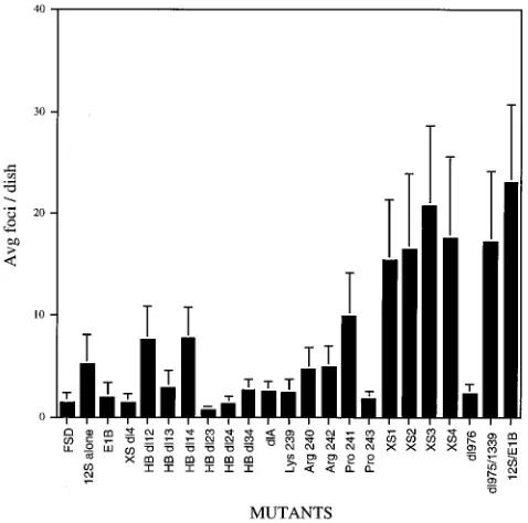
FIG. 5. Quantitation of E1B-cotransformed foci. Three dishes of primary BRK cells were cotransfected with the indicated 12S plasmid and pCMVE1B as indicated in the legend to Fig. 1. The cells were fixed with methanol and stained with Giemsa at 6 weeks posttransfection. The foci were counted and plotted as the average number of foci per dish. Two to five cotransfection experiments were performed for each mutant. FSD, fish sperm DNA; 12S alone, pLE12S alone; E1B, pCMVE1B alone. Error bars indicate standard deviations.
on November 9, 2019 by guest
http://jvi.asm.org/
localized proteins, such as HB dl12 and dl975/1339, and the four point mutants, XS1, XS2, XS3, and XS4, although they were able to cooperate with E1B, did so less efficiently than WT 12S. These results indicate that while efficient 12S nuclear localization may be important for E1B cotransformation, it is not sufficient. It also suggests that the NLS or region of the protein including the NLS is important in a function(s) other than just nuclear targeting. Another possibility is that E1B cooperation may be more sensitive to structural changes than the other two functions. A third possibility is that the E1B cotransformation assay itself is more stringent than the others, especially in a transfection. This would be consistent with the observation that E1B is not a very potent oncogene.
All of these results, taken together, indicate that the E1A second exon encodes several overlapping functions involved in immortalization and transformation. These results also indi-cate that different cooperating oncogenes require different E1A functions to effect the transformed phenotype and that E1A can participate in more than one transformation pathway.
We thank Carolyn Fields for technical assistance, Lee Danley for the graphics work, and the Molecular Resource Center at the University of Tennessee, Memphis, for the synthetic oligonucleotides used for the construction of plasmid pCMVE1B.
This work was supported by Public Health Service grant CA-50540 from the National Cancer Institute and American Cancer Society grants CD-438 and CB-75.
REFERENCES
1. Babiss, L. E., P. B. Fisher, and H. S. Ginsberg. 1984. Effect on transforma-tion of mutatransforma-tions in the early region 1b-encoded 21- and 55-kilodalton proteins of adenovirus 5. J. Virol. 52:389–395.
2. Barker, D. D., and A. J. Berk. 1987. Adenovirus proteins from both E1B reading frames are required for transformation of rodent cells by viral infection and DNA transfection. Virology 156:107–121.
3. Bayley, S. T., and J. S. Mymryk. 1994. Adenovirus E1A proteins and trans-formation (review). Int. J. Oncol. 5:325–444.
4. Bernards, R., P. I. Schrier, J. L. Bos, and A. J. van der Eb. 1983. Role of adenovirus types 5 and 12 early region 1b tumor antigens in oncogenic transformation. Virology 127:45–53.
5. Bokoch, G. M., and C. J. Der. 1993. Emerging concepts in the Ras super-family of GTP-binding proteins. FASEB J. 7:750–759.
6. Boulanger, P. A., and G. E. Blair. 1991. Expression and interactions of human adenovirus oncoproteins. Biochem. J. 275:281–299.
7. Boyd, J. M., T. Subramanian, U. Schaeper, M. La Regina, S. Bayley, and G.
Chinnadurai.1993. A region in the C-terminus of adenovirus 2/5 E1a pro-tein is required for association with a cellular phosphopropro-tein and important for the negative modulation of T24-ras mediated transformation, tumori-genesis and metastasis. EMBO J. 12:469–478.
8. Chinnadurai, G. 1983. Adenovirus 2 lp1locus codes for a 19kd tumor antigen that plays an essential role in cell transformation. Cell 33:759–766. 9. Douglas, J. L., S. Gopalakrishnan, and M. P. Quinlan. 1991. Modulation of transformation of primary epithelial cells by the second exon of the Ad5 E1A 12S gene. Oncogene 6:2093–2103.
10. Douglas, J. L., and M. P. Quinlan. 1994. Efficient nuclear localization of the Ad5 E1A 12S protein is necessary for immortalization but not cotransfor-mation of primary epithelial cells. Cell Growth Differ. 5:475–483. 10a.Douglas, J. L., and M. P. Quinlan. Submitted for publication.
11. Downward, J. 1992. Regulatory mechanisms for ras proteins. Bioessays 14: 177–184.
12. Edbauer, C., C. Lamberti, J. Tong, and J. Williams. 1988. Adenovirus type 12 E1B 19-kilodalton protein is not required for oncogenic transformation in rats. J. Virol. 62:3265–3273.
13. Fukui, Y., I. Saito, K. Shiroki, and H. Shimojo. 1984. Isolation of transfor-mation-defective, replication-nondefective early region 1B mutants of ade-novirus 12. J. Virol. 49:154–161.
14. Gallimore, P. H., P. A. Sharp, and J. Sambrook. 1974. Viral DNA in trans-formed cells. III. A study of the sequences of adenovirus 2 DNA in nine lines of transformed rat cells using specific fragments of the viral genome. J. Mol. Biol. 8:49–72.
15. Gopalakrishnan, S., and M. P. Quinlan. 1995. Modulation of E-cadherin localization in cells expressing wild type E1A 12S or hypertransforming mutants. Cell Growth Differ. 6:985–998.
16. Graham, F. L., A. J. van der Eb, and H. L. Heijneker. 1974. Size and location of the transforming region in human adenovirus type 5. Nature (London)
251:687–691.
17. Harlow, E., P. Whyte, B. R. Franza, Jr., and C. Schley. 1986. Association of adenovirus early-region 1A proteins with cellular polypeptides. Mol. Cell. Biol. 6:1579–1589.
18. Houweling, A., P. J. van der Elsen, and A. J. van der Eb. 1980. Partial transformation of primary rat cells by the leftmost 4.5% fragment of ade-novirus 5 DNA. Virology 105:537–550.
19. Kunkel, T. A. 1985. Rapid and efficient site-specific mutagenesis without a phenotypic selection. Proc. Natl. Acad. Sci. USA 82:488–492.
20. Lillie, J. W., M. Green, and M. R. Green. 1986. An adenovirus E1a protein region required for transformation and transcriptional repression. Cell 46: 1043–1051.
21. Lillie, J. W., P. M. Loewenstein, M. R. Green, and M. Green. 1987. Func-tional domains of adenovirus type 5 E1a proteins. Cell 50:1091–1100. 22. Lowy, D. R., and B. M. Willumsen. 1993. Function and regulation of RAS.
Annu. Rev. Biochem. 62:851–891.
23. Lyons, R. H., B. Q. Ferguson, and M. Rosenberg. 1987. Pentapeptide nuclear localization signal in adenovirus E1a. Mol. Cell. Biol. 7:2451–2456. 24. Moran, E., B. Zerler, T. M. Harrison, and M. B. Mathews. 1986.
Identifi-cation of separate domains in the adenovirus E1A gene for immortalization activity and the activation of virus early genes. Mol. Cell. Biol. 6:3470–3480. 25. Mymryk, J. S., and S. T. Bayley. 1993. Induction of gene expression by exon 2 of the major E1A proteins of adenovirus type 5. J. Virol. 67:6922–6928. 26. Pines, J., and T. Hunter. 1990. Human cyclin A is adenovirus
E1A-associ-ated protein p60 and behaves differently from cyclin B. Nature (London)
346:760–763.
27. Quinlan, M. P. 1994. Enhanced proliferation, growth factor induction and immortalization by adenovirus E1A 12S in the absence of E1B. Oncogene
9:2639–2647.
28. Quinlan, M. P., and J. L. Douglas. 1992. Immortalization of primary epi-thelial cells requires first and second exon functions of adenovirus type 5 12S. J. Virol. 66:2020–2030.
29. Quinlan, M. P., and T. Grodzicker. 1987. Adenovirus E1A 12S protein induces DNA synthesis and proliferation in primary epithelial cells in both the presence and absence of serum. J. Virol. 61:673–682.
30. Rao, L., M. Debbas, P. Sabbatini, D. Hockenbery, S. Korsmeyer, and E.
White.1992. The adenovirus E1A proteins induce apoptosis, which is inhib-ited by the E1B 19-kDa and Bcl-2 proteins. Proc. Natl. Acad. Sci. USA
89:7742–7746.
31. Schneider, J. F., F. Fisher, C. R. Goding, and N. C. Jones. 1987. Mutational analysis of the adenovirus E1a gene: the role of transcriptional regulation in transformation. EMBO J. 6:2053–2060.
32. Shen, Y., and T. Shenk. 1994. Relief of p53-mediated transcriptional repres-sion by the adenovirus E1B 19-kDa protein or the cellular Bcl-2 protein. Proc. Natl. Acad. Sci. USA 91:8940–8944.
33. Subramanian, T., M. Kuppuswamy, R. J. Nasr, and G. Chinnadurai. 1988. An N-terminal region of adenovirus E1A essential for cell transformation and induction of an epithelial cell growth factor. Oncogene 2:105–112. 34. Subramanian, T., M. La Regina, and G. Chinnadurai. 1989. Enhanced ras
oncogene mediated cell transformation and tumorigenesis by adenovirus 2 mutants lacking the C-terminal region of E1a protein. Oncogene 4:415–420. 35. Subramanian, T., S. E. Malstrom, and G. Chinnadurai. 1991. Requirement of the C-terminal region of adenovirus E1a for cell transformation in coop-eration with E1b. Oncogene 6:1171–1173.
36. Telling, G. C., and J. Williams. 1993. The E1B 19-kilodalton protein is not essential for transformation of rodent cells in vitro by adenovirus type 5. J. Virol. 67:1600–1611.
37. White, E., and R. Cipriani. 1990. Role of adenovirus E1B proteins in trans-formation: altered organization of intermediate filaments in transformed cells that express the 19-kilodalton protein. Mol. Cell. Biol. 10:120–130. 38. Whyte, P., K. J. Buchkovich, J. M. Horowitz, S. H. Friend, M. Raybuck, R. A.
Weinberg, and E. Harlow.1988. Association between an oncogene and an anti-oncogene: the adenovirus E1A proteins bind to the retinoblastoma gene product. Nature (London) 334:124–129.
39. Whyte, P., H. E. Ruley, and E. Harlow. 1988. Two regions of the adenovirus early region 1A proteins are required for transformation. J. Virol. 62:257– 265.
40. Whyte, P., N. M. Williamson, and E. Harlow. 1989. Cellular targets for transformation by the adenovirus E1A proteins. Cell 56:67–75.
41. Yew, P. R., and A. J. Berk. 1992. Inhibition of p53 transactivation required for transformation by adenovirus early 1B protein. Nature (London) 357: 82–85.
42. Yew, P. R., X. Liu, and A. J. Berk. 1994. Adenovirus E1B oncoprotein tethers a transcriptional repression domain to p53. Genes Dev. 8:190–202. 43. Zantema, A., J. A. M. Fransen, A. Davis-Olivier, F. C. S. Raemakers, G. P.
Vooijs, B. de Leys, and A. J. van der Eb.1985. Localization of the E1B proteins of adenovirus 5 in transformed cells, as revealed by interaction with monoclonal antibodies. Virology 142:44–58.
44. Zantema, A., P. I. Schrier, A. Davis-Olivier, T. van Laar, R. T. M. J. Vaessen,
and A. J. van der Eb.1985. Adenovirus serotype determines association and localization of the large E1B tumor antigen with cellular tumor antigen p53 in transformed cells. Mol. Cell. Biol. 5:3084–3091.