JOURNALOFVIROLOGY, Feb. 1994,p. 927-932 0022-538X/94/$04.00+0
Copyright ©D 1994,American Society forMicrobiology
Vaccine
Potential
of
a
Herpes
Simplex Virus
Type
1 Mutant
with
an
Essential Glycoprotein
Deleted
H. E. FARRELL,* C. S. McLEAN,2C. HARLEY,' S.EFSTATHIOU,' S. INGLIS,2 ANDA. C. MINSON' Division of Virology, Departmentof Pathology, Universityof Cambridge, Addenbrookes Hospital,
Cambridge CB2 2QQ,' and CantabPharmaceuticals Research Limited, Cambridge CB4 4GN,2 United Kingdom
Received24August 1993/Accepted4November 1993
Several approaches to the production of vaccines to human herpesviruses have been proposed. Subunit vaccines, subunits delivered by live vectors, and rationally attenuated vaccines have all been shown to be
efficacious in animal models but suffer from uncertainties as to the roles of individual genes involved in pathogenesis and the mostrelevantcomponents of the immune response required for protection in humans and thetargetantigens involved. With these problems in mind,weexamined the vaccine potential ofa fully disabledherpes simplex virustype 1mutantthat is capable of onlya single round of replication, sinceavirus ofthistypeshould induce thefullspectrumof immuneresponsesbuthas nopathogenic potential. A virus has beendescribed which lacks essentialglycoprotein H (gH) andcanbepropagated inacellline which supplies gH in trans (A. Forrester, H. Farrell, G. Wilkinson, J. Kaye, N. Davis-Poynter, and T. Minson, J. Virol. 66:341-348, 1992). Infection of normal cells with this mutantisindistinguishable from awild-type infection, exceptthattheresultingprogeny aregH negative andnoninfectious: the virus is self-limiting. Infection of mice
by theearpinnaroutewas similarlyself-limiting in that input infectivity decreased rapidlyattheinoculation siteandnoinfectivitywasdetected insensoryganglia. Animals givenawiderangeof doses of thegH-negative mutantproduced both humoral and T-cellresponsestoherpes simplex virustype1andproved solidly resistant
tochallengewithahigh dose of wild-type virus. The gH-negativemutantis presumably capable ofestablishing alatentinfection, but sincenoinfectious viruswasdetected innumerousattempts toreactivate themutant,the riskofa pathogenicoutcome is minimal.
There is ample evidence, based primarily on studies using
animalmodels, that both the humoral and cell-mediated arms
of the immune response can function in the prevention and control of herpes simplex virus (HSV) primary and recurrent infections (14, 20). Thus, a number ofstudies have
demon-stratedthatadministration ofpassive antibody modulates HSV
type 1 (HSV-1) infection in mice (18, 21); nevertheless, B-cell-suppressed mice recover normally from infection (9,
22). A variety of immunization and vaccinationprotocolshave
been effective in protecting rodents from HSV-1 infection. These includeadministration of inactivated virionsorinfected
cell extracts, immunization with purified or recombinant
gly-coproteins, or vaccination with antigens delivered by live
vectors (reviewed in reference 4), but there is conflicting evidence as tothemajorprotective antigens and the immune mechanism involved in the resultingprotection. Furthermore, given the relative easewith whichprotection canbeachieved,
particularly in inbred mousestrains, there issignificant doubt astothe relevance of themechanismsfor immunitytoHSV-1
in humans. In the face ofthis uncertaintyas to the natureof the protective immune response to HSV-1 in humans, the
development of rationally attenuated vaccines offers some
attraction, because live herpesvirus vaccines have proved
suc-cessfulagainst veterinarydiseasesofeconomicimportance (5, 17) and attenuated vaccines elicitanatural immuneresponse,
thus reducing the need, in vaccine design, for a detailed understanding of the immune response to infection. The
*Corresponding author. Mailing address: Division of Virology,
Department ofPathology, University ofCambridge, Level5, Adden-brookesHospital, Cambridge,CB22QQ,UnitedKingdom.Phone:44
223 336921. Fax: 44 223 336926. Electronic mail address: hef@mbfs. bio.cam.ac.uk.
complexity of the HSV genome and the presence ofa large number of genes that are dispensable for growth in vivo
provide a variety of opportunities for rational attenuation. Deletion or interruption ofanumber ofgenes, including the
thymidine kinase gene (6), several dispensable glycoprotein
genes (2, 24), and the protein kinase gene (16), has yielded
viruses with attenuated phenotypes that elicit a protective
response in experimental animals, but we cannot be certain
thattheseviruses will proveequally attenuatedinhumans. An
alternative approach to attenuation is the use of a fully disabledvirusthatisreplication defective. Althoughviruses of
this type synthesize only a subset of viral proteins and are
unable toproduce virus particles, Nguyen et al. (15) showed
thatHSV-1 mutantslacking eitherimmediate-earlygeneICP4
or ICP27 orearly gene ICP8 were capable ofeliciting T-cell
responsesinBALB/c miceandwerecapableofprotectingmice
from subsequent lethal intraperitoneal challenge with wild-type HSV-1. Replication-incompetent viruses of this type,
however, fail to replicate the viral genome and, in
conse-quence,donotsynthesize significant quantitiesof lateproteins,
many of which are known to elicit protective immune
re-sponses(11, 19, 23).Analternativeapproach istomakeuseof
a virus that is replication competent but produces noninfec-tious particles by virtue of the absence of a single essential
virion protein. A single-cycle virus with these characteristics
shouldpresentall of thevirus-specificproteins,apartfromthe
absent one,to all armsofthe immune response.
We have describedpreviouslythe construction ofan HSV-1 mutant in which glycoprotein H (gH) coding sequences are replaced with the Escherichia coli lacZ gene and which is
propagatedinacell linesupplying gHintrans(7). Infectionof normal cells resultsinacomplete lytic cycleand theproduction
andsecretion of normal numbersofnoninfectious enveloped
927
Vol.68,No. 2
on November 9, 2019 by guest
http://jvi.asm.org/
928 FARRELL ET AL.
virions. Wetherefore decided to investigate the ability of this virus to induce aprotective response in micebya single-cycle
infection.
MATERUILSAND METHODS
Viruses. The parent HSV-1 strain used in all of the experi-ments was the SC16 strain and is hereafter referred to as wtSC16. This virus was propagated on BHK-21 cells as de-scribed previously (6). The construction oftwo HSV-1mutants
derived from the SC16 parental strain in which the E. coli lacZ gene replaced either US5 coding sequences (SC16AUS5) or gH coding sequences (SC16AgH) has also been previously described. The SC16AUS5 mutant is indistinguishable from the parent virus with respect to growth and dissemination in vitro (7, 25) and in vivo (1) and was used to facilitate detection of virus-infected cells in mice. The SC16AgH mutant was propagated on a supporting Vero cell line (F6) which supplies gH in trans upon infection with HSV-1 (7). Thus, progeny virions are phenotypically gH positive (having derived cellular gH) and possess a particle-to-PFU ratio equivalent to that of the wtSC16virus. Viruses used for in vivo experiments were partially purified by the following method. Extracellular virus wasrecovered from cell cultures infected 48 h previously at a
multiplicity ofinfection of0.01 andcentrifuged for 90 min at 85,000 x gfor 1 h. The virus pellets were resuspended in 3 ml of TES (1 mM Tris [pH 7.4], 1 mM EDTA, 100 mM saline,
0.5% fetal calf serum) and loaded onto discontinuous 20 to
40%sucrosegradients. Followingcentrifugation at 25,000 x g for 1 h, the viruses were harvested from the 20 to 40% interface, diluted in TES, and centrifuged at 85,000 x g for 1 h. The virus pellets were resuspended in TES, and aliquots were stored at -70°C. Purified wtSC16 was inactivated with
,B-propiolactone (0.05% [vol/vol]; 30 min at room tempera-ture); the absence of live virus was demonstrated by plaque assay.
Mice. Four-week-old female BALB/c mice were obtained from Tucks Ltd. and inoculated at 5 to 6 weeks of age. Lightly anesthetized mice were vaccinated by scarification of the left
earpinna with an inoculum of either live or inactivated purified virus in 2 ,ul ofphosphate-buffered saline (PBS). Control mice were inoculated with 2 ,ul of PBS only. At 14 days after vaccination, mice were challenged by infection of the right ear with wtSC16 atthe doses indicated in the figure legends.
Measurement of virus titers in mouse tissues. Ear pinnae
and pooled innervating sensory ganglia (CII, CIII, and CIV)
were dissected and stored at -70°C. Samples were later homogenized and assayed for the presence of virus on BHK-21
or F6cells as described previously (6).
Enzatic detection ofSC16AUS5 and SC16AgH mutants
incells infected in vitro and invivo. The method used to detect
,3-galactosidase
in cells infected in vitro with recombinant viruses carrying the lacZ gene with the substrate5-bromo-4-chloro-3-indolyl-13-D-galactopyranoside
(X-Gal) has been pre-viously described (7). The same procedure was used to detect the mutants in 5-,um frozen sections of ear pinnae from inoculated mice, except that as a final step, the tissue was counterstained withhematoxylin and eosin.Detection ofneutralizingantibodies to HSV-1. Neutralizing antibodies were detected by a plaque reduction assay in the presenceof5%freshguineapig serum as asource of comple-ment.Individualserumsamples were serially diluted in 500-,u volumes and incubated with an equal volume ofwtSC16(103 PFU/ml) for 1 h at 37°C. Nonneutralized virus at the end of the incubation was detectedby plaque assay on BHK-21 cells. The titerwascalculated as the reciprocal dilution of serum required
0 2 3 4 5 7 2 3 4
DAY P.1.
FIG. 1. Clearance of HSV-1 lacZ insertional mutants in the ears and ganglia of mice. Groups of fourmice were infected with either SC16AgH (0) orSC16AUS5 (-), and virus titers were measuredon days 1,2, 3,4, 5, and 7 postinfection (P.I.). Virus titers in mice infected withtheSC16AgHmutant were determined on F6 cells, while titers in mice infectedwith the SC16AUS5 mutant were assayed on BHK-21 cells. The data are presented as geometric means with the standard errors of the means. All virus was of the j3-galactosidase-positive phenotype.
to achieve a 50% reduction in plaque numbers relative to the
number of plaques obtained in the absence of mouse serum. Included as a positive standard in each assay was a
comple-ment-dependent neutralizing monoclonal antibody (designat-ed AP7) which is direct(designat-ed against HSV-1 glycoprotein D(13).
Delayed-type
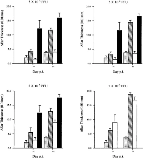
hypersensitivity (DTH) assays. Groups of six mice were vaccinated in the left ear pinna with either the live SC16AgH mutant,inactivated SC16AgH, or livewtSC16 atthe doses indicated in Fig. 4. A control group of mice was inoculated with PBS only. At 14 days postvaccination, mice were challenged with106PFU ofwtSC16by scarification of the right ear pinna. The thicknesses of both ears were measured at24 and 48 h postchallenge. The results are expressed as
differences in thickness between challenged and unchallenged ears.
RESULTS
Determinationof the level of nondefectivevirusin SC16AgH
progeny. Replication-defective deletion mutants are
occasion-ally rescued byrecombination with the viral gene resident in
the helpercell line. Before initiating protection experiments, it
wastherefore important to determine the levels of nondefec-tive virus in SC16AgH virus stocks and to assess the possible impact of this component on the experiments. Following two
rounds of cloning by limiting dilution, working stocks of
SC16AgHvirus were titrated on gH-inducible F6 cells and on
Vero cells and the relative titers were estimated as
approxi-mately 5 x 10⁶:1. Plaques formed in Vero cells were f-galac-tosidase negative, implying that the nondefective phenotype
had resulted from replacement of the lacZ gene by the gH-encoding gene resident in the cell line. Mice were then infected by the ear route with 5 x 10⁷ PFU ofSC16AgHvirus, and titers at the inoculation site and in innervating dorsal root ganglia were measuredby plaque assay on F6 cells. The results
are shown inFig. 1, in which, for comparison, the virus titers
resulting from parallel infections with 5 x 10⁵ PFU
(i.e.,
a 100-fold lower dose) of SC16AUs5 arealso shown. One day afterinfection, less than 0.01% of the inputSC16AgH inocu-lum wasrecovered from the inoculation site and this residual J. VIROL.6
4
W.
on November 9, 2019 by guest
http://jvi.asm.org/
[image:2.612.316.553.69.214.2]VACCINE POTENTIAL OF A gH-NEGATIVE HSV-1 MUTANT 929
inoculum was cleared during the subsequent 7 days. No
infectivity was recovered from dorsal root ganglia. All of the
detectable virus had the f-galactosidase-positive phenotype and was not detected by assay on BHK-21 cells. Thus, although each animal received an estimated 10 PFU of replication-competent virus, this component was neverdetected and was presumably overwhelmed by the huge excess of defective virus.
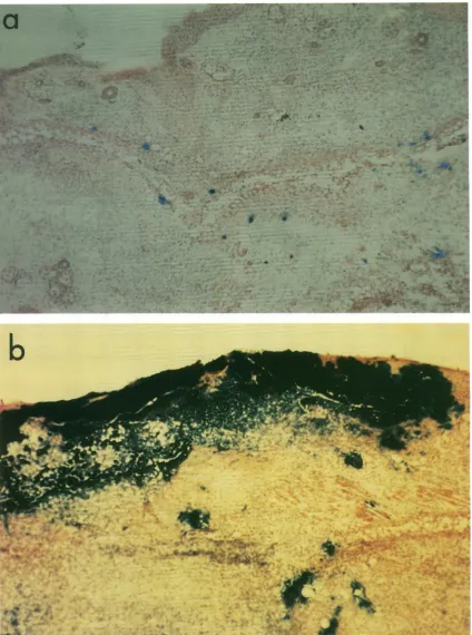
Histologicalexamination revealed that after 5 days, when only about 0.0001% of the input inoculum can be detected, large
numbers of 3-galactosidase-positive cells were present at the
inoculation site (Fig. 2a). Thus, inoculation with phenotypi-cally gH+ virus had successfully established infection but, in
contrast to the SC16AUs5 virus (Fig. 2b), had not spread beyondtheinoculationsite. At 1month afterinfection,theclI, cIll, and cIVdorsal rootgangliaweredissected from 12 mice
infected with SC16AgH and were incubated in tissue culture medium for 5 days to allow reactivation of latent virus. The tissuewasthen homogenized and assayed for infectivity.None was found.
Induction ofprotective immunity bySC16AgHvirus. Exper-iments were designed to compare the protective efficacy of equivalent doses of inactivated virus with that of
replication-defective virus over a broad dose range. Protection was assessedby measuring the growthofwild-type virus in the ear pinna5 daysafterchallenge, thepointatwhichpeak titersare
observed in this model (Fig. 1). Groups of four mice were
vaccinated byleft-earscarification with SC16AgHor ,B-propio-lactone-inactivated virusatdosesrangingfrom 5 x 10²to5 x
107 PFU. The particle-infectivity ratio of the purified virus preparation was approximately 30:1, and the maximum dose
used therefore was about 109virions, equivalenttoabout 1 ,ug
of virion protein. After 14 days, mice were challenged by
inoculationoftherightearwith 2 x 10⁶PFUofwtSC16. The resultsoftwoexperimentsare shown inFig. 3, whichgives the geometric mean titers ofchallenge virus5 days after inocula-tion.Atthe higherdosesused, vaccination withliveSC16AgH
orwith inactivatedvirus resulted incomplete clearanceofthe
challenge virusat5dayspostinfection, whileatthe lower dose no effect was observed with live or killed vaccine. At all intermediate doses, vaccination with the live gH- virus
sup-pressed challenge virus growth with greater efficiency. Com-parison of the data suggests that a 100-fold-higher dose of inactivated virus isrequired to achievean equivalenteffect in thisassay.
Induction ofneutralizing antibodies by the SC16AgH
mu-tant.Although different studieshavesuggesteddifferent
mech-anisms ofimmunity to HSV challenge, a comparison of the
efficacies of different glycoprotein antigens delivered with a
livevector suggested that protection correlated with neutral-izing antibody induction.InjectionwithalivegH-negativevirus
results in a single-cycle infection and significant amplification
of the delivered antigen compared with vaccination with an
equivalentdoseofkilled virus. Wetherefore
predicted higher
neutralizingantibody titersinanimals that receivedSC16AgH
thaninthosethatreceived inactivatedvirions. Serum
samples
taken from individual micepriortochallengeinthe
protection
experiment shown in Fig. 3 (experiment 2) were assayed for
neutralizing antibody to HSV-1 (Table 1). At the lowest vaccine dose for which sera were tested (5 x 10⁴ PFU),
SC16AgH infection induced detectable neutralizing
antibody,
whereas inactivated virions didnot. Nevertheless, the
neutral-izing antibody titers in the sera of mice that received
higher
vaccine doses were not significantly different and it seems
unlikely that preexisting antibody titers can account for the
considerable difference in protection afforded
by
vaccinationwith the live disabled virus and the inactivated virus. For
comparison,theneutralizing antibodytiters of animalsinjected
inparallelwith 2 x 10⁶PFU ofwtSC16are shown. This dose
ofwild-typevirus affords complete protection against
subse-quentchallengewiththesamedose. Asexpected,theantibody
titers induced in these animals were higher although not
dramaticallyso.
Induction of DTH responses bytheSC16AgH mutant.It is
widely acceptedthat T-cell responsesarecrucialincontrolling
HSVinfection,andwhileuncertaintyremainsas tothe relative
contributions of different T-cell subsets, there is strong evi-dence that CD4+T-cell responsesare effective in virus clear-ance in the mouse ear model. DTH responses to wtSC16 in vaccinated mice were therefore measured as described in
Materials and Methods. The
experimental design
was similarto that described for the
protection study
in that a range of dosesof thevaccinating
viruswasemployed
andtheresponsesinduced by equivalent doses of an inactivated vaccine were
included for comparison. In
addition,
groups of mice which receivedequivalent
doses ofwtSC16 as thevaccinating
virus were also included. The results arepresented
inFig.
4. Atvaccinating
doses of5 x 103 and5 x 104PFU,
theSC16AgH
mutant induced DTH responses at levels on
day
2postinfec-tion whichwere
comparable
tothoseinducedby wtSC16,
while the inactivated vaccine at thesedoseswascompletely
ineffec-tive.Inaseparateexperiment,
adoseof102PFUofeither the live-disabled orkilled vaccine failed toelicit a DTH response(data
notshown).
Nevertheless,
DTH responsesweredetected in micegiven
higher
doses of the inactivatedvaccine,
withresponses which were
equivalent
to those inducedby
the SC6IgH
mutantatthehighest
vaccine dose used. While theseresults do not, ofcourse, showthat the basisofthe
efficacy
of alive disabledvirus, by
comparison
withakilledvirus,
lies in itsability
specifically
toinduce aDTH response,they
areconsis-tentwith the
general
viewthat a livevaccine recruits aT-cellresponsemore
efficiently
than doesanequivalent
doseofkilledantigen.
DISCUSSION
It is
generally
accepted
thatlive vaccines offerconsiderableadvantages
overkilledorsubunit vaccinesintermsof both therange ofresponses induced and the
longevity
of theresponse.The
ability
of HSVtoestablishlife-long
latency
is,
however,
asignificant
issue incontemplating
human vaccination with anattenuated HSV. A disabled virus
capable
ofasingle cycle
of infection may be an attractive alternative. Thefindings
pre-sented here demonstrate that the
SC16AgH
HSV-1 mutant is able to establish aself-limiting
infection invivo that inducesboth humoral and T-cell responses and protects mice
against
subsequent
challenge
withwild-type
HSV-1. Lowdoses oftheSC16AgH
mutant induced levels of DTH responses in mice which werecomparable
to those inducedfollowing
infection withwtSC16. Inaddition,
theSC16AgH
viruselicitedneutral-izing
antibodies with titers which remain elevatedwellbeyond
clearance of thevirus from the animals. In
comparison
withaninactivated
vaccine,
theSC16AgH
mutantwas moreeffective ininducing
DTH responses and inconferring
protection
during
acuteinfection.Although
live-virusvaccineshave beenshownto elicit cellular
immunity
morereadily
than killed-virus or subunitvaccines,
themechanism(s) by
which this isachieved isunknown. A
simple
explanation
for our results is that theamplification
ofviral genesproduced
during
thesingle
cycle
ofreplication
of theSC16AgH
mutant in vivo and the naturalpresentation
ofappropriate
viraltargetselicitresponseswhich,
in both
quantity
andquality,
are more effective inconferring
protection
than are the responses inducedby
killed-virusVOL. 68? 1994
on November 9, 2019 by guest
http://jvi.asm.org/
930 FARRELL ET AL.
FIG. 2. Histological demonstration ofcellsinfectedwith HSV-1 lacZinsertionalmutantsinmice(magnification, x160). Sections of the ear
pinnaeofmice infected 5 dayspreviouslywith either SC16AgH (a)or SC16AUS5 (b) were examined microscopically after being stained with X-Gal.
J.VlIROL.
on November 9, 2019 by guest
http://jvi.asm.org/
[image:4.612.99.523.101.671.2]VACCINE POTENTIAL OF A gH-NEGATIVE HSV-1 MUTANT
^ - EXPERIMENTI - 1 EXPERIMENTI
[image:5.612.60.297.88.218.2]5xlO' 5 Vaccinedose (PFU)
FIG. 3. Clearance of wtSC16 in the ears of mice vaccinated with either SC16AgH or ,B-propiolactone-inactivated wtSC16. Groups of four mice were vaccinated at the doses indicated by scarification of the left ear pinna. Control mice were given saline only. Mice were challenged 14 days postvaccination with 2 x 10⁶PFU of wtSC16 in the
rightearpinna,and virus titers were measured at 5 days postinfection. The data areexpressed as geometric means and the standard errors of the means. Asterisks indicate that virus was not detected. In cases in which virus was not recovered from four of four mice, the numbers of mice from which virus was recovered are indicated above the error
bars.
vaccines. However, the protection afforded by the submicro-gramdoses of killed-virus vaccine illustrates the relative ease with which protection may be achieved in the mouse model. Further work is required to quantify the immune responses generated by this single-cycle vaccine and to determine, by
adoptive transfer, which of the responses afford protection. Furthermore, while mice have unique advantages in studies of immune responses,HSV infection in guineapigs isappropriate
for studies of reactivation and recrudescence andexperiments
are now in progress to evaluate the vaccine potential of a
single-cyclevirus in this animal model.
Similarstudiesaddressing the vaccine potential of disabled HSV-1 mutants were reported by Nguyen et al. (15), who demonstrated that cellularimmunity to HSV-1 and protection from lethalchallenge in micecould be achieved byvaccination
[image:5.612.315.554.89.354.2]with replication-defective HSV-1 mutants. The magnitude of
TABLE 1. Quantification of neutralizing antibodies toHSV-1 in
mice vaccinated with either live SC16AgHorinactivated wtSC16 Vaccinedose Neutralizing antibodytiteraat:
Vaccine (PFU) 14days 3 mo
Live SC16AgH 0 <50 <50
5x 10⁴ 79 ±29 NDb
5 x 10⁵ 158 ±58 ND
5 x 10⁶ 400 ±0 500± 178
2 x 10⁷ 630± 162 ND
Inactivated wtSC16 0 <50 <50
5x 10⁴ <50 ND
5 x 10⁵ 100 ±37 ND
5 x 10⁶ 500± 257 912 ± 130
2 x 10⁷ 400 ±0 ND
LivewtSC16 2 x 10⁶ 1,600 + 585 1,600 + 0
aEach individual titer isexpressedas thegeometricmeanof thereciprocal
dilution(plus1standarddeviation)ofserumrequiredtoneutralize 50% of the virusinfectivityobtained in the absence ofantibody.
6ND,notdone.
Day p.i. Day p.i.
FIG. 4. DTH responses in mice vaccinated with PBS (Lii), SC16AgH(0), ,B-propiolactone-inactivated SC16AgH (O),orwtSC16 (-). Micewerevaccinated in the leftearpinnaatthe doses indicated 14days priortochallengewith106 PFUofwtSC16intheoppositeear. Note thatwtSC16wasnot usedatthe highestvaccine dose(5 x 10⁶
PFU), since this is a lethal dose for5-week-old BALB/c mice. Ear thicknesswasmeasured 24 and 48 hpostchallengeandisexpressedas the differencebetween thechallenged andvaccinatedears. The data are presentedasmean differences inearthickness and the standard errorsof themeans.p.i.,postinfection.
T-cell responses to these mutants was proportional to the extent towhich viralgeneexpressionoccurred in infected cells.
However,thereplication-defectivemutantsused in theirstudy weredeficientinthe expressionofallor someof the lategene products,manyofwhichareknowntobetargetsfor antibodies and T cells(3, 8, 10, 12).Incontrast,infection of cells with the
phenotypically gH-positive SC16zAgH mutantresultsin a
rep-licative cycle that is normal in all respects except that the
progenyarenoninfectious.Therefore,we canpresumethat the
SC16AgH mutanthasthecapacityto elicitimmune responses
with arangeofspecificities (withtheexceptionofresponsesto
gH) comparablewith those inducedbythe parental wild-type
virus.
Given that the SC16AgH virus is incapable of producing
infectious progeny in cells that are unable to provide gH in trans,it isunlikelythat infection couldspread beyondthe cells
surrounding the vaccination site in vivo. However, given that
disabledHSV-1mutantshavebeenpreviouslyshownto
estab-lishalatentinfection, it ispossible thattheSC16zAgHviruscan
establish a latent infection as a result of direct entry into
sensorynerve endingsbythe input inoculum.Indeed,
prelim-inary experimentshaveidentifiedthemajorlatency-associated transcript in the cervical ganglia of mice vaccinated with the
SC16AgH virus (data not shown). Although detection of the lacZgeneisrequiredforformalproofthat the
SC16AgH
virus resides in sensory ganglia, numerous attempts to reactivatevirus from infected animals haveproved unsuccessful, implying 5X10³PFU
C~
-0 .2
5X10⁴PFU
E
°; 15.0
Hv: s; 1(0.0
5.0-Day p.i. 5X10⁵PFU
Day p.i.
i
E
C
s
5X1(06PFU E
z:_
-~d
*,,
c
.D
VOL.68, 1994 931
on November 9, 2019 by guest
http://jvi.asm.org/
[image:5.612.56.297.551.700.2]932 FARRELL ET AL.
that the latentvirus is unable toproduceinfectious virusupon
reactivation. Furthermore, attemptsto rescue 3-galactosidase-positive virus by superinfection oflatently infected ganglia in
vitro were also unsuccessful.
In conclusion, our studies demonstrate that the SC16gH mutant is able to stimulate protective immune responses without the risk of virus dissemination associated with live
attenuated HSVvaccines.AlthoughtheSC16AgHmutantmay
have thepropensity to establish a latent infection, reactivation of the latent virus could result only in the production of
noninfectious virus. This vaccination strategy may also be
appliedtosystemsinwhich HSV-1 isused as a live vector for
expression of heterologousgenes.
ACKNOWLEDGMENTS
This work was supported in part by Cantab Pharmaceuticals Re-search Limited. H.E.F. was supported by a grant from the Royal
Society.
REFERENCES
1. Atkinson,H., and A. C. Minson.Personal communication. 2. Balan, P., and A. C. Minson. Personalcommunication.
3. Blacklaws, B. A., and A. A.Nash. 1990. Immunological memory to herpessimplex virus type 1 glycoproteins B and D in mice. J. Gen. Virol. 71:863-871.
4. Burke, R. L. 1992. Contemporary approaches to vaccination against herpes simplex virus. Curr. Top. Microbiol. Immunol. 179:137-158.
5. DeLeeuw, P. W., and J. T. van Oirschot. 1985.Vaccines against Aujesky's disease: evaluation of their efficacy under standardized laboratory conditions. Vet.Q.7:191-197.
6. Efstathiou, S., S. Kemp, G. Darby, and A. C. Minson. 1989. The role ofherpes simplex virustype 1thymidine kinase in pathogen-esis. J. Gen.Virol. 70:869-879.
7. Forrester,A., H. Farrell, G. Wilkinson, J. Kaye, N. Davis-Poynter, and T. Minson. 1992. Construction andpropertiesofamutantof herpessimplexvirus type 1 withglycoprotein H coding sequences deleted. J. Virol. 66:341-348.
8. Johnson,R.M., D. W. Lancki, F. W.Fitch, and P. G. Spear. 1990. Herpes simplex virus glycoprotein D is recognized as antigen by CD4+ and CD8+ Tlymphocytes from infected mice. Character-ization ofTcellclones. J. Immunol. 145:702-710.
9. Kapoor, A. K., A. A. Nash, and P. Wildy. 1982. Pathogenesis of herpessimplexvirus in Bcell-suppressed mice: the relative roles of cell-mediated and humoral immunity. J. Gen. Virol. 61:127-131. 10. Martin, S., E. Cantin, and B. T. Rouse. 1989. Evaluation of
antiviral immunity using vaccinia virus recombinants expressing cloned genes forherpes simplex virustype1glycoproteins. J. Gen. Virol. 70:1359-1370.
11. McDermott, M. R., F. L. Graham, T. Hanke, and D. C. Johnson.
1989. Protection of mice against lethal challenge with herpes
simplex virus byvaccination withanadenovirusvectorexpressing HSV glycoprotein B.Virology 169:244-247.
12. McLaughlin-Taylor, E., D. E. Willey, E. M.Cantin,R.Eberle,B. Moss, and H. Openshaw. 1988. A recombinant vaccinia virus expressing herpes simplex virus type 1 glycoprotein B induces cytotoxic T lymphocytesin mice.J.Gen. Virol.69:1731-1734. 13. Minson, A. C., T. C. Hodgman, P. Digard, D. C. Hancock,S. E.
Bell, and E. A. Buckmaster. 1986. An analysis of the biological properties of monoclonal antibodies against glycoprotein D of herpes simplex virus andidentification of amino acid substitutions that confer resistance toneutralization. J. Gen. Virol. 67:1011-1013.
14. Nash, A. A., and P.Cambouropoulos. 1993. The immune response toherpessimplex virus. Semin. Virol. 4:181-186.
15. Nguyen, L. H., D. M. Knipe, and R. W.Finberg. 1992. Replication-defective mutants of herpes simplex virus(HSV) induce cellular immunity and protect against lethal HSV infection. J. Virol. 66:7067-7072.
16. Nishiyama, Y., Y. Yamada, R. Kurachi, and T. Daikoku. 1992.
Construction ofa US5 lacZinsertion mutant ofherpes simplex
virus type 2 andcharacterization of its phenotype in vitro and in vivo.Virology 190:256-268.
17. Okazaki, W., H. G. Purchase, and B. R. Burmester. 1970. Protec-tionagainstMarek'sdisease by vaccinationwithaherpesvirusof turkeys. Avian Dis. 14:413-429.
18. Openshaw, H., L. V. S. Asher, C. Wohlenberg,T. Sekizawa, and A. L. Notkins. 1979. Acute and latentinfection of sensory ganglia withherpes simplex virus: immune control and virus reactivation. J.Gen.Virol.44:205-215.
19. Rooney, J. F., C. Wohlenberg, K. J. Cremer, B. Moss, and A. L. Notkins. 1988. Immunizationwith a vaccinia virus recombinant expressing herpessimplexvirus type 1 glycoprotein D: long-term protection and effectof revaccination. J. Virol. 62:1530-1534. 20. Schmid, D. S., and B. T. Rouse. 1992. The role of T cell immunity
in control ofherpessimplex virus.Curr. Top.Microbiol. Immunol. 179:57-74.
21. Simmons, A., and A. A. Nash. 1985. Role of antibody in primary and recurrentherpessimplex virus infection.J.Virol. 53:944-948. 22. Simmons, A., and A. A. Nash. 1987. Effect of B cell suppression on
primary and reinfection of mice with herpes simplex virus. J. Infect. Dis. 13:108-114.
23. Stanberry, L.R., D. J. Bernstein, and R. L. Burke. 1987. Vacci-nation with recombinantherpes simplex virus glycoproteins: pro-tectionagainstinitial and recurrent genital herpes. J. Infect. Dis. 155:914-920.
24. Sunstrum, J. C., C. E. Chrisp, M.Levine,and J. C. Glorioso.1988. Pathogenicity of glycoprotein C negative mutants of herpes
sim-plexvirus type 1 for the mouse central nervous system. Virus Res. 11:17-32.
25. Weber, P. C., M. Levine, and J. C. Glorioso. 1987. Rapid
identi-fication ofnonessential genes ofherpessimplex type 1 by TnS
mutagenesis. Science236:576-579.
J.VlIROL.