ROLE OF THE REDOX RESPONSIVE TRANSCRIPTION
FACTOR, NRF2 IN IMMUNE CELL FUNCTION
Thesis submitted in accordance with the requirements of the University of
Liverpool for the degree of Doctor in Philosophy
by
Junnat Hamdam
DECLARATION
This thesis is the result of my own work. The material contained within this thesis has not been presented, nor is currently being presented, either wholly or in part for any degree or
other qualification.
Junnat Hamdam
This research was carried out in the MRC Centre for Drug Safety Science, Department of Pharmacology and Therapeutics,
TABLE OF CONTENTS
PAGE
ABSTRACT i
ACKNOWLEGEMENTS iii
PUBLICATIONS v
ABBREVIATIONS vi
1.0 CHAPTER ONE: GENERAL INTRODUCTION
1.1 The Immune System 2
1.2 Innate immunity 2
1.3 Adaptive Immunity 7
1.4 T cell development 8
1.5 Dendritic cells 11
1.6 Dendritic cells: drivers of the immune response 13
1.7 Dendritic cell antigen presentation 14
1.7.1 Endogenous classical MHCI pathway 15
1.7.2 Exogenous MHCII pathway 16
1.7.3 Cross-presentation 18
1.8 DC-mediated Antigen-specific T cell activation 20
1.9 T cells and the immune response 22
1.10 CD8 T cells 23
1.11 CD4 T helper cells 26
1.11.1 Th1 cells 26
1.11.2 Th2 27
1.11.3 Th17 cells 28
1.11.4 T regulatory cells (Treg) 30
1.11.5 Other effector CD4 T cell subtypes: Tfh, Th9, Th22 31
1.12.1 Th1/Th2 cross talk 33
1.12.2 Th1/Th2/th17 cross talk 33
1.12.3 Treg/Th17 cross talk 34
1.13 Redox and its role in immune cell function 35
1.14 Oxidative stress and immune cell function 39
1.15 Nrf2: Master regulator of the anti-oxidant defence response 40 1.16 The role of Nrf2 in the maintenance of redox homeostasis 44 1.17 The role of Nrf2 in cellular adaptation to xenobiotic incursion 46 1.18 Role of Nrf2 in human immune-mediated diseases 47
1.19 Role of Nrf2 in immune cell function 47
1.20 THESIS AIMS 49
2.0 CHAPTER TWO: METHODS
2.1 Materials and Reagents 51
2.2 Antibodies 51
2.3 Peptides and cytokines 52
2.4 Mice 52
2.5 Complete medium 52
2.6 Cell lines 53
2.7 Generation of Bone Marrow Derived Dendritic cells (BMDCs) 53
2.8 Glutathione assay 53
2.9 Harvesting of thymocytes, splenocytes and lymph node cells 54 2.10 Immunomagnetic separation of CD4+ T cells 54 2.11 Immunomagnetic separation of CD11c+ DCs (For ELISA only) 55 2.12 Murine Nrf2+/+ and Nrf2-/- effector T cell set up 55 2.13 Dendritic cell-mediated F5 antigen-specific T cell proliferation assay 55
2.14 Cross presentation assay 56
2.15.1 Cell surface receptor expression 57
2.15.2 Dendritic cell endocytosis assay 57
2.15.3 Dendritic cell phagocytosis assays 57
2.15.4 Annexin V assay 58
2.15.5 Interferon-γ intracellular staining for DC-mediated 59 re-stimulation of F5 CD8 T cells
2.15.6 Effector T cell IFNγ intracellular staining 59 2.15.7 Effector T cell IL-17A/IFNγ intracellular staining 60 2.15.8 Measurement of reactive oxygen species 60 2.16 Antibody stimulated T cell proliferation assay 60 2.17 Dendritic cell Enzyme-linked immunosorbent assay (ELISA) 61 sample preparation
2.18 Murine T cell ELISA sample preparation 61
2.19 Measurement of cytokine production by ELISA 61 2.20 Isolation of human Peripheral blood mononuclear cells (PBMCs) 62
2.21 Human T cell activation 63
2.22 Detergent lysis of human cells 63
2.23 Nuclear extraction of human T cells 63
2.24 Protein content determination via Bradford assay 64 2.25 Western blotting: Human Nrf2 protein expression 64
2.26 Sample preparation for RNA extraction 65
2.27 Ribonucleic acid extraction 66
2.28 Complementary DNA synthesis 66
2.29 Real-time PCR 67
3.0 CHAPTER THREE: THE ROLE OF NRF2 IN DC IMMUNE FUNCTION
3.1 INTRODUCTION 69
3.2 RESULTS 70
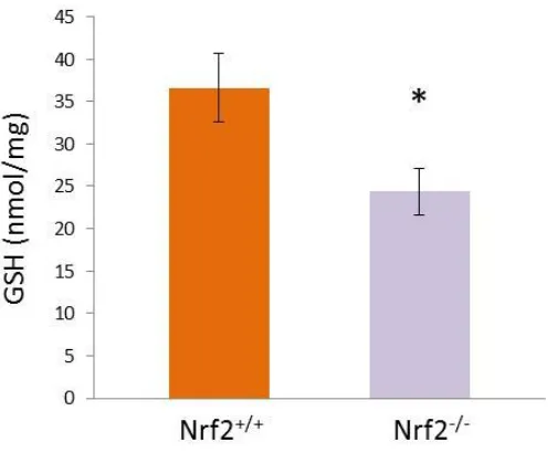
3.2.1 Loss of Nrf2 results in lowered basal GSH content in immature 70 DCs
3.2.2 Enhanced co-stimulatory expression in Nrf2 deficient DCs is 71 not a direct consequence of reduced GSH levels
3.2.3 Loss of Nrf2 impairs endocytic capacity of iDCs 75 independently of lowered GSH levels
3.2.4 Loss of Nrf2 results in impaired capacity of iDCs to phagocytose 76 dying cells independently of lowered GSH levels
3.2.5 Enhanced co-stimulatory receptor expression of Nrf2-/- 79 iDCs is associated with enhanced antigen-specific CD8
T cell stimulatory capacity
3.2.6 Enhanced co-stimulatory receptor expression of Nrf2-/- 82 iDCs is associated with enhanced antigen-specific CD8 T cell
effector function
3.2.7 Loss of Nrf2 enables DCs to cross-present cell-associated 84 peptide antigens to CD8 T cells
3.2.8 Loss of Nrf2 in activated DCs results in a lowered Th1 85 cytokine profile
4.0 CHAPTER FOUR: THE ROLE OF NRF2 IN T CELL IMMUNE FUNCTION
4.1 INTRODUCTION 100
4.2 RESULTS 100
4.2.1 Loss of Nrf2 does not affect T cell development 100
4.2.2 Loss of Nrf2 does not influence nTreg populations within the 103
thymus 4.2.3 Loss of Nrf2 does not influence CD62L, CD69 and CD44 105
expression within the thymus 4.2.4 Loss of Nrf2 does not affect T cell populations in peripheral 107
lymphoid organs. 4.2.5 Loss of Nrf2 results in elevated intracellular ROS levels in 110
peripheral naïve T cells 4.2.6 Loss of Nrf2 does not influence peripheral CD4 T cell 111
activation with respect to CD25 expression 4.2.7 Naïve Nrf2 deficient T cells manifest low levels of T cell 113
activation within lymphoid organs 4.2.8 Loss of Nrf2 results in marginally enhanced effector 118
T cell proliferation. 4.2.9 Loss of Nrf2 results in enhanced effector CD4 T cell 120
IFNγ production 4.2.10 Nrf2 regulates CD4 effector subset differentiation in 122
purified splenic CD4 T cells 4.2.11 Loss of Nrf2 results in enhanced differentiation of 124
CD4+IL-17+ splenic effector T cells 4.2.12 Loss of Nrf2 results in enhanced CD4 effector Th17 128
5.0 CHAPTER FIVE: INTER-INDIVIDUAL VARIATION IN THE CDDO-ME INDUCED NRF2 RESPONSE IN HUMAN T CELLS
5.1 INTRODUCTION 143
5.2 RESULTS 145
5.2.1 Nrf2 was undetectable in whole cell lysates of human T cells 145 5.2.2 CDDO-Me-mediated induction of Nrf2 is detected in the nucleus
of human T cells 147
5.2.3 Inter-individual variation in Nrf2 adaptive response to CDDO-Me is present in human T cells 148 5.2.4 Inter-individual variation in Nrf2 downstream NQO1 gene
expression is present in human T cells 154
5.3 DISCUSSION 158
6.0 CHAPTER SIX: FINAL DISCUSSION
6.1 DISCUSSION 166
LIST OF FIGURES AND TABLES
PAGE 1.0 CHAPTER ONE: GENERAL INTRODUCTION
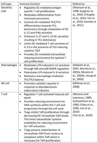
Table 1.1 Innate immune cells and their roles in health and disease. 3
Figure 1.1 T cell development in the thymus 10
Table 1.2 Contrasting roles and features of immature and mature DCs 11
Figure 1.2 DC antigen presentation pathways 17
Figure 1.3 Cross presentation pathways 20
Figure 1.4 Schematic diagram of DC-mediated T cell activation 21 within peripheral lymphoid organs
Figure 1.5 Dendritic cell-mediated CD4 T cell differentiation 25 Table 1.3 The role of the antioxidant glutathione in the maintenance 38
of immune cell function
Figure 1.6 Schematic diagram of the Nrf2-Keap1 pathway 41 Table 1.4 Cytoprotective and antioxidant proteins encoded by 57
Nrf2-related genes and their immunological relevance
3.0 CHAPTER THREE: THE ROLE OF NRF2 IN DC IMMUNE FUNCTION
Figure 3.1 Loss of Nrf2 results in attenuation of basal GSH content in 71 immature DCs
Figure 3.2 Loss of Nrf2 results in a GSH-independent increase in 73 co-stimulatory receptor expression in iDCs.
Figure 3.3 Loss of Nrf2 impairs endocytic capacity of iDCs which is not 76 a direct consequence of lowered GSH levels
Figure 3.4 Dexamethasone induces apoptosis in murine thymocytes 77 Figure 3.5 Loss of Nrf2 results in impaired capacity of iDCs to 78
Figure 3.6 Increased co-stimulatory receptor expression by 81 Nrf2 -/- iDCs is associated with enhanced antigen-specific
CD8 T cell immunostimulatory capacity. 101
Figure 3.7 Nrf2deficient iDCs enhanced maturation phenotype 83 is associated with enhanced DC-mediated antigen-specific
CD8 T cell effector function.
Figure 3.8. Nrf2 deficient iDCs have increased capacity to cross- 85 present cell-associated antigens to naïve CD8 T cells
Figure 3.9 Nrf2 deficient DCs exhibit a lowered Th1 cytokine profile 87
4.0 CHAPTER FOUR: THE ROLE OF NRF2 IN T CELL IMMUNE FUNCTION
Figure 4.1 Nrf2 does not regulate T cell development within the thymus 102 Figure 4.2 Nrf2 does not influence natural occurring T 104
regulatory cell populations within the thymus
Figure 4.3 Nrf2 does not regulate single positive CD4 and CD8 T cell 106 expression of CD62L, CD69 or CD44 cell surface markers
within the thymus
Figure 4.4 Nrf2 does not regulate the composition of CD4 and CD8 108 T cells within the periphery
Figure 4.5 Nrf2 regulates intracellular ROS levels in naïve T cells 111 Figure 4.6 Nrf2 does not regulate the CD4 expression of CD25 112
activation marker within the periphery
Figure 4.7 Loss of Nrf2 results in the induction of low level 116 naïve T cell activation
Figure 4.8 Loss of Nrf2 results in the induction enhanced 119 effector T cell proliferation in response to TCR stimulation
Figure 4.9 Loss of Nrf2 enhances the proportion of CD4 121 effector IFNγ producing T cells during re-stimulation
Figure 4.10 Nrf2 regulates CD4 effector T cell signature cytokine 124 production
Figure 4.12 Nrf2 regulates CD4 effector T cell signature cytokine production 130
5.0 CHAPTER FIVE: INTER-INDIVIDUAL VARIATION IN THE XENOBIOTIC-INDUCED NRF2 RESPONSE IN HUMAN T CELLS
Figure 5.1 Nrf2 is not detected in the whole cell lysates of human T cells 166 upon CDDO-Me induction.
Figure 5.2 CDDO-Me induces nuclear accumulation of Nrf2 in human 168 T cells
Figure 5.3 Inter-individual variation in Nrf2 adaptive response to 170 CDDO-Me is present in human T cells
Figure 5.4 Inter-individual variation in Nrf2 regulated 176 downstream NQO1 expression in response is present in
human T cells
6.0 CHAPTER SIX: FINAL DISCUSSION
i
ABSTRACT
Dendritic cells (DCs) are potent innate antigen presenting cells which are able to sense and engulf pathogens from sites of infection, which are then processed and presented to adaptive T lymphocytes in secondary lymphoid organs. Therefore, they are critical for the initiation and modulation of primary antigen-specific adaptive immune responses. They also play a critical role in the maintenance of T cell tolerance. T cells play vital roles in mediating both cellular and humoral-specific adaptive immune responses. There are various types of T cells present within the immune system including the cytotoxic CD8 T cells and T helper CD4 cells. CD4 T cells can be further subdivided into various subtypes examples of which include T helper 1 cells (Th1), Th2, Th17 and T regulatory cells. Each subset has its own distinctive function, transcriptional regulation and effector cytokine profile.
It is established that appropriate DC and T cell immune function is highly dependent on their intracellular redox status. Cellular redox homeostasis is maintained through a balance between oxidising agents e.g. reactive oxygen species (ROS) and anti-oxidant or reducing agents e.g. glutathione (GSH). Excessive ROS production resulting in oxidative stress is extremely deleterious to the cell and if left unimpeded can result in cell necrosis, tissue damage and the onset of disease. As a result, mammalian cells have evolved an inducible adaptive defence system which provides protection against such oxidative or chemical insult. The functionality of this cellular defence system is principally governed by the activity of the redox-sensitive transcription factor Nrf2. In response to oxidative stress, Nrf2 induces the transcription of a battery of cytoprotective and antioxidant genes involved in GSH synthesis, detoxification of xenobiotics and their reactive metabolites and the maintenance of cellular redox homeostasis. It is now emerging that Nrf2 plays a pivotal role in immunity. However, its precise role in DC and T cell function is unclear.
Using immature bone marrow-derived DCs (iDCs) from Nrf2+/+ and Nrf2-/- mice, the work presented in this thesis demonstrates that Nrf2 deficiency in iDCs resulted in lowered GSH levels, enhanced iDCs co-stimulatory receptor expression, impaired endocytic and phagocytic capacity, and increased iDC-mediated antigen-specific CD8 T cell stimulatory capacity in response to both an antigenic and self-peptide. Furthermore, artificially lowering GSH levels in the iDCs did not recapitulate the Nrf2 deficient iDC phenotype. Moreover, Nrf2-/- DCs exhibited an enhanced capacity to present cell-associated peptide antigens to antigen-specific CD8 T cells, resulting in increased CD8 T cell effector function. Loss of Nrf2 in LPS-stimulated DCs results in a lowered Th1 cytokine profile. These results have implications for Nrf2 in DC-mediated CD8 T-cell immunity, peripheral CD8 T cell tolerance and CD4 effector differentiation.
ii Furthermore, Nrf2 deficiency did not alter the composition of CD4 and CD8 T cell populations within secondary lymphoid organs. It was observed that splenic Nrf2 -/-naïve T cells exhibited enhanced ROS generation, accompanied by low level increases in T cell activation markers. However, the marginal augmentation of Nrf2
-/- naïve T cell activation status did not result in increased T cell receptor
(TCR)-triggered T cell proliferation. In contrast, Nrf2 deficient effector T cells exhibited enhanced TCR/CD3-triggered proliferation, associated with increased Th1 and decreased Th2 effector function. Importantly, we demonstrated that Nrf2-/- effector T cells secreted increased levels of IL-17A and IL-22, a signature cytokine profile indicative of the more recently identified Th17 cell lineage. This was also observed under Th17 polarising conditions, further suggesting that loss of Nrf2 predisposes effector Th17 development. The implications of the latter findings are significant given the pivotal role that Th17 cells play in the pathogenesis of a variety of autoimmune diseases including multiple sclerosis (MS) and Systemic lupus erythematosus (SLE).
Nrf2 plays a critical role in the detoxification of xenobiotics in the liver, which as the primary drug-metabolising organ, is subjected to an array of xenobiotics and their respective metabolites. Individuals vary in their responses to xenobiotic exposure from adaptation to severe adverse drug reactions. However, it is unknown whether this human disparity in drug response is a consequence of inter-individual variation in the Nrf2 adaptive defence system to xenobiotic stress. In light of this, we aimed to firstly investigate whether variation in the Nrf2 adaptive system was present within individuals in response to a chemical inducer of Nrf2, CDDO-Me. To address this issue, basal and induced Nrf2 protein levels and downstream NQO1 expression were measured in activated human T cell blasts, in response to increasing concentrations of CDDO-Me. Examination of various donor-derived T cells, demonstrated that humans vary in their Nrf2 response to CDDO-Me, with respect to nuclear Nrf2 and NQO1 mRNA expression. Therefore we concluded that inter-individual variation does exist in the human’s Nrf2 adaptive system in response to a known Nrf2 probe.
iii
ACKNOWLEGEMENTS
v
PUBLICATIONS
Papers
Laith M A Al-Huseini, Han Xian Aw Yeang, Swaminathan Sethu, Alumeed Naif, Junnat M Hamdam, Laiche Dijouri, Neil Kitteringham, B Kevin Park, Christopher E Goldring and Jean G Sathish. Nuclear factor-erythroid2 (NF-E2) p45-related factor-2 (Nrf2) modulates dendritic cell immune function through regulation of p38-CREB/ATF1 signalling (2013). Journal of Biological Chemistry. 2;288(31):22281-8. Han Xian Aw Yeang*, Junnat M Hamdam*, Laith M A Al-Huseini, Swaminathan Sethu, Laiche Djouhri, Joanne Walsh, Neil Kitteringham, B Kevin Park, Christopher E Goldring and Jean G Sathish. Loss of the transcription factor nuclear factor-erythroid2 (NF-E2) p45-related factor-2 (Nrf2) leads to dysregulation of immune functions, redox homeostasis and intracellular signalling in dendritic cells (2012).
Journal of Biological Chemistry. 287(13):10556-64 (*equal contributors)
Reviews
Junnat Hamdam*, Swaminathan Sethu*, Trevor Smith*, Ana Alfirevic, Mohammad Alhaidari, Jeffrey Atkinson, Mimieveshiofou Ayala, Helen Box, Micheal Cross, Annie Delaunois, Ailsa Dermondy, Karthik Govindappa, Jean-Michel Guillon, Rosalind Jenkins, Gerry Kenna, Björn Lemmer, Ken Meecham, Adedamola Olayanju, Sabine Pestel, Andreas Rothfuss, James Sidaway, Rowena Sison-Young, Emma Smith, Richard Stebbings, Yulia Tingle, Jean-Pierre Valentin, Awel Williams, Dominic Williams, Kevin Park, Christopher Goldring. Safety Pharmacology – current and emerging concepts (2013). Toxicology and Applied Pharmacology. Accepted (*equal contributors)
vi
ABBREVIATIONS
µ; micro
2-ME; 2-Mercaptoethanol
Ag; antigen
ADR; adverse drug reaction AHR; aryl hydrocarbon receptor AKR; aldo-keto reductase ANOVA; analysis of variance AP-1; activator protein 1 AP-3; adaptor protein 3 APC; antigen presenting cell APL; altered peptide ligand
ARE; antioxidant response element ARNT; AHR nuclear translocator ATP; adenosine triphosphate BDCA; blood dendritic cell antigen BCR; B-cell receptor
BCL6; B cell lymphoma-6 BHA; butylated hydroxyanisole
BM; bone marrow
BMDC; bone marrow-derived dendritic cell BSA; bovine serum albumin
BTAF; B cell-Activating Transcription Factor BTB; bric-a-brac/tram-track/broad complex bZip; basic leucine zipper
C; Celsius
Ca2+; calcium CaN; calcineurin
cAMP; cyclic adenosine monophosphate CBP; CREB-binding protein
CCR; chemokine (C-C motif) receptor CCL; chemokine (C-C motif) ligand
CD; cell surface molecules expressed on various cell types cDC; conventional DC
CDDO; 2-cyano-3,12-dioxooleana-1,9(11)-dien-28-oic acid CDDO-Im; CDDO imidazolide
CDDO-Me; CDDO methyl ester also known as Bardoxolone methyl cDNA; complementary DNA
CER; cytoplasmic extraction reagent
CFSE; carboxy fluorescein succinimidyl ester CGD; chronic granulomatous disease
CLEC9A; C-type lectin domain family 9A also known as DNGR-1 CLIP; class II-associated invariant chain peptide
vii CLR; C-type lectin receptor
CM; complete media
CMP; common myeloid progenitor CNC; cap ‘n’ collar
CO2; carbon dioxide CON-A; Concanavalin A
COPD; Chronic obstructive pulmonary disease CpG; C-phosphate-G
CREB; cAMP responsive element binding protein C region; constant region
Csk; C-terminal Src kinase CT; cross threshold
CTL; cytotoxic T lymphocyte
CTLA-4; cytotoxic T lymphocyte antigen 4 CUL3; Cullin 3
CXCR; chemokine (C-X-C motif) receptor CYP450; cytochrome P450
Cys; cysteine
Da; Dalton
DAG; 1,2-diacylglycerol DC; dendritic cell
DC-SIGN; DC-Specific Intercellular adhesion molecule-3-Grabbing Non integrin
DEC-205; C-type lectin receptor (205 kDa) also known as CD205 DGR; double glycine repeat
DHE; dihydroethidium dH2O; distilled H2O
DILI; drug-induced liver injury DL; delta-like
DEM; diethyl maleate DMSO; dimethyl sulphoxide
DN; double negative (CD4-CD8-) DNA; deoxyribonucleic acid DNCB; 2,4-dinitrochlorobenzene
DNTB; 5,5’-dithio-bis(2-nitrobenzoic acid) dNTP; deoxyribonucleotide triphosphate DP; double positive (CD4+CD8+)
dsRNA; double stranded RNA
DTNB; 5,5′-dithiobis(2-nitrobenzoic acid)
EAE; experimental autoimmune encephalomyelitis E. coli; Escherichia coli
EC50; half maximal effective concentration of a drug
ECH; erythroid cell-derived protein with CNC homology ECL; enhanced chemiluminescence
viii ELISA; Enzyme-linked immunosorbent assay
EOMES; eomesodermin
ER; endoplasmic reticulum
ERK; extracellular signal-regulated kinase EtOH; ethanol
FACS; fluorescence activated cell sorting FasL; Fas ligand
FBS; fetal bovine serum Fc; Ig-constant region FcR; Fc receptor FCS; fetal calf serum
FITC; fluorescein isothiocyante
FLT3; Fms-like receptor tyrosine kinase 3 FLT3L; FLT3 ligand
FoxP3; forkhead box P3
Gads; Grb2 related adaptor protein downstream of Shc GAPDH; Glyceraldehyde 3-phosphate dehydrogenase GATA-3; GATA binding protein-3
GCL; γ-glutamylcysteine ligase GCLC; GCL, catalytic subunit GCLM; GCL, regulatory subunit GDP; guanosine diphosphate
GEF; guanine nucleotide exchange factor Gfi-1; growth factor independent-1 GI; gastrointestinal
GM-CSF; granulocyte-macrophage colony-stimulating factor GPI; glycosylphosphatidyl inositol
GPX; GSH peroxidase GS; GSH synthetase GSH; glutathione GSR; GSH reductase
GSSG; oxidised GSH or glutathione disulphide GST; GSH S-transferase
GVHD; Graft-versus-host disease H2O; water
H2O2; hydrogen peroxide
H2SO4; sulphuric acid
HAT; histone acetyltransferase HBSS; Hanks balanced salt solution HBV; Hepatitis B virus
HCl; hydrochloric acid HCV; Hepatitis C virus HDAC; histone deacetylase
ix HES-1; hairy and enhancer of split-1
HIF-1; hypoxia-inducible factor 1 HIV; human immunodeficiency virus HLA; Human leukocyte antigen
HMGB1; High-Mobility Group Box 1 Protein HO-1; heme oxygenase 1
hr; hours
HRP; horseradish peroxidase HSC; haematopoietic stem cell ICCS; Intracellular cytokine staining ICOS; inducible costimulator
ICOSL; ICOS ligand
iDC; immature dendritic cell IFN; interferon
Ig; immunoglobulin
IκB inhibitor of κB IKK; IκB kinase IL; interleukin
iNOS; inducible nitric oxide synthase IP3; inositol 1,4,5-triphosphate
IPEX; immunodeficiency, polyendocrinopathy, enteropathy, X-linked syndrome
IRAK; Interleukin-1 receptor-associated kinase IRF; interferon regulatory factor
IS; immunological synapse
ITAM; immunoreceptor tyrosine-based activation motif Itk; IL-2- inducible T cell kinase
iTreg; inducible T regulatory cell IVR intervening region
JNK; c-Jun N-terminal kinase kDa; KiloDalton
Keap1; Kelch-like ECH-associated protein 1
KO; Knock out
L; litre
LAT; linker for activation of T cells LC; langerhans cell
Lck; lymphocyte-specific protein tyrosine kinase li; invariant chain
LPS; lipopolysaccharide Lys; lysine
m; milli
x
mA; milliamps
mAb; monoclonal antibody
MAPK; mitogen-activated protein kinase
MARCO; macrophage receptor with collagenous structure MEFs; mouse embryonic fibroblasts
MCP-1; monocyte chemotactic protein-1 also known as CCL2 mDC; mature dendritic cell
MHC; major histocompatibility complex MIIC; MHCII–rich compartment
min; minutes
MIP-3β; macrophage inflammatory protein-3β MLN; mesenteric lymph node
MOPS; 3-(N-morpholino)propanesulfonic acid mRNA; messenger RNA
MS; Multiple Sclerosis
MYD88; Myeloid differentiation primary response gene (88)
NAC; N-acetylcysteine
NADPH; nicotinamide adenine dinucleotide phosphate NaH2PO4; monosodium phosphate
NAPQI; N-acetyl-p-benzoquinoneimine Neh; Nrf2-ECH homology
NER; nuclear extraction reagent NFAT; nuclear factor of activated T cells NF-E2; nuclear factor erythroid 2
NF-κB; nuclear factor κB NK; natural killer cell NO; nitric oxide NOX; NADPH oxidase NP-40; nonidet P-40
NQO1; NAD(P)H:quinone oxidoreductase 1 Nrf2; NF-E2 -related factor 2
nTreg; natural occurring T regulatory cell OH; hydroxyl
.
OH; hydroxyl radical O2; molecular oxygen
O2· -; superoxide anion radical
OVA; ovalbumin
PAMPs; pathogen-associated molecular patterns PBMC; peripheral blood mononuclear cell PBS; phosphate-buffered saline
PCR; polymerase chain reaction pDC; plasmacytoid DC
xi PHA; phytohemagglutinin
PI3K; phosphatidyl inositol 3-kinase PIP; phosphatidylinositol 4-phosphate PIP2; phosphatidylinositol 4,5-biphosphate
PIP3; phosphatidylinositol 3,4,5-triphosphate
PKC; protein kinase C PLC; phospholipase C PM; particulate matter
PMA; phorbol 12-myristate-13-acetate PRR; pathogen recognition receptor PRX; peroxiredoxin
PS; phosphatidylserine RA; rheumatoid arthritis
RANTES; Regulated on Activation, Normal T cell Expressed and Secreted RBC; red blood cell
RelA; NF-κB p65 subunit
RIPA; radio-immunoprecipitation assay RO2.; peroxyl radical
ROS; reactive oxygen species
RORγt; retinoic acid-related orphan receptor-γt or human RORC2 RPMI; Roswell Park Memorial Institute-1640
RT; room temperature
RUNX1; Runt-related transcription factor 1 SCA-1; stem cell antigen-1
SCZ; sub-capsular zone SD; standard deviation
SDF-1; stromal cell-derived factor-1 SEM; standard error of the mean SH2; Src Homology 2
SLE; Systemic lupus erythematosus
SLP-76; SH2 domain-containing leukocyte phosphoprotein of 76 kDa SOD; superoxide dismutase
SP; single positive
STAT; Signal Transducer and Activator of Transcription TAP; transporter associated with antigen processing T-bet; T box expressed in T cells
tBHQ; tert-butylhydroquinone TBS; Tris-buffered saline TBST; TBS-Tween
TCCD; 2, 3, 7, 8-tetrachlorodibenzo-p-dioxin TCR; T cell receptor
TCM; central memory T cell
TEM; effector memory T cell
xii Tfh; CD4 T follicular helper cell
TGFβ; transforming growth factor β
Th; CD4 T helper cell TLR; Toll-like receptor
TNFα; tumour necrosis factor α
TNFR-1; tumour necrosis factor receptor-1 TRAF-6; TNF receptor-associated factor 6 Treg; T regulatory cell
TRX; Thioredoxin
TRXR; Thioredoxin reductase
TSLP; thymic stromal lymphopoietin UV; Ultraviolet
V-ATPase; vacuolar-type H+-ATPase
VSIG4; V-set and immunoglobulin domain containing 4
V region; variable region v/v; volume/volume WCL; whole cell lysate
WHIM; Warts, Hypogammaglobulinemia, Infections, and Myelokathexis syndrome
w/v; weight/volume XO; xanthine oxidase
XRE; xenobiotic response element
1
CHAPTER ONE
2 1.0INTRODUCTION
1.1 The Immune System
Multi-cellular animals are continuously threatened by a diverse range of pathogenic and innocuous bacteria, fungi, viruses and parasites. The immune system is geared towards recognising the potential pathogens to which it is exposed to and thus defend the host against infection. Furthermore, this system must be able to discriminate between the animal’s own cellular components (i.e. self) and that of an invading pathogen (i.e. non-self). Failure to do so gives rise to detrimental inappropriate immune responses which underpin the pathogenesis of autoimmune diseases such as diabetes and Multiple Sclerosis (MS).
The immune system is typically divided into two parts, the innate and the adaptive. Each division of the immune system is made up of both cellular and humoral components. Although the innate and adaptive arms functionally complement one another, they differ significantly in their capacity to recognise and subsequently eradiate infectious pathogens. The following introduction gives an overview of the innate and adaptive immune system with a focus on dendritic cells and T lymphocytes.
1.2 Innate immunity
3 Cell type Function and features Pathophysiology Reference Basophil Morphologically similar to
mast cells
Activation via cross-linage of FcεRI with IgE resulting in
degranulation Provide anti-parasitic
immunity:
- Release granules
containing histamine and proteases
- Produce IL-4, IL-13
Helminth infection, asthma, allergic rhinitis, atopic dermatitis (Stone et al., 2010) Dendritic cell (DC)
Most Potent APC Initiate and modulate
adaptive immune responses Involved in viral,
parasitic, bacterial, anti-fungal and anti-tumour immunity
Maintenance of T cell tolerance SLE, systemic sclerosis, RA, asthma, psoriasis, atopic dermatitis, hypersensitivity reactions, measles, cancer, type 1 diabetes
(Ueno et al., 2007; York, 2011; Dorner, 2012)
Eosinophil Activated by cross-linage of FcγRII with IgG resulting in degranulation
Provide anti-parasitic immunity:
- Release cytolytic granule proteins
- Produce IL-4, IL-5, IL-10, TNFα Helminth infection, allergic rhinitis, asthma, hyper-IgE syndrome, atopic dermatitis (Stone et al., 2010)
Macrophage Activated by PAMPs via TLR and opsonised bacteria via FcR
Professional phagocyte and APC capacity
Involved in cellular
homeostasis via ingestion of apoptotic cells
Involved in anti-bacterial immunity:
- Engulfment of microbes and lysis via NOX-respiratory burst mechanisms.
- Produce 1β, TNFα, IL-12, IL-6, IL-8
4 Macrophage
(continued)
Involved in anti-viral immunity:
- Secrete IFNβ preventing intracellular viral
replication
Mast cell Activated by cross-linage FcεRI with IgE resulting in
degranulation
Involved in anti-parasitic immunity
- Release granules
containing histamine and proteases
- Produce TNFα, IL-5, IL-3
Helminth infection, anaphylaxis, allergic rhinitis, asthma, mastocytosis, systemic sclerosis, atopic dermatitis (Stone et al., 2010; York, 2011)
Monocyte Precursor reserve pool for macrophages and DCs (under inflammatory conditions)
Professional phagocyte but low APC capacity as bona fide monocyte
Involved in cellular
homeostasis via ingestion of apoptotic cells
Involved in anti-bacterial immunity:
- engulfment of microbes and killing via iNOS activity and ROS production
- Produce TNFα, 1β, IL-6, IL-10 RA, Atherosclerosis, systemic sclerosis, Crohn’s disease, HIV, sepsis, asthma (Auffray et al., 2009; York, 2011) Natural Killer cell (NK)
Provide anti-tumour and anti-viral immunity: Target and lyse abnormal/ viral infected cells which have low/absent expression of self-MHCI cell surface molecules
Provide anti-bacterial immunity:
Recognise and lyse antibody coated targets via FCR activation.
Lysis via release of cytolytic granules containing perforin and granzyme B
systemic
5 Neutrophil Activated by PAMPs via TLR
via and opsonised bacteria via FcR
Professional phagocyte and APC capacity
Involved in cellular
homeostasis via ingestion of apoptotic cells
Involved in anti-bacterial immunity:
- Engulfment of microbes and lysis via NOX
respiratory burst mechanisms.
- Produce 1β, TNFα, IL-12, IL-6, IL-8
CGD disease, myelokathexis syndrome (WHIM), SLE, sepsis (Dale et al., 2008; Dorner, 2012)
[image:29.595.115.524.72.312.2]Innate immune cells migrate via the blood stream and distribute throughout the body which helps in the rapid identification and efficient removal of potential infectious pathogens. These cells are activated upon recognition of conserved molecular components, known as pathogen-associated molecular patterns (PAMPs) (e.g. lipopolysaccharide (LPS) found in the bacterial cell wall of gram negative bacteria), via the expression of pathogen recognition receptors (PRRs) including Toll-like receptors (TLRs) (Beutler, 2004). These receptors are germline-encoded and recognise a limited number of molecules that are shared by numerous infectious agents. Engagement of PPRs result in a variety of immediate effector responses that endeavour to directly disable the harmful pathogen usually within the first 12 hours of infection (Murphy et al., 2012). A prime example is the activation of dectin-1, a member of the C-type lectin receptor (CLR) superfamily,
Table 1.1 Innate immune cells and their roles in health and disease.
6 that is present on phagocytic myeloid cells including neutrophils and macrophages (Geijtenbeek and Gringhuis, 2009). Dectin-1 binds to β-glucans present in fungal cell walls, certain bacteria and plants, resulting in the phagocytosis and proteolytic destruction of the pathogen (Lee and Kim, 2007). Activation of dectin-1 also triggers the release of proinflammatory cytokines e.g. IL-1β and chemokines e.g. CXCL8, which not only promote inflammation, but also instigate the recruitment of neutrophils and macrophages at the site of infection. This results in the amplification of the inflammatory response and effective removal of the pathogen (Murphy et al., 2012). Neutrophils, macrophages and in particular dendritic cells (DCs) are termed antigen presenting cells (APCs), which possess a unique ability to sense and engulf pathogens from sites of infection that are then processed and presented to adaptive immune cells in secondary lymphoid organs (Table 1.1). Proinflammatory cytokine release from innate immune cells, play a crucial role in priming APCs for efficient antigen presentation, APC-mediated adaptive immune cell activation and subsequent differentiation. This highlights the pivotal role of the innate immune system in complementing and modulating adaptive immune responses (Maldonado-Lopez et al., 1999; Tse et al., 2007; Abi Abdallah et al., 2011).
The innate immune system is not without its limitations. The PPRs are restricted to recognising only common microbial structural features and thus are limited in perceiving a narrow range of potential pathogenic insults. Furthermore, these receptors cannot provide antigen-specific protection to invading pathogens and thus fail to prevent re-infection (Beutler, 2004). In contrast to the innate arm of the immune system, the adaptive immune system specifically recognises and provides protective immunity against a diverse array of invading pathogens, but more importantly, it provides long lasting protection against re-infection due to its unique feature, immunological memory. Although adaptive immune responses are long lasting, they are delayed in their initiation. Therefore, the adaptive arm must work in concert with the rapid responses of the innate immune system to ensure both the immediate and long term protection against pathogenic invasion.
7 The adaptive immune system is organized around B and T lymphocytes, which have the ability to recognise a myriad of invading pathogens and can mount a specific immune response. The great diversity in antigen specificity among lymphocytes is determined by the structure of the B and T cell receptors’ (BCR and TCR) antigen-binding site, whose structural segments are a result of somatic recombination of antigen receptor genes, together with palindromic and random nucleotide addition during development. Naïve T cells and B cells reside in secondary lymphoid tissues such as the spleen, lymph nodes and Peyer’s patches, where they await contact with circulating DCs. The DCs migrate to lymphoid tissues and present captured antigens from the periphery to these adaptive immune cells. The recognition of antigens by the TCR/BCR leads to T/B cell clonal expansion and affinity maturation. Clonal expansion gives rise to a large pool of antigen-specific T or B cells, some of which remain dormant in the body after the infection has been resolved. This gives rise to immunological memory which permits a rapid response upon second exposure to the same antigen (Parkin and Cohen, 2001).
8 antibodies can underlie pathological processes, as is the case for IgE and allergic disease (Table 1.1).
T cells play vital roles in mediating both cellular and humoral-specific adaptive immune responses. There are various types of T cells present within the body as discussed later in this chapter, each with distinctive functions that provide protective anti-viral, anti-tumour, anti-parasitic, anti-bacterial and anti-fungal immunity. However, T cell dysregulation is an underlying cause of a variety of autoimmune and allergic disorders. In order to fully understand the function of the T cell, it is imperative to gain an insight into their development and importantly their activation by the potent innate APC, the DC.
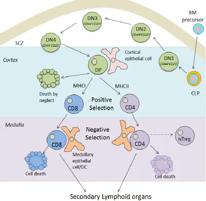
1.4 T cell development
10
Figure 1.1 Schematic diagram of T cell development within the thymus
11 Table 1.2. Contrasting roles and features of immature and mature DCs.
CCR: chemokine (C-C motif) receptor; CD: cell surface molecule; IL-8: interleukin-8; MHCII: major histocompatibility complex II; MIP-3β: Macrophage inflammatory protein-3β also known as chemokine (C-C motif) ligand 19 (CCL19); SDF-1: stromal cell-derived factor-1 Adapted from (Banchereau et al., 2000)
1.5 Dendritic cells
Dendritic cells are specialised APCs present in the blood, skin, peripheral organs e.g. gut, lung and liver, and lymphoid organs e.g. the spleen and thymus. They are responsible for the initiation and modulation of primary adaptive immune responses (Banchereau et al., 2000). They exist in two functional states; immature steady state DCs (iDCs) and mature DCs (mDCs) as determined by their morphology, phenotypic expression of cell surface proteins which promote T cell activation (known as co-stimulatory molecules), chemokine expression and their immune function as shown in Table 1.2 (Banchereau et al., 2000).
Feature Immature DC (iDC) Mature DC (mDC)
Morphology Spherical shape Large cytoplasmic
protrusions (dendrites)
Residence Periphery Secondary lymphoid
organs
Function - Antigen capture
- High phagocytosis - Poorly immunogenic - Maintenance of CD8 T
cell tolerance
- Antigen presentation - Low phagocytosis - Highly immunogenic - Modulation of
adaptive immune responses
Chemokine expression - High CCR1, CCR5, SDF-1, IL-8
- Low CCR7, MIP-3β
- Low CCR1, CCR5, SDF-1, IL-8
- High CCR7, MIP-3β Co-stimulatory receptor
expression
- Low CD80, CD86, CD40, CD83 - High intracellular
MHCII
- High CD80, CD86, CD40, CD83 - High cell surface
MHCII
13 role in inducing tolerance and directing CD4 T regulatory cell polarisation (Maldonado-Lopez et al., 1999; Belz et al., 2005; Scott et al., 2011).
1.6 Dendritic cells: drivers of the immune response
Dendritic cells survey the peripheral environment for potential infectious pathogens subsequently directing cells of the adaptive immune system to execute finely tuned immune responses, ultimately resulting in the clearance of infection and are thus important sentinels of the immune system (Banchereau et al., 2000). In the periphery, DCs reside in an antigen-capturing immature state sampling their environment for potential pathogenic invasion. The iDC can ingest exogenous pathogens, dying and infected cells through engagement of their endocytic and phagocytic receptors. Receptors engaged include dectin-1 for β-glucans, FcγR for IgG opsonised bacteria) and phosphatidylserine receptor (PS), scavenger receptor CD36, and C-type lectin domain family 9A (CLEC9A, also known as DNGR-1) for apoptotic and necrotic cells (Dalgaard et al., 2005; Bratton and Henson, 2008; Geijtenbeek and Gringhuis, 2009; Sancho et al., 2009; den Dunnen et al., 2012). Receptor engagement consequently induces a variety of actin-dependent phagocytic processes facilitating the efficient internalisation of the pathogen and formation of the phagosome (Underhill and Goodridge, 2012).
14 family is comprised of 10 members in humans and 12 in mice and can collectively recognise common constituents derived from multifarious pathogens including bacteria, parasites, viruses and fungi (Kawai and Akira, 2011). Toll-like receptor signalling results in the activation of the mitogen-activated protein kinases (including p38 MAPK, JNK and ERK1/2) and nuclear factor kappa-light-chain-enhancer of activated B cells (NF-κB) signalling pathways which promote the nuclear translocation of transcription factors, activator protein 1 (AP-1) and NF-κB, respectively (Kawai and Akira, 2011). As a consequence, these transcription factors drive the transcription of a variety of genes involved in DC maturation including lymphoid-homing chemokine receptor CCR7, inflammatory cytokines e.g. IL-1β, TNFα, IL-12 and IL-6, cell surface expression of MHCII and the upregulation of co-stimulatory receptors e.g. CD80, CD86 and CD40 (Table 1.2) (Sallusto et al., 1998; Ardeshna et al., 2000; Nakahara et al., 2006; Neves et al., 2009). The phenotypic and morphological changes associated with DC maturation alter the immune function of the dendritic cell from highly phagocytic iDCs efficient at antigen capture, to low phagocytic, highly immunogenic mDCs proficient at antigen presentation (Table 1.2). Consequently, mDCs efficently migrate to lymphoid organs where they present processed antigens to naive T cells in such a context, resulting in the initiation of an adaptive immune response. Importantly, these events enable DCs to greatly influence the nature of the T cell response elicited by guiding naive T cell differentiation into functionally distinct effector subtypes as reviewed later in this chapter.
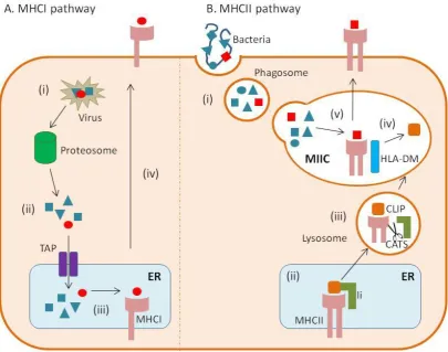
1.7 Dendritic cell antigen presentation
15 from within the cell or from intracellular viruses, parasites or bacteria derived from e.g. intracellular bacteria, viruses, self-defective ribosomal products (DRiPs) or misfolded proteins are processed and loaded onto MHC class I molecules, which are specifically recognised by CD8 T cells (Trombetta and Mellman, 2005). In contrast, internalised exogenous antigens derived from e.g. extracellular bacteria and parasites, are loaded onto MHC II molecules and presented to CD4 T cells (Trombetta and Mellman, 2005). However, there are exceptions to this rule as observed with exogenous dying or viral infected cell MHCI-mediated presentation to CD8 T cells, a unique process known as cross-presentation (section 1.7.3) (Trombetta and Mellman, 2005). The mechanistic basis of the various types of DC antigen processing and presentation are dicussed below.
1.7.1 Endogenous classical MHCI pathway
16 1.7.2 Exogenous MHCII pathway
17 Figure 1.2. DC antigen presentation pathways
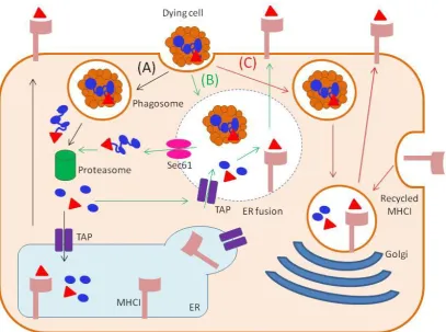
18 1.7.3 Cross-presentation
20
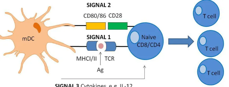
1.8 DC-mediated antigen-specific T cell activation
[image:44.595.117.525.70.373.2]Once the antigen is processed and displayed on the DC cell surface, it is now available for specific recognition by naive T cells in lymphoid organs. In order for competent T cell activation to occur, three activation signals are required (Figure 1.4). The T cell must specifically recognise the peptide:MHC presented by the mDC via its distinct TCR (signal 1) (Murphy et al., 2012). CD8 and CD4 TCRs will specifically recognise peptides complexed to MHCI and MHCII molecules, respectively. Co-stimulatory receptor engagement between CD28 on T cells and CD80/86 on DCs comprises the second critical signal (signal 2) necessary for T cell
Figure 1.3 DC cross presentation pathways
21 Figure 1.4. Schematic diagram of DC-mediated T cell activation within peripheral lymphoid organs
Ag: antigen; IL-12: interleukin-12; mDC: mature DC; MHC: major histocompatibility complex; TCR: T cell receptor.
activation (Murphy et al., 2012). Co-stimulatory receptor signal transduction lowers the threshold for T cell activation, prevents T cell anergy, enhances T cell survival via up regulation of anti-apoptotic protein Bcl-xL and facilitates IL-2 cytokine
[image:45.595.109.511.480.632.2]22 1.9 T cells and the immune response
23 immunological memory of that specific antigen (Zimmerer et al., 2012). Unlike short-lived effector cells, antigen-experienced memory cells can survive for the lifetime of the human body providing long lasting immunity to that particular antigen (Zimmerer et al., 2012). Upon secondary encounter with the same antigen, memory cells undergo robust clonal expansion resulting in the rapid elimination of that specific pathogen (Zielinski et al., 2011).
1.10 CD8 T cells
24 Perforin perturbs the membrane of the target cell allowing entry of granzyme A and B, the latter of which initiates a caspase-dependent apoptotic pathway, ultimately resulting in cell death (Lieberman, 2003). Additionally, CTLs release proinflammatory cytokines such as IFNγ and TNFα (La Gruta et al., 2004). Interferon-γ inhibits viral replication and promotes MHCI expression on infected cells thus increasing their probability of being targeted by activated CTLs. Tumour necrosis factor-α secretion activates innate immune cells and induces caspase-dependent cellular apoptosis via ligation with its cognate receptor TNFR-1 on the target cell (Murphy et al., 2012). Co-stimulation is critical for CTL effector function. Notably, CD28 signalling is associated with induction of IL-2 production. This subsequently enhances functional differentiation of CTLs through increasing granzyme and perforin expression (Janas et al., 2005; Williams et al., 2006).
25 Figure 1.5. Dendritic cell-mediated CD4 T cell differentiation
26 1.11 CD4 T helper cells
CD4 T cells orchestrate complex immune responses against a myriad of pathogens. They can be categorised into a variety of subsets based on their distinctive cytokine profile, chemokine cell surface receptor pattern and immune function such as amplifying CD8 T cell immunity, facilitating B cell activation and preventing autoimmune responses (Figure 1.5). The earliest subtypes to be identified were the classical CD4 T helper 1 (Th1) and T helper 2 (Th2) cells (Mosmann et al., 1986). However, upon further investigation a variety of new distinct subsets were discovered including T regulatory T (Tregs), follicular helper T (Tfh) and the most recent T helper 17 (Th17), T helper 9 (Th9) cells and T helper 22 (Th22) cells (Chen et al., 2003; Harrington et al., 2005; Dardalhon et al., 2008; Eyerich et al., 2009; Craft, 2012). In contrast to CD8 T cells, CD4 T cells have greater plasticity in their differentiation which are not only dependent upon their cytokine milieu and the strength and duration of TCR signalling; but are also further regulated by a network of lineage-specific transcription factors and corresponding activation of specific signalling transducer and activator of transcription (STAT) proteins (Zhu and Paul, 2010). For the purposes of this introduction the main types of CD4 T cells (Th1, Th2, Th17 and Treg) and their dynamic interplay with one another are discussed in greater detail below.
1.11.1 Th1 cells
27 (Shibata et al., 2013). Induction of Th1 IFNγ secretion through DC-derived IL-12 secretion, activates innate immune cells including macrophages and neutrophils, enhancing their phagocytic function and microbial killing (Murphy et al., 2012); whilst Th1 IL-2 production promotes CD8 T cell activation and subsequent cytolytic capabilities (Janas et al., 2005). Multiple transcription factors play important roles in Th1 lineage development including the master transcription factor T-bet, Runx3, EOMES, NF-κB and importantly STAT1 and STAT4 (Szabo et al., 2000; Lighvani et al., 2001; Malmberg et al., 2001; Djuretic et al., 2007; Yang et al., 2008; Kesarwani et al., 2012; Zhu et al., 2012). The strength of the TCR signal is important in determining Th1 lineage commitment. A strong TCR stimulation promotes Th1 differentiation whereas a weak TCR stimulation favours Th2 differentiation. (Tao et al., 1997). Increased Th1 differentiation and effector function has been implicated in the pathogenesis a variety of autoimmune diseases including Crohn’s disease (Fuss et al., 1999; Neurath et al., 2002), MS (Frisullo et al., 2012), type 1 diabetes (Bluestone et al., 2010) and lupus nephritis (Ooi and Kitching, 2012).
1.11.2 Th2
28 inflammatory DCs play vital roles in Th2 differentiation during infection (Kool et al., 2012). Moreover, DCs matured under suboptimal or inflammatory conditions instruct the polarisation of CD4 T cells to Th2 phenotype (Pletinckx et al., 2011). Multiple transcription factors play an important role in Th2 lineage development including the master transcription factor GATA binding protein-3 (GATA-3), c-maf, growth factor independent-1 (Gfi-1), Interferon regulatory factor 4 (IRF4), Notch and importantly STAT proteins, STAT5 and STAT6 (Shimoda et al., 1996; Zhu et al., 2003; Pai et al., 2004; Fang et al., 2007; Luckheeram et al., 2012). The strength of the TCR signal is also important for determining Th2 differentiation. Lower peptide concentration induced activation of the TCR results in the increased production of CD4+ IL-4 producing cells (Yamane et al., 2005). Furthermore, weak TCR stimulation through the use of altered peptide ligands which possess a lower avidity for the receptor, results in the induction of Th2 cytokine production. This is also exclusively dependent upon CD28 co-stimulatory engagement (Tao et al., 1997). Weak TCR stimulation results in reduced ERK activation, favouring Th2 induction (Yamane et al., 2005). Although Th2 cells are vital for mounting humoral responses against helminth invasion, they are notorious for their role in triggering immune responses to innocuous environmental allergens, resulting in chronic inflammation associated with allergic diseases such as eczema, allergic rhinitis and asthma (Holgate, 2012; Murphy et al., 2012).
1.11.3 Th17 cells
29 However, further studies using IL-12p35-/- mice showed a surprisingly enhanced susceptibility to EAE. This indicated that IL-12 was not involved but rather a structurally related cytokine to IL-12, IL-23 was responsible for promoting EAE (Gran et al., 2002). Furthermore, deficiency in IFNγ surprisingly exacerbated disease severity, ruling out Th1 cells as the main culprit behind the inflammatory-driven pathology associated with EAE (Ferber et al., 1996). Upon closer inspection, it was determined that a novel IL-17-producing CD4 effector T cell which is dependent on IL-23 rather than IL-12 for their expansion was in fact the actual offender in question (Harrington et al., 2005; Langrish et al., 2005; Komiyama et al., 2006). This was further clarified through the use of blocking IL-17 antibodies resulting in inhibition of EAE severity (Langrish et al., 2005; Komiyama et al., 2006). This effector subtype designated Th17 cells, play a pivotal role in orchestrating the host’s defence against extracellular microbial and fungal pathogens, but also promote chronic inflammation associated with a variety of autoimmune diseases such as MS, Rheumatoid arthritis (RA), psoriasis and Systemic lupus erythematosus (SLE) (Zhu and Qian, 2012). Effector Th17 cells are characterised by their secretion of IL-17A, IL-17F, IL-21 and IL-22 (Figure 1.5) (Zhu et al., 2010). Th17-mediated cytokine functions include recruitment and expansion of neutrophils to sites of infection, activation of macrophages and DCs and the release of TNFα and IL-1β, which collectively result in a heightened inflammatory response. (Zhu and Qian, 2012). Moreover, IL-17 promotes the activation and subsequent transformation of B cells into antibody secreting plasma cells (Zhu and Qian, 2012). Furthermore, IL-17 in conjunction with B cell activating factor (BAAF) has been shown to synergistically promote self-reactive B cell mediated pathogenesis in autoimmune SLE (Doreau et al., 2009). Functional differentiation into effector Th17 cells is governed by the secretion of TGFβ, IL-6 and IL-23 from DCs (Figure 1.5) (Mangan et al., 2006; Veldhoen et al., 2006). Although IL-23 has been shown to be dispensable for the development of Th17 cells in vitro, it is essential for their phenotypic stability, expansion and effector function against microbial infection in vivo
30 CD28, CD40L and ICOS are also necessary for Th17 generation (Park et al., 2005b; Huang et al., 2012). Multiple transcription factors play an important role in Th17 lineage development including the master transcription factor retinoic acid-related orphan receptor-γt (RORγt or RORC2 human equivalent), nuclear aryl hydrocarbon receptor (AHR), IRF4, B cell-Activating Transcription Factor (BTAF), Runx1 and STAT3 (Ivanov et al., 2006; Mathur et al., 2007; Huber et al., 2008; Veldhoen et al., 2009; Zhu et al., 2010), Expression of RORγt is obligatory for Th17 development which is upregulated by TGFβ and IL-6 (Ivanov et al., 2006; Burgler et al., 2010). It has been shown that DCs are the main drivers of the pathogenic Th17 phenotype (Veldhoen et al., 2006; Huang et al., 2012; Shi et al., 2012). Furthermore, DC signalling via the p38 MAPK pathway is necessary for the promotion of Th17 differentiation and subsequent pathogenesis of EAE as highlighted in p38 MAPK deficient mice (Huang et al., 2012).
1.11.4 T regulatory cells (Treg)
31 killing of CD8 and NK T cells (Cao et al., 2007) and directly outcompeting responder T cell interactions with APCs (Yamaguchi et al., 2011). Additionally, the co-inhibitory receptor Cytotoxic T-Lymphocyte Antigen 4 (CTLA-4) which is highly expressed on nTregs, further enables their suppressive activity. (Wing et al., 2008). Within the appropriate context, peripheral CD4+CD25-Foxp3- T cells can differentiate into CD4+CD25+Foxp3+ T cells which acquire the unqiue characteristics of nTregs; these polarised effector CD4 T cells are termed iTregs (Chen et al., 2003). Upon antigen stimulation and TGFβ exposure, Foxp3 is upregulated in peripheral CD4+ T cells, resulting in their conversion to CD4+CD25+Foxp3+ T cells (Figure 1.5) (Chen et al., 2003; Zheng et al., 2004). Inducible Tregs secrete the immunosuppressive cytokines IL-10 and TGFβ which dampen inflammatory responses, but can also further instruct the differentiation of peripheral CD4 T cells to become suppressor T cells (Figure 1.5) (Zheng et al., 2004). Interleukin-10 secretion can in turn induce tolerogenic DCs, further dampening immune responses (Yamaguchi et al., 2011). Dendritic cells play a pivotal role in the differentiation of peripheral iTregs (Yamazaki et al., 2007). Splenic steady-state DCs presenting low antigen concentrations in the presence of exogenous TGFβ, results in the induction of Foxp3 in peripheral CD4+ T cells (Yamazaki et al., 2007). The gut which is constantly exposed to a myriad of infectious and innocuous microbes is a prime location for DC-mediated iTreg differentiation. Within this region, tolerance to harmless antigens is necessary to avoid regular induction of aberrant immune responses (Scott et al., 2011). In support of this, it has been shown that tolerogenic mesenteric lymph node (MLN) DCs have an enhanced ability to convert CD4 T cells into iTregs in the presence of TGFβ and IL-2 in comparison to skin draining LN DCs (Yamazaki et al., 2007).
1.11.5 Other effector CD4 T cell subtypes: Tfh, Th9, Th22
32 secrete IL-4 and IL-21 which are essential for B cell help and are transcriptionally regulated by B cell lymphoma-6 (BCL6) (Craft, 2012). Dysregulation of Tfh cells have been shown to play a detrimental role in B cell-mediated autoimmune responses observed in SLE (Craft, 2012).
Another subset of T cells namely Th9 (once thought to be Th2 cells) are distinctive in their cytokine production of IL-9 and IL-10 and transcriptional regulation via PU.1, IRF4 and GATA-3 (Dardalhon et al., 2008; Noelle and Nowak, 2010). Furthermore STAT6 is necessary for their development (Noelle and Nowak, 2010). Upon antigen stimulation naive CD4 T cells differentiate into Th9 cells in the presence of IL-4 and TGFβ (Dardalhon et al., 2008). Effector Th9 cells have been shown to promote tissue inflammation in murine colitis model and it is becoming increasingly apparent that this subset plays a key pathogenic role in allergic and autoimmune diseases (Dardalhon et al., 2008; Noelle and Nowak, 2010).
In recent years accumulating evidence of a novel CD4 specific lineage designated Th22 cells has emerged, that can secrete IL-22 independently of IL-17 and is present in a variety of allergic skin disorders (Eyerich et al., 2009). Furthermore, these cells express chemokine receptors CCR6, CCR4 and CCR10 (Duhen et al., 2009). Effector Th22 cells have been shown to be localised in the skin epidermis in patients with psoriasis, atopic eczema and allergic contact dermatitis. They are suggested to be involved in epidermal tissue remodelling and hyperplasia associated with these skin diseases (Eyerich et al., 2009). Differentiation into Th22 cells is dependent upon the presence of IL-6 and TNFα within the T cell microenvironment (Duhen et al., 2009). Furthermore, mature pDCs and not cDCs can efficiently produce high levels of IL-6 and TNFα thus are involved in promoting Th22 differentiation (Duhen et al., 2009). 1.12 Interplay between the CD4 subsets
33 differentiated in that if exposed to different optimal polarising conditions, they can acquire the characteristics of other CD4 subsets (Veldhoen et al., 2006; Lee et al., 2009). The relationships between the differing subtypes are discussed below. 1.12.1 Th1/Th2 cross talk
Numerous studies have dissected the integral relationship between Th1 and Th2 cells. It is obvious that Th1 cells counter regulate the differentiation of Th2 cells and vice versa. Moreover, neutralising antibodies for IL-4 and IFNγ, or IL-12 lineage-specific cytokines are routinely used in in vitro studies in optimal polarising conditions for Th1 and Th2 differentiation, respectively (Nurieva et al., 2007). Indeed, T-bet expression results in the downregulation of GATA-3, the converse also true (Usui et al., 2006; Gu et al., 2012). Furthermore, overexpression of GATA-3 and T-bet in CD4 T cells results in the differentiation of Th2 and Th1 cells, correspondingly (Szabo et al., 2000; Usui et al., 2003; Usui et al., 2006). Additionally, GATA-3 and T-bet inhibit the activity of the lineage specific STAT4 and STAT5, respectively (Usui et al., 2003; Shatynski et al., 2012). Reciprocal regulation of Th1/Th2 cells has been observed in other lineage specific transcription factors such as c-maf mediated IFNγ suppression (Ho et al., 1998), Gfi-1 negative regulation of Th1 differentiation (Zhu et al., 2009), Th2 GATA-3 regulation of Runx3 (Djuretic et al., 2007) and Runx3-mediated suppression of IL-4 (Djuretic et al., 2007). Disease settings in which the Th1/Th2 balance is disrupted permitting a dominant Th2 phenotype, as seen in allergic disorders e.g asthma and eczema highlights the importance of equilibrium between these crucial CD4 effector populations in immune responses (Holgate, 2012).
1.12.2 Th1/Th2/th17 cross talk
In addition to the Th1/Th2 paradigm, it has become increasingly apparent that cross regulation exists between Th1/Th2 and Th17 subtypes. It has been shown that IFNγ and IL-4 both inhibit Th17 development (Harrington et al., 2005). Generally in vitro
34 Both STAT1 and T-bet have been shown to inhibit CD4+IL-17+ producing cells (Harrington et al., 2005). Furthermore, GATA-3 overexpression results in STAT3 and RORγt inhibition and subsequent impairment of Th17 differentiation (van Hamburg et al., 2008). Moreover, increased CD4+IL-17-producing cells are observed in STAT5 deficient mice (Laurence et al., 2007). Th2 transcription factors c-maf and Gfi-1 has also been shown to negatively regulate Th17 differentiation, the latter of which also impairs iTreg differentiation in the presence of TGFβ (Park et al., 2005b; Zhu et al., 2009). Both IL-23 and IL-21 have been shown to inhibit the expression of T-bet (Nurieva et al., 2007; Mus et al., 2010). Interleukin-27 also inhibits RORγt expression and subsequent Th17 differentiation in murine naive CD4 T cells (El-behi et al., 2009). Donor CD4 T cells derived from IFNγ deficient mice exhibit enhanced Th2 and Th17 differentiation in GVHD murine model, further emphasising the cross regulation between the various subtypes (Yi et al., 2009).
1.12.3 Treg/Th17 cross talk
35 STAT3 inhibits Foxp3 expression and subsequent iTreg differentiation whilst promoting Th17 development (Laurence et al., 2012).
1.13 Redox and its role in immune cell function
Cellular redox is a state arising from the combined contribution of oxidising and reducing elements. It impacts on numerous physiological processes involved in cell activation, proliferation, differentiation, cell survival and apoptosis (Valko et al., 2007). The maintenance of cellular redox homeostasis is imperative for proper immune cell functioning (Kesarwani et al., 2012). Cellular redox homeostasis is achieved via an equilibrium between oxidising agents such as electrophiles, reactive oxygen species (ROS), chemical, drugs and their respective metabolites, and reducing systems including enzymatic anti-oxidants e.g. Superoxide dismutase (SOD), ROS scavenging vitamins and the non-protein thiol glutathione (GSH) (Nathan and Cunningham-Bussel, 2013). During cellular metabolic processes, highly reactive oxygen-derived free radicals namely ROS are produced as by-products (Valko et al., 2007). Mitochondrial oxidative metabolism, in which the consumption of oxygen and subsequent oxidation of NADH mediated by NAPDH oxidase (NOX) enzyme, produces the ROS superoxide anion (O2.-) (Ma, 2010). Other ROS include
hydroxyl radical (.OH), peroxyl radical (RO2.) and the non-radical hydrogen peroxide
(H2O2) (Ma, 2010). Reactive oxygen species can also be generated through the
36 2013). ROS operate as important messengers of signal transduction. This is through the oxidative modification of kinases and phosphatases which are present in many signalling pathways including MAPK and NF-κB. Therefore, ROS are crucial for the proper functioning of both the innate and adaptive arms of the immune system (Ryan et al., 2004; Matsuzawa et al., 2005; Ma, 2010; Nathan and Cunningham-Bussel, 2013). With reference to innate immune cell function, activated neutrophils and macrophages are reliant upon the generation of ROS via oxidative metabolism in order to efficiently engulf and destroy noxious pathogens, a process that is highly dependent upon their expression of NOX (Morel et al., 1991). ROS are necessary for efficient TLR signalling in innate immune cells including monocytes and DCs. This results in the elevation of downstream p38 MAPK and NF-κB activation, mediating enhanced proinflammatory cytokine production e.g. IL-1β, IL-8 and IL-6 which in turn shape the adaptive immune response (Ryan et al., 2004; Matsuzawa et al., 2005). The differentiation of DCs from haematopoietic progenitor cells is highly dependent on GMCSF-induced mitochondrial ROS generation (Del Prete et al., 2008; Sheng et al., 2010). Moreover, ROS promotes LPS-induced DC maturation through the up regulation of CD86 subsequently enhancing DC-mediated CD4 T cell proliferation and effector function (Matsue et al., 2003) In contrast, O2.- can induce
IL-37 5 and IL-13 production in response to allergen (Sena et al., 2013). ROS are necessary for mediating antigen-specific CD8 T cell activation, expansion and increased effector IFNγ production in response to Listeria monocytogenes infection (Sena et al., 2013). T cell NOX2-dependent ROS generation is essential for the induction of Th2 differentiation associated with increased STAT5 activation and GATA-3 expression (Shatynski et al., 2012). This suggests that similar to DC-mediated ROS generation, T cell-DC-mediated ROS generation during naïve T cell activation is also essential for modulating CD4 T cell differentiation enabling Th2 dominance over Th1 (Shatynski et al., 2012).
Intracellular ROS accumulation is normally counterbalanced by an array of intricate anti-oxidant defence mechanisms, which effectively detoxify hazardous free radicals, enabling their safe removal from the body. The cellular tripeptide glutathione (γ-L-glutamyl-L-cysteinyl-glycine) (GSH) is the main reductant/antioxidant found ubiquitiously throughout the body (Lu, 2009). This non protein thiol is found mainly in the cytoplasm in its reduced state where it operates as a cellular redox buffer (Lu, 2009). It has many physiological functions including the detoxification of oxidising drugs and their respective metabolites, and the decomposition of H2O2 into water. Furthermore, GSH can participate in