Copyright © 2003, American Society for Microbiology. All Rights Reserved.
G100R Mutation within 4070A Murine Leukemia Virus Env Increases
Virus Receptor Binding, Kinetics of Entry, and Viral
Transduction Efficiency
Chi-Wei Lu, Lucille O’Reilly, and Monica J. Roth*
Department of Biochemistry, Robert Wood Johnson Medical School, University of Medicine and Dentistry of New Jersey, Piscataway, New Jersey 08854
Received 16 July 2002/Accepted 25 September 2002
Passage of 4070A murine leukemia virus (MuLV) in D17 cells resulted in a G-to-R change at position 100 within the VRA of the envelope protein (Env). Compared with 4070A MuLV, virus with the G100R Env displayed enhanced binding on target cells, internalized the virus more rapidly, and increased the overall viral titer in multiple cell types. This provides a direct correlation between binding strength and efficiency of viral entry. Deletion of a His residue at the SU N terminus eliminated the transduction efficiency by the G100R virus, suggesting that the G100R virus maintains the regulatory characteristics of 4070A viral entry. The improved transduction efficiency of G100R Env would be an asset for gene delivery systems.
The successful entry of enveloped viruses requires specific attachment and fusion with the host cell membrane. For ret-roviruses, both steps are mediated by the envelope protein (Env), which is composed of the surface (SU) and the trans-membrane (TM) proteins. With pH-independent viruses, the binding to the cellular receptor triggers the events leading to the fusion between viral and cellular membranes.
Based on receptor interference patterns, murine leukemia viruses (MuLVs) have been classified into subgroups (32, 35). Amphotropic MuLV is able to infect a variety of cells via its cellular receptor, Pit-2 (24), in a pH-independent manner (23). Receptor binding determinants of MuLV have been mapped to the N-terminal half of SU, where two variable regions, VRA and VRB, closely interact (6, 25, 29, 30). The nature of the receptor binding of 4070A MuLV is still obscure, since studies to define the receptor determinants failed to identify a com-mon region that is significant for both receptor binding (5) and interfering with virus entry (14, 38).
The SU protein is attached on the virus surface through covalent (31) and noncovalent (12) interactions between the C terminus of SU and TM. Anchored on the viral membrane, the TM protein contains a fusion peptide to insert into the target membrane (16). Between the fusion peptide and the trans-membrane motif lies the coiled-coil region (11, 44), which brings the virus and cellular membranes into proximity. To activate the fusion function of TM, elements in SU that regu-late fusion have been found at the very N terminus of SU (3, 22, 34) and in the proline-rich region between the N- and the C-terminal domains of SU (1, 19, 42, 43). Regulation of fusion by the His at the SU N terminus depends on receptor binding (4, 20), providing evidence that the receptor binding signal is transmitted through the N terminus of SU.
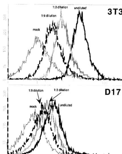
Viral binding on D17 and NIH 3T3 cells. The passage of 4070A MuLV on canine D17 osteosarcoma cells has been effective to identify gain-of-function mutations within wild-type (WT) 4070A as well as chimeric MuLVs (26, 30). There-fore, we speculated that D17 cells might be partially restricted for 4070A MuLV entry and therefore sensitive to variations in Env. To test if the D17 cells were limited in surface receptors, we performed a quantitative binding assay, comparing the virus binding capacities between D17 cells and NIH 3T3 cells (Fig. 1). 4070A virus was obtained from an NIH 3T3 producer cell line (approximate titer, 107) and diluted 1:3 and 1:9 in medium
in the presence of Polybrene. Aliquots of each dilution of virus were incubated with D17 cells or NIH 3T3 cells on ice for an hour and washed, and the total amount of virus bound per cell was analyzed by fluorescence-activated cell sorting (FACS), as described elsewhere (22). Undiluted 4070A virus yielded sig-nificantly more binding on NIH 3T3 cells (mean channel num-ber, 16) than on D17 cells (mean channel numnum-ber, 2.2). Serial dilutions revealed that binding of the undiluted virus on D17 cells yielded the same level as binding of 1:9 diluted virus on NIH 3T3 cells. The lower level of 4070A virus binding on D17 cells could reflect a limited number of available receptors and differences in virus binding affinity. This correlates with the report that the mRNA level of thepit-24070A receptor in D17 cells is lower than that in NIH 3T3 cells (24).
Analysis of 4070A/G100R for binding and titer.Replication of 4070A in D17 cells led to the acquisition of a G-to-R change at residue 100 (numbering from the initiator methionine [28]) in the VRA region of the receptor binding domain of SU (26) (Fig. 2). This variant (G100R) was frequently observed during passage of amphotropic chimeric viruses in D17 cells (more than 80% of over 57 isolates sequenced) and in at least two isolates passaged in human embryonic 293T cells (21; C.-W. Lu and M. J. Roth, unpublished results). Passage of virus with G100R/4070A Env in a human TE671 cell derivative, TelCeB6 cells (8), did not result in counterselection of G100R, indicat-ing that the beneficial effect of G100R/4070A MuLV
replica-* Corresponding author. Mailing address: Department of Biochem-istry, Robert Wood Johnson Medical School, University of Medicine and Dentistry of New Jersey, 675 Hoes Ln., Piscataway, NJ 08854. Phone: (732) 235-5048. Fax: (732) 235-4783. E-mail: roth@waksman .rutgers.edu.
739
on November 8, 2019 by guest
http://jvi.asm.org/
tion in D17 cells is not deleterious for replication in another cell type (data not shown).
In order to identify the function of the G100R, the mutation was subcloned into the 4070A Env expression vector pHIT 456 (36). The DNA encoding the G100R was PCR amplified with primer 9312 (5⬘GGTCTAGAGGCCGACACCCAGAGTG), encoding aXbaI site (underlined), and primer 7500 (5⬘CGAC CGCTACTCCTTCGTAA) and digested to replace the 0.6-kb
XbaI-XhoI fragment of pHIT 456. Virus was produced by tran-siently transfecting 10 g ofenvexpression plasmid into 106
D17/gag-pol/⌿GIP cells. These cells provide the MuLV Gag
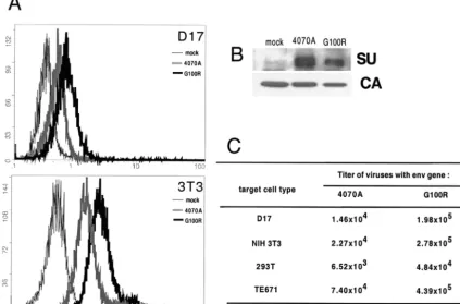
and Pol proteins in trans and package the green fluorescent protein (GFP)-internal ribosome entry site-puromycin resis-tance marker cassette (7). Viral binding assays were performed (Fig. 3A), and virus-associated SU protein was analyzed by Western blotting (Fig. 3B). Compared to WT 4070A, G100R/ 4070A showed an increase in receptor binding. Western blot analysis, in fact, indicated that this increase in binding occurred even in the presence of lower levels of G100R/4070A Env protein in this viral preparation (Fig. 3A and B). Despite being selected only in D17 cells, the binding enhancement of G100R was observed on both D17 and NIH 3T3 cells, suggesting that the improved fitness of G100R is not limited to D17 cells.
The effect of G100R/4070A Env on the viral titer was de-termined by transduction and expression of GFP. The virus utilized in the binding assays was used in parallel to inoculate 106D17, NIH 3T3, 293T, and TE671 cells (Fig. 3C). G100R/
4070A virus demonstrated a higher titer (between 6- and 13-fold) than that of WT 4070A virus in all these cell types tested. Similar fold values of stimulation were observed in indepen-dent assays. This observed increase in titer by G100R on each of the cell types tested suggested that G100R reflected a true gain-of-function Env variant, rather than an adaptation to a cell-specific feature, such as a receptor isotype. Viral interfer-ence assays indicated that G100R entry into D17 cells and NIH 3T3 cells was fully dependent on the Pit-2 receptor (data not shown).
[image:2.603.54.269.72.342.2]Kinetics of viral entry.Studies were performed to address whether the tighter binding by Env bearing G100R resulted in quicker uptake of the virus into the cells, thus yielding the
FIG. 1. Comparison of the 4070A virus binding capacities of NIH 3T3 cells and D17 cells. 4070A virus was obtained from an NIH 3T3 producer cell line (approximate titer, 107), chilled on ice, filtered through a 0.45-m-pore-size membrane, and diluted 1:3 and 1:9 in serum-containing medium in the presence of 8g of Polybrene/ml. One-milliliter aliquots of each dilution of virus were incubated with 2 ⫻105D17 cells or NIH 3T3 cells. After incubation on ice for an hour, the cells were washed with phosphate-buffered saline at 4°C. Virus binding was detected by monoclonal antibody 83A25 (10), which rec-ognizes the SU C terminus (22). Thexaxis is the fluorescence intensity as an indication of the amount of virus (Env) bound per cell, and the yaxis is the cell number. Phycoerythrin-conjugated antibody was ob-tained from Biosource International (Camarillo, Calif.), and FACS was performed at the Environmental and Occupational Health Science Institute, Rutgers University.
FIG. 2. Sequence of G100R mutant compared with that of 4070A. The mature SU is shown as an open box with general features noted. Amino acids were numbered from the first Met residue on the precursor protein (4070A, accession number M33469). The sequence of the VRA region is shown with a potential heparin binding site boxed. Changing of G100 to R resulted in the sequence ARRQR, which matches the heparin consensus sequence, XBBXB (see text). Other positions within VRA which influence receptor interactions of A-MuLV Env are noted, including Y90/V91 (Y60/V61 [39]), marked by #, and A99/Q102 (A71/Q74 [13]), marked by *. PRR, proline-rich region.
on November 8, 2019 by guest
http://jvi.asm.org/
[image:2.603.107.478.540.676.2]higher viral titer. To address these postbinding events, a kinetic assay for viral entry was performed (20). Briefly, at time points postbinding, virus that had not yet entered the cells was inac-tivated by an acid wash (pH 3 saline [17]). The titer of the virus, as scored by GFP expression, is indicative of the productive entry of the virus prior to the acid pulse. One representative set of results from four independent experiments is shown in Fig. 4.
Plotted on a log scale (Fig. 4A), the onset of G100R virus entry initiated after 15 min at 37°C, reached a titer of 103after
an additional 15 min, and eventually reached an end point titer of 104. WT 4070A virus initiated the infection at 20 min, and
103 particles functionally entered after an additional 30 min
and plateaued to the final titer of 103. The earlier initiation of
infection by G100R suggests that the enhanced binding trig-gers virus internalization faster. This may be due to either faster membrane fusion by an Env conformational change or accelerated cellular uptake. The data were also plotted on a linear scale to calculate the rate of postbinding entry by linear regression with Sigma Plot analysis (Fig. 4B). The slope of linear regression indicates the rate of virus entry. By this anal-ysis, G100R has a slope of 200⫾15, which is nearly 10 times the slope of WT 4070A, 17⫾5.7. The faster entry correlates with the 10-fold increase of the final viral titer. Differences in entry kinetics were also observed by alteration of Env density
on amphotropic MuLV (2). Collectively, our results suggest that G100R has evolved to overcome the decreased functional receptors available for virus binding of D17 cells, shown in Fig. 1. Efficient transduction by amphotropic MuLV vector can be achieved by increasing the receptor expression in the target cells (18). Based on our observations, we propose that incor-poration of G100R into the amphotropic MuLV vectors may improve the transduction efficiency and should be useful for gene therapy approaches. This may overcome the limitations of targeting cells expressing low amounts of Pit-2, such as hematopoietic stem cells (27).
[image:3.603.83.507.81.360.2]4070A Env/G100R is regulated by His36.If G100R follows the same fusion activation pathway as the WT 4070A, it should be blocked by mutations known to interfere with the WT 4070A entry process. His36 (H5 if numbered from mature protein) was shown elsewhere to be critical for Env-mediated membrane fusion (3, 20, 43). A deletion of His36 (⌬H36) was incorporated into the WT 4070A Env (⌬H36) and the G100R Env (⌬H36/G100R) within an Env expression vector. Their abilities to mediate virus binding (Fig. 5A) as well as synthesis-incorporation (Fig. 5B) and transduction (Fig. 5C) were exam-ined. Similar to observations with G100R, ⌬H36/G100R showed an increased binding over⌬H36 alone (Fig. 5A and B), without drastically affecting SU expression and incorporation. Consistent with previous reports,⌬H36 was unable to mediate
FIG. 3. Comparison of G100R function with WT 4070A virus. (A) Assays of binding of the G100R Env virus on D17 and NIH 3T3 cells.env expression plasmids were transfected into D17/gag-pol/⌿GIP cells to obtain viruses with desired Env. Binding assays were performed as described in the Fig. 1 legend. Thexaxis is the fluorescence intensity, and theyaxis is cell number. (B) Western blot analysis for virus-associated proteins. Virus was pelleted through a 20% sucrose cushion, separated by sodium dodecyl sulfate–10% polyacrylamide gel electrophoresis, and detected on a polyvinylidene difluoride membrane by anti-SU 79S-842 and anti-capsid CA 75S-287 (Microbiological Associates, Inc.), followed by horseradish peroxidase-conjugated rabbit anti-goat immunoglobulin G and the Supersignal substrate (Pierce). (C) Transduction abilities of G100R on various cell types. Results shown are one representative set of two independent experiments, as numbers of GFP⫹cells per milliliter.
on November 8, 2019 by guest
http://jvi.asm.org/
viral infection.⌬H36/G100R virus also yielded no detectable titer (Fig. 5C). These observations confirmed that G100R ac-tivates virus entry through the fusion activation pathway de-fined for WT virus entry. These results indicated an ordered fusion activation within the SU protein, whereby the receptor binding and stimulation by G100R are followed by the action of His36 at the SU N terminus.
Conclusions.In this report, we demonstrated that the recep-tor binding strength determined the efficiency and rate of viral entry and directly correlated with a functional gain in virus entry. Studies of the receptor binding region of avian sarcoma and leukosis viruses suggested that mutations that interfered with receptor binding did not necessarily correlate with the loss of infectivity (9, 33). Conversely, we showed that tighter initial binding by G100R resulted in faster kinetics of entry and im-proved infectivity.
Mutation of the Gly100 residue to a basic Arg introduces a heparin binding site consensus sequence, XBBXB (X is any amino acid and B is a basic amino acid, boxed in Fig. 2), in the VRA region. G100R may augment the receptor binding by enhancing the virus attachment on cells through heparin. Sim-ilarly, with PVC211, a neuropathogenic variant of Friend
MuLV, an amino acid change at the VRA of Env constitutes a heparin binding site. This mutation may confer the higher titer of PVC211 on brain capillary endothelial cells (15).
Alternatively, G100R may directly facilitate binding to the viral receptor. The region surrounding G100R has been shown to mediate receptor binding. G100R is located 7 amino acids beyond the E80-Y93 motif defined by Battini et al. for inter-acting with receptor (E50-Y64 [5]) and 10 amino acids beyond the Y90 and V91 defined by Tailor and Kabat (Y60/V61 [38]) for determining Pit-2 receptor usage over Pit-1. Simultaneous mutations of A99G and Q102K in 4070A Env, bracketing G100R, were shown to change receptor usage from Pit-2 to Pit-1 in NIH 3T3 cells (13). In contrast, strengthened binding by G100R does not lead to broadened receptor usage, as G100R-containing virus is not able to infect 4070A/D17 cells. The G100R Env mutation was selected as a gain-of-function MuLV Env variant on D17 cells. It is intriguing why this Env variant was not selected on NIH 3T3 cells. It may indeed reflect a balance between the viral fitness and host cell physi-ology. A high-efficiency virus may not be optimal for cells expressing high numbers of virus receptors, since multiple en-try events may create a metabolic burden for the host cell. Multiple rounds of retroviral entry are blocked in many retro-viruses through the process of viral interference (37). It has been hypothesized elsewhere that the level of second-round
[image:4.603.46.280.66.324.2]FIG. 4. Kinetics of postbinding entry. (A) Virus was collected by transient expression ofenvin D17/gag-pol/⌿GIP cells as described in the Fig. 3 legend, diluted 1:10, incubated with 2⫻105D17 cells at 4°C for more than 1 h, and then moved into a 37°C incubator. The infection process was stopped at various time points after 37°C incubation, and the virus remaining on the cell surface was inactivated by an acid wash (pH 3 saline [17]). The cells were then refed with fresh medium, and the viral titers were scored after an additional 48 h. Viral titers were plotted on a log scale as a function of the time allowed for postbinding entry. The titer at 110 min is noted at the end of each curve. Shown is a data set representative of four independent experiments. (B) Linear regression of the entry kinetics (same data as shown in panel A) analyzed by Sigma Plot. The linear fit of equation between entry time (t) and titer (f) isf⫽c⫹(a䡠t), wherearepresents the slope as a reference of the entry rate, shown on the top left corner of the chart.
FIG. 5. Functional analysis of⌬H36 mutants. The⌬H36 mutation was generated by PCR with mutagenic primers A⌬H5 (5⬘AGCCCCC AGGTCTTTAATGTAACC) and A⌬H3 (5⬘TACATTAAAGACCTG GGGGCTCTC) on the 4070A or G100R provirus DNA template and subcloned into Env expression vector (pHIT 456). (A) Binding analy-sis, performed as described in the Fig. 3 legend. (B) Virus-associated proteins analyzed by Western blotting, also as described in the Fig. 3 legend. (C) Transduction ability was assayed on D17 cells, and results shown were averaged from a triplicate experiment.
on November 8, 2019 by guest
http://jvi.asm.org/
superinfections yielding unintegrated viral DNA correlates with the cytopathic effects of the avian retroviruses (40, 41). Therefore, the selection for G100R might occur only in host cells in which the entry of the virus is limited by factors such as low numbers of functional receptors on the cell surface.
We thank Keith Bupp for critical reading of the manuscript, Mikhail Levin and Natalie Stano for Sigma Plot analysis, and Numan Rashid for FACS analysis.
This work is supported by NIH grant RO1 CA49932 to M.J.R.
REFERENCES
1. Andersen, K. B.1994. A domain of murine retrovirus surface protein gp70 mediates cell fusion, as shown in a novel SC-1 cell fusion system. J. Virol. 68:3175–3182.
2. Bachrach, E., M. Marin, M. Pelegrin, G. Karavanas, and M. Piechaczyk. 2000. Efficient cell infection by Moloney murine leukemia virus-derived particles requires minimal amounts of envelope protein. J. Virol.74:8480– 8486.
3. Bae, Y., S. M. Kingsman, and A. Kingsman.1997. Functional dissection of the Moloney murine leukemia virus envelope protein gp70. J. Virol.71: 2092–2099.
4. Barnett, A. L., R. A. Davey, and J. M. Cunningham.2001. Modular organi-zation of the Friend murine leukemia virus envelope protein underlies the mechanism of infection. Proc. Natl. Acad. Sci. USA98:4113–4118. 5. Battini, J.-L., O. Danos, and J. M. Heard.1998. Definition of a 14-amino
acid peptide essential for the interaction between the murine leukemia virus amphotropic envelope glycoprotein and its receptor. J. Virol.72:428–435. 6. Battini, J.-L., P. Rodrigues, R. Muller, O. Danos, and J. M. Heard.1996.
Receptor-binding properties of a purified fragment of the 4070A ampho-tropic murine leukemia virus envelope glycoprotein. J. Virol.70:4387–4393. 7. Chen, C.-C., A. Rivera, N. Ron, J. P. Dougherty, and Y. Ron.2001. A gene therapy approach for treating T-cell-mediated autoimmune diseases. Blood 97:886–894.
8. Cosset, F.-L., Y. Takeuchi, J.-L. Battini, R. A. Weiss, and M. K. L. Collins. 1995. High titer packaging cells modulating recombinant retroviruses resis-tant to human serum. J. Virol.69:7430–7436.
9. Damico, R., L. Rong, and P. Bates.1999. Substitutions in the receptor-binding domain of the avian sarcoma and leukosis virus envelope uncouple receptor-triggered structural rearrangements in the surface and transmem-brane subunits. J. Virol.73:3087–3094.
10. Evans, L. H., R. P. Morrison, F. G. Malik, J. Portis, and W. Britt.1990. A neutralizable epitope common to the envelope glycoprotein of ecotropic, polytropic, xenotropic, and amphotropic murine leukemia viruses. J. Virol. 64:6176–6183.
11. Fass, D., R. A. Davey, C. A. Harmson, P. S. Kim, J. Cunningham, and J. M. Berger.1997. Structure of a murine leukemia virus receptor-binding glyco-protein at 2.0 angstrom resolution. Science277:1663–1666.
12. Gray, K. D., and M. J. Roth.1993. Mutational analysis of the envelope gene of Moloney murine leukemia virus. J. Virol.67:3489–3496.
13. Han, J.-Y., P. M. Cannon, K.-M. Lai, Y. Zhao, M. Eiden, and W. F. Ander-son.1997. Identification of envelope protein residues required for the ex-panded host range on 10A1 murine leukemia virus. J. Virol.71:8103–8108. 14. Han, J.-Y., Y. Zhao, W. F. Anderson, and P. M. Cannon.1998. Role of variable regions A and B in receptor binding domain of amphotropic murine leukemia virus envelope protein. J. Virol.72:9101–9108.
15. Jinno-Oue, A., M. Oue, and S. K. Ruscetti.2001. A unique heparin-binding domain in the envelope protein of the neuropathogenic PVC-211 murine leukemia virus may contribute to its brain capillary endothelial cell tropism. J. Virol.75:12439–12445.
16. Jones, J., and R. Risser.1993. Cell fusion induced by the murine leukemia virus envelope glycoprotein. J. Virol.67:67–74.
17. Kizhatil, K., and L. M. Albritton.1997. Requirements for different compo-nents of the host cell cytoskeleton distinguish ecotropic murine leukemia virus entry via endocytosis from entry via surface fusion. J. Virol.71:7145– 7165.
18. Kurre, P., H.-P. Kiem, J. Morris, S. Heyward, J.-L. Battini, and D. A. Miller. 1999. Efficient transduction by an amphotropic retrovirus vector is depen-dent on high-level expression of the cell-surface virus receptor. J. Virol. 72:495–500.
19. Lavillette, D., M. Maurice, C. Roche, S. J. Russell, M. Sitbon, and F.-L. Cosset.1998. A proline-rich motif downstream of the receptor binding do-main modulates conformation and fusogenicity of murine retroviral enve-lopes. J. Virol.72:9955–9965.
20. Lavillette, D., A. Ruggieri, S. J. Russell, and F.-L. Cosset.2000. Activation of
a cell entry pathway common to type C mammalian retroviruses by soluble envelope fragments. J. Virol.74:295–304.
21. Lebkowski, J. S., R. B. DuBridge, E. A. Antell, K. S. Greisen, and M. P. Calos.1984. Transfected DNA is mutated in monkey, mouse, and human cells. Mol. Cell. Biol.4:1951–1960.
22. Lu, C.-W., and M. J. Roth.2001. Functional characterization of the N termini of murine leukemia virus envelope protein. J. Virol.75:4345–4366. 23. McClure, M. O., M. A. Sommerfelt, M. Marsh, and R. A. Weiss.1991. The pH independence of mammalian retrovirus infection. J. Gen. Virol.71:767– 773.
24. Miller, D. G., R. H. Edwards, and A. D. Miller.1994. Cloning of the cellular receptor for amphotropic murine retrovirus reveals homology to that for gibbon leukemia virus. Proc. Natl. Acad. Sci. USA91:78–82.
25. Morgan, R. A., O. Nussbaum, D. D. Muenchau, L. Shu, L. Couture, and W. F. Anderson.1993. Analysis of the functional and host range-determining regions of the murine ecotropic and amphotropic retrovirus envelope pro-teins. J. Virol.67:4712–4721.
26. O’Reilly, L., and M. J. Roth.2000. Second-site changes affect viability of amphotropic/ecotropic chimeric enveloped murine leukemia viruses. J. Vi-rol.74:899–913.
27. Orlic, D., L. J. Girard, C. T. Jordan, S. M. Anderson, A. P. Cline, and D. Bodine.1996. The level of mRNA encoding the amphotropic retrovirus receptor in mouse and human hematopoietic stem cells is low and correlates with the efficiency of retrovirus transduction. Proc. Natl. Acad. Sci. USA 93:11097–11102.
28. Ott, D., R. Friedrich, and A. Rein.1990. Sequence analysis of amphotropic and 10A1 murine leukemia viruses: close relationship to mink cell focus-inducing viruses. J. Virol.64:757–766.
29. Ott, D., and A. Rein.1992. Basis for receptor specificity of nonecotropic murine leukemia virus surface glycoprotein gp70SU. J. Virol.66:4632–4638.
30. Peredo, C., L. O’Reilly, K. Gray, and M. J. Roth.1996. Characterization of chimeras between the ecotropic Moloney murine leukemia virus and the amphotropic 4070A envelope proteins. J. Virol.70:3142–3152.
31. Pinter, A., R. Kopelman, L. Zhiyong, S. Kayman, and D. A. Sanders.1997. Localization of the labile disulfide bond between SU and TM of the murine leukemia virus envelope protein complex to a highly conserved CWLC motif in SU that resembles the active site sequence of the thiol-disulfide exchange enzymes. J. Virol.71:8073–8077.
32. Rein, A.1982. Interference grouping of murine leukemia viruses: a distinct receptor for the MCF-recombinant viruses in mouse cells. Virology120:251– 257.
33. Rong, L., K. Gendron, B. Strohl, R. Shenoy, R. J. Wool-Lewis, and P. Bates. 1998. Characterization of determinants for envelope binding and infection in Tva, the subgroup A avian sarcoma and leukosis virus receptor. J. Virol. 72:4552–4559.
34. Rothenberg, S. M., M. N. Olsen, L. C. Laurent, R. A. Crowley, and P. O. Brown.2001. Comprehensive mutational analysis of the Moloney murine leukemia virus envelope protein. J. Virol.75:11851–11862.
35. Sommerfelt, M. A., and R. A. Weiss.1990. Receptor interference groups of 20 retroviruses plating on human cells. Virology176:58–69.
36. Soneoka, Y., P. M. Cannon, E. E. Ramsdale, J. C. Griffiths, G. Romano, S. M. Kingsman, and A. J. Kingsman.1995. A transient three-plasmid expression system for the production of high titer retroviral vectors. Nucleic Acids Res.23:629–633.
37. Steck, F., and H. Rubin.1966. The mechanism of interference between an avian leukosis virus and Rous sarcoma virus II: early steps of infection by RSV of cells under conditions of interference. Virology29:642–653. 38. Tailor, C. S., and D. Kabat.1997. Variable regions A and B in the envelope
glycoproteins of feline leukemia virus subgroup B and amphotropic murine leukemia virus interact with discrete receptor binding domains. J. Virol. 71:9383–9391.
39. Tailor, C. S., A. Nouri, and D. Kabat.2000. A comprehensive approach to mapping the interacting surfaces of murine amphotropic and feline subgroup B leukemia viruses with their cell surface receptors. J. Virol.74:237–244. 40. Temin, H.1988. Mechanisms of cell killing/cytopathic effects by nonhuman
retroviruses. Rev. Infect. Dis.10:399–405.
41. Weller, S. K., A. E. Joy, and H. M. Temin.1980. Correlation between cell killing and massive second-round superinfection by members of some sub-groups of avian leukosis virus. J. Virol.33:494–506.
42. Wu, B. W., P. M. Cannon, E. M. Gordon, F. L. Hall, and W. F. Anderson. 1998. Characterization of the proline-rich region of murine leukemia virus envelope protein. J. Virol.72:5385–5391.
43. Zavorotinskaya, T., and L. M. Albritton.1999. Suppression of a fusion defect by second site mutations in the ecotropic murine leukemia virus surface protein. J. Virol.73:5034–5042.
44. Zhao, Y., L. Zhu, C. A. Benedict, D. Chen, W. F. Anderson, and P. Cannon. 1998. Functional domains in the retroviral transmembrane protein. J. Virol. 72:5392–5398.