Vol.66, No. 4 JOURNAL OF VIROLOGY,Apr. 1992,p. 2240-2250
0022-538X/92/042240-11$02.00/0
Copyright C1992,AmericanSocietyforMicrobiology
A
Novel
Herpes
Simplex
Virus
Glycoprotein,
gL,
Forms
a
Complex with Glycoprotein
H
(gH)
and
Affects
Normal
Folding
and
Surface
Expression
of gH
L. HUTCHINSON,'H.
BROWNE,2
V.WARGENT,2
N. DAVIS-POYNTER2S.PRIMORAC,1
K.
GOLDSMITH,'
A. C.MINSON,2ANDD. C.JOHNSONlt*
MolecularVirologyandImmunologyProgram, McMaster University, Hamilton, Ontario, Canada L8N
3Z5,1
andDivision ofVirology, Department ofPathology, University ofCambnidge, CambridgeCB2IQP, United
Kingdom2
Received13November1991/Accepted10January 1992
A
glycoprotein
encoded by theULlgene ofherpessimplexvirustype
1(HSV-1) was detected in infected cellswithantipeptide sera. TheULlgene has
previously
beenimplicated in virus-induced cell fusion (S. Little and P. A. Schaffer, Virology 112:686-697, 1981). Two protein species, a 30-kDa precursor form and a 40-kDa matureform of theglycoprotein,both of which weremodifiedwith N-linkedoligosaccharides, were observed. Thisnovelglycoprotein
is the 10thHSV-1glycoprotein to bedescribed and was named glycoprotein L (gL). A complex was formed between gL and gH, aglycoproteinknown to beessential forentry
ofHSV-1 into cells and for virus-induced cell fusion.Previously,
it had been reported that gH expressed in theabsence of other viral proteins wasantigenically
abnormal, notprocessed,andnot expressed at the cell surface (U. A. Gompels and A.C. Minson, J. Gen. Virol.63:474 4755,1989; A. J.Forrester, V. Sullivan, A. Simmons, B. A. Blacklaws, G. L. Smith, A. A. Nash, and A. C. Minson, J. Gen. Virol.72:369-375, 1991). However,giH
coexpressed with gLby using vaccinia virusrecombinants wasantigenically
normal, processednormally,
andtransported to the cell surface. Similarly, gL was dependent on gH for proper posttranslational processing and cell surface expression. These results suggest that it is a hetero-oligomer of gH and gL which is incorporated intovirions andtransportedtothe cell surface and which acts duringentryof virus into cells.Herpesviruses frequentlyinduce fusion of cells. Fusion of
virus-infected cells with uninfected cells may allow for
spread of herpesviruses and reduce contact with compo-nents of the immune system. The mechanisms by which
theseviruses influence infected cells tofuse arethought to beanalogous tothe processesbywhich thevirionenvelope
fuseswith cellular membranesduringviruspenetration into host cells.
Cell fusion induced by herpes simplex viruses
(HSV)
appears to involve a number of membrane glycoproteinswhich are also essential for entry of viruses into cells. Studies utilizing monoclonal antibodies
(MAbs),
liposomes containingviralproteins, andtemperature-sensitive
mutants haveimplicatedglycoproteins B, D, andH(gB,
gD,
andgH)
invirus-inducedcellfusion and in virus entry(6, 14, 15, 19, 22, 25, 26, 34, 38,42,50).
Inaddition,virusmutantsunable toexpressgB, gD,orgHareunabletoentercells(7, 17, 35),
andgB-andgD- virusesareunabletocausecellfusion(7, 35). Mutations inatleast three otherregionsof theHSV type 1 (HSV-1)genome, includingtheULl (36), UL24(29), and UL53 (4,13,
45, 46) genes, can give rise to thesyncytial
phenotype in which massive cell fusion is observed, in contrast to the low level of cell fusion observed with wild-type viruses. Theproteinproductsoftheseviralgenes may act to regulate membrane fusion events duringvirus entry oregress, perhaps by interactingwithviralproteins,
suchasgB,gD, andgH, whichplayadirect role in the fusion process.Mutations in the UL53gene, includingthosewhich affect
* Correspondingauthor.
tPresentaddress
(until
July, 1992):RoomQ3-17,ChironCorpo-ration,4560HortonSt.,Emeryville,CA 94608.
residue 40 of thepredicted protein, frequentlygiverisetothe
syncytial phenotype (13, 46). We haverecently detectedan HSV-1 glycoprotein encoded by the UL53 gene and have
named thisproteingK(27).The HSV-1
ULl
gene wasalsoimplicatedin thesyncytial phenotype by Little andSchaffer (36),who performedmarkertransferexperiments involving
the syncytial mutatnt 804. The syncytial phenotype was transferred to awild-type viruswith theHpaI 0 fragment
frommutant 804.
Unfortunately,
these marker transfer ex-periments were performed before cloned fragments ofHSV-1 DNA were available and, therefore, it is formally possiblethatcontaminatingDNAfragmentswerepresentin
their
preparations
of DNA. TheHpaI 0 fragment containsthe first 94 codons ofthe ULl open readingframe and no other open reading frames (40), and it thus appears likely
that thesyncytialmutation in 804 mapstothe
NH2-terminal
halfof theULl
gene.Here, we have utilized
antipeptide
sera toidentify
andcharacterize theprotein productof theULlgene,which isa glycoproteinthatwehave namedgL.Alargefraction of the gL found in infected cells was discovered to be tightly
associated with gH. Previously, it had
been
found that gH expressed in the absence of other HSVproteins
wasanti-genicallydistinct from gHin infected cells, was not
proc-essedproperly, andwas not
transported
tothe cell surface (18, 23). Similarly, gL was not properly processed whenexpressedinthe absence of other HSV
polypeptides
and in cells infected with a mutant virus unable to expressgH.
However, when gLandgHwerecoexpressed,
theproteins
were antigenically similarto those found in infectedcells,
were processed, and were transported to the cell surface. These observations areespecially
noteworthy becausegH,
2240
on November 9, 2019 by guest
http://jvi.asm.org/
NOVEL HSV GLYCOPROTEIN COMPLEX 2241 which
plays
a central role in membrane fusion and virusentry into
cells,
appears torequire
gL
foritsactivity.
MATERIALS
ANDMETHODSCells and viruses. Vero
cells,
143cells,
CV-1cells,
and human R-970 cells(47)
weregrowninalpha
minimalessen-tial medium
(GIBCO Laboratories, Burlington,
Ontario,
Canada) supplemented
with 7% fetalbovine serum(FBS).
HSV-1
strains
F(obtained
fromP. G.Spear, University
ofChicago),
KOS(obtained
fromJ.Smiley,
McMasterUniver-sity,
Hamilton, Ontario, Canada),
and17(obtained
fromH.Marsden,
University
ofGlasgow)
and thesyncytial
mutant804
(obtained
fromP.Schaffer,
Dana-FarberInstitute,
Bos-ton,
Mass.)
werepassaged,
and titerswere determined onVero or BHK cells. The HSV-1 mutant
SCgHZ, lacking
structural sequencesencoding
gH
whichwerereplaced
withthe LacZ gene, was
propagated
on F6 cells that expressHSV-1
gH
in aninducible fashion(17).
The HSV-1 mutantK082,
which is unable to expressgB,
waspassaged
on D6 cells(8).
An adenovirus vectorexpressing
gL
wascon-structed
by
firstexcising
anEcoRVfragment containing
the ULl genefromplasmid pSG-1,
whichcontainstheEcoRI
JKfragment
of HSV-1(KOS)
(20),
andinserting
thefragment
into
the EcoV site ofpBR322
togenerateplasmid pULl.
A 1.95-kbAvrII-BamHI
fragment
derived frompULl
wasinserted between the XbaI and BamHI sites of
plasmid
pAB26,
which contains theright
end ofadenovirus type 5(Ad5)
DNA(3),
yielding pABgL.
Human 293 cells werecotransfectedwith
pABgL
andpFG173,
which contains the left end of AdS DNA(24),
and 14days later,
adenovirusplaques
arose. Recombinantviruses
wereplaque purified,
viral DNAwas
analyzed,
andavirus isolateabletoexpressgL
(AdgL)
wasused in further studies. Vaccinia virus strainWR and
thymidine
kinase(TK)-negative
recombinantsde-rived fromitwere
propagated
inCV-1orVerocellsandwereassayed
in Vero cellmonolayers.
Arecombinant vacciniavirus
expressing
gL
wasconstructedby
thegeneral
strategy describedby
Mackett et al.(37)
as follows. TheKpnI
Bfragment
of HSV-1 strain 17 clonedinpAT153
wasdigested
with
StyI,
and an825-bp fragment corresponding
tounique
long
nucleotides 9329to 10154(40)
waspurified.
Theinitiat-ing
ATG ofULl lies atunique
long
nucleotide 9339. Thisfragment
wasendrepaired
andsubcloned intopRK19
suchthat the
gL coding
sequence, under the control of thevaccinia4b late promoter,wasinserted withinthevaccinia virusTK gene. TK-viruseswereselected from the progeny
by growth
in 143 TK- cells in the presence ofbromodeox-yuridine,
individualplaques
werepicked,
andrecombinantswere identified
by hybridization
withtheStyI fragment.
Ahybridization-positive
viruswassubjected
tothreeroundsofplaque purification,
and its genotype was confirmedby
restriction
analysis.
This virus was namedVac4b-gL.
Recombinant vaccinia viruses expressing
gH
from the 4bpromoter,
namedVac4b-gH,
and the 7.5 promoter, namedVac7.5-gH,
have been describedpreviously (18).
Synthetic
peptides, antipeptide
sera, and otherantibodies.Two
synthetic
peptides,
UL1-1(YVIRSRVAREVGDILK
VPC)
and UL1-2(CATKSRRRRPHSRRL),
were synthe-sized andpurified
by
Bachem Inc.(Torrance,
Calif.).
Thepeptides
wereconjugated
tokeyhole limpet
hemocyanin
orbovineserumalbumin
(BSA)
via thecysteine
residueswithmaleimidobenzoic
acid-N-hydroxysuccinimide
ester asde-scribed
previously
(16).
New Zealand White rabbits wereinjected initially
with 1.0 mg each ofpeptide conjugated
tokeyhole limpet
hemocyanin
in Freund'scomplete adjuvant
and then subsequently (after 4 to 6 weeks) with peptide conjugated to BSA in RIBI adjuvant (RIBI Immunochemi-cals, Hamilton, Montana). The animals were bled 9 to 10 days after each injection, and sera from animals which reactedwith gL were pooled. MAbs LP11 and52S
against
HSV-1 gH (5, 51) and 151B3 against HSV-1 gB (32) have been describedelsewhere.Rabbitpolyclonalserumspecific
forgH (anti-gH) purifiedby immunoaffinitychromatography was a kind gift of Roselyn Eisenberg and Gary Cohen. Rabbit antiserum directed to atrpE-gH fusion protein(anti-trpE-gH) has been described elsewhere
(15).
Radiolabelling of cells, immunoprecipitation, and gel elec-trophoresis. R-970 or Vero cells were infected with HSV-1 with 10 or 20 PFU per cell. At 7 h after infection, the cells were washed three times with medium lacking methionine andcysteine and containing 1% FBS(labellingmedium)and incubated for 4 to 5 h with labelling medium containing 100
RCi
of[35S]methionineand 100 ,Ci of [35S]cysteine per ml. Pulse-chase experiments were performed by first washingcells three times with labelling medium and labelling cells with 200 ,uCiof
[35S]methionine
and 200,uCiof[35S]cysteine per ml inlabellingmedium for 20min,and then cell extracts weremade(pulses)or the cells werewashed oncewithalphaminimal essential mediumcontaining 1% FBS and the cells wereincubated for 100or240 min inalphaminimalessential medium containing 1% FBS (chases). Cell extracts were made with NP40-DOC buffer (1% Nonidet P-40 [NP40],
0.5% sodium deoxycholate [DOC], 50 mM Tris-HCI [pH
7.5],
100 mMNaCl) containing2mg of BSA per ml and 1.0mM phenymethylsulfonyl fluoride and stored overnight at -70°C.Theextractswerethawed, centrifugedat82,000 x g for 60min, and insomeinstancesprecleared byincubation with rabbitseradirectedtogEandgIandprotein A-Sepha-rose. Extracts derivedfrom 5 x 10⁵to10 x 105 cellswere mixed with rabbitseraor mouseascites fluids(5 to 15 ,ul) for 90to120minonice. Insomeexperiments, antipeptidesera (5to15 ,ulofpooled sera)werepreincubatedwith100 to150
p,g of synthetic peptide for 30 to 60 min at 4°C. Protein
A-Sepharose
(Pharmacia Chemicals, Dorval, Quebec,Can-ada)wasadded, and thesampleswererotated end overend forafurther 60to90min.Immunoprecipitateswerewashed either three times withNP40-DOCbuffer or with the
follow-ingmorestringentwashes: twice with RIPAbuffer (50mM
Tris-HCl [pH 7.5], 150 mM NaCl, 0.1% sodium dodecyl sulfate
[SDS],
0.5% DOC, 1% NP40), once with 2 MNaCl-50mMTris-HCl(pH
7.5)-0.5%
DOC-1%NP40, oncewith
phosphate-buffered
saline(PBS) containing 1%NP40-0.5%
DOC,
once with 1 M MgCl2, and once with 1 mMTris-HCl
(pH 7.5).
Theprecipitated proteinswere eluted byadding
50 mM Tris-HCl(pH
6.8) containing 2% SDS, 10%glycerol, bromophenol blue,
and2%3-mercaptoethanol
andboiling
thesample
for5min.Electrophoresis involved10 or12%
polyacrylamide
gels as described elsewhere (30). Thegels
wereimpregnated
with Enhance (Dupont, Montreal,Quebec, Canada)
and exposedtoKodak XARfilm.Endoglycosidase
treatmentofimmunoprecipitatedproteins.Immunoprecipitated
proteins were eluted from proteinA-Sepharose by boiling
in 0.1 M sodium phosphate buffer,(pH 7.5) containing
0.5%3-mercaptoethanol
and 1% SDSand thendiluted with 2 volumes of 0.1 M sodium phosphate buffer
containing
1% octyl glucoside, 150 p,Mphenanthro-line,
and 10 mMEDTA. EndoglycosidaseF(2 or 10 U/ml;Boehringer
Mannheim) was added, and the material wasincubated for 2 hat37°C; then2% SDS-2% ,-mercaptoeth-anol-10%
glycerol
was added to the samples, which werethen
subjected
toelectrophoresis.VOL.66, 1992
on November 9, 2019 by guest
http://jvi.asm.org/
Cleveland partial proteolysis of glycoproteins. Proteins were immunoprecipitated, eluted, and electrophoresed on preparative polyacrylamide gels. Bands corresponding to gB, gH, gL, or proteins associated with either gL or gH were
located by using X-ray film, excised, and rehydrated as
described previously(30). Samples were digested with 5, 20, or 100 p,g of V8 protease per ml during early stages of electrophoresis through 18% polyacrylamide gels as
de-scribed previously(30).
Western immunoblot
analysis.
Extracts from Vero cellsinfectedwith HSV-1 (KOS) or SCgHZ were made 16 h after
infection and then in some cases immunoprecipitated with
thefollowingMAbs: LP11 or 52S, anti-gH rabbit serum, and
anti-UL1-2oranti-UL1-2inthe presence ofUL1-2peptide. Cell extractsor immunoprecipitatedproteins were
electro-phoresed with 4 to 15% gradient polyacrylamide minigels
(Bio-Rad, Mississauga, Ontario, Canada) or 10% linear
polyacrylamidegels.Theproteins were transferred to
nitro-cellulose membranes, and the membraneswereblocked for 10 h with PBS containing 1% skim milk and 1% BSA and thenfor 1 h with PBS containing 1% BSA and 0.5% gelatin. The blots were incubated for 2 h with anti-trpE-gH serum
diluted 1:200inPBScontaining 1%BSA orwithanti-UL1-2
serum diluted 1:50 in PBS containing 1% BSA followed by
five washes with PBS containing 0.2% NP40. The mem-braneswereincubated with 0.15 p,Ciof
1251I-labelled
protein A(New EnglandNuclear, Montreal, Quebec, Canada) per ml for1.0handthen washedfivetimes with PBS containing0.2% NP40, dried, andexposed tofilm.
Immunofluorescence. 143 cells were seeded and infected on glass coverslipsin24-welltrays. The cellswerefixed in 2% formaldehyde inPBSfor10 min andwashed three times with PBS containing 1% FBS. Thefixed cells were
perme-abilized by incubation in PBS containing 1%Triton X-100,
10%sucrose, and1% FBS for5 minand then washedthree
times withPBScontaining 1% FBS. All subsequent incuba-tionsand washes were inPBS containing 1% FBS. Cover-slipswereincubatedfor 1 h in antibody52S(culture super-natant diluted 1:2), washed three times, incubated with
fluoresceinisothiocyanate-conjugated rabbit anti-mouse
im-munoglobulin G (IgG) (DAKO) at adilution of 1:50for 45 min, and then washed three times. All treatments were at roomtemperature.
RESULTS
Identification and characterization of theprotein productof theULI gene, gL. Inordertoidentify and further character-ize the protein predicted to be encoded by the ULl gene
(40), we had synthesized two peptides: UL1-1, which in-cludes residues 26to44, andUL1-2,whichcontainsresidues
200to 224(the carboxy terminus)of theULl openreading
frame. Sera from rabbits injected with either UL1-1 or UL1-2conjugated tocarrierproteinsreacted with two pro-tein species, of 30 and 40 kDa, which were labelled with
[35S]methionine and
[35S]cysteine
in cells infected withHSV-1 (KOS)(Fig. 1). The 30- and 40-kDa protein species were not detected in uninfected cells, with
preimmune
serum,orwhen immuneserum wasmixedwith the relevantpeptide. The 30- and 40-kDaproteinswere also detected in cells infected with thesyncytialmutant,804. The anti-UL1-2 serum reproducibly precipitated a larger fraction of the 40-kDa protein than did the anti-UL1-1 serum, and
there-fore, the anti-UL1-2 serum wasused in mostof the subse-quentexperiments. Prominent
protein
bands inthe range ofJ.VIROL.
pre anti-ULI-2 pre anti-ULl-2 anti.-ULl-2 anti-ULl-1
peptide_lo- - -+ Il - +L - + i - t
_ 4_
_t
wt _gE 97
91
-W~~~~~~~~. :: ^ .53-5
40K- _
30K- m
a
m
30KOS UNINFECTED 804 KOS
FIG. 1. Immunoprecipitation of the ULl gene product from HSV-infected cells with antipeptidesera. Human R-970 cellswere left uninfected or infected with wild-type HSV-1 (KOS) or the HSV-1
syncytial
mutant804. After 4.5 h, the cells were radiola-belled with[35S]methionineand[35S]cysteineuntil 10.5 h postinfec-tion. Cellextractswerepartiallyprecleared witharabbitanti-gE/gI serum andprotein A-Sepharose and then mixed with anti-UL1-2 serum(-),anti-UL1-2serumpreincubatedwithUL1-2peptide (+), pooled preimmune serum(pre), or anti-UL1-1 serumwith (+) or without(-)UL1-1 peptide.Antigen-antibody complexeswere pre-cipitated with protein A-Sepharose and washed under stringent conditions (see Materialsand Methods), and the precipitated pro-teins wereelectrophoresed by using12%polyacrylamide gels.The positions of the 30- and 40-kDa proteins, HSV IgG Fc receptor proteinsgE and gI,andmarkerproteins of 97, 68, 46, and 30 kDa are indicated. The arrowindicates the presenceofa 100- to 110-kDa proteinassociated withgL.60 to 90 kDa were derived from the HSVIgG Fc receptor which isprecipitated inassociationwith rabbit Ig(30, 31).
We investigated the relationship between the 30- and 40-kDa species by performinga pulse-chase experiment in which infected cellswereradiolabelled for 20 min, the label was then chased for 100or240 min, and cellextracts were immunoprecipitatedwithanti-UL1-2serum.After thepulse,
the 30-kDa species was detected, and during subsequent chase periods, a large fraction of the 30-kDa protein was convertedtothe 40-kDa species (Fig. 2, right panel).
To further demonstrate that the 30- and 40-kDa proteins were derived from the ULl gene and toconfirm the speci-ficities ofourantipeptidesera,weexpressed the ULl gene
product using an adenovirus vector, AdgL, in which the ULl gene was inserted into the E3 region of AdS. The anti-UL1-2 serumrecognizeda30-kDaprotein expressedin AdgL-infectedcells whichwasnotobserved when antisera were preincubatedwith the appropriate peptide; however, the 40-kDaproteinwas notdetected(seeFig. 6).Itappeared that the 30-kDa protein was not processed to the 40-kDa form in AdgL-infected cells (see below). Three proteins
rangingin molecular massesfrom 75 to 115 kDawere also precipitatedfrom AdgL-infected cells; however, these pro-teins did not disappear when the sera were preincubated with peptides. The largest and most
prominent
of theseproteins(marked byan arrowin Fig.
6)
wasapparently
the adenovirus hexonprotein (115to120kDa),which has been a commoncontaminant inimmunoprecipitation
experiments involving rabbitserum (54).TheULl geneproduct containsonlyasingle regionnear
on November 9, 2019 by guest
http://jvi.asm.org/
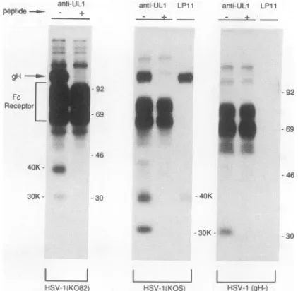
[image:3.612.335.564.76.247.2]NOVEL HSV GLYCOPROTEIN COMPLEX 2243
LPI 1 anti-gH anti-gL anri-gL
P Cl C2 P Cl C2 P Cl C2 P Cl C2
as
Gos
U.-200
-97
.53.5
a.
antlU_ ail2;
+n.>
_...
02
ts^l______3E
01 agE
giPgE p4g
40K<
arlt'. 3rti.:_
-pept,dl
t _ t
g92- _ _
.69
Sls
4.
46
30
-30
I
_I
I
L 1 IKOS KOS KOS
gH-FIG. 2. Pulse-chaseanalysisof the 30- and 40-kDa ULlproteins inextractsfrom cells infected withwild-typeHSV-1orgH-HSV-1. HumanR-970 cellswereinfected with HSV-1 (KOS) orthe gH-mutantvirusSCgHZ,and after 6 hwerelabelled with
[35S]methio-nine and[35S]cysteinefor 20min. The cellswereimmediately lysed withdetergents (P)or werewashed with mediumcontaining
methi-onine andcysteineand incubated for 100min (Cl)or240min(C2)in
this medium before cell extracts were made. Cell extracts were
mixed with MAb LP11 specificfor gH, anti-gH rabbitserum, or
anti-UL1-2 (anti-gL), and immunoprecipitated proteinswere
sub-jectedtoelectrophoresison10%polyacrylamide gels.Thepositions of the 40- and 30-kDagL proteins, gH,HSV Fcreceptorproteins, gE, gI/pgE, pgI, and molecularmassmarkers of200, 97, 53.5,and 30 kDaareindicated.
the NH2 terminus which would be predicted to act as a
signal or transmembrane spanning domain. If this region werecleaved,then theprotein mightbe secreted.However,
in a number of experiments (results not shown) we were
unable to immunoprecipitate proteins using either anti-UL1-1 oranti-UL1-2 serum from cell culture supernatants (fromwhich virus particles had been removed), suggesting that the ULl gene product was not secreted from cells in detectableamounts.In thesesameexperiments,wedetected
cleaved formsof other HSVproteins (gEandgI) whichwere
secreted from the cells in smallquantities.
The ULl gene product, gL, is glycosylated. It has been noted that the ULl gene product hasonepotential site for attachment of N-linked oligosaccharides (40). We treated samples containingthe 30- and 40-kDaspecies with endogly-cosidase F and observed a shift in the electrophoretic
mobilities of both protein species (Fig. 3), suggesting that bothare modified with N-linked oligosaccharides. In other experiments (datanotshown),asingle protein species of 25
kDawasimmunoprecipitated byanti-UL1-2antibodies from HSV-infected cells which had been treatedwith tunicamy-cin.This observationwasconsistentwith the predicted size
of the ULlprotein beforeglycosylation (40). Furthermore, both the 30- and 40-kDa species were labelled with
3H-glucosamine (28). Together, these results suggest that a
25-kDa ULlgeneproduct is initially modified by additionof
anN-linkedoligosaccharidesothataprotein species withan
apparentmolecularmassof30-kDa is observed immediately
DarkerExposure Llght??rExposure
FIG. 3. EndoglycosidaseFtreatmentofproteins immunoprecip-itatedwith anti-UL1-2serum.Human R-970cellswereinfected with
HSV-1(F)and then radiolabelled with[35S]methionineand [35S]cys-teinebetween 5 and 10 hpostinfection.Cellextractswere
immuno-precipitatedwith anti-UL1-2serum oranti-UL1-2serummixedwith
UL1-2peptide. Immunoprecipitated proteinswereelutedand incu-bated in buffer(0)or weretreatedwithendoglycosidaseF(endo F) by using 2 or 10 U/ml for 2 h at 37°C and then subjected to
electrophoresisin12%polyacrylamide gels.Alighterexposure(B) ofpanelA is includedsothat heavier bandsnearthetopof thegel
canbemorereadilyvisualized. The doublearrowsindicateapairof
protein bands which disappearedwhenserum wasincubated with peptide,in additiontothe 30- and 40-kDaproteins. Componentsof theHSV-1IgGFcreceptor, gE, gI/pgE, pgI,and molecular mass
markers of92, 69, 46, and 30 kDaareindicated.
after synthesis. Subsequent modifications to N-linked oli-gosaccharides and addition of 0-linked oligosaccharides duringtransporttothe cell surfaceapparentlycontributeto the reducedelectrophoretic mobilityof the 40-kDa species,
as has been noted for other HSVglycoproteins (33).
Evi-dencesuggestingthatgLis modified with 0-linked oligosac-charides comesfrom the observation that treatment of the 40-kDa form with endoglycosidase F produced a 37-kDa species(Fig. 3)rather thana25-kDaspecies,whichmightbe expected if alltheoligosaccharideswereremoved.
We propose to name thisnewly described HSV-1
glyco-proteingL, following the customs agreed uponatthe 1983 Herpesvirus Workshop in Oxford, England, and at subse-quentworkshops.AnHSVglycoprotein, gJ,encodedbythe
US5 gene,hasbeenpreviously referred toinareview(48).
Further, we recentlyidentified a novel HSVglycoprotein,
denotedgK,which is encodedbytheUL53gene(28). Onthe
basis ofpast experience, it wouldseem highly likely thata
protein analogous to the HSV-1 gL protein is encoded by HSV-2. Unfortunately, the anti-ULl-1 and UL1-2 sera do
not reactwith ananalogous protein in extractsfrom HSV-2-infectedcells,mostprobably because ofextensive
differ-encesin the amino acidsequences intheregionschosen to
produceantipeptide sera(39).
gLformsacomplexwithgHininfected cells.Twoprotein
species, with apparent molecular masses ofapproximately
100 and 110kDa,wereconsistentlyobserved in
immunopre-A
gH
-gE
-B
gpgE- _ pg9
-40K- a
30K-VOL.66, 1992
40.0 ?:A;-.4 400
on November 9, 2019 by guest
http://jvi.asm.org/
[image:4.612.58.297.75.297.2] [image:4.612.321.549.77.297.2]anti-ULl
peptide-- - + antrULl 153B2 anti-gH
4ie,,T
An_
iagH
Fctr
ReceptorL B
anti-ULl LP11 anti-ULl LPI1
* of
V8protease 0 5 20 100 0 5
.ux/mO)
--9
20 100 0 5 20 100
6933
-46 40K- *
30K- -30 *1 -40K
_0w -30K
-L
HSV-1(KOSi HSV-1(gH-)
I I I 1
gB gL associated gH
glycoprotein
FIG. 4. Clevelandpartial proteolysis of gB, gH, anda
glycopro-tein associated withgL. (A) Radiolabelled extracts from HSV-1-infected cellswereimmunoprecipitated with MAb 151B2, which is
specific for gB, anti-gH rabbit serum, or anti-UL1-2 serum, and
were electrophoresed in polyacrylamide gels. (B) Gel fragments
containing gB, gH, or the 100- to 110-kDa protein which was
precipitated by anti-UL1-2were excised and subjected to partial proteolysiswith 0, 5, 20, or 100 pgof V8 protease permlduring electrophoresis through 18%polyacrylamide gels.
cipitations involving the UL1-2 serum (Fig. 1, 2, and 3,
arrows). This protein was notobserved when anti-gL anti-bodies were preincubated with the UL1-2 peptide. Anti-UL1-1 antibodies also precipitated this
higher-molecular-mass protein in some experiments, although in mostcases
there was less protein than that observed with anti-UL1-2
serum(Fig. 1). The 100- and 110-kDaprotein species were
converted to a single faster-migrating protein species by
endoglycosidase F (Fig. 3) and, thus, were apparently de-rived froma singleHSVglycoprotein. Two obvious
candi-dates for thegL-associated proteinweregBandgH,which have similaroridenticalelectrophoreticmobilities in thisgel system.Todetermine which of theseglycoproteins mightbe associated withgL,we immunoprecipitated gB (usingMAb 1513B2), gH (using polyclonal anti-gH serum), and the gL--associatedprotein (using anti-UL1-2), subjectedtheproteins toelectrophoresis, excised theprotein bands,andsubjected theproteinstoV8partialproteaseanalysis (9).Thisanalysis clearlydemonstrated that thegL-associated proteinis struc-turally similartogHandnot togB (Fig. 4).
Tofurther examine theoriginofthegL-associated protein,
we precipitated gL from extracts of cells infected with HSV-1 mutants unable to express either gB or gH. The gL-associated proteinwas readilydetected inextractsfrom cellsinfectedwithHSV-1(K082),which is unabletoexpress
[image:5.612.78.300.72.343.2]gB(8) (Fig. 5).Incontrast, thegL-associated proteinwasnot detected inextracts from cells infectedwith thegH- virus,
FIG. 5. Expression of the gL-associated protein in cells infected with HSV-1mutantsunable toexpressgHorgB. Vero cellswere
infected with wild-type HSV-1(KOS), the HSV-1 gB-mutantK082,
ortheHSV-1gH-mutantSCgHZandlabelled with[35S]methionine and[35S]cysteinefor6h, beginning 5hafterinfection. Cellextracts wereprepared andmixedwith anti-UL1-2 serum(-), anti-UL1-2 serumwhichhadbeenpreincubated with the UL1-2 peptide (+),or
MAb LP11 specific for gH. Immunoprecipitated proteins were
separatedon12%polyacrylamide gels. The positions of the IgG Fc
receptorproteins, gH,the 30-and40-kDagLproteins, and
molec-ularmassmarkersof92, 69, 46, and 30kDaareindicated.
SCgHZ (17).Theseobservations furthersupportedthe
con-clusionthatgHwasthegL-associated protein.
It was formally possible that anti-gL antibodies
cross-reacted with gH, especially because these antibodies were
antipeptide antibodies. To examine this possibility, we in-fected cells with Vac7.5-gH, avaccinia virus recombinant which expresses gH (18). The anti-UL1-2 serum did not precipitate gH from Vac7.5-gH-infected cells, yet this gH could be precipitated with anti-gH serum (Fig. 6). These
results indicate thatgHisprecipitated by anti-gLantibodies becausegLandgHareassociated in infected cells. Further supportfor thishypothesis came from the observation that the gH-specific MAb LP11 precipitated two proteins with electrophoretic mobilities equivalentto the30- and40-kDa gLspeciesfrom extractsofHSV-infected cells (Fig. 3 and 6). The 40-kDa protein immunoprecipitated by LP11 was
subjectedtoV8 protease analysis andfound to be identical tothe40-kDa gLspecies (Fig. 7).LP11 didnotdirectlyreact withgLbecausegLwasnotprecipitatedfromcellsinfected with AdgL bythis antibody (Fig. 6). Therefore, itappears
that both the anti-gL polyclonal serumand thegH-specific MAb LP11precipitateacomplexofgH andgL.
Tofurther examine thehypothesisthatgHandgLforma
complex,weperformedWestern blotanalysis using anti-gH
serumto detectgH inmaterial whichwas first
immunopre-cipitated with anti-gL antibodies and, conversely, using anti-gLserum to detect gLin the material which was first
immunoprecipitated with anti-gH antibodies. Anti-trpE-gH fusion protein serum (15) reacted with gHprecipitated by usingtheanti-gH MAbLP11 and rabbit polyclonal anti-gH and also detected gH in material immunoprecipitated with anti-UL1-2 serum (Fig. 8). Further, the gH bandwas not _
40''s
-69
-46
L
HSV-1 IK0821
-30
J. VIROL.
A
on November 9, 2019 by guest
http://jvi.asm.org/
[image:5.612.343.554.77.283.2]NOVEL HSV GLYCOPROTEIN COMPLEX 2245
an- ariti- UL1 LP1I
pephide-__!H - +
92
&9- "
0.0
antiULI LP11
anti-- + gH
anti-UL1 LPI11
arili-- _ gH
__
w~~~~~-
92....
.r w ~
~~~~
-46-QH
-46
--40K
30
-S
Vac gH HSV-1 (KOS) AdgL
FIG. 6. Expression of gH and gLin cells infected withavaccinia viruscarryingthegHgene orwithAdgL. Human R-970 cellswere
infected withVac7.5-gH, HSV-1 (KOS),orAdgL.At 6 h after infection withHSV-1andVac7.5-gHorat24 h after infection withAdgL,the cellswerelabelled with[35S]methionineand[35S]cysteinefor5 h and then cellextractsweremade. Cellextractsweremixed with MAbLP11, anti-gH serum, anti-UL1-2 serum (-), or anti-UL1-2 serum preincubated with the UL1-2 peptide (+) and subsequently with protein A-Sepharose. Immunoprecipitated proteinswereeluted andanalyzedwith12%polyacrylamide gels.ThepositionsofgH,the 30- and 40-kDa
gL protein species, and molecular mass markers of 92, 69, 46, and 30 kDa are indicated. Also indicated in immunoprecipitates from
AdgL-infected cells (arrow)was acontaminating protein of approximately 120 kDawhichisroutinely observedin extracts fromwild-type
adenovirus-infected cellsimmunoprecipitatedwithrabbitsera.
observed when extracts from cells infected with the gH-mutantSCgHZwereused. The dark bandscentrallylocated onthese blots with anapparentmolecularmassof
approxi-mately 55-kDaweremostprobably IgG heavy chainswhich
canbindprotein A. Anti-UL1-2 serumreacted withthe
30-and 40-kDagL speciesinmaterialimmunoprecipitated with
ariti-ULl LP11 0
gH
eitheranti-gLserum or anti-gH antibodies fromextractsof wild-type HSV-infected cells. With extracts from cells in-fected with thegH-mutantSCgHZ, gLwasnotdetectedin material immunoprecipitated with anti-gH antibodies, and only the 30-kDa form of gLwas observed when the
immu-noprecipitation reactions involved anti-gL antibodies.
To-420 100 0 4 20 100 -V8protease
(il/nV)
woo-
692-46
40K- * ' g
30K
--30 **
L I
gL
-30
L I
gHassociated
[image:6.612.155.452.78.284.2]glycoprotein
FIG. 7. Clevelandpartial proteolysis ofgL andaproteinassociated with gH. Radiolabelledextractsfrom cells infectedwithHSV-1 (KOS)
wereimmunoprecipitated with anti-UL1-2orMAb LP11,specificfor gH, and the immunoprecipitated proteinswerethenelectrophoresedby
using12%preparative polyacrylamide gels. Gel fragments containingthe 40-kDa gL species precipitated with anti-UL1-2 and the 40-kDa
species precipitatedwithLP11 inconjunction withgHwereexcised,rehydrated, and treated with 0, 4, 20,or100,ugof V8proteaseperml
during electrophoresis through 18% polyacrylamide gels.
- 30
VOL. 66,1992
30K
is-f ""
on November 9, 2019 by guest
http://jvi.asm.org/
[image:6.612.147.459.466.678.2]anti anti-gL anti anti gL
LP1I gH - + P LPt1 gH - + P LP11anti-gH -anti-gL+ P LP1t anti-gH anti.gL- + P
*wK
i
gH= _
IgG 4 j
KOS JL
gH-IL
-IgG
=gL-KOS
anti-gH
II
gH-anti-gL
FIG. 8. Western blot analysis ofproteins coprecipitated with gH or gL. Vero cells infected with wild-type HSV-1 (KOS) or the gH- mutant SCgHZwereharvested 16 h after infection, and cell extracts were immunoprecipitated with anti-gH MAb LP11,anti-gHserum,anti-UL1-2 serum(-), anti-UL1-2serum preincubated with theUL1-2 peptide (+), or serum from preimmune rabbits (P). Theimmunoprecipitated proteinswereseparated by using 4 to15%polyacrylamide minigels, and the proteins weretransferredtonitrocellulose membranes. gH was detectedonthe blots with rabbit anti-trpE-gHserum, and gL was detected with anti-UL1-2serum, followed in each case by "2I-labelled proteinA.The major protein band in the central portion of these blots appears to be derived from the IgG heavy chain which can bind protein A.
gether, these dataprovide strong evidencethat gH and gL form a complexin infected cells. It appears likely that the
complex formed between gH and gL does not depend on interchaindisulfide bonds, becausethe electrophoretic
mo-bilities ofgH and gL observed in the absence of reducing agents
(P-mercaptoethanol)
were similaralthough not iden-ticaltothose observedinthe presence ofreducingagents.Expression and processing of gL in cells infected with an HSVmutantunable to expressgH.WhengLwasexpressed in the absence of other HSVproteins by using AdgL, we observed thatgLwas notprocessedfrom the 30-kDa imma-tureformtothe 40-kDamatureform. To determine whether
this lackofprocessingwasrelatedtothe absence ofgH, gL was examined in extracts of cells infected with the gH-mutantSCgHZ. The 30-kDa form ofgLbut notthe 40-kDa mature formof gLwasobserved in extractsfrom SCgHZ-infected cells that had been labelled for 5 h (Fig. 5). Simi-larly, in a pulse-chase experiment, the 30-kDa immature
formofgLwasnotprocessedtothe 40-kDamatureform in the absence ofgH(Fig. 2). Further, the 30-kDa rather than the 40-kDa form of gL was detected on Western blots
involvingextracts fromSCgHZ-infectedcells (Fig.
8).
CoexpressionofgHandgL leadstointracellular transport andprocessingof bothproteins. Our results establish thatgL
forms acomplexwith gHand that processingofgLto the 40-kDa species is dependent onthe formationof this com-plex. It has
previously
been demonstrated that gH, when expressed in the absence of other HSV proteins,wasanti-genically abnormal and was not transported to the cell surface.
Specifically,
gHexpressed
in COS-1 cells or bya recombinantvacciniaviruswasrecognized by antibody
52S but not by antibody LP11, whichrecognizes
a complex epitope (18, 21, 23). The obviousimplicationis that forma-tion of thegL-gHcomplexisaprerequisite
fortheprocess-ingand transportof both molecules. To confirmthis,andto demonstrate thatnoother
HSV-specific protein
isinvolved,gL and gHwere coexpressed. Thiswas first attempted by coinfecting cells with Vac4b-gH or Vac7.5-gH and AdgL
recombinantviruses, but itwas apparent that these viruses wereincompatible.Therefore,arecombinant vacciniavirus, Vac4b-gL, which expresses gL under the control of the vaccinia virus 4b promoter,wasconstructed, and cellswere
infected withVac4b-gHalone, Vac4b-gL alone, or with the tworecombinants together.Lysatesweresubjectedto West-ernblotanalysis with anti-UL1-2serum(Fig.9). gL synthe-sizedbyVac4b-gL is primarily in the30-kDa form, consis-tent with the results of experiments with AdgL (Fig. 6),
althoughasmall fraction of the gL wasobserved to be in the 40-kDa form when darker exposures of these blots were made. CoexpressionofgL and gH resulted in thesynthesis
ofapproximately equalamountsof the 30-and 40-kDa forms
of gL, a result similar to that observed for HSV-infected
cells. We note that the electrophoretic mobilities of gL
species from recombinant vaccinia virus-infected cells are slightlydifferent fromthose ofgLinHSV-infected cells. At present, we assume that this is due to small differences in
glycosylation in vaccinia virus-infected and HSV-infected
cells. We have previously noted minor differences in the
2 3 4
92
68
46
[image:7.612.164.481.77.211.2]3 0
FIG. 9. Glycoprotein Lexpressedwith arecombinant vaccinia virus.Vero cellswereinfected at amultiplicityofinfection of 10 with HSV strain 17(lane1), Vac4b-gL(lane 2),Vac4b-gH(lane3)or
Vac4b-gH plus Vac4b-gL(lane 4). Lysates corresponding to 105 cellswereprepared16 h afterinfection andsubjectedto electropho-resison10%polyacrylamide gels,andtheproteinsweretransferred
tonitrocellulose.gLwasdetectedwithanti-UL1-2serum.Numbers
ontherightindicatemolecularmassmarkers of92.5, 68,46,and 30 kDa.
VIROL.
L
on November 9, 2019 by guest
http://jvi.asm.org/
[image:7.612.398.504.496.641.2]NOVEL HSV GLYCOPROTEIN COMPLEX 2247
1 2 3 4 5 6 78
A
1 2 3 4 5 67 8
B
[image:8.612.315.547.77.406.2]30
FIG. 10. Antigenicproperties of gHexpressed in thepresence
and absence of gL. Vero cells were infected as described in the legendtoFig.9 with HSV-1(lanes1and2), Vac4b-gH(lanes3and 4), Vac4b-gL(lanes5and6),andVac4b-gH plus Vac4b-gL(lanes7 and8). Lysateswereimmunoprecipitatedwith MAbLP11(lanes 1, 3, 5, and7)or52S(lanes 2, 4, 6, and8), precipitated proteinswere
separated on 10% polyacrylamide gels, and the proteins were
transferred to nitrocellulose. gHwas detected onthe membranes with rabbit anti-trpE-gH serum (A), and gL was detected with anti-UL1-2 serum (B), followed in both cases with
"2I-labelled
proteinA.electrophoretic mobilitiesofgBsynthesized incellsinfected
withHSVversusgBexpressed byanadenovirusvector(53).
To examine the influence of gL on the structure and transportof gH, lysates of infected cellswere
immunopre-cipitated with antibodies 52S and LP11, and theprecipitation productswere subjected toelectrophoresis and transferred tonitrocellulose membranes. gHwasdetectedonthe mem-branes with anti-trpA-gH fusionproteinserum, andgLwas detected with anti-UL1-2 serum. Vac4b-gH synthesized a form ofgHrecognized byMAb 52S but notbyMAbLP11, aspreviously reported
(18),
butcoexpression ofgHandgLresults inaproductthatwas recognized byboth antibodies andwhich is electrophoretically indistinguishable from gH synthesizedinHSV-infected cells(Fig. 10A).Inaddition, gL was immunoprecipitated in conjunction with gH by both
anti-gH MAbs (Fig. 10B), demonstrating that the gH-gL complexcanform in the absence of otherHSVpolypeptides.
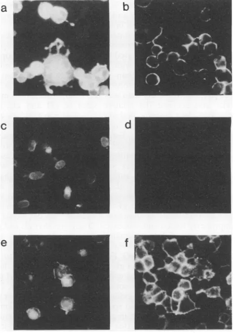
The cellularlocalization ofgHwasexaminedby immunoflu-orescence.Incellsexpressing gH alone,the distributionwas exclusivelyintracellular andlargely perinuclear, asreported previously (18),butcoexpressionofgLresulted in efficient cell surfaceexpression(Fig. 11). These results confirm that gH and gL formacomplex,that theprocessingofgL into the 40-kDa form is gH dependent, and that formation of the authentic antigenic structureofgH and its transport to the cell surface isgLdependent. In otherimmunofluorescence experiments, the cell surfaceexpression ofgLwasfoundto dependoncoexpressionwithgH, although asmallfraction
ofgLdid reach the cell surface in the absence ofgH(results notshown).
DISCUSSION
Anovel HSV-1 glycoprotein, encoded by the ULl gene and designated gL, was detected in extracts from virus-infected cellsby using antipeptidesera.Nucleotide sequence
analysisof theULlgenehadpreviously predictedaprotein of 224 aminoacids(25 kDa)with aputative signal sequence
c
a
e
f
FIG. 11. Cell surface expression of gH requires gL. 143 cells wereinfectedat amultiplicity of infection of 10 with HSV-1(a and b), Vac4b-gH (candd),orVac4b-gH plus Vac4b-gL(e and f).After 16h, the cellswerefixed and the cells shown inpanelsa, c,ande werepermeabilized. gHwasdetectedby incubation with MAb 52S followedby fluorescein-conjugated rabbit anti-mouse IgG.
andasinglesite for attachmentof N-linkedoligosaccharides (40). Two different antipeptide sera
immunoprecipitated
a 30-kDa precursor form of the protein as well as a 40-kDa mature form of the protein. Both protein species were sensitivetoendoglycosidase F, suggestingthat both containN-linkedoligosaccharides. Itwould appear thatgLbehaves inafashionanalogous tothebehavior ofanumber of other HSVglycoproteins,in which the fastermigrating,immature
protein species is converted to more slowly migrating, maturespecies duringtransport of theglycoprotein through
theGolgiapparatus,frequentlywith theadditionof0-linked
oligosaccharides
andtheprocessingof N-linkedoligosaccha-rides(33, 43).
In several experiments,we were unable tolabelgL with [35S]methionine under conditions in which gLwas heavily
labelled with [35S]cysteine (28). Since sequence analysis predicts thatgL hasonly a single methionine at the amino
terminus of the predicted signal sequence (40), it appears that theputative signal peptide maybe removed. Thereare nootherhydrophobicdomains presentingLwhich couldact as putative transmembrane domains, and this observation
coupled with our inability to detect secreted forms ofgL
suggests thatgLmaybe membrane associatedbyvirtue of its interactions withgH. However, it is alsopossiblethat the
a
92.5
68
46
b
_ - 'IF
L_.n~~0
VOL. 66,1992
-A
on November 9, 2019 by guest
http://jvi.asm.org/
[image:8.612.54.291.79.226.2]amino-terminal methionine is removed without removal of the entire signal peptide and that this hydrophobic region acts to anchor the protein in the membrane.
gL was immunoprecipitated from HSV-infected cells in conjunction with a higher-molecular-weight viral glycopro-tein which was demonstrated to be gH. The gH immunopre-cipitated in conjunction with gL by anti-gL sera was more intensely radiolabelled than gL. Similarly, gL present in immunoprecipitations involving anti-gH antibodies was la-belled less extensively than was gH. Comparisons here must take into account the relative sizes of gH and gL; gH contains eight cysteine and seven methionine residues and would thus be expected to incorporate approximately three times more label than gL. Estimates of the relative amounts of label incorporated into gH and gL suggest that approxi-mately equal molar quantities of gH and gL were present when the gH-gL complex was immunoprecipitated with either anti-gH or anti-gL antibodies. Thus, we expect that a
1:1 complex of gH and gL exists in infected cells and probably in virions. Furthermore, it would appear that any gH not associated with gL in infected cells or in virus particles would be nonfunctional because this gH would be improperly folded and processed. It appears that gL and gH are relatively tightly associated, because the complex was stable in washes with 2M NaCl, 0.1% SDS, and 1 MMgCl2. Previously, the HSV IgG Fc receptor was found to be composed of a complex of gE and gI (30, 31); however, here it was found that infected cells contained an excess of gE, which apparently retains some Fc receptor activity (2, 24). At this time, we have no evidence for an excess of eithergH or gL in infected cells and speculate that a large fraction of both proteins are present in gH-gL complexes. Pulse-chase experiments suggested that the gH-gL complex forms shortly after synthesis of the proteins.
Expression of both gH and gL was required for the proper processing of both proteins, and the correct folding and cell surface expression of gH was entirely dependent on coex-pression with gL. Furthermore, gL excoex-pression on the cell surface, evaluated by immunofluorescence experiments, was dependent on the expression of gH(results not shown), although a small fraction of gL reached the cellsurface in the absence of gH, consistent with the observation that a minor fraction of gL was processed in the absence of gH.
The observation that gH and gL form acomplex solves a long-standing problem involving gH, which when expressed in the absence of other HSV proteins is not properly folded, processed, and transported to the cell surface. A mammalian cell line expressing gH produced a form of gH which was not recognized by conformationally sensitive MAbs, and this protein was not processed and expressed on the cell surface (23). Infection of the cells with HSV led to normal folding, processing, and transport. Similar results were obtained when gH was expressed with a recombinant vaccinia virus vector (18). Together, these results suggested that other HSV proteins intervened during or after synthesis of gH to alter folding and transport of the polypeptide. Previous attempts to detect gH-associated proteins met with little success, partially because gL cannot be labelled with
[35S]methionine. The results reported here suggest a meth-odology for studying thefunctional andimmunogenic prop-erties of gH: gH and gL can be coexpressed by using transfection or virus vectors so that both proteins are properly folded, processed, and localized in the cells.
The role of gL in membrane fusion and virus entry is still by no means clear. However, theobservations that gL forms a complex with gH and that complex formation is essential
for normal folding and transport of gH, coupled with the well-established role of gH in virus entry and cell fusion (5, 15, 17, 22, 23), suggest that gL also plays a central role in these processes. Mutant 804, which apparently possesses a
mutation in the NH2-terminal half of the gL gene and forms
syncytial plaques, grew tolower titers in a number of cell
lines and displayed a reduced level of gB dimers (36). Furthermore, imbalances ingH levels or in the timing of gH
synthesisincellsmayalsoproduce the syncytial phenotype, because wild-type and gH- viruses produced syncytial
plaques on F6 cells which express gH earlier than is ob-served in other cells (17).
Further evidence that a complex of gH and gL plays a central role in penetration into cells or in some other
essential process during infection comes from the observa-tion that geneshomologoustoHSV gHand gL are observed in most other well-studied herpesvirus families. Homologs of the HSV-1 gH and gL genes have been observed for vari-cella-zoster virus (11, 41), human cytomegalovirus (10,52),
and Epstein-Barr virus (1, 12, 41). The human cytomegalo-virus gH homolog was not expressed on the cell surface in the absence of other human cytomegalovirus proteins (10,
52). Further,the Epstein-Barr virus homolog of gH, gp85, is improperly folded and transported when the protein is expressed in the absence of other Epstein-Barr virus pro-teins(54), and an Epstein-Barr virus gene (BKRF2) has been suggested as a positional homolog to the HSV-1 ULl (1, 12, 41).
Recently, we replaced the HSV-1 ULl gene with a lacZ gene construct usinga cell lineexpressing gL to complement the mutantvirus(49). The mutant virusis unable to replicate in the absence of gL, suggesting that, like gH, gL is essential for HSVreplication. This result might have been expected, given that gLis required for proper folding and cell surface expressionof gH, a protein which is essential forvirus entry into cells and cell-to-cell spread of virus. Given the results reported here, it will be more appropriate in the future to
considergH and gL as a complex or hetero-oligomer. ACKNOWLEDGMENTS
We thank Frank Graham and John Rudie, who facilitated the construction of AdgL,Andrew Davison for the
KpnI
Bfragment of HSV-1, and Roselyn Eisenberg and Gary Cohen for rabbit sera specificfor gH. We are also indebtedtoPriscillaSchafferformutant 804 andStan Person for mutant K082and toDuncanMcGeoch for communication of unpublished work.The work in the United Kingdom wassupportedby the Medical Research Council and the Wellcome Trust and in Canada bythe National Cancer Institute of Canada (NCIC) and the Medical Research Council (MRC).D.C.J. is a researchscholar of theNCIC, and L.H. holds a studentship from the MRC.
REFERENCES
1. Baer, R., A. T. Bankier, M. D. Biggin, P. L. Deininger, P. J. Farrell,T. J. Gibson, G. Hatfull, G. S. Hudson, S. C.Satchwell, C. Seguin, P. S. Tuffaell, and B. G. Barrell. 1984. DNA sequence and expression of the B95-8 Epstein-Barr virus ge-nome. Nature (London) 310:207-211.
2. Bell, S., M. Cranage, L. Borysiewicz, and T. Minson. 1990. Induction ofimmunoglobulin G Fc receptors by recombinant vaccinia viruses expressing glycoproteins E and I of herpes simplex virus type 1.J. Virol. 64:2181-2186.
3. Bett, A.,and F. L. Graham. Unpublished results.
4. Bond, V. C., and S. Person. 1984. Fine structurephysicalmap locationsofalterations that affectcell fusion in herpessimplex virus type 1. Virology 132:368-376.
5. Buckmaster, E. A., U. Gompels, and A. C. Minson. 1984. Characterization and physical mapping ofan HSV-1 glycopro-J.VIROL.
on November 9, 2019 by guest
http://jvi.asm.org/
NOVEL HSV GLYCOPROTEIN COMPLEX 2249 tein of approximately 115 x 103 molecular weight. Virology
139:408-413.
6. Bzik, D. J., B. A. Fox, N. A. Deluca, and S. Person. 1984. Nucleotide sequence of a region of the herpes simplex virus type 1gB glycoprotein gene: mutations affecting rate of virus entryandcell fusion. Virology137:185-190.
7. Cai, W., B.Gu,andS. Person.1988. Roleofglycoprotein Bof herpes simplex virus type 1 in viral entry and cell fusion. J. Virol. 62:2596-2604.
8. Cai,W.,S. Person,S. C. Warner, J. Zhou, andN. A.DeLuca. 1987. Linker-insertion nonsense and restriction-site deletion mutations of thegBglycoprotein geneofherpes simplex virus type 1. J.Virol. 61:714-721.
9. Cleveland, D. W., S. G. Fischer, M. W. Kirschner, and U. K. Laemmli. 1977. Peptide mapping by limited proteolysis in so-dium dodecyl sulfate and analysis by gel electrophoresis. J. Biol.Chem. 252:2202-2206.
10. Cranage, M. P., G. L. Smith, S. E. Bell, H. Hart, C. Brown, A.T.Bankier, P. Tomlinson, B. G. Barrell, and T. C. Minson. 1988.Identification andexpressionof ahumancytomegalovirus glycoproteinwith homologytothe Epstein-Barr virus BXLF2 product,varicella-zostervirus gpIII, and herpes simplexvirus type 1glycoproteinH. J. Virol. 62:1416-1422.
11. Davison, A. J., and J. E. Scott. 1986. The complete DNA sequenceof varicella-zostervirus. J. Gen. Virol.67:1759-1816. 12. Davison,A.J.,and P. Taylor. 1987. Geneticrelations between varicella-zoster virus and Epstein-Barr virus. J. Gen. Virol. 68:1067-1079.
13. Debroy, C., N. Pederson, and S. Person. 1985. Nucleotide sequence ofherpes simplexvirus type 1 gene that causes cell fusion. Virology145:36-48.
14. Deluca,N., D. J. Bzik, V. C. Bond, S. Person, and W. Snipes. 1982. Nucleotide sequences of herpes simplex virus type 1 (HSV-1) affecting virus entry, cell fusion, and production of glycoprotein gB(VP7). Virology122:411-423.
15. Desai,P.J., P. A. Schaffer, and A. C. Minson. 1988. Excretion of non-infectious virus particles lacking glycoprotein H by a temperature sensitive mutant of herpes simplex virus type 1: evidence thatgHis essential for virioninfectivity. J.Gen. Virol. 69:1147-1156.
16. Doolittle, R. F. 1986. Of Urfs and ORFs: a primer on how to analyze derived amino acid sequences, p. 63-79. University ScienceBooks, MillValley, Calif.
17. Forrester, A., H. Farrell, G. Wilkinson, J. Kaye, N. Davis-Poynter,and T.Minson. 1992.Constructionandproperties ofa
mutant of herpes simplex virus type 1 with glycoprotein H codingsequences deleted. J. Virol. 66:341-348.
18. Forrester,A.J., V. Sullivan, A. Simmons, B. A. Blacklaws, G.L. Smith, A. A. Nash, and A. C. Minson. 1991. Induction of protectiveimmunity withantibody to herpes simplex virustype 1glycoproteinH (gH) and analysis of the immune responseto gH expressed in recombinant vaccinia virus. J. Gen. Virol. 72:369-375.
19. Fuller, A. O., and P. G. Spear. 1987. Anti-glycoprotein D antibodiesthatpermitadsorption but block infection byherpes simplexvirus 1 prevent virion-cell fusion at the cell surface. Proc. Natl. Acad.Sci. USA84:5454-5458.
20. Goldin, A. L., R. M. SandrinGoldin, M. Levine, and J. C. Glorioso. 1981. Cloning of herpes simplex virus type 1 se-quencesrepresenting thewholegenome. J. Virol.38:50-58. 21. Gompels, U. A., A. L. Carss, C. Saxby, D. C. Hancock, A. C.
Forrester, and A. C. Minson. 1991. Characterization and se-quenceanalysis ofantibody-selected antigenic variants of her-pessimplexvirus show aconformationallycomplex epitopein glycoproteinH.J. Virol. 65:2393-2401.
22. Gompels, U. A., and A. Minson. 1986. The properties and sequence of glycoprotein H of herpes simplex virus type 1. Virology153:230-247.
23. Gompels,U. A., and A. C. Minson.1989. Antigenicproperties andcellular localizationof herpes simplex virusglycoprotein H synthesized in a mammalian cell expression system. J. Gen. Virol. 63:4744-4755.
24. Hanke, T., F. L. Graham, V.Lulitanond, and D. C. Johnson.
1990. Herpes simplex virus IgG Fc receptors induced using recombinantadenovirusvectorsexpressingglycoproteinsEand I. Virology 177:437-444.
25. Highlander, S. L., S. L. Sutherland,P.J. Gage, D.C.Johnson, M.Levine, and J. C. Glorioso. 1987. Neutralizing monoclonal antibodies specific for herpes simplex virus glycoprotein D inhibit viruspenetration. J. Virol. 61:3356-3364.
26. Holland, T. C., R. M. Sandri-Goldin, L. E. Holland, S. D. Marlin, M.Levine, and J. C. Glorioso. 1983. Physical mapping of the mutation inan antigenic variant ofherpes simplexvirus type 1 by use of an immunoreactive plaque assay. J. Virol. 46:649-652.
27. Hutchinson, L. M., K. Goldsmith, D. Snoddy, H. Ghosh, and D. C. Johnson. Unpublished results.
28. Hutchinson, L. M., and D. C. Johnson. Unpublishedresults. 29. Jacobsen, J. G., S. L. Martin, and D. M. Coen. 1989. A
conserved open reading frame that overlapsthe herpessimplex virus thymidine kinase geneisimportant for viralgrowth in cell culture.J. Virol. 63:1839-1843.
30. Johnson,D.C.,and V. Feenstra. 1987. Identification ofa novel herpes simplex virus type 1-induced glycoprotein which com-plexes with gE and binds immunoglobulin. J. Virol. 61:2208-2216.
31. Johnson, D. C., M. C. Frame, M. W. Ligas, A. M. Cross,and N. D. Stow. 1988. Herpes simplex virus immunoglobulin G Fc receptor activity depends on a complex of two viral glycopro-teins, gE andgI. J. Virol. 62:1347-1354.
32. Johnson, D. C., G. Ghosh-Choudhury, J. R. Smiley, L.
Fallis,
and F. L. Graham. 1988. Abundant expression ofherpes sim-plex virus glycoprotein gB usinganadenovirus vector.Virology 164:1-14.33. Johnson, D. C., and P. G. Spear. 1983. 0-linked oligosaccha-rides are acquired by herpes simplex virus glycoproteins in the Golgi apparatus. Cell32:987-997.
34. Johnson, D. C., M. Wittels, and P. G. Spear. 1984. Binding to cells of virosomes containing herpes simplex virus type 1 glycoproteins and evidence for fusion. J. Virol. 52:238-247. 35. Ligas, M. W., and D. C. Johnson. 1988. A herpes simplex virus
mutant in which glycoprotein D sequences are replaced by p-galactosidase sequences binds to but is unable to penetrate into cells. J. Virol.62:1486-1494.
36. Little, S. P., and P. A. Schaffer. 1981. Expression of the syncytial (syn) phenotype in HSV-1, strain KOS: genetic and phenotypic studies of mutants in two syn loci. Virology 112: 686-697.
37. Mackett,M., G. L. Smith, and B. Moss. 1984. A general method for the production and selection of infectious vaccinia virus recombinants expressing foreign genes. J. Virol. 49:857-864. 38. Manservigi, R., P. G. Spear, and A. Buchan. 1977. Cell fusion
induced by herpes simplex virus is promoted and suppressed by different viral glycoproteins. Proc. Natl. Acad. Sci. USA 74: 3913-3917.
39. McGeoch, D. J. Unpublished results.
40. McGeoch, D. J., M. A. Dalrymple, A. J. Davison, A. Donlan, M. C. Frame, D. McNab, L. J.Perry,J. E.Scott,and P. Taylor. 1988. The complete DNA sequence of the long unique region in the genome of herpes simplex virus type 1. J. Gen. Virol. 69:1531-1574.
41. McGeoch, D. J., and A. J.Davison. 1986. DNA sequence of the herpes simplex type 1 gene encoding glycoprotein H, and identification of homologues in the genomes of varicella zoster and Epstein-Barr virus. Nucleic Acids Res. 14:4281-4292. 42. Minson, A. C., T. C. Hodgman, P. Digard, D. C. Hancock, S. E.
Bell, and E. A. Buckmaster. 1986. An analysis of the biological properties of monoclonal antibodies against glycoprotein D of herpes simplex virus and identification of amino acid substitu-tions that confer resistance to neutralization. J. Gen. Virol. 67:1001-1013.
43. Oloffson, S., J. Blomberg, and E. Lycke. 1981. 0-glycosidic carbohydrate-peptide linkages of herpes simplex glycoproteins. Arch. Virol. 70:321-329.
44. Para, M. F., R. B. Bauke, and P. G. Spear. 1980. Immunoglobin G (Fc)-binding receptors on virions of herpes simplex virus type VOL. 66,1992
on November 9, 2019 by guest
http://jvi.asm.org/
1 and transfer of thesereceptors tothe cell surface by infection. J. Virol. 34:512-520.
45. Pogue-Geile, K. L., G. T.-Y. Lee, S. K. Shapira, and P. G. Spear. 1984. Fine mapping of mutationsin thefusion-inducing MP strain ofherpes simplex virustype1.Virology 136:100-109. 46. Pogue-Geile, K. L., and P. G. Spear. 1987. The single base pair substitution responsible for the synphenotype of herpes sim-plex virustype1, strain MP. Virology 157:67-74.
47. Rhim, J. S., H. Y. Cho, and R. J. Huebner. 1975. Non-producer human cells induced by murine sarcoma cells. Int. J. Cancer 15:23-29.
48. Roizman, B., and A. E. Sears. 1990. Herpes simplex viruses and theirreplication,p.1795-1842. InB. N.Fields,D.M.Knipe,et
al.(ed.), Virology. Raven Press, New York.
49. Roop, C., L. M. Hutchinson, and D. C. Johnson. Unpublished data.
50. Sarmiento, M., M. Haffey, and P. G. Spear. 1979. Membrane
proteins specified by herpes simplex virus. III. Role of glyco-protein VP7 (B2) in virion infectivity. J. Virol. 29:1149-1158. 51. Showalter, S. D., M. Zweig, and B. Hampar. 1981. Monoclonal
antibodiestoherpes simplex virustype1proteins, including the immediateearly protein ICP4. Infect. Immun. 34:684-692. 52. Spaete, R. R., K. Perot, P.I.Scott, D.Bauer, A. S.Lee,M. H.
Scott, L. Rasmussen, W. J. Britt, and C. Pachl. 1991. CMV (Towne) glycoprotein H (gH) is complexed with GRO78 and GRP94, p. 133-136. In M. P. Landini(ed.), Progressin
cyto-megalovirus research.ElsevierSciencePublishers, Amsterdam. 53. Witmer, L. A., K. L. Rosenthal, F. L. Graham, H. M. Friedman, A. Yee, and D. C. Johnson. 1990. Cytotoxic T lymphocytes specificforherpes simplexvirus(HSV)studiedusing
adenovi-rusvectors expressing HSV glycoproteins. J. Gen. Virol. 71: 387-396.
54. Yaswen, L. R., and L. Hutt-Fletcher. 1991. Abstr. 16th Int. Herpesvirus Workshop,abstr.no.44.Asilomar, Calif.