Copyright© 1990, AmericanSociety forMicrobiology
Identification of
a
Glucocorticoid-Responsive
Element
in
Epstein-Barr Virus
STUART R. KUPFERt* ANDWILLIAM C. SUMMERS
Radiobiology Laboratories, Yale University Schoolof Medicine, NewHaven, Connecticut06510-8039 Received 22 November1989/Accepted 17January1990
Immortalizationof Blymphocytes by Epstein-Barrvirus(EBV)iscomplexandpoorlyunderstood.However,
someevidence suggeststhatglucocorticoids influence this process. We identified a glucocorticoid-responsive
element in the BamHI Cfragmentof EBV whichwecallES-1. Inglucocorticoid-treated cells, ES-1enhanced chloramphenicol acetyltransferasegeneexpressionfrom theherpes simplexvirusthymidinekinasepromoter,
as well as the EBV Bam-C promoter, from which several latent viral gene products are transcribed. By Northern blotanalysis, glucocorticoid treatmentenhanced transcriptionfrom theBam-C promoter in Jijoye
cells, a Burkitt's lymphomacell line. In addition, the DNA-binding domain of the glucocorticoid receptor
boundspecificallytothe ES-1region. TheseglucocorticoideffectsontheBam-C promoterregionmayprovide someinsight into theprocessof EBV immortalization.
Epstein-Barr virus (EBV), a human herpesvirus, is the etiologicagentof mononucleosisand is associated withtwo forms of malignant disease, Burkitt's lymphoma and
na-sopharyngeal carcinoma (11, 25). When EBV infects B lymphocytes in vitro, cellimmortalization occurs and viral DNA islatently maintainedasmultiple copiesof extrachro-mosomal, circular plasmids (31). Several EBV proteins expressed during viral latency, such as EBNA-1 (33),
EBNA-2 (16), and latent membrane protein (15, 22), appear
toplay important rolesincellimmortalization; however,the
process is still poorly understood. Some in vitro evidence suggests that glucocorticoids influence this process,
al-though the dataare conflicting asto whether the effect on
viral latency is positive or negative (2, 10, 23, 34). In
addition, EBV immortalization of B lymphocytes is associ-ated with increases in theconcentration and absolute
num-ber ofglucocorticoid receptors (GR) (35).
Glucocorticoidsexerttheirphysiological effectson
devel-opment anddifferentiationthrough theGR, whichactsas a
ligand-dependent transcription factor (3, 12). Glucocorti-coid-bound receptor complexes activate transcription by binding to specific DNA sequences, called glucocorticoid-responsive elements (GRE). The GRE consensus sequence
generally consists of variations of the perfect palindrome
AG'ACAnnnTGTTCT.
Many of the glucocorticoid-induc-iblegeneswhich have beenidentifiedarecharacterized byacluster ofmultiple GREs at various distances upstream of thepromoterregion (8, 17, 24,38). Examples ofsystems in which GREs enhance transcription of viral promoters in-cludetheMoloney murinesarcomavirus(24) and themouse mammary tumorvirus (38).
Wehypothesized that the EBVgenomemight containone or more GREs and that localization of these regulatory
elements might contribute to a better understanding ofthe
influence of glucocorticoids on EBV immortalization. By
sequencesearching, we identified threeregionsof the EBV genome which could potentially possess GRE-like
proper-ties. One region, which we call ES-1, in the BamHI-C
*Corresponding author.
tPresent address: DepartmentofPediatrics, Division of Endo-crinology, University of North CarolinaatChapel Hill, CB7220, 509 Bumett-Womack, Chapel Hill, NC27599-7220.
region, provedto have GREpropertiesand enhanced tran-scription from the latent-cycle Bam-C promoter (BC-R2), which regulates the synthesis of the highly spliced EBNA family of RNAs (4). This regulatory activity of glucocorti-coidsmay providean explanationforsomeof the observed effects ofglucocorticoids on the EBV-lymphocyte interac-tion.
MATERIALS AND METHODS
Plasmid constructions. All DNA constructions were done by standardprocedures and verifiedby restriction enzyme
analysis (21). PlasmidpCTGre was constructed by ligating
the 36-base-pair (bp) XbaI fragment of pmGTCO (which containsasingleGRE derived frommousemammarytumor virus [Fig. 1C]) intothe BamHI site ofpBLCAT2 (19) after the cohesive ends were made blunt by filling in with the Klenow fragment of DNA polymerase I. Plasmid pCTC14
wasconstructed in thesame manner aspCTGreexceptthat the insert was the 197-bp TaqI-AvaI fragment (Fig. 1B) of
pSV2neoBamHI-C, which contains the BamHI Cfragment of EBV (provided by G. Miller, Yale University). Plasmid pCTC13isidenticaltopCTC14except that the insert is inthe
reverse orientation. Plasmid pCEC9 was constructed by
ligating the Sau3AI fragment (1,426 bp, see Fig. 4A) of pSV2neoBamHI-C into the HindIII site ofpSVOCAT (14) after thecohesiveendsweremade bluntby fillingin with the Klenowfragmentof DNApolymeraseI. PlasmidpCP11was
constructed by ligating the SacI-Sau3AI (240 bp, see Fig.
4A) fragment ofpSV2neoBamHI-C intothe HindlIl site of pSVOCATafter the cohesive endsweremade blunt with the
Klenowfragmentof DNApolymeraseI and T4 DNA
poly-merase. Plasmid pCPG4 was constructed by inserting the 197-bp TaqI-AvaI fragment of pSV2neoBamHI-C into the NdeI siteofpCP11 after thecohesive endsweremade blunt
by fillingin with theKlenowfragmentof DNApolymeraseI,
thus positioningtheformer AvaI site of the insert approxi-mately300bpupstreamof thechloramphenicol acetyltrans-ferase(CAT)initiation codon.
Cell lines. Thefollowingcelllineswereused in thisstudy:
HeLa, a human epidermoid carcinomaline, originally des-ignatedtheS3 subline andgrowninourlaboratory formany years; HepG2, a human hepatoma line recently obtained from M. Karin (University ofCalifornia, San Diego); D98/
1984
on November 10, 2019 by guest
http://jvi.asm.org/
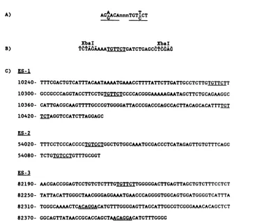
A)
AGA-ACAnnnTG,T-CT
XbaI XbaI
B) TCTAGAAAATGTTCTGATCTGAGCdtddid
C) ES-1
10240- TTTCGACTGTCATTTACAATAAAATGAAACCTTTTATTCTTGATTGCCTCTTGTGTTCTT
10300- GCCGCCCAGGTACCTTCCTGTGTTCTCCCCACGGGAAAAAGAATAGCTTCTGCAGAAGGC 10360- CATTGACGCAAGTTTTGCCCGTGGGGATTACCCGACCCAGCCACTTACAGCACATTTTGT
10420- TCTAGGTCCATCTTAGGAGC
ES-2
54020- TTTCCTCCCACCCCTGTCCTGGCTGTGGCAAATGCGACCCTCATAGAGTTGTGTTTCAGG
54080- TCTGTGTCCTGTTTGCGGT
ES-3
82190- AACGACCGGAGTCCTGTCTCTTTGTGTTCTTGGGGGACTTGAGTTAGCTGTCTTTCCTCT
82250- TATTACATTGGGCTAACGGGAGGAAATGAACCCAGGGGTGGCAGTGGATGGGGTCATTTA
82310- TGGGCAAAACTCACAGGACATGTTTGGGGAGTTAGCATTGGCGTCGGGAAACACAGCTCT
82370- GGCAGTTATAACCGCACCAGCTAACAGGACATGTTTGGGG
FIG. 1. EBVsequences selectedaspotentialGREs. (A)Perfectpalindrome GRE consensus sequence. (B) Single GRE sequence from mousemammary tumorvirus subcloned intopBLCAT2 forthe construction of pCTGre (Fig. 2A). (C) Three EBV sequences selected for constructionof CATreporterplasmids. Underlinedregionsrepresenttheveryhighlyconserved hexanucleotide sequence common to most GREs. Numbers correspondtothe EBV nucleotide sequencecoordinates.
HR1, an adherent human line constructed as a hybrid betweentheDetroit-98 epidermal celllineand the Burkitt's lymphoma line P3HR1; Jijoye and Raji, human cell lines derivedfrom Burkitt's lymphoma biopsy specimens;FF41, a spontaneously productivemarmosetlymphoblastoidcellline containingEBVfromhumanpatients; X50-7,a lymphoblas-toid cell line made by in vitro immortalization of human B lymphocytes byEBV. These lastfive lineswereallrecently obtainedfromG. Miller(Yale University), in whose labora-torytheX50-7lineoriginated. The HeLa andD98/HR1lines weregrowninminimumessential mediumwith5%fetal calf serum. In addition, D98/HR1 cells were maintained in lx MAGGT(6x 10⁻⁷Mmethotrexate, 5 x
10-'
Madenosine, 5 x10⁻⁵
M guanosine, 1 x 10⁻⁴ M glycine, 1.6 x 10⁻⁵ M thymidine) (27). The HepG2 line was grown in Dulbecco modified Eagle medium with 10% fetal calfserum. Jijoye, Raji, FF41, and X50-7 cells were grown in RPMI 1640 medium with10%fetal calfserum. Penicillinand streptomy-cinwere addedtoall media.Transienttransfections.D98/HR1, HeLa, andHepG2cells were transfectedat 30% confluency in 10-cm dishes witha totalof18pmol ofDNAbythe DEAE-dextrantechniqueas described previously (1). The DEAE-dextran solution was removed after 30 min and the cellsweretreated with medium containing 0.1 mM chloroquine diphosphate. After a 12-h recoveryperiodin normalmedium,the cellswereincubated in serum-free medium for 24 h with or without 1 ,uM dexamethasone before harvesting. Jijoye, Raji, FF41, and X50-7cellsweretransfectedat acelldensityof 3 x 10⁵cells permlwithatotal of18pmol ofDNAbythe DEAE-dextran
technique. The DEAE-dextran solution was removed after 30min, and the cellswereincubated innormal medium for5 h. The cellswerethenincubatedin normal medium withor
without 1 ,uM dexamethasone and harvested after 36 h. Whenindicated, Rajiand FF41cellswerecotransfectedwith 3 pmolof thep6RGR, aplasmid whichencodes the GR.
Enzyme assays. Cells were washed twice in phosphate-buffered saline, scraped from the dish in the case of D98/ HR1cells,centrifugedat1,000x g,andsuspendedin 150 ,ul of250mMTris chloride(pH7.8). Cellswerelysed bythree freeze-thaw cyclesand thenincubatedat60°C for10 minto
inactivate endogenous acetylases. The cellular debris was
removed by centrifugation at 7,000 x g for 5 min at 4°C. Equal amounts of protein from the crude extracts were incubatedat37°C forvarying timecourses(1or2h)in 150
,ul
ofan assay mixture
containing
250 mM Tris chloride(pH
7.8), 4 mM acetyl coenzyme A, and 0.125
1jRCi
of[14C]chloramphenicol.
Reaction mixtures were extracted withethyl acetate, spottedontothin-layer chromatography plates, anddeveloped inamixture of chloroform-methanol (19:1). The acetylated and nonacetylated forms of[14C]chloramphenicol
wereexcised from theplate
and quan-titatedby liquidscintillation counting.Northern(RNA)blotanalysis.JijoyeandX50-7 cellswere seeded approximately 24 h
preinduction
in RPMI 1640 medium with10%fetal calfserum.Ataconcentration of 3 x 10⁵cells perml, cells weretreated with dexamethasonefor varioustimes. The cellswereharvested, andtotal RNAwas extractedas describedpreviously (6).
Total RNAwaselec-trophoresed
on a 1%agarose-6%
formaldehyde gel
andon November 10, 2019 by guest
http://jvi.asm.org/
[image:2.612.128.494.64.384.2]transferred to a nylon membrane (Gene Screen Plus; Du-pont, NEN Research Products, Boston, Mass.). RNA blots were hybridized with a 32P-labeled probe produced by random primer synthesis (13). Hybridization was done at 42°C for 15 h in 50% deionized formamide-1 M
NaCl-1%
sodium dodecyl sulfate-10% dextran sulfate. Membranes werewashed twice in 2x SSC(lx SSC is 0.15 M NaCl plus 0.015Msodiumcitrate) at room temperature for 5min, twice in 2x SSC-1% sodium dodecyl sulfate at
60°C
for 30 min, andtwicein 0.1x SSC at room temperature for 30min.
Mobility shift assay. DNA probe was end labeled with
[at-32P]dCTP
and Klenow fragment of DNA polymerase I to aspecificactivity of107
cpm/[Lg.
In a15-,u reaction volume, 0.5 ngoflabeled DNA probe and 3 ng of purified T7X556, a truncatedproteincontaining the DNA-binding domain of theGR,
wereincubated under the following conditions: 100,uM
Tris chloride (pH 7.8), 50 mM KCl, 1 mM EDTA, 1 mM dithiothreitol, 10% glycerol, and 300,ug of bovine serum albumin per ml for 15 min at 20°C. For the competition assay,various amounts of unlabeled specific and nonspecific
probe
wereadded to each reaction 15 min before the addition of labeledprobe. Specific probe was the 197-bp ES-1 DNA(Fig.
1), and nonspecific probe was the 275-bpHinclI-BamHI fragment of pBR322. A 4% acrylamide gel (acryl-amidetobisacrylamide ratio of 80:1) was preelectrophoresed for1hat20 mAinlow-ionic-strengthbuffer (5). Electropho-resiswasdone at 30 mA for 3 h while recirculating the buffer. Thegel wasthen vacuum dried and autoradiographed.
Microdensitometry. Microdensitometry readings of the autoradiographs from Northern analyses and mobility shift assays weremeasured onaJoyce-Loebl Mark III recording microdensitometer. Theareas under the curves were deter-mined by planimetry.
RESULTS
Selecting potential GREs. Initially, we devised a strategy using various criteria for selecting specific EBV sequences which might possess GRE-like activity. Although GRE consensus sequences are variations of the palindrome
AGAACAnnnTGT'CT,
the most highly conserved motif is the hexanucleotide TGTTCT. Since the majority of gluco-corticoid enhancers consist of a cluster of two or more consensus sequences, we selected three regions from the EBV genome which contained a cluster of hexanucleotide motifs locatedadjacent to promoters proved to be functional(Fig.
1).EBV sequence 1 (ES-1) is located in theBamHI C region
approximately
850 bp upstream of the latent-cycle Bam-C promoter, BC-R2. EBV sequence 2 (ES-2) is located in the BamHI H region, approximately 1,230 bp upstream of the BH-Ll promoter. EBV sequence 3 (ES-3) is located in the BamHIMregion,approximately1,280 bp downstream of the BM-R2 promoter.To determine whether these EBV sequences enhanced transcription ina
glucocorticoid-dependent
manner, we con-structed CAT reporter plasmids with these various se-quences inserted directly upstream of the herpes simplex virus thymidine kinase (HSV-TK) promoter. Figure 2Adepicts
a set ofconstructs forES-1and the results of CATactivity
from transiently transfected D98/HR1 cells with these constructs. CAT activity was highly inducible by dexamethasone in pCTC14 (47-fold) and pCTC13 (30-fold). ThisdemonstratedthatES-1 possessesorientation-indepen-dent,
GRE-like activity. As expected, pCTGre, which con-tains a single GRE derived from mouse mammary tumorvirus, wasalso inducible by dexamethasone
(18-fold);
how-ever,absolute levelsofinduced CATactivity
werehigher
inpCTC14
andpCTC13.
Similar experiments in transient transfections of
HepG2
cells and HeLa cells also demonstrated dexamethasone-responsive activityby
ES-1
(datanotshown). ES-2 andES-3 didnotdemonstrate induction with dexamethasonein these CAT assay experiments in any ofthe above cell lines(data
not shown).GRE activity of
ES-1
withlatentBam-C
promoter.Having
identified a GRE in EBV,
ES-1,
which was active with the heterologous HSV-TK promoter, weattemptedtodetermine whether this activity existed in the context ofthe homolo-gousEBV promoter, theBam-C
promoter (BC-R2), 850bp
downstream. We constructed CAT reporter
plasmids
con-tainingES-1
and various components of downstream se-quence which contained theBam-C
promoter (Fig. 3). The level of CAT activity after transient transfection of these constructs in Jijoye cells and X50-7 cells is alsodepicted.
Afterdexamethasone treatment, athreefoldenhancementof CAT activity in Jijoye cells and fourfold enhancement in X50-7 cells occurred with
ES-1
in natural context 850 bp upstream of theBam-C promoter (pCEC9).The specific effect of glucocorticoid responsiveness was delineated furtherby inserting
ES-1
directly upstreamof the promoter region. Deletion of intervening sequences betweenES-1
and the promoter regions resulted in an even greater degree (ninefold) of CAT activity induced inJiUoye
cells (pCPG4). In X50-7 cells, the degree of dexamethasone induction increased to 14-fold with pCPG4; however, the absolute level of CAT activity in induced and uninduced cultures was substantially decreased compared with that with pCEC9. This phenomenon possibly suggests that a generalized enhancer or other transcriptional regulatory domain present in pCEC9 has been deleted in pCPG4.Transient transfections of pCEC9 were done in HepG2 and
D98/HR1
cells. No CAT activity could be detected, suggesting that the Bam-C promoter is not functional in these celllines. Transient transfections of pCEC9 weredone also in Raji cells and FF41 cells. Glucocorticoid inductionof CAT activity occurred in FF41 cells, but only if cotrans-fected with p6RGR, a plasmid encoding the GR. In Rajicells,
glucocorticoid induction of CAT activity was not observed, even when cotransfected with p6RGR (data not shown).Glucocorticoid induction of transcription from latent Bam-C promoter. From the CAT assay data, we predicted that transcription of native mRNAs from the
Bam-C
pro-moter could be induced by dexamethasone. The specific coordinates of the first exon transcribed from theBam-C
promoter have been well characterized, and deduced struc-tures of mRNAs transcribed from this promoter are pre-sented in Fig. 4A. Therefore, we performed Northern anal-yses on total RNA and probed with a
DdeI
fragment of EBV corresponding to exon 1 to evaluate the level of specific mRNA expression (Fig. 4A and B). We treated cultures ofJijoye
cellsatthemid-log growth phase
with dexamethasone at various times and extracted total RNA. With exon 1 as the probe, Northern analysis demonstrated induction of specific mRNA beginning at6 h (Fig. 4B, lane 4). Microdensitometry readings of the autoradiograph from this Northern blot revealed a 10-fold induction of specific mRNA 15 h after dexamethasone treatment (Fig. 4B, lane 5). These results correlate with the CAT assay data and probably represent a more physiological determination of glucocorticoid-induced enhancement of transcription from the Bam-C promoter.on November 10, 2019 by guest
http://jvi.asm.org/
A
Plasmid Constructs
no DNA
CAT e
CAT
H--CAT e
CAT
H
pBLCAT2
pCTGre
pCTC1 4
pCTC13
0 10 20 30
B
%/acetylation[14C]chloramphenicol
DEXAMETHASONE
AcCMK
_ + + - + +
.4
CML **.
-Cl
Z F
a <E
0
O cn
QL
a) T rn)
CD C3 U
H~~~~F
C) u Q)
QL Q
Q-FIG. 2. Effect ofdexamethasoneonCATreporterplasmidscontaining ES-1 and the HSV-TKpromoter.(A)CATplasmidconstructsfor
testing ES-1andcorrespondingCATactivityfrom crudeextractsoftransientlytransfected D98/HR1cells,determinedaspercentacetylation of[14C]chloramphenicol.Numberscorrespondtothe EBVnucleotidesequencecoordinates. The solidareaindicatesthe HSV-TKpromoter
region.Greferstothe singleGRE frompmGTCO, originallyderived frommouse mammarytumorvirus.Hatched barsindicatetreatmentwith 1 ,uM dexamethasone; open bars indicate notreatment. Equal amounts of crudeextract were used for eachassay within anindividual experiment.The resultspresentedarethemeanof threeseparatetransfections.(B) Representative exampleofthin-layerchromatographyof aCATassaydescribed above. CM, chloramphenicol; AcCM, acetylated chloramphenicol.
Similarexperiments weredone in X50-7 cells with
polyade-nylated RNA, but induction of mRNA with dexamethasone couldnotbe detected (datanotshown).
Specific bindingof GR toES-1sequence.Basedonthe CAT
assaydata, ES-1functioned as aGRE in vivo. We
hypoth-esized that if the enhancer function of ES-1 was acting
throughthe GR, thenthisfragment should bindspecifically to the GR protein. To determine whether specific binding
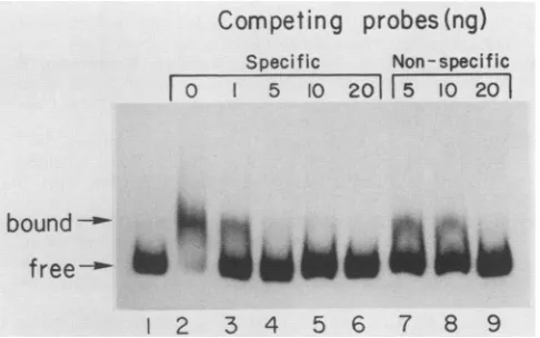
wasoccurring,weperformed mobilityshiftassayswithES-1 and theDNA-bindingdomain of the GR(Fig. 5). Inlane2,a
shifted band migrated more slowly than the protein-free
probe in lane 1 and probably represents a DNA-protein
complex. To determine whether therewas sequence speci-ficitytothisbinding,we added eithernonspecificorspecific
(ES-1) DNA to the binding reaction. In this competition
assay(Fig. 5),approximatelyninefoldmorenonspecificthan specific probewasrequiredtocompetewith thelabeledES-1
probeto equivalent levels. Microdensitometry ofthe
auto-radiographs allowedquantitation of thefraction of radioac-tive ES-1 sequence bound. This experiment demonstrated that ES-1 contains a nucleotide sequence(s) to which the DNA-bindingdomain of the GR binds specifically.
DISCUSSION
Our results demonstratedthat the EBVgenomecontainsa
GRE, which we call ES-1, located between coordinates 10,240 and 10,440, approximately 850 bp upstream of the Bam-C promoterregion. ES-1enhancedcat geneexpression under the control ofthe HSV-TK promoter in dexametha-sone-treated D98/HR1 cells by asmuch as47-fold. In
dex-amethasone-treated Jijoye and X50-7 cells, ES-1 enhanced cat gene expression under the control of the Bam-C pro-moter by three- to fourfold. In addition, dexamethasone
-E
SI
10240 10440
10440 10240
-I,ZZ22ff'e3
I I I I
40 50 60
on November 10, 2019 by guest
http://jvi.asm.org/
[image:4.612.142.470.78.480.2]Plasmid
Constructs
noDNA
CAT pSVOCAT
I CAT
9910 11336
CAT
10240 10440 11091 11336
JIJOYE
pCEC9
pCPG4
0 10 20 30 40
X50-7
I I I I I
---I
.I I I I
15-f
0 20 40 60
%
acetylation
[14C]
chloramphenicol
FIG. 3. EffectofdexamethasoneonCATactivityfrom CATreporterplasmidscontaining ES-1and the latentBam-C promoter.Left,CAT plasmid constructs containing elementsofES-1and/orvariouscomponentsofdownstreamEBV sequence. ro- representsthetranscription initiationsiteidentifiedfor theBam-C promoter. Subscripts correspondtotheEBVnucleotide sequencecoordinates.Right, CAT activityof crudeextractsfromJijoyeand X50-7 cellsafter transienttransfectionwith the aboveconstructswasdeterminedbypercentacetylationof
[14C]chloramphenicol. Assayconditionswere 8 ,ugofcrude extract and reaction time of 60 min. Hatched barsindicate treatmentwith 1 ,uM dexamethasone;openbarsindicatenotreatment.
induced transcription from the Bam-C promoter in Jijoye cells by as much as 10-fold as measured by Northern blot analysis. Further supportforES-1 possessing GREactivity comesfrom mobilityshift datademonstrating that the
DNA-A
0 EBV(kbp)
EBNA2
3ABC Ibinding
domain of the GR bindsspecifically
to the ES-1 region.Although
ES-1 containsmultiple
GRE consensusse-quences, further
analysis
will be necessary to determine170 EXONS
B
DEX
Time-hrs
0
I0
3 6 1510,200
I t
I t
I I I
TGTTCT
ES-
I11,000 EXON I 11,800
TACAAAA
CCAAT 28 S
--18
Slane 2 3 4 5
LI
3.8
2.34.8
9.81relative
amounts
ofspecific
mRNAFIG. 4. Effect of dexamethasoneontranscriptionofEBV-specificmRNA. (A)Transcriptionmap for theBam-C promoter.Thelocations oftheexonsfor EBNA-1, EBNA-2,andEBNA-3A,-B, -Careshown on the complete EBV map. Theexpandedmapshows thelocationof the threeTGTTCTmotifs ofES-1,theCAAT andTATA motifs, the RNA start site, and the first two exons. kbp,Kilobasepairs. (B)Northern blotanalysis oftotal RNA (10
p.g
perlane) from Jijoye cellsinduced with 1 ,uMdexamethasone(DEX) for the indicated times. The blot was probed withthe 193-bpDdeIfragment(EBVcoordinates11293 to11486)correspondingtoexon 1 of the highly splicedtranscriptsfrom the Bam-C promoter. The relative amount of specific mRNA per lane was determined by microdensitometry of the autoradiograph and standardized totheintensityof the actinmRNAhybridizationon thesame blots.-
|ES-11
r-i
I a I
on November 10, 2019 by guest
http://jvi.asm.org/
[image:5.612.96.539.76.254.2] [image:5.612.94.542.392.662.2]Competing probes(ng)
Specific Non-specific
0 51020 5 10 20
boundI
free
1 2 3 4 5 6 7 8 9
FIG. 5. BindingofGRto ES-1sequence. Bindingwas detected
by the shift in mobility of the DNA fragment incubated in the
presenceofGR protein. a-32P-labeled ES-1 DNA(0.5 ng)wasadded
to each reaction. Except for lane 1 (no protein control), 3 ng of purifiedT7X556 protein (DNA-bindingdomain peptideof the GR)
was added to each reaction. The nonspecific probe is the 275-bp HincII-BamHI fragment ofpBR322, and the specific probe is the ES-1 DNAfragment.
whetheronlyone or multiple consensus sequences actually possess significant GRE activity. Furthermore, our studies do not preclude the possibility of other functional GREs within the EBV genome. The lack of demonstrable
gluco-corticoid induction by ES-2 and ES-3 in our experiments does not eliminate the possibility of GRE function of these
sequences in vivo. The construction of these respective reporter plasmids may have deleted promoter or enhancer elements that are necessary for GRE activity. Also, our
inclusion criteria for screening potential GREs may have been too stringent (for example, regions containing single GRE motifs werenot selected).
Other cis- or trans-acting factors also may be critical for glucocorticoid-induced enhancementofgene expression by ES-1. This possibility is suggested by the fact that in
transient transfections ofpCEC9 into Rajicells, glucocorti-coid induction of cat gene expression does not occur.
Although othertranscriptionfactors besides the GRare not absolutely requiredforglucocorticoid induction, a
synergis-tic increase of promoter activity by GREs in combination with other transcription factor-binding sites has been dem-onstrated in vitro (30). EBNA-1 can act as atranscription
factor for thelatent-cycleBam-Cpromoter(32); however, in
thepresentexperiments,this effectwasprobablynot impor-tantbecause EBNA-1activityappearstorequireoriP in cis.
In addition, it has been shown that specificity of some
hormone receptor elements is not highly stringent and that
these elementscanmediate induction byseveral hormones, including progestins, androgens,andglucocorticoids (9, 37). It is possible that ES-1 modulates transcription through other hormone receptors; however, additional experiments willberequired to determinethis.
Most ofthe efforts towardunderstandingEBV
immortal-ization have concentratedontheroles oflatently expressed EBVproteins. Amongthe EBNAfamilyofproteins,
EBNA-1 isrequiredin transtomediatereplicationofviralplasmids in EBV-immortalized cells (20, 39), and EBNA-2 may be
involved in stimulating proliferation of these cells(31). The
function of ENBAs 3 to 6 in latency is unknown. The
latent-cycle Bam-C promoter (4) may be essential to the
immortalization process since transcripts of at least two
latently
expressed
proteins,
EBNA-1 andEBNA-3,
aretranscribed from this promoter.
Since,
as we haveshown,
glucocorticoids
enhancetranscription
from theBam-C
pro-moter, one could
speculate
thatglucocorticoids,
likewise,
enhance
expression
of one or severalspecies
of EBNAproteins.
If this provestobetrue, thenglucocorticoids
may beintimately
involved in theregulation
of EBV immortal-ization.One
possible
scenario forphysiological
EBNAexpression
based on this novel GRE could be thatglucocorticoids
induce increasedexpression
ofEBNA-1,
resulting
intighter
virallatency.
Thishypothesis,
however,
iscontradictory
to evidence thatcyclosporin
A-glucocorticoid-induced
immu-nosuppression
results in an increased incidence of EBV reactivationand EBV-associatedlymphoproliferative
disor-ders(7, 29).
Anotherpossibility
could be thatglucocorti-coids enhance
transcription
from theBam-C promoter
in such a way that a differentpattern
of EBNAproteins
isexpressed,
contributing
to adisruption
ofvirallatency.
Analtered
pattern
of EBNAexpression
mayaffect thecytotox-ic-T-cellresponsetoEBV-transformed
cells,
thusimpairing
immunosurveillance(18, 26, 28, 36).
A glucocorticoid-in-duced alteration of EBNAexpression
and glucocorticoid-inducedimmunosuppression
maybeimportant
factorsin theetiology
of EBV-inducedlymphoproliferative
lesions. Fur-ther studies to examine the effects ofglucocorticoids
onEBNA
protein
expression
and viralreplication
will bere-quired
to determine whether theserelationships
areplausi-ble.
ACKNOWLEDGMENTS
Weare
grateful
toWilmaSummers forproviding
HeLacells and for crucialhelp,
Michael Karin fortheHepG2
cell line,George
Miller for the D98/HR1 and
Raji
cell lines and theplasmid
pSV2neoBamHI-C,
Hal Jensonfor theJijoye
andFF41cell lines,Saumyen
Sarkar for theplasmid pBLCAT2,
andQingyun
Liu fortheplasmid
pSVOCAT
and valuable advice.Wearealsoverygrateful
toKeithYamamotoandLen Freedman for advice andfor
providing
theplasmids
pmGTCO
andp6RGR
andthepeptide
T7X556.This work was
supported by
Public Health Service grantCA-16038 from the National Institutes of Health. S.R.K.was
supported
by
Public Health Service grant 5T32CA-09159 fromthe National Institutes of Health.LITERATURE CITED
1.
Banerji,
J., L. Olson, and W. Schaffner. 1983. Alymphocyte-specific
cellularenhancerislocateddownstream ofthejoining
region
inimmunoglobulin heavy
chaingenes.Cell33:729-740. 2. Bauer,G. 1983. Induction ofEpstein-Barr
virusearly
antigens
by
corticosteroids: inhibitionby
TPAandretinoic acid. Int.J. Cancer 31:291-295.3. Beato, M. 1989. Gene
regulation by
steroid hormones. Cell 56:335-344.4. Bodescot, M., M. Perricaudet, and P. J. Farrell. 1987. A promoter for the
highly
spliced
EBNAfamily
of RNAs ofEpstein-Barr
virus.J. Virol. 61:3424-3430.5. Chodosh, L. A. 1987.
Mobility
shiftDNA-binding
assayusing
gel
electrophoresis,
p. 12.2.1-12.2.10. In F. M. Ausubel, R. Brent, R. E. Kingston, D. D. Moore, J. G. Seidman, J. A. Smith, and K. Struhl (ed.), Currentprotocols
in molecularbiology.
GreenePublishing
Associatesand JohnWiley
&Sons, Inc.,NewYork.6.
Chomczynski,
P., and N. Sacchi. 1987.Single-step
method of RNA isolation by acidguanidium
thiocyanate-phenol-chloro-formextraction. Anal. Biochem.162:156-159.
7. Cleary, M. L.,M. A. Nalesnik, W. T. Shearer, andJ. Sklar. 1988.Clonal
analysis
oftransplant-associated
lymphoprolifera-tionsbasedonthestructureofthegenomic
termini ofEpstein-Barrvirus. Blood 72:349-352.
IL!"
.f
-4
lo
...on November 10, 2019 by guest
http://jvi.asm.org/
[image:6.612.53.296.80.232.2]8. Danesch,U., B. Gloss, W. Schmid, G. Schutz, and R. Renkawitz. 1987. Glucocorticoid induction ofthe rat tryptophan oxygenase gene is mediated by two widely separated glucocorticoid-re-sponsiveelements. EMBOJ. 6:625-630.
9. Darbe, P., M. Page, and R. J. B. King. 1986. Androgen regulation bythe longterminalrepeatofmousemammary tumor virus. Mol. Cell. Biol. 6:2847-2854.
10. Dietrich,J. B., S. Chasserot-Golaz, G. Beck, and G. Bauer. 1986. Antagonismof glucocorticoid induction ofEpstein-Barr virus earlyantigens by different steroids in Daudilymphoma cells.J. Steroid Biochem. 24:417-421.
11. Epstein,M. A., and B. G. Achong (ed.). 1986. TheEpstein-Barr virus:recentadvances. Wiley MedicalPublications,New York. 12. Evans, R. M. 1988. The steroidand thyroidhormone receptor
superfamily. Science240:889-895.
13. Feinberg, A. P., and B. Vogelstein. 1982. A technique for radiolabeling DNArestriction endonucleasefragments tohigh specific activity. Anal. Biochem. 132:6-13.
14. Gorman, C. M., L. F. Moffat, and B. H. Howard. 1982. Recombinant genomeswhich express chloramphenicol acetyl-transferase in mammalian cells. Mol. Cell. Biol. 2:1044-1051. 15. Hennessy,K., S. Fennwald,M.Hummel, T. Cole, and E. Kieff.
1984. A membrane protein encoded by Epstein-Barr virus in latent growth transforming infection. Proc. Natl. Acad. Sci. USA81:7207-7211.
16. Hennessy, K., and E. Kieff. 1985. A second nuclearprotein is encoded by Epstein-Barr virus in latent infection. Science 227:1238-1240.
17. Jantzen,H.-M., U. Strahle, B. Gloss, F. Stewart, W. Schmid, M. Boshart, and R. Miksicek. 1987.Cooperativity of glucocorticoid responsive elements located farupstreamof the tyrosine ami-notransferasegene. Cell49:29-38.
18. Klein, G.1989. Viral latency and transformation: the strategy of Epstein-Barrvirus. Cell 58:5-8.
19. Luckow, B., and G. Schutz. 1987. CAT constructions with multiple unique restriction sites forthefunctional analysis of eukaryoticpromoters andregulatory elements. Nucleic Acids Res. 15:5490.
20. Lupton,S., and A. J. Levine. 1985.Mappinggeneticelements of Epstein-Barr virus that facilitate extrachromosomal persistence of Epstein-Barr virus-derived plasmids in human cells. Mol. Cell. Biol. 5:2533-2542.
21. Maniatis, T., E. F. Fritsch, and J. Sambrook. 1982. Molecular cloning: alaboratorymanual. ColdSpringHarborLaboratory, ColdSpring Harbor,N.Y.
22. Mann, K. P., D. Stanton, and D. A. Thorley-Lawson. 1985. Epstein-Barr virus-encodedprotein found inplasmamembranes of transformed cells. J.Virol. 55:710-720.
23. McGrath,I.T., P. A. Pizzo, L.Novikos, and A. S. Levine. 1979. EnhancementofEpstein-Barrvirusreplicationinproducer cell lines by combination of low temperature and corticosteroids. Virology97:477-481.
24. Mikscicek, R., A. Heber, W.Schmid, D. Ulrich, G. Posseckert, M. Beato, and G. Schutz. 1986. Glucocorticoid responsiveness of the transcriptional enhancer of Moloney murine sarcoma
virus. Cell46:283-290.
25. Miller, G. 1985. Epstein-Barr virus,p.563-589. In B. N.Fields (ed.), Virology. WileyMedicalPublications, New York. 26. Moss, D. J., I. S. Misko, S. R. Burrows, K. Burman, R.
McCarthy, and T. B. Sculley. 1988. Cytotoxic T-cell clones discriminate between A- and B-type Epstein-Barr virus trans-formants. Nature (London) 331:719-721.
27. Munyon, W., E. Kraiselburd, D. Davis, and J. Mann. 1971. Transfer ofthymidinekinase tothymidinekinaseless L cellsby infection with ultraviolet-irradiated herpes simplex virus. J. Virol. 7:813-820.
28. Murray, R. J., D. Wang, L. S. Young, F. Wang, M. Rowe,E. Kieff, and A. B. Richardson. 1988. Epstein-Barr virus-specific cytotoxic T-cell recognition of transfectants expressing the virus-coded latent membraneproteinLMP. J. Virol. 62:3747-3755.
29. Nalesnik, M. A., R. Jaffe, T. E. Starzl, A. J.Demetris, K.Porter, J. A.Burnham, L. Makowa, M. Ho, and J. Locker. 1988. The pathology of posttransplant lymphoproliferative disorders oc-curring in the setting of cyclosporine A-prednisone immunosup-pression.Am. J. Pathol. 133:173-192.
30. Schule, R., M. Muller, C. Kaltschmidt, and R. Renkawitz. 1988. Manytranscription factorsinteract synergistically withsteroid receptors.Science 242:1418-1420.
31. Sugden, B. 1989. An intricate route toimmortality. Cell 57:5-7. 32. Sugden, B., and N. Warren. 1989. A promoter of Epstein-Barr virus that can functionduringlatentinfectioncanbe transacti-vated by EBNA-1, a viral protein required for viral DNA replication during latent infection. J. Virol. 63:2644-2649. 33. Summers, W. P., E. A. Grogan, D. Shedd, M. Robert, C.-R. Liu,
andG. Miller. 1982. Stable expression in mouse cells of nuclear neoantigen after transfer of a 3.4 megadalton cloned fragment of Epstein-Barr virus DNA. Proc. Natl. Acad. Sci. USA 79: 5688-5692.
34. Sundar, S.K., D.V.Ablashi,G. R.Armstrong, M.Zipkin, A. Faggione, and P. H. Levine. 1981. Steroids inhibittumor pro-moting agent-induced Epstein-Barr virus antigens in Raji cells. Int.J.Cancer.28:503-507.
35. Tomita, M., G. P.Chrousos, D. D. Brandon, S. Ben-Or, C. M. Foster, L. DeVougn, S.Taylor,D. L.Loriaux, and M. B. Lipsett. 1985. Glucocorticoid receptors in Epstein-Barr virus-trans-formed humanlymphocytes. Horm. Metab. Res. 17:674-678. 36. Townsend, A. R. M., J. Rothbard, F. M. Gotch, G. Bahadur, D.
Wraith, and A. J. McMichael. 1986.The epitopes ofinfluenza nucleoprotein recognized by cytotoxic T-lymphocytes canbe defined with shortsynthetic peptides. Cell44:959-968. 37. von derAhe, D., S.Janich,C.Schneidereit,R.Renkawitz, and
M.Beato.1985.Glucocorticoidand progesterone receptors bind to the same sites in two hormonally regulated promoters. Nature(London)313:706-709.
38. Yamamoto, K. R. 1985. Steroid receptorregulated transcription ofspecific genes and gene networks. Annu. Rev. Genet. 19: 209-252.
39. Yates, J. L., N. Warren, and B.Sugden.1985.Stablereplication of plasmids derived from Epstein-Barr virus in a variety of mammalian cells. Nature(London)313:812-815.
![FIG. 2.testingofaexperiment.region.1 ,uM CAT [14C]chloramphenicol. Effect of dexamethasone on CAT reporter plasmids containing ES-1 and the HSV-TK promoter](https://thumb-us.123doks.com/thumbv2/123dok_us/1321102.85779/4.612.142.470.78.480/testingofaexperiment-chloramphenicol-effect-dexamethasone-reporter-plasmids-containing-promoter.webp)
![FIG. 3.dexamethasone;plasmidcrudeinitiation[14C]chloramphenicol. Effect of dexamethasone on CAT activity from CAT reporter plasmids containing ES-1 and the latent Bam-C promoter](https://thumb-us.123doks.com/thumbv2/123dok_us/1321102.85779/5.612.94.542.392.662/dexamethasone-plasmidcrudeinitiation-chloramphenicol-dexamethasone-activity-reporter-containing-promoter.webp)