by
W.D. Boucher, B.Agr.Sc.(Hons.) Tas.
Submitted in partial fulfilment of the requirements for the Degree of Doctor of Philosophy
University of Tasmania Hobart
This thesis contains no material which has been accepted for the award of any other degree or diploma in any University and to the best of my knowledge contains no copy or paraphrase of material previously published or written by any other person except where due reference is made in the text of the thesis.
W.D. Boucher.
University of Tasmania, Hobart.
1 ACKNOWLEDGEMENTS 00 O. 00 00 00 00 00 00 041 110 04 00 00 .0 00 60
SUMMARY 00 •0 •0 OD 00 00 00 00 00 00 00 00 0. 00 400 OS 00 00
LITERATURE REVIEW
VIRUS-HOST BIOCEEMCAL INTERACTIONS. • 00 00 SO SO •• •• • 0 0• ••
A. ENZYMES AND SUBSTRATES OF CELLULAR METABOLISM.
1. Respiration Rates of Virus-Infected Tissues._...." • (a) Respiration rates following infection • •.
(b) Respiration rates in tissues systemically • infected 00 00 Co 00 •0 00 OS •• 00 0• OS
(c) Respiration rates of local necrotic lesion
hosts • 00 00 •0 00 SO ••00 00 ID. 00 0•
0. ..
0. oo
go
2. Respiration Substrate Concentrations 0. CO 3. Carbohydrates OW CC •. 00 CC •0 00 0. 11. 0.0 • 0 .0 041
4
, Organic AcidS • .. 00 00 00 00 00 00 00 00 OS 0• w.• (i)
(ii
4
5
5
• 7
5.
Enzyme Systems Associated with "Dark" Respiration. .. •. 8(a) EMbden-Dbyerhof-Parnas pathway and pentose
. phosphate pathway enzymes • .. 00 SO 0• OD 00 00 •0
(b) Tricarboxylic acid Cycle and enzymes associated with the mitochondrion • OC •1 •1 •• •1 WAL
8
10
6. Phosphorylated Compounds. •
00•. CO •0 00 00 • 0 00. 0011
7. Host Nucleic Acid Synthesis... 0.• •-.11. J. 401 .1.4. • • • 1 CS_ 138. Non-Protein Nitrogen .. 00 00 011 00 00.00 OS 00 SO 00 00
15
9. Photosynthesis and other Functions Associated_with
Chloroplasts .. 00 00 WO 00 01. 00. •• • •1 CO 01 i.e. • 1 Op 18
10. Enzymes of Photorespiration • 00 00 00 OC OC 01 00 C. OW 23
11. PolYphenol Oxidases and Peroxidases .. .,... 00 01 CO 00 WO 24 12. Acid Hydrolases • 00 00 00 00 .0 00 00 00'00 00 • CO 01 27
13. Behydrogenases and Miscellaneous Cellular Enzymes •. .. 29
B. VIRUS-HOST HORMONAL INTERACTIONS.. 30 •
1. Auxins .. 00 • •• •• 00 0.0 00 • • •..• • • AL 0..• • .• • • • • •
31
2. Auxin -Scopoletin Interactions • • • • • • • • • • • • 0 • • • • 032
3. Gibberellins..
0. •• 00 0 • • 0 .11•• 00 • . 11 • .• 00 . 0 • 0 •33
4. Cyt okinins • • • 0 • • • • • 0 0 0 • •O 00 0 • 00
■
• • • • • • 0 0 •35
5.
Abscisic Acid and other Growth Inhibiting Substances.36
VIRUS-CELL ULTRASTRUCTURE INTERACTIONS.. .. •0 •W •• 0 • • • 00 • 037
1. Crystalline Inclusions. .0 • .. 00•.. 041 410 00 38 2. X -Bodies .0 0. 00 • 01, 00 .0
39
3. The Nucleus • .. 00 00 • 00 00 040 O. o. oo • 00 00 041
42
•
Table of _Contents cont.) Page Number
SECTION
VIRUS-HOST GROWTH AND PMEIOLOGICAL INTERACTIONS INTRODUCTIONO• 00 00 O• O• •• •0 0% 00 00 O• 00 04 •• •• O• 00
0. 00 . 00 0. 00 51 52 52
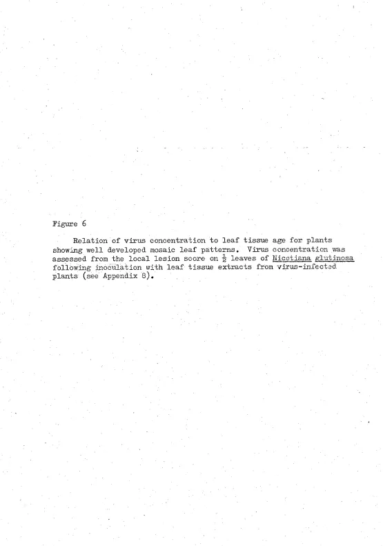
MATERIALS AND METHODS.. 00 0. 0. 0. .0 Os ol .06 0. T. Virus concentration and tissue distribution in leaves
showing well developed systemic symptoms 00 0•• (a) Virus concentration. • • • • • 00 0• 0• O• O•
(b) Tissue distribution 0. 00 00 • O• 04 •0 • 00 00 ea 52
2, Trials to Determine the Effects of Virus Infection on
Plant Growth •O O• O• 00 00 O• O• 0•.00 O• O• O• 00
(a) Plant height, internode length, leaf length,
O• O• 53
leaf number and root weight '40 00 00 . 0 . 0 53 .(b) Leaf growth during disease development. 0. • o6 op 54
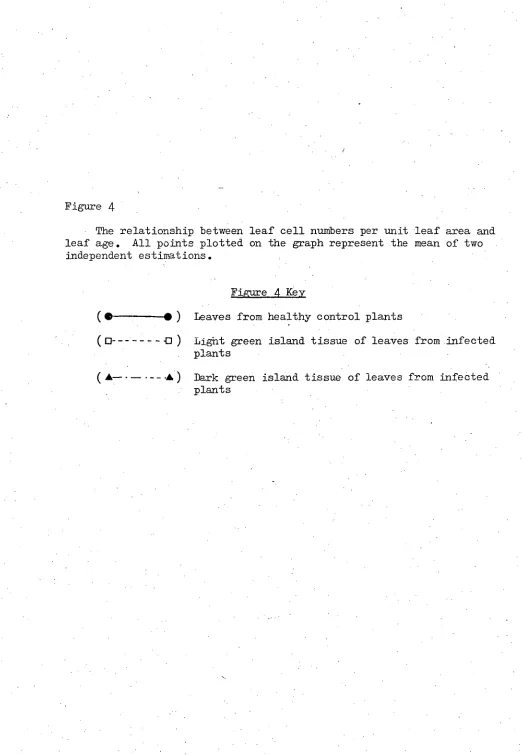
3. Determination of Fresh Weight per ,Unit Leaf Area .. 0• 54 4. Estimation of Leaf Cell Numbers per Unit Leaf Area. -54
5. Leaf Tissue Preparation for Light Microscopy. 01 OS 00 00 55
6. Trial to Determine the Effect of Virus Infection on
Flowering Response 0 O• 00 •• •0 •• •• O• •• •• •CP •• 00 55
RESULTS AND OBSERVATIONS.. 0. .0 0. 64. 00 00 04 04 00 00 40 00 57
1.: Plant Size as Affected by Virus Infection. •. 0. •. .. .. 57
2.. Leaf Growth and Development Following Infection. .. 00 00 60
3. Effect of Infection on Leaf Cell Sizea• .• •0 •• •• •0 •e 64
4. Effect of Infection on Leaf Fresh Weight 0. 00 0• 05 0. 00 64 5; Leaf Tissue Age and Virus Concentration .. .. •0 0. •. .. 64
6. Virus distribution Within the Mosaic .• .. .. .. .0 .. • 0 69
7. Observations on Flowering Response as Affected by Virus
I
nfection 0. 00 .. 0. 00 .0 0. .. .0 .. .. .. ..a a 0. 60 69SECTION II
VIRUS-HOST BIOCHEMICAL INTERACTIONS
A. ENZYMES AND REACTIONS OF CELLULAR METABOLISM
I
NTRODUCTION., oo •o oo oo a. as is OS o• o• so .4 so,o. a. es 73 MATERIALS AND NEFHODS.. 0. •0 . 041 00 00 •0 00 00 01 . 11... 00 00 0 . 0 • 0 00 76 1, Crude Leaf-Tissue-Enzymes Preparation .. 00 00 00 00 00 00 76 a) Purification and Concentration by Acetone. •• .0 •. 76.. b) Purification and Concentration by Dialysis .0 00 OS ' 77
2,Acrylamide Gel Electrophoresis 40 40 00 00 40 •0 •0 '00 .0 77
3. Assays for Enzymes Separated : by Electrophoresis . 40 •• 0 : 0 80
(a) phosphatase•O •.0 O• O• Cf• •0 •0 •0 •• O• Oa is 80
(b) Rib onuclease 0. .. e• 00 00 00 00 • •0 00 00 04 00 81 (c) 04.amylase... .0 0. 00 00 40 04, 04 00 00 111. .•• 00 • 0 81 . (d) Esterase • • • .0 O• 50 O• 140 O• O• O• •• 11.0 O• •• •• 81
_ • .... O• •• •0 O•
(e) Leucine amino peptidase. .. . O• •• 82
Table of Contents (cont.)
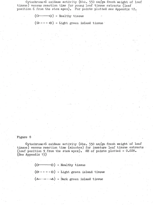
4. Direct Assays for Enzymes in Crude Tissue Extracts. (a) Phosphoglucoisomerase •. •• .• • .. • 00 (b) Glucose-6-phosphate dehydrogenase • .. .. .0 - .. (c) Alcohol dehydrogenase .. .. .. 4.0 •• 4•0 se moo (d) Gytochrome-C oxidase. •• .. 0. 0. 04 00 Os 00 (e) Glutamate dehydrogenase and NADH oxidase.. 0. (0 Ribulose 1,5-diphosphate carboxylase • • 0.,
5. Enzymes Assayed In Situ. 00 00 •0 OS •0 •0 •• 00 •0
Page Number .. .. 83 00 00 83 00 83 .so 0. 84
Se 00 84 041 00 85 P. os 86 00 0• 87 .• 0• 87 .. .• 87
0 •• 89
(a) Nitrate reductase. .• .• • •. •0 •. ...•
6. Chlorophyll Determinations. •• 0• .• .. .• •• .... RESULTS. • 0 O• •• O• O • •0 •• • • • • 0 • •• •• •Cr •• •• 0 •
1. Effect of Virus Infection on Enzymes of the Respiratory
Chain and Mitochondria.. .. .. 0. 00 00 00 00 00 00 00 • 0 89 a) Cytochrome-C oxidase. •• • • •. •• •• ••
b) NADH oxidase
0 0. .
.0 •• •• .• •a •• a,. 0.. •• .• .•
.• •0 •• •• 89 93
(c) Glutamate dehydrogenase. 00 00 00 00 SO 00 00 00 040 93
2. Effect of Infection on Enzymes Associated with
Carbohydrate Metabolism. GO 041 00 00 •0 04 00 00 00 • 0 . 100 (a) Phosphoglucoisomerase • .. fre 00 04 00 00 00 00 0 0 100 (b) Glucose-6-phosphate dehydrogenase •. •44 SO 00 00 00 100
(c) Alcohol dehydrogenase •.. 0. .. .. 00 04 00 •0 00 Oe 104
3. Effect of Infection on the Photorespiratory Enzyme,
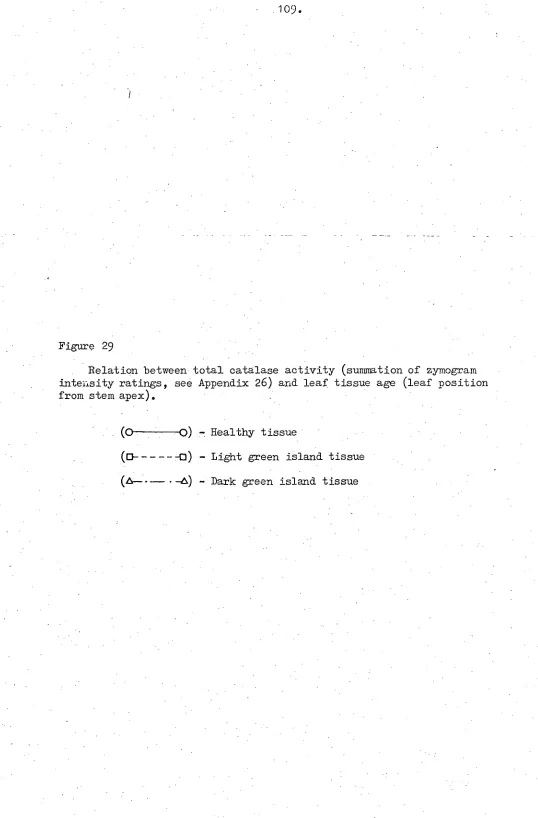
Catalse.. .• .. •. .• a• •• .• o• .. a• •• 0..• .. as .• 107
4._ Peroxidase.. • • 0. .. .. .. 00 00 00 00 00 00 00 00 0.o 107
5. Effect of Infection on the Host Photosynthetic Apparatus. 111 (a) Rdbulose 1,5-diphosphate carboxylase .. 00 00 00 00 111 (b) Chlorophyll "a" 00 •• •• 00 410 00 00 •0 00 30 00 00 111 (c) Chlorophyll a : chlorophyll b ratios " " e . 0. OS 115
6. Effect of Infection on Nitrate Reductase Activity 0. .. 0. 117
70 Effect of Infection on the Acid Hydrolase Group of
E
hzymes. • .. 0. 04. •o OS 0• o• 00 00 00 00 O. 00 00 00 00 119 (a) Acid phosphatase.. .. 00 00 00 • 00 00 0• 0• 00 0• 119 (b) Ribonuclease .. .. .. .. • .. .. .. .. .. 00 00 00 122 (c) c.4-amylase.. 04 00 • 00 00 041 00 041 Op 00 00 00 00 122 (d) Acid esterase 00 00 •0 00 00 00 00 00 00 00 00 00 00 127 (e) Specific protease-leucine amino pepiidase • 00 00 00 127B. ENDOGENOUS HOST HORMONES
INTRODUCTION Op 00 OS •0 00 00 00 00 00, 00 410 00 • '00 00 . 00 .410 00 130 •• MATERIALS AND METHODS..
1. Extraction and Partial Purification of Endogenous
Growth Regulators. • .. 00 04 00 00 00 00 00 00 00 00 00 133 (a Gibberellic acid and abscisic acid .0 00 00 00 041 00 133 (b Cytokinins 0. toe oe *As • too se Odi 0411 04 oo oo es 0. 134
(c) Auxins .. •. .. .. 0. 0. of. •• 00 0. oil 00 00 50 09 135
• Separation of Growth Regulators by Thin Layer -
Chromatography • 00 fse si, 00 so es 0* 00 0... 0* se flo
(a) Gibberellic acid and abscisic acid.. ... .. 0. s• .. ol, oil 135 135
.(i) General conditions of chromatography. .. (ii) Chromatography of pure compounds and
Table of
RESULTS.,
3. Physiochemical
4. Biochemical -
Contents.(cont.) Page Number
Cytokinins. • •0 •0 • 00 00 •0 00 0• 00 00a00 00 00 137 (c) Auxins 00 0• 00 00 00 SO OS •0 00 00 00 •6 •0 60 137 Determinations of Growth Regulators. 00 0.0 138 (a) Gibberellin A3 0 • .. 0. .. .0 00 60 00 00 00 ea 138 b) Abscisic acid.. • 0. 00 00 • • 00 • 00 00 • 00 138 c) Indole acetic acid 00 00 • • 00 00 00 00 00 Os 00 138 Assays of Growth Reguators. 00 00 00 00 00 00 139 (a) Gibberellic acid. 0 0. .. 0. 00 Os 0a 00 00 00 00 Oe 139. (i) Barley endosperm bioassay .. 0. 00 SO 00 00 00 139 (ii) Rumex leaf disc bioassay ... .0 ••• •• • •. .. 140 (b) Abscisic acid.. .0 00 0. .. 00 00 SO 00 Oe ee 00 Oe 141 (c) Cytokinins00 • 00 66 60 00 00 00 00 00 06 00 00 00 142
i) Amaranthus-betacyanin production bioassay.. .. 142
(ii) Tobacco pith callus bioassay. • -.0 0 • • • 00 • • 143
(d) Auxins (Indole Acetic Acid) Oe 00 •e •0 •0 00 04. so 144 0. 00•00 .0 0. 0. 00 0 00 00..0 00.0. 00.00 0.0 00.146 I: Gibberellin A3-like Activity in Extracts from Healthy
and Virus-Infected Plant Tissues. 0 • 00 00 • 0 . • 0 • 0 • 00 148 (a) GA3 -like activity in extracts from young leaves .
and shoots •. 0. .. .. 00 00 SO 0* 00 • 00 00 00 149 (b) GA3 -like activity in tissue extracts from mature
leaves • .. 0. 0• 0. 00 •e SO 04, fre 00 00 00 00 00 149 - 2.'ABA -like Activity in Nature Leaf Extracts from Healthy
_and Virus-Infected Plants.. .. .. •• 0. • • 00 00 •0 •0 149
3, Cytokinin Activity in Extracts from Healthy and
Virus-Infected Plant Tissues.. •• •e .• 0• 00 .• o• •• ... 0• •• 157 (a) Cytokinin activity in young leaf and shoot
extracts. 0. • 0. .., 410 00 *0 00 00 00 • 00 • 00 158 b) Cytokinin activity in mature leaf tissue extracts.. 162 c) Cytokinin activity in root tissue extracts •• 00 .0 165
4, The Effect of Virus Infection on the Concentration of an Indole Acetic Acid-like Component in Young Leaf and
Shoot Tissue Extracts • 0• 0. .. .. 00 60 66 00 00 00 00 170 SECTION III
VIRUS-HOST-CELL UITRASTRUCTURAL INTERACTIONS
INTRODUCTION° • 0• ••.• a• a• •0 •• •• 0••a• ••.•• •• •• o• ••.• •• •• 174
MATERIALS AND VETHODS 0. 0. • 60 00 • .00 00 00 • 00 00 00 06 175 I: Tissue Preparation •• •.0 00 04. Oe 0.0 00 00 00 00 00 00 es, 175
a) Araldite -embedded tissues.. 0. 0. 0. .... 0. 00 •0 0e 175 • I)) Glycol methacrylate-embedded tissues 0. 0. 0. .. 00 175 I ....
2. Sectioning and Post-Staining Techniques • 3o 00 00 00 176
RESULTS AND OBSERVATIONS.,
1. Chloroplast Structure and General Cellular Organization
• (d) Senescing leaf tissues.. • 0 • 0 • 0 • 00 • 0 • • • •
Some Miscellaneous Observations of Virus-Infected and
6 0 1
99
Healthy Tobacco Leaf Cells. .. 0 0. .0 00 • •. 2.02 DISCUSSION'. .. 00 • 4 00 • o op• •• SO • 0 •• 0.6-00 205 BIBLIOGRAPHY.. 0. 0. 0 • 0 • 0 • • S • • 0 • 0 • 0 • 0 • • • 0 • •• •• •• 00 226
LIST OF PLATES Page Number Plate Plate Plate Plato Plate Plate Plate
7 Uninfected and TMV infected tobacco plants - Mature leaves from uninfected and infected
tobacco plants
3 Nature leaves from uninfected and infected tobacco plants
4 , Young and immature leaves from uninfected and infected tobacco plants
Section through leaf of uninfected tobacco plant
- Section through leaf of infected tobacco plant
7 .Section through border between light green - island and dark green island tissue of
infected tobacco leaf
58 62 62 63 66 66 71 71 71 72 72 78 78 179 180 181 182 183 183
Plate 8 - Apical meristem from virus infected tobacco plants, 12 weeks old
Plate 9 - Apical meristem from uninfected tobacco plant, 12 weeks old
Plate 10 - Apical meristem from an uninfected tobaueu plant, 12 weeks old
Plate 11 - Uninfected tobacco plants, 15 weeks old Plate 12 - Infected tobacco plants 15 weeks old Plate 13 - Electrophoresis power unit and buffer tank Plate 14 - Electrophoresis gel mould
Plate 15 to 50 electronmicrographs of virus infected and uninfected tobacco leaf cells.
Plate 15 - Virus infected leaf, 1 cm long Plate 16 - Uninfected leaf, 1 cm long Plate 17 - Young infected leaf, 2 cm long
Plate 18 - Virus inclusions in cells of young infected leaf, 2 cm long
Plate 19 - Plastids in virus-infected tobacco leaf cells, 2 cm long
•
Plate 20 - Epidermis of uninfected leaf, 2 cm long Plate 21 - Cells of dark green island tissue in virus
Plate 22 - Cells of uninfected leaf, 2'cm long 185
Plate 23 - Plastids in cells of uninfected leaf , 2 cm
long 186
Plate 24 - Plastids in cells of infected tobacco leaf,
2 cm long 187
Plate 25 - Plastids in cells of uninfected tobacco leaf,
2..cmlong 187
Plate 26 - X-body in cell of infected tobacco leaf, 2 cm
long 188
Plate 27 -,X-body in cell of tobacco leaf, 2 cm long 188
Plate 28 - Cells on the border between light green and
dark green island of infected, immature leaf 190
Plate 29 - Cell wall boundary between cells of adjoining
light green and dark green islands 191-
Plate 30 - Plasmodesmata connections between cells of
adjoining light green and dark green islands 191
Plate 3 - Cells of dark green island tissue of immature
infected leaf 192
Plate 32 - Chloroplasts in cells of uninfected immature
.leaf 193
Plate 33 7 Chloroplasts in cells of uninfected, immature
leaf 193
Plate 34 Chloroplasts in cells of uninfected immature
leaf 194
Plate 35 - Chloroplasts in cells of dark green island
tissue of infected, immature leaf 194
Plate 36 - Chloroplasts in cells of light green island
tissue of infected, immature leaf 194
Plate 37 - X -body in cells of infected, immature leaf 195 Plate 38 - Cells of mature, virus-infected leaf 197
Plate 39 - Cells of mature uninfected leaf 197
Plate 40 - Chloroplasts in cells of mature, virus-infected
leaf 198
Plate 41 t Chloroplasts_in cells of mature, uninfected leaf 198
Plate 42 - Chloroplasts in cells of senescing, virus-
List of Plates (cont.)
Plate 43 - Chloroplasts in cells of senescing, virus-infected - leaf
Plate 44 - Chloroplasts in cells of senescing, virus-infected leaf
Page Number
200
. 201
Plate 45 Plate 46 - Chloroplasts in cells of senescing,.
uninfected leaf 201
Plate 47, Plate 48, Plate 49 - Single membrane-bound
I wish.to express my gratitude to my supervisor, Professor G.C. Wade,
Mean of the Faculty of Agricultural Science and Lecturer in Plant Pathology,
for his guidance and constructive criticism throughout the course of this
study.
I express my thanks to Professor K.C. Marshall, former Reader in
Microbiology, for beneficial discussion and Dr. R.C. Mnary, Senior
Lecturer_ in Horticultural Science, for constructive criticism
on_the_pres-entation of certain aspects of this study. I also wish to express my thanks
to Mr. R. Cruickshank, Research Assistant to Professor G.C. Wade for many
relevant discussions on the content of this work and for technical advice
and assistance.
Vany other people contributed to the work of this thesis. In
partic-ular, I wish to thank Mr. R. Davies former Electron Microscope Technician
for the Department of Zoology and IL: 4 D. Munro Plant. Pathologist in the
Tasmanian Department of Agriculture for valuable technical assistance with
respect to electron microscopy. I also wish to thank Mr. J. Groot,
Glasshouse Technician, for his services in maintaining plants used during
the course of this study and Mr. W. Peterson, Head of the Technical Staff,
for his willingness to help at all times.
Finally, to my wife Robyn, I extend my special thanks for her support
(ii)
SUNWARY
A Susceptible tobacco variety infected with a mosaic-inducing strain of tobacco mosaic Virus displayed certain alterations in normal growth patterns. This work confirmed that such plants, as a result of infection, were reduced in plant height, internode length and leaf size. Infected plan -Es also had reduced root systems. The rate of leaf growth., that is the time taken for leaves to reach full expansion size, was similar for infected and virus-free plants although the number of leaves formed over
a 6 'week period following inoculation was slightly greater for virus- _
• infected plants. Infection of young plants delayed both flower initiation
and the appearance of inflorescences. A reduced rate of cell division in sub-apical regions appeared to be the major factor in reducing plant size.
The survival of plant supporting virus synthesis appeared to be rel-ated to certain metabolic changes. Specific enzymes associrel-ated with pho-torespiration and "dark" respiration had lower activities in
Virus-infected plants.
mosaics.- Biochemical and ultrastructural studies revealed that over-mature leaves onvirus-infected plants were metabolically active and com-posed of cells containing intact membrane systems. Most enzymes studied
in similarly. aged leaves from uninoculated plants had greatly reduced activities and cells in these tissues contained membrane components that showed signs of deterioration and disorganization. Virus-free areas of
-mosaic diseased leaves, referred to as dark green island tissues, were
Metabolically more active than comparably aged tissues from uninfected plants. Enzymes associated with photosynthesis and carbohydrate metabo-lism and photosynthetic pigments were at greatest levels in these tissues. Of the three tissue types compared, virus-containing and virus-free
tis-sues from infected plants and leaf tistis-sues from uninfected plants, dark green island tissues had the greatest potential for photosynthesis and Carbohydrate metabolism.
VIRUS-HOST BIOCHEMMAL INTERACTIONS
Plants systemically infected with viruses generally undergo changes in pigment production, growth development or both. In some cases virus infec-.tions.are lethal. It is well documented that infections of host plant's with
viruses produce disturbances in host-plant metabolism. However the nature of Such disturbances is almost as varied as 'thenumber of virus/host plant combinations reported in the literature - . Some attempts have been made to correlate symptom expression with disturbances to various metabolites, cel-lular enzyme activities, host physiological changes and the reflected mor-phological changes'. The major problem associated with a review of this nat-ure is that most reports in the literatnat-ure concerning measnat-urements on cel-lular biochemical functions have tried to relate concentrations of various metabolites or enzyme activities to host tissue resistance or susceptibility to infection.
The purpose of this review is to attempt to express the findings of these various .reports in terms of host reponse to infection.
A. ENZYMES AND SUBSTRATES OF CELLULAR METABOLISE
1. The Respiration of Tissues Infected by Virus
Porter (1959) concluded that different viruses have different effects on:respiration and that effects on respiration can vary between different hosts infected with the same virus. The physiological state of the tissue
involved is So important that it requires a very strict definition of exp-erimental conditions when evaluating the findings' of such studies. However, provided some differentiation is made with respect to age of_infection and type, some broad generalizations can be reached.
) Respiration rates following infection
Increased respiration rate occurs for inoculated tobacco leaves for three weeks following inoculation, whereas young leaves present at the time of inoculation showed no increase (Owen, 1956). Initial increases In resp-iration rate, following infection of tobacco epidermis with tobacco mosaic virus, have been reported by Yamaguchi and Takahashi (1964) and Takahashi and Hirai (1964). Burroughs, Goss and Sill (1966) measured increased resp-iration for barley following infection with brome-grass mosaic virus. They also reported
a
similar increase in respiration rate for mature barley lea-vest seven days after infection with barley yellow dwarf virus. However, Jensen (1968) showed that respiration, of leaves not fully mature at the time of infection, was unaltered.(b) Respiration rate of tissues systemically infected by virus Mny researchers have reported divergent and apparently contradictory results on this type of infection.. M./Tett and Bayley (1969) suggested that the greatest reason for this variation was the nature of expression of res-ults, that is, whether final respiration rates were expressed on a dry or fresh weight basis or on protein-nitrogen, carbohydrate content or some
other parameter. All Of the preceding parameters can be secondarily changed as a result of Infection. Owen (1955b) pointed out that infection could alter water content, dry-matter content and, growth rate of infected leaves. The effect of virus on respiration would therefore depend on the basis used
to express results.
of cucumber infected with cucumber mosaic virus (Rubin and Zeleneva, 1904) and for barley leaves formed after infection.of plants with barley yellow dwarf virus (Jensen, 1908).
For some virus/host combinations the initial increase in respiration, following inoculation, declined with time after infection. With Maize dwarf mosaic virus on maize Tu and Ford (1968) found that respiration rate inc-reased by
0%
six days after inoculation. After nine days, the rate was 32% higher. At day eighteen the rate had declined but still remained higher compared with healthy tissue. However, the authors pointed out that much of the respiration increase was due to mechanical damage caused at the time of inoculation. A similar increase in respiration, followedby
a decline was reported for N. rustica L. Infected with alfalfa mosaic virus (Verhoyen, 1966; as cited by'Vbrrett and Bayley, 1909).In some cases, the decline in respiration rate with age of infection has proceeded until respiration rate had fallen to below that of comparable healthy tissues (Owen, 1956; Takahashi and Hirai, 1964). Although'respir-ation rate of barley yellow dwarf virus-infected barley plants increased, following inoculation, it subsequently declined and from thirty five to sixty nine days after infection it was below that for comparable healthy tissues (Orlob and Amy, 1961 . ).._ Respiration rates of virus-infected tis-sues that are below rates for comparable healthy tistis-sues have been recorded for young tobacco leaves that were formed after infection by tobacco mosaic virus and were showing systemic symptoms (Owen, 1955b; Owen, 1956). Resp-iration rate was lower in tomato stem tissue infected with tomato aucuba mosaic virus (Rbrrett, 1960).
• types of respiration cOUld be,affected differently
by
infection, the wayin which respiration has been measured could.also be a factor contributing to the conflicting reports concerning the effect of virus infection on respiration rate of tissue's systemically infected by virus.
(c) Respiration rates of local necrotic lesion hosts
The investigation of several host-plant/virus combinations that res-ulted in local lesion formation has shown that at some stage in lesion dev-elopment there is an enhancement of host-plant respiration (Nferrett and Bayley, 1969). Increased oxygen consumption in leaves of Nicotiana
sylvestri6 L. occurred following inoculation with tobacco mosaic virus (Parish, Zaitlin and Siegel, 1965), Similar increases in oxygen uptake', following tobacco mosaic'. infection have been recorded for the host plants N. putinosa L. (Yamaguchi and Hirai., 1959), and N. tabacum L. Xanthi
(Sunderland and Perrett, 1965). Increases in respiration rate have also been reported for-Phaseolus vulgaris L. infected with southern bean mosaic virus (Chant, 1967) and for potato virus X-infected N. tabacum L. cv. White Burley (Owen, 1958). The rise in respiration appears to be more related to the development Of disease symptoms than to an increase in virus concen-tration (Nbrrett and Bayley,
1969).
This has been most conclusively dem-onstrated with those virus/host combinations where, under certain condit-ions, the combination does not produce a necrotic lesion. Under these cir-cumstances the respiratory increase is small (Sunderland and Nferrett, 1965; Parish, Zaitlin and Siegel, 1965). It has been suggested that the increa-sed respiration rates of infected tissues was due to increaincrea-sed numbers of mitochondria (Weintraub, Ragetli and Ewurazna, 1964), However, more rec-ent work byPierpont (1968) failed to confirm this report.A recent report by Simons and Ross (1971) suggests that respiratory increases following inoculation are not permanent. Tobacco mosaic virus 'infection, of the tobacco variety Samsun NN, resulted in increases in
respiration. However, with older infections these_i=reases and activities - declined,
2. Respiration Substrate Concentration in Virus-Infected Tissues Mhitehead (1934) suggested that the .higher respiration rate of pot-atoeLi infected with potato leaf toll virus, Was related to the amount of • available substrate. He pointed out that the accumulation of starch in leaves of diseased plants occurred at a very early stage of development.. Such accumulations could be delayed by exposing plants . to continuous light Of low intensity. Under these conditions the respiration of diseased tis-sues approximated to that of healthy ones.
Mich work has been published since then concerning substrates which . can be utilized in respiration for the' ultimaterelease of carbon dioxide.
As pointed out by Vbrrett and Bayley (1969) a major problem in correlating substrate concentration with respiration rate is determining which of the number of compounds present in the plant is providing the carbon ultim-ately released as carbon dioxide. Most polysaccharide reserves can be bro-'. ken down to their constituent sugars which, in turn can - be converted to car-bon dioxide . via the EMbden-N6yerhof-Parnas pathway, or the pentose, mono-phosphate- shunt pathway, followed by the tricarboxylic acid cycle. .Infor-mation on the concentration of ntermediates in these pathwaysiparticul-arly the sugar phosphates, would provide Useful information. However, Werrett and Bayley (1969) pointed out that determinations of sugar.phos- . phates, in plant tissues, is difficult because of their low concentrations..
Troteins can be broken down to' their constituent amino acids - and their.car-bon skeletons released as cartheir.car-bon dioxide following deamination or trans-amination. Fats can .ultimately be broken down to acetyl co-enzyme A which • in turn can .be fed into the tricarboxylic acid cycle.
3. Carbohydrates
6 .
carbohydrates in the cell. Photosynthetic rate and translocation have a direct effect on carbohydrate concentration and both rates may be affected by virus infection (Nbrrett and Bayley, 1969).
Bowden (1964) observed that, with yellows diseases of plants, the Carbohydrate/nitrogen ratio increased, whereas mosaic diseases often dec-reased the ratio. Whitehead (1934) reported incdec-reased starch in leaves of potato infected with potato leaf roll virus. A similar increase in carb-ohydrates was reported for sugar beet leaves infected with beet yellows virus (Watson and Watson, 1951).' In barley, leaves infected with barley yellow dwarf virus, - accumulation of starch and soluble carbohydrates, esp-ecially reducing sugars', occurs. Soluble carbohydrates were reduced in the roots of infected plants (Orlob and Amy, 1961). - Diseases of the yel-lows type affect mostly, the phloem of the host (Esau, 1967). It has been suggested that for these diseases, 4 virus-induced stimulation of respir-ation could be maintained. in the leaf because of increased availability of substrate(Nbrrett and Bayley, 1969).
Mynd (1943) reported a decrease in carbohydrates for plants infected with mosaic virus diseases. -However, he concluded that no definite conc-lusions.dould be reached concerning the general behaviour of the total car-bohydrate fraction of plants to virus infections. Alfiatthews (1973) conclu-, ded that turnip yellow mOsaic virus infection of Chinese cabbage reduced sugars by diverting carbon fixed in the Calvin cycle from sugars to org-anic -acids. Increased rate of carboxylation of phosphoenol pyruvate to - form oxalacetate, resulted in higher levels of malate and aspartate being
forMed.
There are few reports concerning the effect of virus infection on -pentose sugars. Dunlap (1931) found that healthy tobacco leaves contained
0.27% pentoses (expressed on a leaf fresh weight basis) while infected leaves contained 0.21%. Pentoses were reported to accumulate in areas
sur-rounding local necrotic lesions caused by tobacco mosaic virus on tobacco (Farkas-and Solymosy, 1962).
4.
Organic AcidsThe oxidation of organic acids in the tricarboxylic acid cycle and the associated coupling of this cycle to respiratory chain phosphorylation . provides the main source of ATP for plants in the dark. Organic acids also
provide carbon skeletons for amino acids.
Increases in organic acids following virus infection have been repor-ted. Porter and Weinstein (1957) found more malic and citric acid in tob-acco leaves seven days after inoculation by cucumber mosaic virus. This
increase appeared to be correlated with 4 virus-induced stimulation of 14
growth. When plants were exposed to CO
2 following inoculation, less iso-tope was incorporated into malic'acid and about the Same amount into cit-ric acid compared with healthy leaves (Porter, 1959). For Physalis floridana, N. tabacum L. cv. Mite Burley and Lycoperisicum esculentum systemically infected with potato virus X potato virus Y and tobacco mos-aic virus increases in leaf oxalate, malate and citrate occurred .(Venekamp, 1959). Similarly, Schuster (1964, as cited by Mrrett and Bayley, 1969) reported increases in.succinic malic and citric acids in Nicotinia species
infected with tobacco mosaic virus and Hyocyamus species infected with pot ato virus. X.
Some reports suggest that virus multiplication is stimulated by org-anic acids. Tobacco mosaic virus multiplication increased in tobacco leaf discs floated on distilled water plus one of the organic acids, citrate,
8.
(1956a) found that the susceptibility of French beans to tobacco necrosis virus increased when the plants were kept in the dark. Dark treatment dec-reased the content of malic fumaric, succinic and glycolic acids and inc-,reased citric acid without affecting oxalic and malonic acids. None of
these acids had much effect on virus synthesis when infiltrated into leaves. Dark treatment of leaves of French bean and tobacco increased their Suscep-tibility to tobacco necrosis virus and tomato aucliba mosaic virus respec- tively, but decreased the level of ascorbic acid. However, infiltration of
_
leaves with ascorbic acid also increased their susceptibility to these vir-uses (Wiltshire, 1956b). Schlegel (1957, as cited by Porter , 1959) found
that tobacco mosaic virus concentration increased 50% in tobacco leaf discs floated on solutions of organic acids. The effectiveness of organic' acids . was dependent on adequate nitrogen fertilization of plants prior to inoc-ulation. It was not known whether the organic acids were effective indir-ectly as energy sources or dirindir-ectly as precursors in virus synthesis.
5.
Enyme Systems Associated with "Dark" Respiration(a) Embden-Neyerhof-Parnas pathway and pentose phosphate pathway enzymes
Generally little change has been reported for the activities of enz-ymes of 'glynolysis, following virus infection'. Boser (1958) looked at pot-ato leaves and tubers systemically infected with potpot-ato leaf roll, streak virus, or mosaic virus and:found that streak virus had no effect on phos-phoglucomutase, hexokinase and .enolase activities. Leaf roll and mosaic viruses caused slight increases in activity of hexokinase in tubers, and enolase in leaf tissues. The activities of phosphoglunomutase and enolase: were reduced in leaf roll infected tubers„
In leaves of N. tabacum L. cv,.white Burley locally infected with
tob-acco mosaic virus, the areas immediately surrounding lesions showed inc-reased glucose -6-phosphate dehydrogenase and 6-phosphogluconate
dehydrog-enase activities. No change in activities occurred for phosphohexoisom-
'phosphate pathway could not cope with strongly activated early stages ,(Farkas and Bolymosy, 1962). For potato leaves systemically infected with
potato leaf roll virus, decreased activity of glucose-6-phosphate dehydro-genase was found to occur (Boser, 1959). Takahashi (1971) found that in: tobacco leaf epidermis, systemically infected with tobacco mosaic virus, the activities of glucose-6-phosphate dehydrogenase and 6-phosphoglucose dehydrogenase were unaltered. Reddy and Stahmann (1970) reported new iso-enzymes for glucose-6-phosphate dehydrogenase in plants infected With pea wilt virus. No quantitative or qualitative changes in 6-phosphogluconate dehydrogenase were observed.
Some attention has been given to the relative roles of the Embden-Neyerhof-parnas pathway and the pentose phosphate pathway in tissues inf-ected with virus. Solymosy and Farkas (1963) reported that while key enz-ymes of the pentose phosphate pathway (G-6-P dehydrogenase, 6-phosphoglu-donate dehydrogenase) increased in activitiesi enzymes of the glycolytic pathway (hexokinaSe, glucose phosphate isomerase) showed unaltered activ-ities in local-lesion hosts. The greatest increasein'pentoae phosphate pathway enzymes appeared to occur in tissues surrounding lesions.
Bell (1964) found that whenlesions appeared on bean plants'inocu-lated with southern bean mosaic virus, the GO/C1 ratio . (ratio of sugars of the Ebden-Neyerhof-Parnas pathway to sugars of.the pentose phosphate pathway) decreased sharply, suggesting that more glucose was being
meta-bolised via the pen-Lose phosphate pathway. However, although Nerrett and 1
Sunderland (1967) reported increased release of 4C0
2 from glucose-6- 14C and glucose-1- 140 for a local-lesion tobacco host infected with tobacco mosaic virus, there was no change in the ratio COO'', with virus infection.
10.
the period of maximum virus increase for tobacco leaves infected with tob-acco mosaic virus. Bell (1964)1 on the other hand, found that the C6/C1 ratio increased when beans were systemically invaded with southern bean mosaic virus. No change in the G6/C1 pathway ratio was reported by Baur et al....(1967)s,- for the systemic infection of tobacco by tobacco mosaic virus. They found that approximately 80% of respiration was mediated via the Embden-Nbyerhof-Parnas pathway and 20% via the pentose phosphate path-way. Nerrett and Bayley (1969) concluded that sufficient evidence did not exist to support the hypothesis that there was a shift in respiratory met-abolism from the Embden-Neyerhof-Parnas pathway to the pentose phosphate pathway.
(b) Tricarboxylic acid cycle and enzymes associated with the mitochondrion
Isolated mitochondriaare capable of carrying out all the reactions of the tricarboxylic acid cycle. However, counterparts of several of the mitochondria' enzymes are present in the soluble fraction of the plant cell. Nerrett and Bayley (1969) pointed out that in general the levels of a num-ber of mitochondria' enzymes and carriers, such as cytochromes, succinate dehydrogenase, malate dehydrogenase, appeared to be present in constant relative proportions in mitochondria from widely different sources. .They .suggested that a virus-induced change in the total amount of one of the
enzymes could result in a change in the amount of other enzymes if the constant relative proportions of the enzymes was to be maintained.
Takahashi and Hirai (1966) pointed out that, although chloroplasts were the major site of ATP generation and amino acid synthesis in green leaves, mitochondria were also capable of amino acid synthesis and incorporation • into proteins. In fact in those tissues devoid of chlproplasts, mitochon-dria were the major source of amino acid synthesis.
of this cycle suppressed synthesis of tobacco mosaic virus (Ryzhkov,
1957,
as cited by Diener, 1963); Takahashi and Hirai 1966) reported that mito-- -
-
chond ria of infected plants gave increased uptake of C-leucine. The- 14 authors suggested that amino acid uptake represented increased protein syn-thesis by the mitochondria, although it was not known whether this increa-sed protein was a bound, fixed mitochondrial type or asoluble type. No changes in mitochondrial protein-nitrogen were reported for N. glutinosa plants infected with tobacco mosaic virus (Pierpont, 1968). However, Nambier and Ramakrishnan (1970) found increases in mitochondrial-nitrogen and succinic oxidase activity for Capsicumand
aucurbida infected with tob-acco mosaic virus and cucumber mosaic virus.Nartin (1958, as cited by Diener, 1963) observed an increase in cyto-chrome-oxidase in tobacco mosaic virus-infected tobacco leaves. Rubin and Ladygina (1972) found that for tobacco plants systemically infected with tobacco mosaic virus, cytochrome-C-oxidase.activity decreased 24 hours after infection. For the local-lesion host N. glutinosa, cytochrone-C oxidase and succinic oxidase increased following inoculation. Pierpont (1968) on the other.handreported a slight decrease in cytochrome,oxidase activity for N. glutinosa infected with tobacco mosaic virus..
6.
Phosphorvlated CompoundsPhosphorylated nucleotides provide the energy necessary for most cel-lular functions. In addition they constitute a nucleotide - pool on Which nucleic acid synthesis is dependent.
For virus-host interactions of the local-lesion.type,.an increase in ATP levels seems characteristic. Increased ATP following tobacco mosaic virus infection of resistant tobacco varieties has been reported by . Sunderland and Vbrrett (1964; 1965 and 1967) and for tobacco - etch virus
12.
of ADP appeared to occur with increased levels of ATP (Sunderland and Nerrett, 1967). Local lesion hosts treated with the oxidative phosphoryl-ation uncoupler, 2 4-dinitrophenol (DNP), which increased respirphosphoryl-ation rate of uninfected tissues, characteristically showed no further increase in respiration rate following infection (Sunderland and Nbrrett, 1967); These Authors suggested that virus infection brough about an uncoupling of res-piration from oxidative phosphorylation, as reflected by a decreased ADP/ _ ATP.ratio, Nbrrett_and Bayley (1969) proposed that in local lesion
reac-tions, high respiration rate could be due to either ADP availability not being limiting or respiration proceeding without oxidative, phosphorylation. Certain pathogenic fungi have been shown to produce toxins that have a
sim-ilar uncoupling effect on respiration to DNP (Krupa, 1959). However, as
-lbrrett and Bayley (1969) pointed out, no such uncoupling agents have been
demonstrated in virus-infected tissues. It has been suggested that ATP concentration plays a role in viral resistance for local-lesion hosts. Bayley and Nbrrett (1969), in an investigation of the effects of tobacco etch virus on tobacco found that addition of adenine increased the level of ATP in healthy tissues and increased tissue resistance to virus infec-tion. No increase in respiration rate occurred following adenine treat-ment. Also, high ATP levels and viral resistance were 'characteristic of tissues surrounding local lesions. :Rubin and Ladygina (1972) postulated that in resistant tobacco varieties, such as N. plutinosa, a maintenance or activation of energy conversion processes of mitochondria are character-istic, being Associated with increased mitochondrial ATP Content and a maintenance of inorganic-phosphorous Uptake, 24 hours after infection. The chloroplasts in such diseases also displayed enhanced. phosphorylation, ATP content and light-induced - ATPase activity.'
organic phosphorous in tobacco, leaves,
7
days following inoculation with • cucumber mosaic virus. Decreases in ADP and ATP levels were also reported in tobacco callus cultures systemically infected with tobacco mosaic virus (Sunderland and Nbrrett 1963b). Bozarth and Browning (1970) studied the diurnal fluctuations of the nucleotide pool of virus-infected and, healthy bean leaves. The concentration of ATP varied in a similar : manner for bothinfected and healthy tissues, with the lowest quantities occurring during the light and. highest quantities during the dark periods. A higher conc-entration was found in diseased tissues at all times of sampling
5
days after inoculation. No consistent differences were found in the nucleotide pool or phosphorylated nucleotides of guanosine, uridine , cytidine, AN2and ADP; Unlike local-lesion tissues, DNP has been shown to stimulate res-piration in both healthy and systemically infected tobacco tissues
(Takahashi and Hirai, 1965):
7;
Host Nucleic Acid SynthesisMost studies on host nucleic acid synthesis in virus-infected tissues have been concerned with changes that occurred immediately following inoc-ulation. The earlier reports gave details on total extractable nucleic acids, which usually included viral nucleic acid. Consequently, increases
in total nucleic acids were often reported. Both ribose nucleic acid and deoxyribosenucleic acid-phosphorous increased in tobacco leaves infected with cucumber mosaic virus (Porter and Weinstein,
1957,
as cited by Diener,1963);
A similar increase in nucleic-acid-phosphorous : for tobacco mosaic , virus-infected tobaccos was reported by Elbertzhagen (1958): Basler and Commoner (1956) divided the nucleic acids extracted from TNW-infectedat a level comparable with healthy tissues.
More recent reports suggest that the nucleic acid fractions most affected by systemic infections are associated with the chloroplasts. A decrease in this fraction has been widely reported. Mohamed and Handles (197I:) investigated the effects of tomato spotted wilt virus infection on the chloroplast ribosomal nucleic acids (rRNA) of tobacco leaves. It was found that infection led to a reduction in chloroplast (700 ribosomes. However,. chloroplast rRNA still incorporated 32R-orthophosphate while their concentration declined Incorporation of radioactive label ceased 2 days after infection. It was concluded that for this virus and lettuce necro-tic yellows virus iffErtiaas some 70 S&gradatialcf 70S ribesorml RNA =mired. On the other hand, 12 hours after the appearance of TMV symptoms chloroplast ribosomes ceased to incorporate label. Although only small net losses of
70$ rRNA occurred, synthesis of 70$ rRNA was inhibited.
Hirai and Wildman .(1969) showed that isolated chloroplasts, from TM . - infected tobaccos had a reduced capacity for the incorporation of nucleo-side triphosphates into RNA. They concluded that during the period of maX7 imum virus synthesis, chloroplast rRNA and messenger RNA (MRNA) production from chloroplast DNA ceased. Oxefelt (1971) studied the effects of 2 strains of tobacco mosaic virus on the RNA type content of tobacco leaves. Both strains were reported to strongly inhibit chloroplast rRNA synthesis although inhibition was more complete in flavum-infected than in
vulgare-infected leaves. Substantial degradation of chloroplast rRNA was Shown to occur as :a result of infection, wi .ch the amount of RNA degraded greatly exceeding the total amount of viral RBA synthesised. 'Cytoplasmic rRNA was not affected by infection, but the content of transfer RNA (tRNA) was slightly reduced.
•leaves, virus reduced host REA•synthesis competition and in older leaves . maintained REA synthesis which was necessary for viral - protein.synthesiS.
• 1/ •
•It was found, that once leaves had reached about of their final length, most parameters of growth were resistant to TNV infectioni.especially cyt-oplasmic rRNA and tREA accumulations. Ghloroplast.rREA was the only type • to show a reduction with infection. There was some evidence that
cytopla-smic rRNA'was actually higher in older leaves following infection but this was thought to be due to an inhibiting effect on rRNA breakdown rather
than a stimulating effect on synthesis.
In contrast to the above reports, Kato and Misawa (1971) suggested that cucumber mosaic virus infection of tobacco leaves suppressed nuclear DNA-dependent RNA synthesis in affected cells. It was found that cellular ribonucleic acid synthesis ceased following inoculation. They did not differentiate between cytoplasmic and organelle ribonucleic acids.
Few reports have appeared concerning types of nucleic acids other than 'ribosomal nucleic acids. Johnson and Young (1969) analysed for transfer
RNA's (RNA) in tobacco leaves., systemically infected with tobacco mosaic' virus, for periods of up to
9
days following inoculation. In maturetis-sues, infection decreased the total tRNA level, while in young tissues no change in the total amount of tRNA was found. The only tRNA species to show a change in elution profile, following infection, was that for phen-ylalanine, in young tissues. However , with increasing time after
inocul-ation, the elution profile for this tRNA tended to that which was typical for healthy tissue.
8. Non-Protein Nitrogen
16.
Both increases and decreases in a range of nitrogenous compounds have been reported for a number of host-virus Combinations. Andreae and Thompson (1950) demonstrated greatly reduced levels of tryptophane and
tyrosine in leaf roll-infected potato tubers. Allison (1953) on the other hand suggested that the only consistent differences for a number of potato varieties infected with leaf roll, were increases in glutamine and glutamic acid. Results for tyrosine and tryptophane varied between pot- ato varieties. Accumulation of pipecolic acid appeared to be a character-istic of Western-X diseased peach leaves (Diener and Dekker , 1954). Inc -
reases in free amino acids and amides, following infection have been reported by Bozarth and Diener (1963) for N. tabacum systemically infec-ted with either potato virus X, potato virus Y, or a combination of both. Studies on free amino acids and amides of intact tobacco plants after inoculation with tobacco mosaic virus, revealed net increases in . serine, glutamine and asparagine, maximum concentrations being reached 48-120 hours after infection (Porter, 1959); Harpaz and Applebaun (1961) found
increases in asparagine in maize seedlings infected with maize dwarf mos-aic virus. Increases in asparagine and glutamine were also reported, for the first foliage leaves of tomato infected with tomato spotted wilt virus
(Selman et al. 1961). Higher levels of glutanic acid glutamine and asp-aragine occurred in N. glutinosa one to two days after symptoms of lettuce necrotic yellows appeared (Handles 1971). Decreases in ammonia, free amino acids and amides, during the period of tobacco mosaic virus synthe-sis, were reported by Commoner and Dietz (1952). This report was confir-med by Commoner and Nehari (1953) who described deficiencies in glutamine, glutamic acid, aspartic acid asparagine and serine following infection with tobacco mosaic virus.
appeared in the literature. The nitrogen status of plants prior to
infec-tion may have an important effect on Whether virus synthesis occurs at the
expense of host protein synthesis or concomitantly with it. Commoner and
-Nehari (1953) and Commoner et al. (1953) concluded that most nitrogen for
tobacco mosaic virus synthesis comes from free ammonia within the cell and
that changes in non-protein nitrogen, such as amides and amino acids,
ref-lected the withdrawal of ammonia for tobacco mosaic virus synthesis. This
further substantiated an earlier report by Wildman et al. (1949) who
con-cluded from electrophoretic'studies, that TNT was synthesised at the
dir-ect expense of . a normal protein fraction. On the other hand 'Holden and
Tracey (1948) have shown that for well fertilized tobacco plants, the total
nitrogen content of TNV-infected plants was higher than that of healthy
plants. Non-protein nitrogen content has been shown to vary with the age
of infection. Elbertzhagen (1958) found deficiencies of total-nitrogen
and alcohol-soluble-nitrogen in TNV-infected and potato virus .X-infected.
tobacco leaves during the early stages of infection. However, at later
stages of infection total-nitrogen, proteinrnitrogen and lcohol-soluble",
nitrogen were higher in infected leaves. For tobacco plants infected with
potato virus X, AIR concentrations of free amino acids and amides were
lower at: the time of rapid systemic spread of the virus. In symptomless
leaves and leaves With older infection's, these pools were higher than in
•comparable healthy leaves (Nlczynski, 196'0. ! Raraseck (1963) has shown
that for tobacco systemically infected with 'tobacco mosaic virus., increa-.
sea in free amino acids, and amides occur immediately following inoculation,
followed by decreases after 216 - hours from inoculation.
' The effect of virus infection on the measured levels of nitrogenous
pools in the hot can vary with the tissue type determined. In virus
dis-eases of the yellows type, infection induced opposite effects on levels of
• nitrogenous compounds in roots and. leaves. Sugar beet plants infected
18.
in leaves compared with healthy plant.. leaves. Infected 1oo6s-
- on the other hand had higher levels of all classes of nitrogenous comp-.
• ounds especially soluble-N (Diener, 1965)„ Orlob and Arny (1961) repor-
ted similar findings for plants infected withbarley yellow dwarf Virus..
The severity of the infecting virus strain has also been shown to
induce different host responses in terms of altered amino acid composition
and inorganic nitrogen content.- For .maize infected with-maize dwarf
mos-aic virus, twelve amino acids increased, three Showed no consistent rela- .
tionship three decreased and all ammonium compounds and amides increased
compared with healthy plants.
A
more severe strain of maize dwarf mosaicvirus induced greater changes in amino acids than a milder strain(Ford
and..Tu, 1968).
Diener (1965) warned that the method of tissue extraction for amino .
acids and amides could introduce a further variability in reported.
res-ults. There are several reports of both - asparagine and glutamine accum- •
ulations in virus-infected tissues. However, glutamine.is - easily hydro-.
lysed . and since hot water or hot ethanol was used to extract amino acids
and amides, in some of the Studies where only asparagine accumulation was
reported Lhese results for asparagine may be misleading.
9.
Photosynthesis and Other Functions Associated with Chioroplasts •Chlorosis which is typical of many virus diseases, makes•it obvious
that either chlorophyll is not synthesized at the sane rate as in healthy
plants or is destroyed (Diener, 1965).. Reduced chlorophyll as a result
of mosaic infection of tobacco has been reported by Dickson 1922, as .
cited by Peterson and M6K4nney, 1938) Elmer (1925, as Cited by Peterson
and McKinney, 1938), Dunlap (1928) and Peterson and McKinney .(1938).
The effect ofvirus infection on thehost pigment system can depend •
on the type of infection.. Natthews (1973). investigated six leaf pigments
in Chinese cabbage infected with turnip yellow mosaic virus. The effects ,
viruS. Eight days after inoculationithe concentration of pigments fell to 68-85 .percent the levels in healthy tissues. This fall was attributed to a net loss of pigments.. For leaves present at the time of inoculation but systemically invaded to induce vein-clearing . symptoms u the six pigments - remained unchanged until 10 days after inoculation. Beyon4 this period pigment production became less, compared with healthy tissues due to a. . _ cessation in pigment synthesis. For tissues systemically infected and sho-wing clearly defined mosaic symptoms of light green and dark green island
tissues, dark green island tissue formed contributed to a renewed increase in chlorophyll, on a per plant basis. It was concluded that effects on leaf pigments were a secondary consequence of infection and not essential for virus replication. Kato and MiSawa (1974) found, that for tobacco leaves systemically infected with cucumber mosaic virus, chlorophyll was reduced but carotehoids increased 10 days following inoculation. An inc-rease in carotene content and a decinc-rease in chlorophyll was also reported for tobacco. plants infected With TMV (Elmer , 1925, as cited by Bawden,
1956). Peterson and McKinney (1938) on the other hand reported that car-otene content decreased following mosaic infections of tobacco.
The mechanism by which leaf pigments are reduced, following infection of a systemic nature, is probably also dependent on the type of infection and age of infection, Peterson and RbKinney (1938) reported drops in chl
orophyll, carotene and xanthophyll in mosaic diseased tobacco. Associated with reduced chlorophyll was a higher chlorophyllase activity. In healthy plants, however, the level of chlorophyliase activity was directly propor- tional to chlorophyll content.
20.
catalysing the breakdown of chlorophyll to chlorophyllide and phytol it • . was also pointed out that this enzymealso probably catalyses the syn-
thesis of chlorophyll during early leaf development.• It was proposed that the enzyme was bound in the chloroplast lamellae and that virus infection resulted in a disruption of the chloroplast, releasing the bound enzyme for chlorophyll degradation.- However, for cucumber mosaic virus induced yellowing of systemically infected tobacco leaves, Kato and Vlsama (1974) suggested that the reduction in chlorophyll was not due to the action of the enzyme chlorophyllase. The amounts of. chlorophyllides and pheophor-bibes were negligible in infected tissues. Infection also greatly red-uced . the activity of chlorophyllase. From their results it appeared that, - in tobacco leaf tissues, chlorophyll was converted to pheophytin, a form lacking magnesium in chlorophyll. The appearance of pheophytin was pre-ceded by a stimulated proteolytic activity which released chlorophyll from ajprotein-chlorophyll complex.
VITUS infection is generally accompanied by a reduction in photosyn-thetic rate.. Owen (1957) 'reporteda 20 percent lowering of photosynphotosyn-thetic rate .,for tobacco plants infected with tobacco etch virus. Infection of tobacco by either potato virus X (systemic infection) or tobacco mosaic . virus (local lesion infection) reduced photosynthetic rate 'by 20 percent,
but only after . the appearance of symptoms (Owen, 1957). Reductions in photosynthetic rate have also been reported for corn infected with maize.
dwarf mosaic virus (Tu and Ford, 1968) and barley infected with barley yellow dwarf virus (Orlob and Amy, 1961).
A possible consequence of infection is a reduced number of chloro-plastsi rather than joifiefre4(chloroplast activity. Iitiagyarosy et al. (1973) concluded that although squash mosaic virus infection resulted in fewer chloroplasts, isolated chloroplasts showed no differences with respect to products of photosynthetic CO
ribulose 1,5-diphosphate carboxylase and malate dehydrogenase. However, most reports suggest that virus infection affects directly photosynthetic pigment content and/or rates of raactions associated with carbon dioxide fixation and ATP formation by the chloroplast.
Earlier work by Robert's et al -. (1952) for potato virus X-infected potatoes and Spikes and Stout (1955) for chloroplasts isolated from sugar beets infected with beet yellows virus implied that reduced photosynthetic activity of infected tissues reflects more than reduced photosynthetic pigment. For tobacco infected with tobacco mosaic virus, the Hill reac-tion was reduced in chloroplasts isolated from diseased leaves (Zaitlin and Jagendorf, 1960). An instance of increased chloroplast activity as a result of virus infection has been reported by Gxffeau and Bove' (1965). The Hill reaction rate and both cyclic and non-cyclic phosphorylation inc- reased, in Chinese cabbage infected with turnip yellow mosaic virus during the period of rapid virus replication following inoculation. More recent work, reported by Matthews (1973) for turnip yellow mosaic virusinfection of Chinese cabbage, has shown that in young, systemically infected leaf tissues, a stimulation of phosphoenol pyruvate carboxylase and aspartate amino transferase. activity occurred. When infected plants were exposcd to
14
CO21 more label entered organic and amino acids and less label entered sugars and sugar phosphates. However, it was also found that the follow-ing proteins and enzymes remained unaltered until virus concentration had reached its maximum,'when they fell substantially:
.6es
ribosomes, Fraction I protein, ribulose diphosphate carboxylase and the overall rateof carbon fixation. Matthews (1973) also pointed out that these changes were shown to occur only in turnip yellow mosaic virus-infected Chinese cabbage. Increases in phosphoenolpyruvate carboxylase and aspartate amino trans-'ferase did not occur in tobacco mosaic virus-infected tobaccos.Narked reductions in the concentration of CO 2 -fixing enzyMe,ribulose- -
4. •
shown to occur as a result of systemic infection with some viruses. -Pratt .(1967) investigated chlorophyll content and CO 2 -fixing enzyme levels in
healthy and virus-infected leaves of five plant speciesinfected with six viruses. Reduced levels of chlorophyll in infected leaves were. always accompanied by similar reductions in 18S protein. It was suggested that these reductionsresulted from:a partial repression of the genetic mechan-ism of the chloroplast and that the repression was not necessarily a func-tion of virus concentrafunc-tion. A similar mechanism for the effect of virus infection on chloroplast function was suggested by Hirai and Wildman (1969) who concluded that ribosomal RNA and messenger RNA production, from chlo-roplast DNA, was switched off by 'unknown regulators during the period of grand TENT accumulation. The Flavum strain of tobacco mosaic virus stron-gly inhibited Fraction I protein synthesis in systemically infected tob-acco while the Vulgare strain caused less inhibition (Oxefelt, 1971). Kato and Elsawa (1974) found a reduction in Fraction I protein following
infection of tobacco leaves with cucumber mosaic virus. They proposed that the reduction was due mainly to a suppression in synthesis of the 'smaller subunit of this protein, the smaller subunit being translated from
nuclear DNA-dependent RNA.
Within leaves, chloroplasts are a prime site for ATP formation. The Hill reaction and cyclic, and non-cyclic phosphorylation in leaves increa-sed following the inoculation of Chinese cabbage with turnip yellow mosaic virus .(GoeffeaU and:Bove' 1965). However, phosphorylation and the Hill reaction were decreased in isolated chloroplasts of tobacco mosaic
virus-infected tobaccos (Zaitlin and Jagendorf, 1960). Rubin and Ladygina (1972)
uncoupling with virus-infection. They concluded that with a susceptible
.tobacco host, infection had its greatest effect on non-phosphorylation electron flow. That is, the long wavelength photochemical reaction con ,,
cerned with light-induced electron- transport and involving cytochromes "f" and"b
6 ' " was inhibited.
10.. Enzymes of PhotoresPiration
Photorespiration in a sequence : of enzymatic reactions which converts the end product of photosynthetic CO 2 fixation, _glycolate, to glycine
(Kisaki and Tolbert, 1968; Vigil, 1973a; 1973b). These enzymatic reac- - tions are compartmentalized, within leaf cells, in single membrane-bound organelles termed peroxisomes or microbodies (de Dave and Baudhuin, 1966; Kisaki and Tolbert 1968; Frederick and NewcoMb, 1969; Baker et al., 1973;
Bibby and Dodge l 1973). The initial step of photorespiration is the cxid-ation of glycolate by glycolate oxidase l. with the loss of carbon dioxide. This step is also responsible for the generation of hydrogen peroxide. Catalase, a major enzyme constituent of microbodies is involved in the breakdown of hydrogen peroxide (de Dave and Baudhuin, 1966; Tolbert
.1968; Feierabend and Beevers, 1971; Murry. et al., 1972; Baker et al. f 1973). For diseases of a systemic nature, some reduction in phOtorespiration app-ears to be associated with obvious symptom development..
Wynd (1942,. 1943) found that catalase was greatly reduced in leaves of tobacco showing mosaic symptoms. Dark green leaves were found to have
24.
1964) .. Healthy tissues and green, virus-free tissues of the mosaic had
a higher enzyme activity. Virus-infected tissues and yellow tissues of .
the variegated mutant also had reduced levels of enzyme substrate and
flavin mononucleotide (FNN).. It was suggested that chloroplasts played
an active role In the regulation of the level of glycolic. acid oxidase.
No changes in the isoenzyme pattern of catalases were detected in peas
infected with pea wilt Virus (Reddy and Stahmann, 1970).
For infections of the local lesion type, there is conflicting evidence
for the effect of infection on photorespiration. Solymosy and Farkas..
(1964) found decreased activity of glycolate oxidase in the chlorotic halo
around local-lesions resulting from infection of N. tabacum L. cv.471hite Burley's with the para-strain of tobacco mosaic virus. Pierpont (1968) on
the other hand, found no changes in activity of glycolate oxidase 40-68
hours after inoculation of leaves of N. glutinosa L. with tobacco mosaic virus. Nbasurement of enzyme activity coincided with the time of maximum
.synthesis of the virus. Although most respiratory enzymes increased then
. decreased, following infection of a local-lesion tobacco host with tobacco
mosaic virus, catalase, together with peroxidase remained higher in
act-ivity (Simons and Ross 1971).
11. PolyphenoLOxidaseS-and'Peroxidases
These two enzyupsystems'have. been extensively investigated in
stud-ies on metabolic changes resulting from virus infection. Polyphenol
oxid-..ases'were once thought to be terminal respiratory oxidases, transferring
electrons 'directly. to oxygen. However, it has since been shown that this group of enzymes has a very low affinity for oxygen (Ebrrett and Bayley,
1969). Bonner (1957) suggested that phenol oxidases functioned by
trans-ferring electrons from phenols to a cytochrome.
There have been many reports of increases in phenol oxidases following
infection of plant tissues with viruses, especially for the local lesion
type Of infection. Best (1937) reported that expressed sap from tomato
• levels of an oxidase enzyme, tentatively identified as tyrosinase. This enzyme Oxidized phenol, .catechol .quinol and tyrosine in the presence of oxygen. A. local lesion infection of Nicotiana tabacum L. - cv .."White Burley" with tobacco mosaic* virus resulted in increases in NADPH
2-dependent quin-one oxidoreductase l NADPH
2 oxidase and 0-diphenol oxidase activities (Farkas and Saymosy, 1962).
Reports of such increases following infection led to the early_hypo-thesis that quinones, produced as a consequence of polyphenol oxidase
act-ivity,.were responsible for the observed tissue necrosis (e.g.' Solymosy et al. 1959). However, more recent evidence suggests that increases in polyphenol oxidases following infection, are more a consequence of the hypersensitive reaction than the cause of it. Kikuchi and Yamaguchi (1960) demonstrated the appearance of increased 0-diphenol oxidase activity one
day after the first signs of visible lesion formation in N. glutinosa inf-ected with tobacco mosaic virus. Van Kamen and Brouwer (1964) found that 0-diphenol oxidase activity not only increased in the area of the necrotic lesion but increased throughout, leaves Of N. tabacum cv. "Samsun" locally infected with a strain of tobacco mosaic virus. Fritig and Hirth (1971) -concluded from their studies on phenyl propanoids andccUlliarins in tobacco
mosaic virus infected tobacco leaves showing a local lesion type reaction, that no significant changes in the biosynthesis of chlorogenic acid, umb- elliferone, scopoletin and scopolin occurred until at least two days after infection. High increases in these compounds appeared after lesion forma-tion. Van Loon and Geelen (1971) also concluded, from studies of tobacco mosaic virus infection of a local lesion tobacco cultivar, that changes in polyphenol oxidase activity did not precede lesion formation.
Enhanced. 0-diphenol oxidase activity has also been reported for,tis- .- sues systeMically infected with ViTUB. Vbrrett (1962) reported a peak in -
0-diphenol oxidase activity eight days following inoculation of
26.
tobacco etch virus. They recorded the greatest increase in the chloroplast fraction but suggestedthat infection resulted in the activation of inac-tiVe forms of the enzyme rather than new synthesis..
The other group of enzymes most reported in metabolic studies of virus-infected tissues is the peroxidases. Peroxidases catalysc the oxi-dation of various metabolites especially phenolics using hydrogen pero-xide as the oxidizing agent. However, little isknomn_ of the importance of peroxidases in overall cellular metabolism. Their location is mainly in the cytoplasm with some activity in the chloroplast and mitochondrial fractions.
Increased perosidase activity as a result of virus infection is well documented. Vager (1955) reported increased peroxidase activity for virus-infected leaves of tomato and tobacco. Orlob and Amy (1961) recorded sim-ilar increases for barley leaves infected with .barley yellow dwarf virus.. In addition to a general increase in peroxidase activity there are several reports of increases in the number of isoenzymes of peroxidases following
infection. TWo new isoenzymes appeared in young bean leaves infected with southern bean mosaic virus (Farkas and Stahmann 1966); The number of isoenzymes of peroxidase, in Nicotiana 'Flutinosa, increased from five to six with infections of tobacco mosaic virus and potato virus X (Bates and Chant 1969; Chant and Bates, 1970). The nuMber'of positively charged iso-enzymes of peroxidase increased in pea plants infected with pea wilt virus
(Reddy and Stahmann, 1970); However, investigations of peroxidase isoen-zyMe changes with infection for tissues of various ages haa led to-the.con-elusion that no new isoenzynes are induced as a result of infection.
Solymosy et al. (1967) stated that changes in isoenzyme patterns are a function Of the host and not the virus. Novacky and Hampton (1968)