The Zebrafish Mutants
dre, uki,
and
lep
Encode Negative Regulators
of the Hedgehog Signaling Pathway
Marco J. Koudijs1, Marjo J. den Broeder1, Astrid Keijser1, Erno Wienholds1, Saskia Houwing1, Ellen M. H. C. van Rooijen1, Robert Geisler2, Fredericus J. M. van Eeden1*1Hubrecht Laboratory, The Netherlands Institute for Developmental Biology, Utrecht, The Netherlands,2Max-Planck-Institut fu¨r Entwicklungsbiologie, Tu¨bingen, Germany
Proliferation is one of the basic processes that control embryogenesis. To identify factors involved in the regulation of proliferation, we performed a zebrafish genetic screen in which we used proliferating cell nuclear antigen (PCNA) expression as a readout. Two mutants,hu418Bandhu540A, show increased PCNA expression. Morphologically both mutants resembled the dre (dreumes), uki (ukkie), and lep (leprechaun) mutant class and both are shown to be additional uki alleles. Surprisingly, although an increased size is detected of multiple structures in these mutant embryos, adults become dwarfs. We show that these mutations disrupt repressors of the Hedgehog (Hh) signaling pathway. Thedre, uki,andleploci encode Su(fu) (suppressor of fused), Hip (Hedgehog interacting protein), and Ptc2 (Patched2) proteins, respectively. This class of mutants is therefore unique compared to previously described Hh mutants from zebrafish genetic screens, which mainly show loss of Hh signaling. Furthermore,su(fu)andptc2mutants have not been described in vertebrate model systems before. Inhibiting Hh activity by cyclopamine rescuesukiandlep mutants and confirms the overactivation of the Hh signaling pathway in these mutants. Tripleuki/dre/lep mutants show neither an additive increase in PCNA expression nor enhanced embryonic phenotypes, suggesting that other negative regulators, possibly Ptc1, prevent further activation of the Hh signaling pathway. The effects of increased Hh signaling resulting from the genetic alterations in theuki, dre,andlepmutants differ from phenotypes described as a result of Hh overexpression and therefore provide additional insight into the role of Hh signaling during vertebrate development.
Citation: Koudijs MJ, den Broeder MJ, Keijser A, Wienholds E, Houwing S, et al. (2005) The zebrafish mutantsdre, uki,andlepencode negative regulators of the hedgehog signaling pathway. PLoS Genet 1(2): e19.
Introduction
During development, proliferation is one of the key processes in the formation of an embryo, but how it is controlled spatiotemporally is still poorly understood. A tight regulation of proliferation is necessary during development and the remaining lifespan of an organism, as disrupted regulation might result in tumorigenesis. Several essential developmental signaling pathways are reported to control embryogenesis and many of these are involved in regulating proliferation in vertebrates and invertebrates. These basic developmental pathways all involve receptor ligation of
highly conserved sets of secreted peptides like the TGF-b
superfamily [1], FGF [2], Wnt [3], and Hedgehog (Hh) [4]. The Hh signaling pathway is highly conserved throughout evolution and has been documented to control proliferation [5]. In our current understanding, Hh proteins are expressed in a signaling cell, secreted and bound to the 12-trans-membrane receptor Ptc (Patched) on a neighboring cell. Upon this binding, Ptc is thought to be internalized into endosomes where it is unable to repress the activity of Smo (smoothened) [6,7]. The signal is transmitted to the down-stream proteins Cos2 (Costal2), Fused, Su(fu) (suppressor of fused), and one of the at least three members of the Gli family of zinc finger transcription factors [4]. In the presence of Hh, the Gli protein can be activated and transported to the nucleus where it activates genes mainly involved in pattern-ing, proliferation, and cell structure [8]. Multiple genes are
described to limit the activity of Hh signaling. Besides its own receptors Ptc1 and Ptc2, Hip (Hedgehog interacting protein) [9] is also expressed at the membrane in response to Hh activity. All three are involved in sequestering Hh to limit its effective range [10]. Further down the pathway, casein kinase
I (CKI), glycogen synthase 3b (GSK3b), and protein kinase A
(PKA) are involved in the processing or degradation of the Gli transcription factor [4]. The nuclear activity of Gli proteins is inhibited by Cos2 (Costal2) [11–13] and Su(fu) [14–18], which are both reported to be involved in tethering Gli in the cytoplasm, preventing overactivation of the pathway.
Hh signaling regulates multiple developmental processes in specific tissues in vertebrates and invertebrates [4]. In
Received February 24, 2005; Accepted June 23, 2005; Published August 19, 2005 DOI: 10.1371/journal.pgen.0010019
Copyright:Ó2005 Koudijs et al. This is an open-access article distributed under the terms of the Creative Commons Attribution License, which permits unrestricted use, distribution, and reproduction in any medium, provided the original author and source are credited..
Abbreviations: CMZ, ciliary marginal zone; ENU, ethyl-nitrosourea; GH, growth hormone; Hh, Hedgehog; hpf, hours post fertilization; Ihh, Indian hedgehog; ISH, in situ hybridization; LG, linkage group; MO, morpholino antisense oligonucleotide; PAS, Periodic Acid Schiff; PCNA, proliferating cell nuclear antigen; POMC, proopiomelanocortin; PRL, prolactin; PTHrP, parathyroid hormone-related protein; PTU, phenylthiourea; SSLP, simple sequence length polymorphism; TSH, thyroid stimulating hormone; TUNEL, terminal transferase dUTP nick-end labeling
Editor: Mary Mullins, University of Pennsylvania School of Medicine, United States of America
addition to the role of Hh during development, it is necessary to tightly regulate its activity during adulthood, where its aberrant activation is reported to predispose to malignant types of tumors in bone [19], pancreas [20], gut [21], skin, and brain [22,23]. Mutations in the negative regulator Su(fu) have been reported to predispose to medulloblastomas [24]. The formation of medulloblastomas has also been observed in patients suffering from Nevoid basal-cell carcinoma syn-drome (NBCCS), where mutations in the Ptc protein have been identified [25].
Here we report about a forward genetic screen, performed to identify proliferation mutants. In this screen we used an in situ hybridization (ISH) approach in which we used PCNA expression levels as a specific marker for proliferation. Two
zebrafish mutants, calledhu418Bandhu540A,were identified
showing increased levels of proliferation at 40 h post fertilization (hpf). After 4 d, a combination of phenotypes was observed, similar to a known class of mutants from the Tu¨bingen large-scale zebrafish screen [26]. These mutants,
known as dre (dreumes), uki (ukkie), and lep (leprechaun), were
identified based on their eye [27], ear [28], and pectoral fin [29] phenotypes. Both proliferation mutants from our screen
are shown to be additional alleles of the uki mutation. In
addition to the previously described phenotypes, adult dre
mutants specifically show a disturbed regulation of chondro-cyte differentiation in the branchial arches.
Positional cloning of this class of mutants identified
mutations in negative regulators of Hh signaling. The dre,
uki,and lep mutants encode the zebrafish homologs of the
negative regulators Su(fu) [30], Hip [9], and Ptc2 [31]. As a result, the Hh signaling pathway is aberrantly activated. Treating mutant embryos with cyclopamine, a specific
inhibitor of Hh signaling, rescues the phenotypes ofukiand
lep mutants. In an attempt to enhance the level of
proliferation, double and triple mutants were generated
showing equal levels of proliferation, compared toukihu418B
and ukihu540A, and no additive effect on the embryonic phenotypes. This suggests that additional regulators are still capable of inhibiting the Hh pathway, preventing further activation. In this report we describe the identification of the
first vertebrate su(fu) and ptc2 mutants, and three nonsense
mutations inHip,all showing similar phenotypes. All mutants
demonstrate the effects of aberrant activation of Hh signal-ing, which differs from all previously described Hh mutants in the zebrafish, which mainly show inhibition of Hh activity. This class of mutants will therefore contribute to the understanding of the role of Hh signaling during vertebrate development.
Results
A Genetic Screen for Proliferation Mutants
To identify mutants showing altered levels of embryonic proliferation, we used proliferating cell nuclear antigen (PCNA) as a readout, a commonly used marker in prolifer-ation studies. PCNA is a protein that cooperates with DNA
polymerase d during DNA replication and repair [32]. We
found that it was difficult to use the standard antibody (PC10, Novocastra Laboratories Ltd, Newcastle upon Tyne, United Kingdom) in a whole-mount procedure, but PCNA ISH gave robust results. Early in development, all cells are PCNA positive and expression gradually diminishes as patterning and differentiation proceed. At 40 hpf, a number of tissues are still positive, and these correspond to ones known to develop late, e.g., the pectoral fins, the gut, and the branchial arches. Furthermore, the cells that line the lumen of the neural tube, or cells in several brain folds that are derived from that region, are PCNA positive (Figure 1A). Finally, a ring of cells around the lens, called the ciliary marginal zone (CMZ), is PCNA positive (Figure 1B). The CMZ is known to contain stem cells that continue to generate retinal cells throughout life in lower vertebrates [33]. BrdU (bromo-2-deoxy-uridine) labeling experiments have shown that these regions indeed contain actively dividing cells (Figure 1C) [34]. Later in development, expression further decreases and at 120 hpf, PCNA expression becomes almost undetectable.
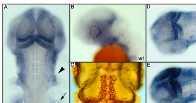
We screened 100 mutagenized genomes for mutations that affect the level of PCNA expression. Several mutants with an increased expression were noted, but the majority showed typical degeneration/apoptosis phenotypes. Two mutants, however, showed an increase in PCNA expression, most prominently visible in the tectum, and did not show
degeneration. These mutants, named hu418B and hu540A,
show an increased expression of PCNA in the peripheral retina and the tectum (Figure 1D and 1E). These regions are known to be highly proliferative and are thought to contain stem cells. The increase can be observed at 36 hpf, but not at 24 hpf when a larger proportion of the embryo is PCNA positive. Furthermore, expression still undergoes a general reduction as the embryo ages. At 96 hpf, PCNA is difficult to detect in both mutant and wild-type embryos using an ISH approach. To exclude the possibility that a low level of apoptotic cell death is responsible for the increase of PCNA expression, we performed a whole-mount terminal
trans-ferase dUTP nick-end labeling (TUNEL) assay on hu418B
mutants and siblings at 40 hpf. No difference in the level of apoptosis could be observed (data not shown), indicating that
Hedgehog Repressors in Zebrafish
Synopsis
In a screen aimed at finding genes that control proliferation in the zebrafish embryo, three mutants were identified. Mutants showed an increase in size of several structures including the brain, the retina, and the fins. Surprisingly, although size was increased in the embryos, adults remained small. Cloning of these genes revealed that increased Hedgehog signaling was at the basis of the phenotype, because all three genes encoded known repressors of the Hedgehog signaling pathway: Ptc2, Su(Fu), and Hip.
Hedgehog is known to play a role in proliferation. For instance, ectopic Hedgehog signaling can lead to several tumors including basal cell carcinoma and medulloblastoma. However, the pheno-types were still a surprise, because earlier experiments in zebrafish embryos suggested that activation should lead to patterning rather than proliferation defects. Current models of the pathway predict that these genes act independently to inhibit the signal but curiously, redundancy amongst these genes was not found, because triple mutants looked like the single mutants.
these mutants purely display increased levels of PCNA, independent of apoptosis.
Altered Level of Proliferation Affects Several Structures of the Developing Embryo
Morphological analysis of hu418B and hu540A mutants
shows abnormalities that correspond with an increased level of cell proliferation. At 72 hpf, the volume of the head is increased (Figure 2A and 2B), mainly in the region of the tectum, where an increased level of proliferation is observed.
Additionally, hu418B and hu540A mutant embryos show a
reduced size of the pupil, whereas the overall size of the eye is unaffected (Figure 2C and 2D). Measurements revealed that both length and width of the pupil are significantly decreased (Figure 2E). However, the formation of the lens is normal (data not shown). This eye phenotype might result from the increased rate of proliferation in the CMZ. The retina is reported to grow as a result of several division steps of retinal stem cells in the CMZ [35]. An increased level of proliferation of these cells might cause the retina to overgrow the lens, reducing the size of the pupil. Additionally, the pectoral fins
of thehu418Bandhu540Amutants were enlarged (Figure 2F
and 2G). Dorsoventrally the fins have approximately
in-creased in size by 50% (0.01,p,0.02,n¼6), the area has
increased by 65% (p,0.001,n¼3) (Figure 2H and 2I).
An additional phenotype is observed in the otic vesicle in the hu418B and hu540A mutants, in which the dorsolateral septum is missing (Figure 2J and 2K). However, the otoliths are correctly positioned, indicating that the anlage of the ear is correct.
This phenotypic combination was already observed in a class of mutants identified in a large-scale screen [26],
covering thedre, uki,andlepmutants [27–29].
Complementa-tion analysis revealed thathu418Bandhu540Aare additional
alleles ofuki(now referred to as ukihu418B and ukihu540A). Of
this complementation group, theukihu418Bmutant shows the
most consistent and strongest phenotype and is therefore
used for further experiments. The morphological phenotype
of dre and lep is slightly weaker and no clear difference in
PCNA expression can be detected in thedreandlepmutants
using an ISH approach.
Adult Mutants Show Additional Phenotypes
Raising homozygousuki, dre,andlepmutants demonstrates
that approximately 10% of theuki andlep mutants reach 2
mo of age, and all die before the third month. Only dre
mutants can be raised in significant numbers (50%) for 3 mo, with a maximum lifespan of 9 mo (5%). All mutants stay infertile and show a dwarfism phenotype [29]. This could be a result of the absence of growth hormone, which is secreted by
the pituitary gland. However, sectioning an adultdremutant
revealed the presence of a pituitary gland (data not shown). The adenohypophysis secretes multiple hormones that are reported to play a role in the development of a dwarfism phenotype [36]. However, ISH experiments show that expression levels of growth hormone (GH), proopiomelano-cortin (POMC), prolactin (PRL) and thyroid stimulating
hormone (TSH) are not altered in dre mutants (data not
shown).
Histological analysis of a 5-mo-olddremutant (n¼4) and
sibling (n¼3) reveals an additional phenotype concerning the
gills of the adult fish. The gills contain branchial arches that are composed of multiple primary lamellae, formed by a stack of single chondrocytes (Figure 3A and 3B). To increase the area for sufficient oxygen uptake, these primary lamellae are branched into a large number of secondary lamellae (Figure
3A). However, in thedremutant, the degree of branching to
[image:3.651.46.384.51.229.2]form secondary lamellae is severely diminished. The primary lamellae contain large clusters of cells, which morphologically resemble chondrocytes (Figure 3C). The strictly organized stacks of single chondrocytes are mainly absent. Occasionally, lines of chondrocytes appear to branch instead of the epithelium (Figure 3D). To investigate whether these clusters are indeed composed of chondrocytes we performed an
Figure 1.PCNA and Proliferation Patterns
(A) PCNA pattern as scored during the screen. In a dorsal view at 40 hpf, PCNA staining is observed in the medial and posterior part of the tectum, and in the cerebellum, the neural crest (arrowhead), and the pectoral fin (arrow).
(B) In a sideview (42 hpf), a ring of positive cells is visible around the lens.
(C) In a whole-mount BrdU labeling from day 3.5 to 4.5, similar regions are labeled indicating that PCNA RNA expression prefigures where BrdU will be incorporated.
Alcian Blue staining (Figure 3E and 3F) staining differ-entiated chondrocytes. In a wild-type fish the presence of differentiating chondrocytes could be confirmed in these single cell stacks. However, in the mutant, the clusters of cells were shown to be Alcian Blue negative (Figure 3F). On the other hand, the chondrocytes in the region where the primary lamellae are attached to the skeleton are Alcian Blue positive. This suggests that the chondrocytes in the gills
are specifically affected in the dre mutant. One of the
possibilities is that these Alcian Blue–negative cells are not able to properly differentiate during the development of the branchial arches. To examine whether these cells are in an earlier stage of cartilage formation, a Periodic Acid Schiff (PAS) staining was performed to detect ovotransferrin, a glycoprotein transiently expressed by differentiating hyper-trophic chondrocytes, before they become Alcian Blue positive [37]. Both the mutant and the wild-type sections are negative for the PAS staining (data not shown). We therefore suggest that the chondrocyte-like cells in the mutant are prehypertrophic chondrocytes, which are ar-rested in their proliferative stage, which are therefore unable to finally differentiate into mature chondrocytes. This
branchial arch phenotype appeared to bedrespecific because
primary lamellae in ukiand lepmutants are able to branch
(Figure 3G and 3H) and do not contain these foci of chondrocytes.
Thedre Locus is Encoding the Suppressor of Fused Protein
To identify the genes responsible for the observed phenotypes, we intended to positionally clone this class of
mutants. The dre mutation was roughly mapped to linkage
[image:4.651.47.548.50.373.2]group (LG) 13 near z5395. Linkage analysis on 765 mutants reveals that the mutation was positioned close to z5395, leaving nine recombinants (0.6 cM) and z25745, leaving six recombinants (0.4 cM) (Figure 4A). Both markers mapped north of the mutation (referring to the MGH mapping panel at the Zebrafish Information Network at http://zfin.org). We identified an assembled contig of the Zv2 zebrafish genome assembly containing these markers, called ctg11890 (http:// www.ensembl.org). We assumed that, based on the physical distance between these two markers, the mutation could be located on this contig. Several simple sequence length polymorphisms (SSLPs) mapping to this contig were tested
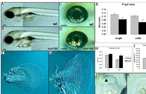
Figure 2.Phenotypes ofukihu418BMutant Embryos
(A and B) Lateral view of a wild-type (wt) andukihu418Bmutant.
(B) Showing an increased volume of the head. The size of the pupil is reduced in theukihu18Bmutant without affecting the size of the eye. (C–E) The size of the pupil is reduced in theukihu18Bmutant compared to wild-type, without affecting the size of the eye. Measurements revealed that
the length and width of the pupil is significantly reduced in theuki418Bmutant (n¼5, *p,0,001).
(F–H) Pectoral fins showing the increased size of anuki418Bmutant in which the dorsal/ventral (D/V) size of the pectoral fin is increased by 50%, (n ¼3, *0.01,p,0.02).
(I) The anterior/posterior size is not significantly affected, but the fin area has increased by 65% (n¼3,p,0.001). (J and K)ukihu418Bmutants lack the dorsolateral septum in the ear (arrowhead). Scale bar is 100lm.
DOI: 10.1371/journal.pgen.0010019.g002
for linkage, leaving zero recombinants with 11890.2A (Figure 4A). However, no marker was identified on this contig that would enclose the mutation on the south side. The closest marker was positioned in a region containing four predicted
genes (Figure 4A). One of those, theb-mannosidase precursor
gene,manba,was not considered to be a likely candidate. The
other three candidates were screened for mutations by direct sequence analysis of all 28 predicted exons. This analysis revealed several silent mutations and one missense mutation in the third exon of Su(fu) [30], changing a threonine (ACG) to a lysine (AAG) at amino acid position 111 (Figure 4B), indicated as T111K. This residue is highly conserved in a stretch of eight amino acids: GFELTFRL, from bacteria
(Bacillus circulans) to human (Figure 4C). No additional mutations affecting protein sequence could be identified in the other predicted coding regions, so we expected this
mutation to be responsible for thedrephenotype. To test the
hypothesis whether a loss of function of Su(fu) was able to phenocopy the mutants, we injected morpholino antisense oligonucleotides (MO) targeted against the predicted initia-tion codon of Su(fu) (Figure 5). The characteristic eye (Figure
5A and 5B) and ear (Figure 5E and 5F) phenotype of thedre
mutant could be phenocopied effectively (75% phenocopy in
two different strains, n¼58), in contrast to a control MO.
Besides the eye and ear phenotype, the MO also induced a somite phenotype (Figure 5I and 5J). The normally chevron-shaped somites become partially flat, an effect previously described [30]. This could be due to a maternal component, which can be inhibited by the MO, enhancing the phenotype.
Alternatively, the dretm146d allele may be a partial loss of
function allele. To distinguish between these possibilities, we injected up to 25 ng of MO against a splice site, thereby affecting only the zygotic component of Su(fu). This results in
a clear phenocopy of thedremutant without a somite defect
(.90% phenocopy in two different strains, n¼66).
Addi-tionally, injecting the same amount of splice MO into dre
mutants, does not enhance the phenotype (.95% phenocopy,
n¼71), suggesting this allele of Su(fu) to be a severe loss of
function.
TheukiandlepLoci Encode Negative Regulators of the Hh Signaling Pathway
The similarity of phenotypes between dre, uki, and lep
mutants suggested that all the mutants encode negative
regulators of Hh signaling. Positional cloning of theukiand
lep mutations was therefore initiated by linkage analysis of
SSLPs neighboring 14 candidate genes, all members of the Hh
signaling pathway. The ukihu418B mutation is tightly linked
with marker z13452 and z27361 on LG 1, enclosing Hip. Sequence analysis of all predicted coding sequence of Hip revealed a nonsense mutation in exon 5, changing a tyrosine to a stop codon at position 295 (Y295STOP) of the transcript encoding 694 amino acids (Figure 6A). Sequence analysis of the ukihu540A mutant identified a stop codon in exon 5 at
position 285 (Y285STOP) of the Hip protein. The ukitc256d
allele contained a premature stop codon at position 406 (Y406STOP) in exon 7 (Figure 6A). Amino acids 285, 295, and 406 of the zebrafish homolog of Hip corresponds to amino acids 293, 303, and 414 in human Hip. The identified
mutation in theukihu418Bmutant should lead to a truncated
protein without a membrane anchor, possibly resulting in a malfunctioning protein.
Positional cloning of theleptj222mutants was performed in
a similar way. No recombination events were detected with marker z11948 and four newly identified SSLP markers on contig 10160 in genome assembly Zv2, containing Ptc2 (http:// www.ensembl.org). The zebrafish Ptc2 protein has a transcript of 3,732 base pairs, encoding for 1,244 amino acids [31]. Exon sequencing identified a T to A substitution changing a tyrosine to a premature stop at position 590 (Figure 6B) directly after the sixth transmembrane domain. When this transcript is expressed, Ptc2 misses the second large extracellular domain known to be necessary for Hh binding and probably the inhibitory capability on Smo. We therefore expect this truncated protein to be a functional null.
[image:5.651.48.289.49.389.2]To confirm that the identified mutations in these genes are responsible for the phenotypes, we injected wild-type embryos with an MO against the initiation codon of Hip
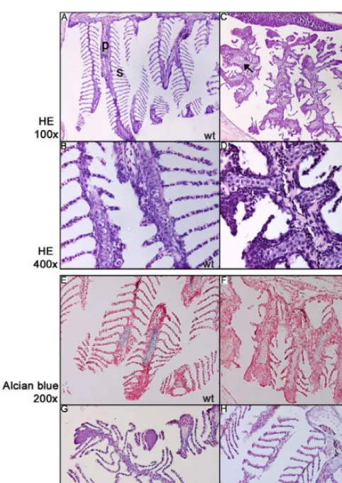
Figure 3.Patterning of the Branchial Arches in a 5-Mo-OlddreMutant (A and B) The strict organization of the brachial arch into primary (p) and secondary (s) lamellae in a wild-type situation (1003magnification). Higher magnification shows stacks of single chondrocytes in the primary lamellae.
(C and D) Sectioning of a dre mutant shows disturbed patterning, resulting in the absence of secondary lamellae and the presence of foci of chondrocyte-like cells in the primary lamellae (arrowsHE, hematoxylin and eosin stain; wt, wild-type.
(E and F) Alcian Blue staining reveals the presence of differentiated chondrocytes in the wild-type (wt) primary lamellae, but not in thedre mutant, indicating that the differentiation of these chondrocytes is affected (2003).
(G and H) Branchial arches ofukiandlepmutants appear to be wild-type (wt).
and a splice MO for Ptc2. For the uki mutant, a clear phenocopy could be observed after 4 d, affecting the head, eyes, and ears (see Figure 5C and 5G) (60% phenocopy in two
different strains,n¼64). Injection of wild-type embryos with
20 ng of Ptc2 splice MO resulted in a phenocopy of the ear and eye phenotype, however with a lower success rate (12/44; 28.5%) (see Figure 5D and 5H). This might be an effect of the positive feedback loop on Ptc2 when the Hh signaling pathway is activated, counteracting the efficiency of the
MO. These experiments show that the uki and lep mutant
phenotypes are indeed caused by Hip and Ptc2.
Loss of a negative regulator of Hh signaling should increase Hh activity, for which Ptc1 expression is generally used as a
readout. An ISH was performed ondre, uki,andlepmutants,
resulting in a slight increase in Ptc1 expression only foruki
and lep mutants (Figure 6C). dre mutants do not show a
significant increase in Ptc1 expression (data not shown). Taken together, we conclude that aberrant activation of
the Hh signaling pathway is responsible for theuki, dre,andlep
mutant phenotypes.
Double and Triple Mutants Do Not Enhance the Phenotypes
Because the increase in proliferation can only significantly
be detected in uki mutants, we initiated the generation of
double and triple mutants in an attempt to enhance the level of proliferation. Current models suggest that the three genes should independently inhibit Hh signaling, therefore a higher level of proliferation could be expected. Analyzing PCNA
expression in the progeny of two ukiþ//dreþ//lepþ/carriers
shows increased PCNA expression for a small subset, which
upon genotyping turned out to be mainly uki mutants.
Additionally, after sorting 96 genotyped embryos into all the possible genotypic combinations, it turned out that double
and triple mutants do not show an obvious increase of the strength of the morphological phenotypes (Figure 7A–7E). The only morphological difference in double or triple mutants, compared to single mutants, comprises the ear (Figure 7F–7I). In a wild-type situation, the semicircular canal is formed after the ingrowth of the epithelial projections from the outline of the otic vesicle, which fuses in the center
of the ear to form the ear lumen (Figure 7F). In the uki/lep
double anduki/dre/leptriple mutants, all epithelial projections
fail to grow toward the lumen of the ear (Figure 7G and 7I). These findings suggest that other negative regulators might still be present to prevent further activation of the Hh signaling pathway.
TheukiandlepMutants Can Be Rescued by Cyclopamine Treatment
To further prove that the described mutants are a result of increased Hh signaling, we attempted to inhibit Hh signaling, and thereby rescue the mutant phenotypes, by cyclopamine treatment. Cyclopamine is an inhibitor of Hh signaling acting on the level of Smo [38] at the initial stage of the signal
transduction pathway. Treating lep mutants with limited
amounts of cyclopamine (3lM) clearly rescued the eye and
ear phenotype. Genotyping of all apparent wild-type embryos
identified the presence of lep mutants (Table 1). Wild-type
embryos were unaffected using this concentration. However,
a treatment using 25lM of cyclopamine clearly mimicked the
syuphenotype [39], showing the functionality of the
cyclop-amine.
The eye phenotype ofukimutant can be partially rescued
using 10 or 15lM cyclopamine, and fully rescued using 25lM
(Table 1). However, using 25lM of cyclopamine, a subset of
both siblings and mutants in the same clutch are affected,
[image:6.651.50.418.55.262.2]shown by a curly tail. Apparently, uki mutants are not
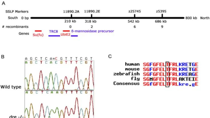
Figure 4.Positional Cloning of thedreMutant
(A) Schematic representation of assembled contig 11890 of the Zv2 genome assembly. SSLP markers z5395 and z25745 and newly identified SSLPs 11890.2A and 11890.2 were closely linked with thedrelocus. Remaining recombinants of a complete panel of 765 mutant embryos are indicated. Four genes were predicted in the region of marker 11890.2A that encode Su(fu), TRC8, ubiquitin conjugating enzyme E2, andb-mannosidase precursor protein.
(B) Thedremutation is a C to an A substitution, changing a threonine to a lysine.
(C) Multiple alignment of Su(fu) homologs revealed that the induced mutation changes an amino acid in a highly conserved region of Su(fu). DOI: 10.1371/journal.pgen.0010019.g004
protected against the effects of cyclopamine. This shows a
limitation of using cyclopamine for rescuing theukimutant,
which is not the case for rescuing lep mutants, in which a
much lower concentration is able to fully rescue without any side effects.
Whendremutants are treated with 25lM cyclopamine, the
mutant eye phenotype can be observed in combination with a curly tail, which is a result of the cyclopamine (data not shown). Increasing the cyclopamine concentration to 50 or 75
lM severely affected the development of all embryos, and
therefore the eyes and ears could not be analyzed. Thusdre
mutants are not protected against the effects of cyclopamine, but they also cannot be rescued by a cyclopamine treatment.
The latter is expected becausedre/Su(fu) acts downstream of
Smo. These results emphasize that the phenotypes in this class of mutants are a result of an increased level of Hh activity.
Discussion
Proliferation Is Increased in thehu418B andhu540A Mutants
In our forward genetic screen, we were able to identify two proliferation mutants based on altered expression levels of
PCNA and detected by an ISH approach. The identified mutants show an increase in the level of PCNA expression after 40 hpf, which is ectopically expressed in the developing tectum and in the CMZ of the eye. Unfortunately, no mutants were identified with a decreased proliferation rate. This could be explained by the fact that this screen covered only 1%– 10% (100 genomes) of the zebrafish genome, leaving several additional genes to be identified in a larger screen. The limited amount of identified mutants suggests that there might be a high level of redundancy in controlling prolifer-ation. Mutants showing an altered level of proliferation associated with increased apoptosis were excluded due to their frequent occurrence. We speculate that an increase in the amount of apoptosis results in an increase in prolifer-ation/PCNA expression as part of a wounding/repair response [40], on which altered levels of proliferation are a secondary effect. Possibly, this secondary defect has obscured some interesting early defects.
TUNEL experiments have shown that the increase in
proliferation in the hu418B and hu540A mutants is not
[image:7.651.46.382.48.395.2]associated with increased apoptosis. The increased PCNA expression could therefore be a result of impaired regulation
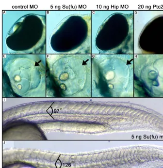
Figure 5.MO Injection Experiments against Su(fu), Hip, and Ptc2 (A) Dorsal view of the eye showing the lens in the eye chamber.
(B–D) Dorsal view of embryos injected with the indicated MOs, resulting in a phenocopy ofdre, uki,andlepmutants.
(E) A wild-type ear showing the presence of the dorsolateral septum (arrow), which is not present after injections with the indicated MOs (F–H, arrow). (I) Injections with control MOs against the initiation codon of Su(fu) results in chevron-shaped somites with an angle of 978.
of proliferation. The phenotypes observed after 4 d are similar to a previously described class of mutants identified in
the large-scale Tu¨bingen zebrafish screen [26], containingdre,
uki, and lep. Complementation analysis revealed that both
hu418Bandhu540Aare additional alleles of theukimutation.
dre and lep mutants are weaker than uki as judged from
morphology and do not show a comparable increase of PCNA
expression. Possibly, the increase in proliferation in theuki
mutant might reflect a specific function for Hip, but considering the overall morphological similarity of the mutants, it is more likely that the difference is due to other factors (see below).
Aberrant Hh Activation Is Responsible for thedre, uki,and lepMutants
We show thatdre, uki,andlepencode components of the Hh
signaling pathway. The lesion in thedremutant is a missense
mutation in the su(fu) gene. The mutation, changing a
threonine to a lysine, has been proposed as a potential PKC target site [41]. It is positioned in a highly conserved N-terminal region shown to be involved in binding the Gli protein [42]. Crystal structure analysis revealed that this threonine is buried and therefore suggested to be unim-portant for the activity of Su(fu) [42]. However, our data indicate that this residue is crucial for the proper functioning of Su(fu), therefore it might become accessible for certain kinases due to conformational changes.
By injecting MOs against Su(fu), the mutant phenotype of
dre could be copied, confirming that the dre locus encodes
Su(fu). However, Su(fu) MOs against the initiation codon of Su(fu) induce a somite phenotype [30], which could be explained in a situation in which the Su(fu) MO also affects a maternal contribution. This is confirmed by the finding that an MO against a splice site does not result in a somite
phenotype. Additionally, the phenotypes of dre can not be
enhanced by the splice MO, suggesting this allele of Su(fu) to
be a strong loss of function or a null. The similarity of the
phenotypes within this mutant class suggested that the uki
and lep mutants are also a result of aberrant activation of
Hh. Linkage analysis of markers near multiple candidate genes confirmed this. Premature stop codons in Hip and
Ptc2 were identified to be responsible for the uki and lep
mutants, respectively, which was confirmed by the
MO-induced phenocopies. In the leptj222 mutant, the identified
premature stop is positioned at amino acid 590 directly after the sixth putative transmembrane domain [31], only
producing the N-terminal half of the protein. In Drosophila,
multiple alleles of Ptc have been analyzed, showing that expression of either the N- or C-terminal half alone will abolish its function [43]. We therefore expect that this severe truncation will abolish Ptc2 protein function in the mutant. The three nonsense mutations in the Hip protein result in all cases in a comparable phenotype, suggesting these alleles to be nulls. As a result, the Hh signaling pathway will be aberrantly activated, confirmed by the
increase in Ptc1 expression inukiandlepmutants. However,
the effect of the overactivation of the Hh pathway is subtle compared to Hh overexpression data. This might be a result of a restricted expression pattern of these negative regulators. ISH experiments showed that Su(fu) is ubiqui-tously expressed until 24 hpf [30] and becomes more anteriorly restricted at 42 hpf (data not shown). The expression pattern of Ptc2 is generally overlapping Ptc1 expression, with some minor differences [31]. Hip tran-scripts can be detected in the adaxial cells in the developing trunk and in the head, generally resembling the expression pattern of Ptc1 (Figure S1A). Within the developing trunk, two rows of adaxial cells are shown to be Hip positive at 24
hpf (Figure S1A and S1B). Hip expression is reduced in uki
[image:8.651.47.389.50.251.2]mutants at 24 hpf, which is probably due to nonsense-mediated decay (data not shown). At 40 hpf the pectoral
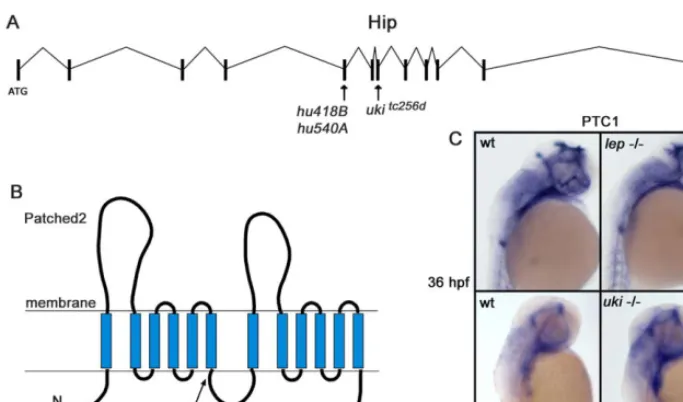
Figure 6.Premature Stop Codons Were Identified in theukiandlepMutant in Hip and Ptc2, Respectively
(A) Schematic representation of the genomic organization of the Hip gene. All three alleles of theukimutation contain premature stop codons positioned in exon 5 and exon 7.
(B) Representation of the protein structure of Ptc2 shows that the identified nonsense mutation is positioned after the sixth transmembrane domain, probably resulting in a malfunctioning protein.
(C) ISH experiments show that Ptc1 expression is increased inukiandlep(arrow), confirming the aberrant activation of the Hh signaling pathway. DOI: 10.1371/journal.pgen.0010019.g006
fins, some branchial arches, and the tectum opticum are Hip positive, linking the expression pattern with the observed embryonic phenotypes (Figure S1C and S1D). Combining these expression patterns suggests that the subtle pheno-types observed in these mutants are not a result of a restricted pattern of one of these negative regulators, but
are probably due to other negative regulators, most likely Ptc1, preventing further activation of the pathway.
Phenotypic Consequences of Aberrant Hh Activation Theukihu418Bandukihu540Amutants were picked up showing an increased level of proliferation in the developing brain.
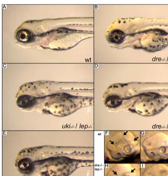
Figure 7.Phenotypic Analysis ofdre/uki/lepTriple Mutants (A) Wild-type (wt) embryo at 96 hpf.
(B–E) The indicated double and triple mutants do not show severe enhancement of the phenotype.
(F–I)dre/lepdouble mutants have an ear phenotype comparable with a single mutant. In theuki/lepanddre/uki/leptriple mutants, the epithelial projections (arrows) fail to grow out to fuse in the middle of the ear to form the ear lumen.
[image:9.651.46.383.49.401.2]DOI: 10.1371/journal.pgen.0010019.g007
Table 1.Rescuing Experiment Using Cyclopamine onuki, dre,andlepMutants
Mutant Concentration (lM)
Phenotype Genotype
Wild-Type Curly Tail Wild-Type Curly Tail Mutant Weak Mutant Mutant Wild-Type Heterozygotes Mutant Total
lep 3 23 0 0 0 0 5 12 6 23
25 13 10 0 0 0 10 10 3 23
uki 10 25 2 1a
13 7 11 17 20 48
15 7 8 5a
4 0 3 12 9 24
25 8 15 0 0 0 9 11 3 23
dre 10 20 0 0 0 4 10 10 4 24
25 10 5 1 0 8 5 10 9 24
Data are the numbers of embryos. Rescue experiment foruki, dre,andlepby inhibiting Hh activity using cyclopamine.lepmutants can be fully rescued using 3lM cyclopamine, a concentration not affecting siblings.ukimutants can be rescued using 25lM cyclopamine. However, the cyclopamine affects the development of the siblings, shown by the curly tail phenotype. Cyclopamine treatment did not rescuedremutants.
aWeak mutant and curly tail.
Hh activity is reported to be involved in the proliferation of cells in the central nervous system [44–49]. The increase in
the volume of the head of theukihu418Bmutant is therefore in
line with previous studies in which the growth of the brain is shown to be regulated by the activation of Shh-Gli1 signaling [50,51].
All the affected structures and tissues in the described mutants are known to be under control of Hh signaling during development. Hh signaling is one of the key regulators in the development of the eye, in which the formation of the retina is driven by a wave of Hh signal, secreted by the cells of the ganglion cell layer [52]. As a result, Hh controls proliferation of multiple cell types of the eye like photo-receptors and glia [53]. All described mutants show a decreased size of the pupil, which might be due to an overgrowing activity of the cells of the retina. Therefore the lens is not visible from a dorsal view, but no defects are observed in the lens itself.
The increased fin size in the ukihu418B mutant embryos
could be linked to impaired Hh signaling. It is the opposite of
the phenotype of syu mutants, in which finbuds are
established, but fail to grow out, due to the absence of Shh
signaling [39]. The observed phenotypes in thedre, uki,andlep
mutants can therefore be linked to aberrant activation of the Hh signaling pathway.
Surprisingly, dre, uki, and lep mutants can be grown for
several months, but remain small and are infertile. One explanation for the dwarfism phenotype involves the absence of growth hormone secreted by the pituitary gland. The formation of the pituitary gland is reported to be regulated by Hh signaling [54,55], indicating that its functioning might be hampered in these mutants and resulting in the observed
small phenotype. However, sectioning of adremutant embryo
revealed that the pituitary gland is morphologically present. The secreted hormones of the adenohypophysis are reported to be involved in the development of a dwarfism phenotype
[36]. However,dremutants do not show obvious altered levels
of POMC, TSH, PRL, and GH expression, indicating that the formation of the adenohypophysis is not affected in this class of mutants. Currently, we are examining a potential role for the IGF signaling pathway in the development of the dwarfism phenotype.
Thedremutants show an abnormality in the development
of the branchial arches. Normally, the primary lamellae are strictly patterned and intensely branched into secondary lamellae. Occasionally, the chondrocyte-like cells appear to form the secondary lamellae itself instead of the branching of epithelial cells. Branching of the mammalian lungs is reported to be regulated by Shh activity [56], in which increased Shh activity disrupts branching and increases the level of proliferation. The primary lamellae gain their rigidity
by stacks of single chondrocytes. However, in thedremutant
the primary lamellae contain large clusters of prehypertro-phic chondrocytes, which might be unable to start the differentiation process. The formation of cartilage is reported to be tightly regulated by the action of Indian hedgehog (Ihh) and parathyroid hormone-related protein (PTHrP) [57,58]. In this process the amount of Ihh acts like a sensor, thereby limiting the group of cells that are stimulated to enter the differentiation stage [57]. This might be
deregulated in thedremutant, in which an aberrant activation
of Ihh signaling increases the amount of PTHrP, thereby
preventing hypertrophic differentiation. The prehypertro-phic chondrocytes therefore remain in their proliferative stage and might form the observed clusters in the primary lamellae. The branchial arch phenotype has not been
observed inuki/Hip andlep/Ptc2 mutants, suggesting that this
is a unique function for Su(fu). Therefore Su(Fu) could be modulating signals via Ptc1 as well. This is in agreement with results on Su(Fu) morphants that mimic Ptc1 morphants in their somite phenotype [30]. If the Su(fu) allele is a strong loss of function and affects signals via Ptc1 and Ptc2, why is the phenotype not any stonger? In addition to rescue by maternal protein, the role of Su(fu) in Hh inhibition may be accessory rather than absolutely central. This has been shown in
Drosophila, in which complete inactivation of Su(Fu) does not lead to a full Hh overactivation phenotype [59].
Complex Regulation of Hh Activity
We have shown that aberrant activation of the Hh signaling
pathway is responsible for the dre, uki, and lep mutants.
Nevertheless, none of the mutants that was identified shows a typical phenotype described for aberrant Hh activation as was obtained by the overexpression of dnPKA or Shh [30,60].
Surprisingly, triple mutants ofsu(fu), Hip,andptc2still do not
show a further increased PCNA expression or a strongly enhanced phenotype. This could be explained in a scenario in which a slight activation of the Hh pathway exceeds a certain threshold upon which Ptc1 will be expressed via an autoregulatory loop, preventing further activation of the pathway. Inhibiting Ptc1 functioning in these triple mutants could probably result in the expected Hh-overexpression phenotypes. Ptc1/Ptc2 double morphants were described, confirming this idea [30].
The ability of cyclopamine to rescue theukiandlepmutants
is in line with expectations. Cyclopamine acts on the level of Smo and can revert the effect of upstream components such as Hip and Ptc2. The ability of cyclopamine to rescue the effects of mutations in Ptc is also documented for cell lines [38]. Because Su(fu) acts downstream of Smo (the point where cyclopamine acts), it is likely to be independent of the presence of an upstream signal. Similar results have been reported in a system with Gli2 overexpression, in which cyclopamine was unable to revert the effects [38]. Indeed, we
find that dre mutants cannot be rescued by cyclopamine
treatment.
Aberrant postnatal activation of the Hh signaling pathway is implicated in various types of neuronal and epithelial tumors. However, no obvious tumors have been observed in the mutants and heterozygotes. Future experiments using additional mutants, like a p53 knockout, might induce tumorigenesis in a background with aberrant Hh activity.
Materials and Methods
Strains and screening methods.ukitc256d, leptj222,anddretm146dstocks were obtained from the Max Planck Institute for Developmental Biology stock center in Tu¨bingen, Germany. ENU mutagenesis was performed on TL males as described [61]. F1families were generated and interbred to obtain F2families. Inbreeding generated F3embryos for screening. Embryos were incubated in PTU and dechorionated by pronase treatment according to standard protocols (http:// zfin.org). Staging of embryos was according to Kimmel et al. [62]. Embryos were fixed in 4% paraformaldehyde/PBS at 40 hpf. During screening, PCNA whole-mount ISH (WISH) was performed on an Abimed 96-wells ISH robot (Intavis Bioanalytical Instruments, Cologne, Ger-many; settings available on request). Mutants that were detected by
morphological screening at 24 hpf were processed separately for WISH, along with two wild-types as controls. In addition, morpho-logical screening was performed at 72 hpf on a duplicate clutch.
In situ hybridization.Manual ISH was carried out as described [63]. An antisense probe for PCNA was generated by linearizing EST clone fc43g05 (MPMGp609L0932, RZPD Deutsches Ressourcenzentrum fu¨r Genomforschung, Berlin, Germany; http://www.rzpd.de), using SalI and transcription using SP6 polymerase. Ptc1 probe synthesis was performed according to Concordet et al. [64]. ISH for POMC, TSH, GH, and PRL was performed according to Herzog et al. [65]. A 2-kb fragment of Hip was amplified from cDNA, using primers 59 -AATTTGTGCTCTTGTTAGCC-39and 59 -AGTGAGGTCCAGCAGG-TAAG-39, cloned and subsequently transcribed.
TUNEL assay and BrdU labeling.To determine the amount of apoptosis, a whole-mount TUNEL assay was performed on 40 hpf embryos as described [66]. The presence of mutants was confirmed by genotyping the analyzed embryos. BrdU labeling was performed according to a previous report [67].
Histology.Adult fish were fixed in 4% paraformaldehyde at 48C for 4 d and subsequently decalcified in 0.25M EDTA (pH 8) for 2 d. Paraffin sections (6lm thick) were stained with eosin, Alcian Blue, or PAS in combination with hematoxylin using standard protocols.
Measurements.The size of the fins and pupils was determined on a Zeiss Axioplan microscope (Carl Zeiss, Jena, Germany) using a micrometer. The area of the pectoral fins was measured by determining the amount of pixels of a scanned photograph using Paint Shop Pro version 5 (Jasc Software, Corel Corporation, Ottawa, Ontario, Canada).
Genetic mapping and positional cloning ofdre.The rough genome mapping ofdretm146dto LG 13 was performed by bulked segregation
analysis of F2embryos and genome scanning with SSLPs [68]. To fine map the mutation, a mapping strain was created by crossing adre/þ
male in a Tu¨bingen background to a wild-type WIK female. F2fish carrying thedremutation were crossed; 765 mutant F3embryos were collected and genomic DNA was isolated. In total, 35 SSLPs from the Massachusetts General Hospital [69] were used for positional cloning on LG 13 in the region around 40 cM. Marker names and primer sequences can be obtained on request. Additionally, 12 SSLPs were identified on assembled contig 11890 of version Zv2 of the zebrafish genome assembly (http://www.ensembl.org) that contained the two most closely linked markers (z5395 and z25743) from the MGH database. Linkage analysis of these newly identified markers was performed to enclose the mutation. All predicted exon sequences south (corresponding to the MGH map) of the most closely linked SSLP marker were amplified and sequenced using DyeNamic ET (Amersham Biosciences, Little Chalfont, United Kingdom) protocols. All used zebrafish genome sequence data were produced by the Sanger Institute (http://www.sanger.ac.uk).
Candidate gene approach to positional cloneukiandlep.Mapping crosses were generated crossing a femaleukihu418Borleptj222 carri-er in Tu¨bingen background, with a WIK or TL male, respectively. Fourteen known regulators of the Hh signaling pathway were selected, and homologs were identified in the Zv2 zebrafish genome build at http://www.ensembl.org. A likely map position of the identified contigs was determined using the comparative map at http:// zfin.org and http://www.sanger.ac.uk. Forty SSLPs surrounding these candidates were analyzed for linkage on 10ukiorlepmutants and two siblings. In the case of linkage to a candidate gene, all predicted exons were amplified and sequenced. Predicted Hip exons were obtained using EST clone fc52e12 and additional homology-based assembly of the transcript.
MO antisense knockdown. MOs (Gene Tools, LLC, Philomath, Oregon, United States) were designed against the predicted initiation
codon or splice donor site of Su(fu), Hip, and Ptc2, along with a five-mismatch MO as a control. Their sequences are as follows (mismatches in lower case): Su(fu) MO: 59 -GCTGCTAGGCCGCATCT-CATCCATC-39, Su(fu) mismatched control: 59 -GCTGgTAcGCC-G a A T C T g A T a C A T C - 39, S u ( f u ) s p l i c e M O : 59 -TGACATTCTTACTCGTGAACTCTGT-39; Hip MO: 59 -AATGCTT-CATTTTTGCAGGGATGA-39, Hip mismatched control: 59 -AATGgTTgATTTTTcCAGcGATcA-39, Ptc2 splice MO: 59 -CTAG-CAAATAAGCCATACCTGTTGT-39, Ptc2 control 5-mm–splice MO: 59-CTAcCAAAaAAcCCATAaCTGaTGT-39. MOs were diluted in water to a stock concentration of 50 ng/nl. Ranges from 0.33 to 25 ng of MO were injected into one- to four-cell stage TL or ABxTL embryos and screened for the expected phenotypes 4 d after fertilization.
Cyclopamine treatment.Progeny from a cross of twouki, dre,orlep
heterozygotes were grown in embryo medium in the presence of various concentrations of cyclopamine (from a 10 mM stock in 96% ethanol), ranging from 2 to 75lM, administered at 5.5 hpf. Control embryos were treated with equal amounts of 96% ethanol. Genotyp-ing was performed to identify mutants in the clutch of treated embryos.
Supporting Information
Figure S1.Expression Pattern of Hip in Wild-Type Embryos (A and B) At 24 hpf, Hip is expressed in the brain and in two lines of adaxial cells in the developing tail.
(C and D) At 42 hpf, Hip expression is reduced in the somites. The mid-hindbrain boundary, some branchial arches, and the pectoral fins are Hip positive. Hip expression is also detected in the tectum (arrowhead).
Found at DOI: 10.1371/journal.pgen.0010019.sg001 (9.8 MB TIF).
Accession Numbers
The GenBank (http://www.ncbi.nlm.nih.gov/Genbank) accession num-bers for the described ESTs, genes, and proteins areBacillus circulans,
(CAD41946), zebrafish Hip EST clone fc52e12 (AI878265), PCNA EST clone fc43g05 (AI794381),su(fu)(NP_958466), andptc2(CAB39726), and human Hip (AAH25311).
Acknowledgments
We would like to thank Peter Lanser, Chris Jopling, Carina van Rooijen, Gerwen Lammers, and Marit Kosters for help during the screen. We thank Dr. Dana Jongejan-Zivkovic for advice and suggestions, and J. Korving, Harry Begthel, and Evelyn Groot for the sectioning experiments. Probes for the analysis on the adenohy-pophysis were kindly provided by M. Hammerschmidt. RG was supported by the German Human Genome Project (DHGP Grant 01 KW 9919), and MJK was supported by NWO genomics grant 050-10-024.
Competing interests.The authors have declared that no competing interests exist.
Author contributions.MJK and FJMV conceived and designed the experiments. MJK, MJDB, AK, SH, EW, EMHCV, and FJMV performed the experiments. MJK, MJDB, SH, and FJMV analyzed the data. AK, SH, and RG contributed reagents/materials/analysis tools. MJK wrote the paper. &
References
1. Massague J, Chen YG (2000) Controlling TGF-beta signaling. Genes Dev 14: 627–644.
2. Szebenyi G, Fallon JF (1999) Fibroblast growth factors as multifunctional signaling factors. Int Rev Cytol 185: 45–106.
3. Wodarz A, Nusse R (1998) Mechanisms of Wnt signaling in development. Annu Rev Cell Dev Biol 14: 59–88.
4. Ingham PW, McMahon AP (2001) Hedgehog signaling in animal develop-ment: Paradigms and principles. Genes Dev 15: 3059–3087.
5. Wetmore C (2003) Sonic hedgehog in normal and neoplastic proliferation: Insight gained from human tumors and animal models. Curr Opin Genet Dev 13: 34–42.
6. Denef N, Neubuser D, Perez L, Cohen SM (2000) Hedgehog induces opposite changes in turnover and subcellular localization of patched and smoothened. Cell 102: 521–531.
7. Incardona JP, Gruenberg J, Roelink H (2002) Sonic hedgehog induces the segregation of patched and smoothened in endosomes. Curr Biol 12: 983–995. 8. Yoon JW, Kita Y, Frank DJ, Majewski RR, Konicek BA, et al. (2002) Gene expression profiling leads to identification of GLI1-binding elements in target genes and a role for multiple downstream pathways in GLI1-induced cell transformation. J Biol Chem 277: 5548–5555.
9. Chuang PT, McMahon AP (1999) Vertebrate Hedgehog signalling modu-lated by induction of a Hedgehog-binding protein. Nature 397: 617–621. 10. Nybakken K, Perrimon N (2002) Hedgehog signal transduction: Recent
findings. Curr Opin Genet Dev 12: 503–511.
11. Robbins DJ, Nybakken KE, Kobayashi R, Sisson JC, Bishop JM, et al. (1997) Hedgehog elicits signal transduction by means of a large complex containing the kinesin-related protein costal2. Cell 90: 225–234. 12. Sisson JC, Ho KS, Suyama K, Scott MP (1997) Costal2, a novel
13. Wang G, Amanai K, Wang B, Jiang J (2000) Interactions with Costal2 and suppressor of fused regulate nuclear translocation and activity of cubitus interruptus. Genes Dev 14: 2893–2905.
14. Ding Q, Fukami S, Meng X, Nishizaki Y, Zhang X, et al. (1999) Mouse suppressor of fused is a negative regulator of sonic hedgehog signaling and alters the subcellular distribution of Gli1. Curr Biol 9: 1119–1122. 15. Kogerman P, Grimm T, Kogerman L, Krause D, Unden AB, et al. (1999)
Mammalian suppressor-of-fused modulates nuclear-cytoplasmic shuttling of Gli-1. Nat Cell Biol 1: 312–319.
16. Methot N, Basler K (2000) Suppressor of fused opposes hedgehog signal transduction by impeding nuclear accumulation of the activator form of Cubitus interruptus. Development 127: 4001–4010.
17. Murone M, Luoh SM, Stone D, Li W, Gurney A, et al. (2000) Gli regulation by the opposing activities of fused and suppressor of fused. Nat Cell Biol 2: 310–312.
18. Dunaeva M, Michelson P, Kogerman P, Toftgard R (2003) Characterization of the physical interaction of Gli proteins with SUFU proteins. J Biol Chem 278: 5116–5122.
19. Jones KB, Morcuende JA (2003) Of hedgehogs and hereditary bone tumors: Re-examination of the pathogenesis of osteochondromas. Iowa Orthop J 23: 87–95.
20. Thayer SP, di Magliano MP, Heiser PW, Nielsen CM, Roberts DJ, et al. (2003) Hedgehog is an early and late mediator of pancreatic cancer tumorigenesis. Nature 425: 851–856.
21. Berman DM, Karhadkar SS, Maitra A, Montes De Oca R, Gerstenblith MR, et al. (2003) Widespread requirement for Hedgehog ligand stimulation in growth of digestive tract tumours. Nature 425: 846–851.
22. Vorechovsky I, Unden AB, Sandstedt B, Toftgard R, Stahle-Backdahl M (1997) Trichoepitheliomas contain somatic mutations in the overexpressed PTCH gene: Support for a gatekeeper mechanism in skin tumorigenesis. Cancer Res 57: 4677–4681.
23. Vorechovsky I, Tingby O, Hartman M, Stromberg B, Nister M, et al. (1997) Somatic mutations in the human homologue of Drosophila patched in primitive neuroectodermal tumours. Oncogene 15: 361–366.
24. Taylor MD, Liu L, Raffel C, Hui CC, Mainprize TG, et al. (2002) Mutations in SUFU predispose to medulloblastoma. Nat Genet 31: 306–310. 25. Fujii K, Kohno Y, Sugita K, Nakamura M, Moroi Y, et al. (2003) Mutations in
the human homologue ofDrosophilapatched in Japanese nevoid basal cell carcinoma syndrome patients. Hum Mutat 21: 451–452.
26. Haffter P, Granato M, Brand M, Mullins MC, Hammerschmidt M, et al. (1996) The identification of genes with unique and essential functions in the development of the zebrafish,Danio rerio. Development 123: 1–36. 27. Heisenberg CP, Brand M, Jiang YJ, Warga RM, Beuchle D, et al. (1996)
Genes involved in forebrain development in the zebrafish, Danio rerio. Development 123: 191–203.
28. Whitfield TT, Granato M, van Eeden FJ, Schach U, Brand M, et al. (1996) Mutations affecting development of the zebrafish inner ear and lateral line. Development 123: 241–254.
29. van Eeden FJ, Granato M, Schach U, Brand M, Furutani-Seiki M, et al. (1996) Genetic analysis of fin formation in the zebrafish, Danio rerio. Development 123: 255–262.
30. Wolff C, Roy S, Ingham PW (2003) Multiple muscle cell identities induced by distinct levels and timing of hedgehog activity in the zebrafish embryo. Curr Biol 13: 1169–1181.
31. Lewis KE, Concordet JP, Ingham PW (1999) Characterisation of a second patched gene in the zebrafishDanio rerioand the differential response of patched genes to Hedgehog signalling. Dev Biol 208: 14–29.
32. Lu X, Tan CK, Zhou JQ, You M, Carastro LM, et al. (2002) Direct interaction of proliferating cell nuclear antigen with the small subunit of DNA polymerase delta. J Biol Chem 277: 24340–24345.
33. Perron M, Harris WA (2000) Retinal stem cells in vertebrates. Bioessays 22: 685–688.
34. Mueller T, Wullimann MF (2002) BrdU-, neuroD (nrd)- and Hu-studies reveal unusual non-ventricular neurogenesis in the postembryonic zebra-fish forebrain. Mech Dev 117: 123–135.
35. Wetts R, Serbedzija GN, Fraser SE (1989) Cell lineage analysis reveals multipotent precursors in the ciliary margin of the frog retina. Dev Biol 136: 254–263.
36. Nica G, Herzog W, Sonntag C, Hammerschmidt M (2004) Zebrafish pit1 mutants lack three pituitary cell types and develop severe dwarfism. Mol Endocrinol 18: 1196–1209.
37. Gentili C, Bianco P, Neri M, Malpeli M, Campanile G, et al. (1993) Cell proliferation, extracellular matrix mineralization, and ovotransferrin transient expression during in vitro differentiation of chick hypertrophic chondrocytes into osteoblast-like cells. J Cell Biol 122: 703–712. 38. Taipale J, Chen JK, Cooper MK, Wang B, Mann RK, et al. (2000) Effects of
oncogenic mutations in Smoothened and Patched can be reversed by cyclopamine. Nature 406: 1005–1009.
39. Schauerte HE, van Eeden FJ, Fricke C, Odenthal J, Strahle U, et al. (1998) Sonic hedgehog is not required for the induction of medial floor plate cells in the zebrafish. Development 125: 2983–2993.
40. Smith PG, Liu M (2002) Impaired cutaneous wound healing after sensory
denervation in developing rats: Effects on cell proliferation and apoptosis. Cell Tissue Res 307: 281–291.
41. Stone DM, Murone M, Luoh S, Ye W, Armanini MP, et al. (1999) Characterization of the human suppressor of fused, a negative regulator of the zinc-finger transcription factor Gli. J Cell Sci 112: ( Pt 23): 4437–4448. 42. Merchant M, Vajdos FF, Ultsch M, Maun HR, Wendt U, et al. (2004) Suppressor of fused regulates Gli activity through a dual binding mechanism. Mol Cell Biol 24: 8627–8641.
43. Johnson RL, Milenkovic L, Scott MP (2000) In vivo functions of the patched protein: Requirement of the C terminus for target gene inactivation but not Hedgehog sequestration. Mol Cell 6: 467–478.
44. Goodrich LV, Milenkovic L, Higgins KM, Scott MP (1997) Altered neural cell fates and medulloblastoma in mouse patched mutants. Science 277: 1109–1113.
45. Wallace VA (1999) Purkinje-cell-derived Sonic hedgehog regulates granule neuron precursor cell proliferation in the developing mouse cerebellum. Curr Biol 9: 445–448.
46. Wechsler-Reya RJ, Scott MP (1999) Control of neuronal precursor proliferation in the cerebellum by Sonic Hedgehog. Neuron 22: 103–114. 47. Dahmane N, Ruiz-i-Altaba A (1999) Sonic hedgehog regulates the growth
and patterning of the cerebellum. Development 126: 3089–3100. 48. Rowitch DH, S-Jacques B, Lee SM, Flax JD, Snyder EY, et al. (1999) Sonic
hedgehog regulates proliferation and inhibits differentiation of CNS precursor cells. J Neurosci 19: 8954–8965.
49. Kenney AM, Rowitch DH (2000) Sonic hedgehog promotes G(1) cyclin expression and sustained cell cycle progression in mammalian neuronal precursors. Mol Cell Biol 20: 9055–9067.
50. Dahmane N, Sanchez P, Gitton Y, Palma V, Sun T, et al. (2001) The Sonic Hedgehog-Gli pathway regulates dorsal brain growth and tumorigenesis. Development 128: 5201–5212.
51. Palma V, Ruiz i Altaba A (2004) Hedgehog-GLI signaling regulates the behavior of cells with stem cell properties in the developing neocortex. Development 131: 337–345.
52. Neumann CJ, Nuesslein-Volhard C (2000) Patterning of the zebrafish retina by a wave of sonic hedgehog activity. Science 289: 2137–2139.
53. Jensen AM, Wallace VA (1997) Expression of Sonic hedgehog and its putative role as a precursor cell mitogen in the developing mouse retina. Development 124: 363–371.
54. Sbrogna JL, Barresi MJ, Karlstrom RO (2003) Multiple roles for Hedgehog signaling in zebrafish pituitary development. Dev Biol 254: 19–35. 55. Herzog W, Zeng X, Lele Z, Sonntag C, Ting JW, et al. (2003)
Adenohypophysis formation in the zebrafish and its dependence on sonic hedgehog. Dev Biol 254: 36–49.
56. Pepicelli CV, Lewis PM, McMahon AP (1998) Sonic hedgehog regulates branching morphogenesis in the mammalian lung. Curr Biol 8: 1083–1086. 57. Vortkamp A, Lee K, Lanske B, Segre GV, Kronenberg HM, et al. (1996) Regulation of rate of cartilage differentiation by Indian hedgehog and PTH-related protein. Science 273: 613–622.
58. Lanske B, Karaplis AC, Lee K, Luz A, Vortkamp A, et al. (1996) PTH/PTHrP receptor in early development and Indian hedgehog-regulated bone growth. Science 273: 663–666.
59. Preat T (1992) Characterization of Suppressor of fused, a complete suppressor of the fused segment polarity gene ofDrosophila melanogaster. Genetics 132: 725–736.
60. Hammerschmidt M, Bitgood MJ, McMahon AP (1996) Protein kinase A is a common negative regulator of Hedgehog signaling in the vertebrate embryo. Genes Dev 10: 647–658.
61. van Eeden FJ, Granato M, Odenthal J, Haffter P (1999) Developmental mutant screens in the zebrafish. Methods Cell Biol 60: 21–41.
62. Kimmel CB, Ballard WW, Kimmel SR, Ullmann B, Schilling TF (1995) Stages of embryonic development of the zebrafish. Dev Dyn 203: 253–310. 63. Thisse C, Thisse B, Schilling TF, Postlethwait JH (1993) Structure of the
zebrafish snail1 gene and its expression in wild-type, spadetail and no tail mutant embryos. Development 119: 1203–1215.
64. Concordet JP, Lewis KE, Moore JW, Goodrich LV, Johnson RL, et al. (1996) Spatial regulation of a zebrafish patched homologue reflects the roles of sonic hedgehog and protein kinase A in neural tube and somite patterning. Development 122: 2835–2846.
65. Herzog W, Sonntag C, Walderich B, Odenthal J, Maischein HM, et al. (2004) Genetic analysis of adenohypophysis formation in zebrafish. Mol Endo-crinol 18: 1185–1195.
66. Cole LK, Ross LS (2001) Apoptosis in the developing zebrafish embryo. Dev Biol 240: 123–142.
67. Kimmel CB, Miller CT, Kruze G, Ullmann B, BreMiller RA, et al. (1998) The shaping of pharyngeal cartilages during early development of the zebrafish. Dev Biol 203: 245–263.
68. Geisler R (2002) Mapping and cloning. In: Dahm CN-VaR, editor. Zebrafish: A practical approach. Oxford (United Kingdom): Oxford University Press. pp. 175–212
69. Shimoda N, Knapik EW, Ziniti J, Sim C, Yamada E, et al. (1999) Zebrafish genetic map with 2000 microsatellite markers. Genomics 58: 219–232.