for Reverse Transcription during HIV-1 Replication
Shewit S. Tekeste,aThomas A. Wilkinson,aEthan M. Weiner,bXiaowen Xu,aJennifer T. Miller,cStuart F. J. Le Grice,cRobert T. Clubb,b Samson A. Chowa
Department of Molecular and Medical Pharmacologya
and Department of Chemistry and Biochemistry,b
University of California, Los Angeles, Los Angeles, California, USA; Basic Research Laboratory, National Cancer Institute, Frederick, Maryland, USAc
ABSTRACT
Human immunodeficiency virus type 1 (HIV-1) replication requires reverse transcription of its RNA genome into a
double-stranded cDNA copy, which is then integrated into the host cell chromosome. The essential steps of reverse transcription and
integration are catalyzed by the viral enzymes reverse transcriptase (RT) and integrase (IN), respectively.
In vitro
, HIV-1 RT can
bind with IN, and the C-terminal domain (CTD) of IN is necessary and sufficient for this binding. To better define the RT-IN
interaction, we performed nuclear magnetic resonance (NMR) spectroscopy experiments to map a binding surface on the IN
CTD in the presence of RT prebound to a duplex DNA construct that mimics the primer-binding site in the HIV-1 genome. To
determine the biological significance of the RT-IN interaction during viral replication, we used the NMR chemical shift mapping
information as a guide to introduce single amino acid substitutions of nine different residues on the putative RT-binding surface
in the IN CTD. We found that six viral clones bearing such IN substitutions (R231E, W243E, G247E, A248E, V250E, and I251E)
were noninfectious. Further analyses of the replication-defective IN mutants indicated that the block in replication took place
specifically during early reverse transcription. The recombinant INs purified from these mutants, though retaining enzymatic
activities, had diminished ability to bind RT in a cosedimentation assay. The results indicate that the RT-IN interaction is
func-tionally relevant during the reverse transcription step of the HIV-1 life cycle.
IMPORTANCE
To establish a productive infection, human immunodeficiency virus type 1 (HIV-1) needs to reverse transcribe its RNA genome
to create a double-stranded DNA copy and then integrate this viral DNA genome into the chromosome of the host cell. These
two essential steps are catalyzed by the HIV-1 enzymes reverse transcriptase (RT) and integrase (IN), respectively. We have
shown previously that IN physically interacts with RT, but the importance of this interaction during HIV-1 replication has not
been fully characterized. In this study, we have established the biological significance of the HIV-1 RT-IN interaction during the
viral life cycle by demonstrating that altering the RT-binding surface on IN disrupts both reverse transcription and viral
replica-tion. These findings contribute to our understanding of the RT-IN binding mechanism, as well as indicate that the RT-IN
inter-action can be exploited as a new antiviral drug target.
T
o establish an infection after entry into a susceptible cell,
hu-man immunodeficiency virus type 1 (HIV-1) has to reverse
transcribe its RNA genome to double-stranded DNA, followed by
integration into the host genome. Reverse transcriptase (RT) and
integrase (IN) are the viral enzymes responsible for catalyzing the
essential steps of reverse transcription and integration,
respec-tively. Both enzymes are synthesized as part of the Gag-Pol
poly-protein, which is later processed by the viral protease to produce
active RT and IN during HIV-1 maturation (
1
,
2
). RT is a
het-erodimeric enzyme consisting of 66- and 51-kDa subunits and
catalyzes the RNA- and DNA-dependent reverse transcription of
the viral RNA genome into double-stranded cDNA through a
complex cascade of events (
3
,
4
). The 32-kDa IN has three
mains: an N-terminal zinc-binding domain, a catalytic core
do-main, and a C-terminal domain (CTD) that binds DNA
nonspe-cifically. IN catalyzes the integration of the viral cDNA into the
host genome in two steps: an initial 3
=
-end processing step that
removes two nucleotides at each 3
=
end and exposes a highly
con-served CA 5
=
overhang, followed by a strand transfer step that
inserts both processed viral DNA ends into the host cell genome
(
5
,
6
).
In vitro
, IN can also catalyze a reverse reaction, termed
disintegration, resolving a DNA mimic of the viral-host DNA
in-termediate to products corresponding to a 3
=
processed viral DNA
end and a target duplex DNA (
7
). IN can multimerize and forms a
complex with viral DNA ends, termed the intasome (
8–10
).
Struc-tural studies of the prototype foamy virus (PFV) intasome found
the tetramer to be the active IN configuration (
9
,
11
,
12
). HIV-1
IN has also been proposed to function as a tetramer (
10
,
13–17
).
Mutations in IN can result in pleiotropic effects throughout
the viral life cycle (
18
,
19
), suggesting that it participates in other
events during the viral life cycle. Depending on their effects on
viral processes, IN mutants are grouped as either class I or class II
(
20–23
). Class I IN mutants are enzymatically inactive in
catalyz-Received4 June 2015Accepted10 September 2015
Accepted manuscript posted online23 September 2015
CitationTekeste SS, Wilkinson TA, Weiner EM, Xu X, Miller JT, Le Grice SFJ, Clubb RT, Chow SA. 2015. Interaction between reverse transcriptase and integrase is required for reverse transcription during HIV-1 replication. J Virol 89:12058 –12069. doi:10.1128/JVI.01471-15.
Editor:K. L. Beemon
Address correspondence to Samson A. Chow, [email protected].
Copyright © 2015, American Society for Microbiology. All Rights Reserved.
on November 7, 2019 by guest
http://jvi.asm.org/
ing the 3
=
end processing and strand transfer, and viruses
harbor-ing such mutations are specifically blocked at the integration step.
Class II IN mutants display partial or full IN activity
in vitro
but are
nonetheless replication defective due to pleiotropic effects at
rep-licative steps other than integration. Several mutagenesis studies
have described the effect of IN mutation upon both the early
stages of viral replication, such as uncoating (
24
,
25
), reverse
tran-scription (
22
,
26–34
), and nuclear import of preintegration
com-plexes (
26
,
35–37
), and the late steps postintegration, including
polyprotein processing, packaging, and maturation (
19
,
22
,
29
,
31
,
38–41
).
Mutational studies support IN involvement during reverse
transcription. The IN mutations that disrupt reverse transcription
steps are scattered throughout its three domains (
19
,
29
,
30
),
sug-gesting that the effect of IN upon reverse transcription may rely on
multiple mechanisms that are yet to be fully understood. Some IN
pleiotropic effects, especially those associated with late events
postintegration, may result from the effect of IN mutations upon
proper Gag-Pol polyprotein processing and packaging. The direct
involvement of IN on reverse transcription during HIV-1
replica-tion has been demonstrated through transcomplementareplica-tion
ex-periments using the HIV-1 accessory protein Vpr fused to
wild-type (WT) IN, which can be packaged into virions to rescue
reverse transcription-defective viruses bearing certain IN
muta-tions (
25
,
33
).
One straightforward explanation for the influence of IN on
reverse transcription is that IN binds RT and facilitates viral cDNA
synthesis. Under
in vitro
conditions, HIV-1 IN physically interacts
with RT and facilitates the early steps of reverse transcription (
27
).
Deletion analyses of different IN domains and their ability to
co-immunoprecipitate with recombinant RT revealed that amino
acid residues 220 to 270 within the CTD (IN220 –270) are necessary
and sufficient to bind RT (
28
,
34
). An RT-binding surface on IN
has been previously determined by nuclear magnetic resonance
(NMR) spectroscopy using IN
220 –270and RT lacking bound
prim-er-template substrate (apo-RT) (
42
). However, various
experi-mental data suggest that IN can interact with RT in the context of
bound nucleic acid substrate:
in vitro
reverse transcription assays
have shown that IN enhances synthesis of early products,
stimu-lating both the initiation and elongation phases of reverse
tran-scription by increasing RT processivity and suppressing the
for-mation of pause products (
27
,
43
). Other studies demonstrate that
IN can interact with nucleic acid-bound RT to inhibit
DNA-de-pendent DNA polymerization (
44
). The precise RT conformation
and functional form (either free or nucleic acid bound) that IN
initially targets during any particular IN-mediated reverse
tran-scription pathway step remain largely unexamined. To explore the
RT-IN interaction under conditions where RT is bound to nucleic
acid, we have performed NMR chemical shift perturbation
exper-iments to fully characterize a binding surface on the IN CTD in the
presence of RT prebound to a duplex DNA construct that mimics
the primer-binding site in the HIV-1 genome.
To determine the functional relevance of the RT-binding
sur-face mapped by the NMR experiment, we analyzed the
conse-quence of selectively substituting different IN residues that form
the putative binding surface for both free and DNA-bound RT
and characterizing IN functional properties
in vitro
and in viral
replication assays. We found that disruption of the putative
bind-ing surface on IN impaired viral replication due to the inability to
synthesize viral cDNA, indicating the functional significance of
the RT-IN interaction during viral replication.
MATERIALS AND METHODS
Construction of IN mutant viral clones and protein expression plas-mids.Mutations within the IN coding sequence were introduced into a pBluescript cloning vector bearing the AgeI-EcoRI fragment from the infectious HIV-1 NL4-3 molecular clone (nucleotide positions 3486 to 5744) (45) using mutagenic primers and PCR-based site-directed mu-tagenesis (46). The IN gene containing the desired mutation was then excised from this cloning vector with AgeI and EcoRI and ligated into AgeI/EcoRI-digested NL4-3. Competent TOP10 cells (Invitrogen) were transformed by NL4-3 plasmids encoding the mutant IN sequences, and the resulting colonies were verified for desired constructs by DNA se-quencing (Laragen, Inc., Los Angeles, CA). PCR-based site-directed mu-tagenesis (46) was also used to introduce mutations directly into either a His-tagged pT7-7 expression plasmid containing the full-length HIV-1 IN (pT7-7/H-IN) gene (47) or an expression plasmid encoding HIV-1 IN220 –270, which was provided by Robert Craigie, National Institutes of Health (NIH).
Protein expression and purification.CodonPlusEscherichia colicells (Agilent) were transformed by expression constructs encoding either full-length IN or amino acids 220 to 270 of IN with an N-terminal His tag for facilitating protein purification. All full-length IN and IN220 –270 con-structs were purified under nondenaturing conditions as previously de-scribed, with few modifications (34). Briefly, transformed cells were grown in LB medium at 32°C until the optical density at 600 nm (OD600) was between 0.8 and 1.0. Protein expression was then induced by adding 0.4 mM isopropyl-1-thio--D-galactopyranoside (IPTG), and cells were left to grow for an additional 4 h. Pelleted cells were suspended in a lysis buffer containing 20 mM HEPES-Na, pH 7.5, 5 mM 2-mercaptoethanol, 1 M NaCl, 0.2 mM EDTA, 10% glycerol, 0.5% IGEPAL CA-630 (Sigma-Aldrich), and EDTA-free protease inhibitor tablets (Roche; 1 tablet/10 ml lysis buffer), and further disrupted by sonication. Lysates were then clar-ified by centrifugation at 100,000⫻gfor 1 h at 4°C, followed by 16 to 20 h of dialysis at 4°C in 1 L buffer C (20 mM HEPES-Na, pH 7.5, 1 M NaCl, 10% glycerol, 5 mM 2-mercaptoethanol, and 0.1% IGEPAL CA-630). Purified IN or its mutant derivatives were then obtained from clarified lysates using Ni2⫹-nitrilotriacetic acid (NTA) immobilized metal affinity chromatography (IMAC) and cation exchange chromatography. Purified proteins were quantitated using extinction coefficients that were calcu-lated based upon amino acid sequence, and purity was determined by SDS-PAGE.
For NMR studies, the IN220 –270construct was expressed in CodonPlus E. colicells at 37°C in minimal M9 medium containing15N-labeled am-monium chloride.15N-labeled IN
220 –270expression was induced using 0.4 mM IPTG for 4 h after an OD600of 0.8 had been reached. Purified 15N-labeled IN
220 –270was obtained using Ni2⫹-NTA IMAC, followed by dialysis for 10 to 12 h at 4°C in NMR buffer (50 mM sodium phosphate buffer, pH 6.5, 100 mM NaCl, and 0.5 mM EDTA). Concentration of 15N-labeled IN
220 –270for subsequent NMR analysis was performed using a 3,500-molecular-weight-cutoff (MWCO) centrifugal filter unit (Milli-pore). Recombinant RT was expressed using a plasmid (p6HRT-PROT) with dual inducible expression cassettes for 6⫻His-tagged p66 and un-tagged HIV-1 protease to generate the His-un-tagged p66/p51 RT het-erodimer, which was then purified using Ni2⫹-NTA IMAC and cation exchange chromatography (42,48). The purity (95%) of the RT het-erodimer was confirmed by SDS-PAGE.
Generation of viruses.Virus stocks were generated by transient trans-fection of 293T cells with NL4-3 constructs encoding the desired IN mu-tation. Cells were supplemented with 10% fetal bovine serum in RPMI 1640 medium with 10,000 IU/ml penicillin and 10,000g/ml streptomy-cin and transfected at 80 to 90% confluence in T-75 flasks using 8g of each viral DNA construct according to a PolyFect-based protocol (Qia-gen). Virus-containing medium was collected at 36 to 48 h
on November 7, 2019 by guest
http://jvi.asm.org/
tion and gravity filtered through 0.45-m-pore-size cellulose acetate fil-ters. Harvested virus particles were concentrated through a 20% (wt/vol) sucrose cushion by ultracentrifugation at 100,000⫻gfor 3 h at 4°C, suspended in Cellgro sterile phosphate-buffered saline (PBS; Corning), and stored at⫺80°C. Viral titers were quantified by levels of HIV-1 p24 antigen, as determined by enzyme-linked immunosorbent assays (ELISA; PerkinElmer).
Western blot analysis of viruses.SDS-PAGE loading buffer was added to 100 ng of the p24 equivalent of viruses and boiled for 5 min at 95°C. The samples were then electrophoresed on 12% SDS-polyacryl-amide gels and transferred onto nitrocellulose membranes (Thermo Fisher). Membranes were blocked with 5% nonfat milk in TBS-T buffer (20 mM Tris-HCl, pH 7.5, 150 mM NaCl, and 0.05% Tween 20) for 1 h and then probed for 1 h with either a 1:500 dilution of anti-HIV human serum (Scripps Laboratory, Inc.), a 1:1,000 dilution of a polyclonal an-ti-RT antibody, or a 1:500 dilution of a polyclonal anti-IN antibody in TBS-T containing 1% nonfat milk. Blots were subsequently washed three times for 10 min each in TBS-T and then incubated for 1 h with a 1:10,000 dilution of anti-human or anti-rabbit secondary antibodies conjugated with horseradish peroxidase (Santa Cruz Biotechnology, Inc.) in TBS-T with 1% nonfat milk. The signal was detected using a chemiluminescence substrate kit (SuperSignal West Pico; Pierce) according to the manufac-turer’s instructions. The following reagents were obtained through the NIH AIDS Reagent Program, Division of AIDS, NIAID, NIH: polyclonal anti-HIV-1 HXB2 IN amino acid residues 23 to 34 from Duane P. Grand-genett (49) and polyclonal anti-HIV-1 RT antibody.
Replication kinetics assays.Infection assays were carried out as pre-viously described (42). Briefly, 100 ng of the p24 equivalent of viruses was used to infect 2⫻106CEM cells, and virus-containing medium was care-fully collected without disturbing the sedimented cells at 48-h intervals for 10 days postinfection. Harvested viruses were quantified by ELISA against HIV-1 p24 antigen. Following virus collection at each time point, infected cells were split 1:3. Negative controls included mock-infected cells (no virus added) as well as cells infected with heat-inactivated (HI) virus, produced by heating wild-type virions at 95°C for 10 min.
Pulldown assay using RT-conjugated magnetic beads.RT was con-jugated directly to Dynabeads M-270 epoxy using a coupling kit (catalog number 14311D; Life Technologies) at a ratio of 4g (34 pmol) of RT to 1 mg of magnetic beads according to the manufacturer’s instructions. RT-conjugated beads were stored at a concentration of 1 mg/100l in SB buffer (supplied with the kit) at 4°C until use. For the pulldown assay, 100
l of the RT-conjugated beads (1 mg) was initially washed with 900l of binding buffer (20 mM HEPES-Na, pH 7.5, 2 mM MgCl2, 150 mM NaCl, 0.5% Triton X-100, 27.5 mM potassium acetate) and then incubated with 20 pmol of IN in 500l of binding buffer at 4°C for 0.5 h. Following incubation, the supernatant fraction containing unbound IN (unbound fraction) was collected. The RT-coupled beads were then washed once with 200l of binding buffer, and the supernatant fraction was collected (wash fraction). Any bound IN was then eluted from the RT-conjugated beads by addition of 40l of elution buffer (100 mM glycine, pH 2.5) and incubation at room temperature for 5 min. The eluate (bound fraction) was neutralized by the addition of 4l of 1 M Tris-HCl, pH 9.5, and resuspended in 30l of loading buffer. For ease of analysis, the unbound fractions of some samples were precipitated with 20% trichloroacetic acid (TCA). All samples were heated in loading buffer at 95°C for 3 min before analysis by SDS-PAGE and immunoblotting using polyclonal anti-IN an-tibody for full-length IN and anti-HIV human serum for IN220 –270. In all the samples analyzed, amounts of WT or mutant IN present in the wash fractions were either undetectable or negligible. The percentage of IN binding to RT in these reactions was obtained by the following calcula-tion: [amount bound/(amount bound⫹amount unbound)]⫻100%. The data are expressed as the means⫾standard deviations. Differences between WT and mutant means were identified by a two-tailed Studentt
test, and aPvalue of 0.01 or less was the criterion for statistical signifi-cance.
Viral attachment and entry assays.Attachment and entry assays were performed as described previously (34). A total of 100 ng of the p24 equiv-alent of virus was added to 2⫻106CEM cells, which were then placed at 4°C for 30 min to synchronize virus attachment to the cell surface. Virus-exposed cells were then incubated at 37°C for 4 h to allow infection, pelleted, and washed three times in 1 ml of ice-cold sterile PBS to remove any loosely bound virus from the cell surface. To quantify viral attach-ment, infected cells were disrupted in a lysis buffer consisting of PBS containing 1% Triton X-100 and 0.1 mM phenylmethylsulfonyl fluoride, and HIV-1 p24 levels in the resulting lysate were subsequently measured by ELISA.
To quantify viral entry, cells were initially treated similarly to the viral attachment assay, except that virus-exposed cells were washed only once in 1 ml of ice-cold sterile PBS following the 37°C incubation step to re-move loosely bound virus. Infected cells were then incubated at 37°C for 5 min in 0.5 ml of 0.25% trypsin (Invitrogen) to remove any stably attached virus. Cells were then washed twice more in 1 ml of PBS and then resus-pended in 0.5 ml of lysis buffer before viral entry was quantified by mea-suring the p24 levels using an ELISA.
Real-time qPCR analysis of reverse transcription products.WT or mutant IN viruses collected from 293T producer cells were treated with 2 U/ml DNase I (New England BioLabs) in the presence of 10 mM MgCl2 for 2 h at room temperature to remove contaminating plasmid DNA. Two million CEM cells were infected with 100 ng of the p24 equivalent of DNase-treated viruses. Infected cells were pelleted by centrifugation at 16 h postinfection, washed twice in 5 ml of PBS, and then lysed in a buffer containing 6.7 mM Tris-HCl, pH 8.0, 0.23 M NaCl, 0.67 mM EDTA, 4.7 M urea, and 1.3% (wt/vol) SDS. Total DNA from the lysed cells was obtained by phenol-chloroform extraction and ethanol precipitation and subsequently used in TaqMan real-time quantitative PCR (qPCR) assays (Applied Biosystems) as previously described (50,51). The primer pair M667 (5=-GCTAACTAGGGAACCCACTG-3=) and AA55 (5=-CTGCTA GAGATTTTCCACACTGAC-3=) that amplifies the HIV-1 R-U5 region along with the probe ZXF (5=-FAM-TGTGACTCTGGTAACTAGAGAT CCCTCAGACCC-TAMRA-3=, where FAM is 6-carboxyfluorescein and TAMRA is 6-carboxytetramethylrhodamine) (34) was used to measure early reverse transcription product levels. The primer/probe pair BGF1 (5=-CAAC CTCAAACAGACACCATG-3=) and BGR1 (5=-TCCACGTTCACCTTGCC C-3=) that amplifies the-globin gene, along with the probe BGX1 (5=
-FAM-CTCCTGAGGAGAAGTCTGCCGTTACTGCC-TAMRA-3=), was used as
an internal standard for each DNA sample (34). All amplifications were per-formed in parallel with a set of linearized HIV-1 DNA standards of known concentrations. Viral DNA copy numbers of samples were calculated based on the standard curves and normalized to the internal control.
Endogenous RT activity assay.Equal amounts of the p24 equivalent of viruses were resuspended in lysis buffer (16.7 mM Tris-HCl, pH 7.8, 80 mM KCl, 0.17 mM EDTA, 6.7 mM dithiothreitol [DTT], 33% glycerol, and 0.3% Triton X-100). All reaction mixtures were incubated at 37°C for 1 h in a final volume of 50l containing 0.05 U of poly(rA) · poly(dT) (Amersham Pharmacia) and 2.5Ci of [3H]dTTP (Amersham Pharma-cia). The sample was then spotted on Whatman DE81 cellulose chroma-tography paper, which was then air dried, washed three times with 10 ml of 2⫻SSC (0.03 M sodium citrate, 0.3 M NaCl; 1⫻SSC is 0.15 M NaCl plus 0.015 M sodium citrate), and finally washed with 10 ml of 95% ethanol. Levels of radiolabeled reverse transcription products were deter-mined by scintillation counting.
NMR sample preparation, data collection, and analysis.Synthetic 18-mer (5=-GTCCCTGTTCGGGCGCCA-3=) and 19-mer (5=-ATGGCG CCCGAACAGGGAC-3=) DNA oligonucleotides with sequences corre-sponding to the 18 3=-terminal nucleotides of the tRNALys3primer and the viral RNA primer-binding site, respectively, were purchased from Inte-grated DNA Technologies. Concentrations of these oligodeoxynucle-otides were determined using extinction coefficients that were provided by the manufacturer (158,700 and 185,600 M⫺1cm⫺1for the 18-mer and 19-mer oligonucleotides, respectively). Equimolar amounts of the 19-mer
on November 7, 2019 by guest
http://jvi.asm.org/
and 18-mer were annealed by heating to 90°C and slow cooling in 10 mM Tris-HCl, pH 8.0, 100 mM NaCl, and 0.02 mM EDTA to form a DNA duplex with a single A nucleotide overhang. Purified RT heterodimer and the 19-mer/18-mer DNA duplex were each dialyzed at 4°C for 12 to 14 h in NMR buffer, concentrated using centrifugal filter units (10,000 and 3,500 MWCOs, respectively; Millipore), and combined in equimolar amounts to give a final RT-DNA complex concentration of 200M.
Chemical shift perturbation experiments were performed by adding aliquots of this unlabeled RT-DNA complex to 150M 15N-labeled IN220 –270(concentration calculated assuming monomeric IN220 –270) in NMR buffer with 93% H2O–7% D2O. To facilitate IN220 –270peak assign-ments, the same buffer and temperature conditions used in the NMR structure determination of IN220 –270(52) were employed in these NMR experiments. An initial two-dimensional15N-1H heteronuclear single quantum coherence (HSQC) spectrum (42) of free15N-labeled IN
220 –270 was recorded at 25°C on a Bruker 800 MHz Avance spectrometer. Cross-peaks in the NMR spectrum were readily assigned using archived chemi-cal shifts found in the Protein Data Bank (PDB code1IHV). Successive aliquots of the RT-DNA complex were then added to a solution of 15N-labeled IN
220 –270to produce IN220 –270:RT-DNA molar ratios of 10:1, 5:1, and 2:1 (calculated assuming monomeric amounts of IN220 –270). A15N-1H HSQC spectrum was acquired for each molar ratio. Spectra were examined, and peak intensities were quantified using the program Sparky (53). Structural representations for IN220 –270were produced using the program MacPyMOL (54).
IN activity assays.Enzymatic activities of IN mutants were analyzed following previously published protocols (55). Both 3=-end processing and disintegration assays were performed at 37°C for 1 h in 20-l reaction volumes containing 75 nM purified IN, 5 nM32P-labeled substrate, 30 mM NaCl, 10 mM MnCl2, 10 mM DTT, and 0.05% IGEPAL. Reaction products were separated on a 15% polyacrylamide–7 M urea gel and quantified using a Typhoon 9410 scanner (GE Healthcare).
RESULTS
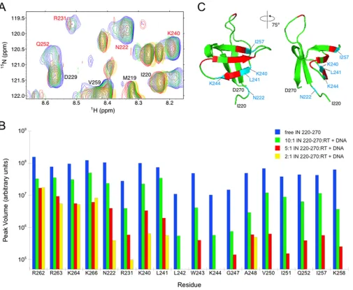
Mapping an RT-DNA binding surface on IN by NMR
spectros-copy.
An RT-binding surface on IN was previously determined by
NMR spectroscopy using recombinant IN220 –270
and apo-RT
(
42
). To determine whether the IN-RT interaction changes
signif-icantly when RT is bound to nucleic acid, we performed NMR
chemical shift perturbation experiments to characterize the
bind-ing surface on IN220 –270
in the presence of RT that had been
pre-bound to a duplex DNA segment corresponding to the
primer-binding site in the HIV-1 genome. X-ray crystal structures of this
particular duplex DNA construct bound to RT are available (
56
,
57
), and a dissociation constant of 38
⫾
16 nM for RT bound to
this DNA duplex has been measured (
58
). Such a dissociation
constant, along with the RT-DNA complex concentrations used in
the NMR experiments (14 to 54
M), suggests that
⬎
91% of RT is
DNA bound in our NMR experiments.
Successive aliquots of unlabeled RT prebound with duplex
DNA (1:1 molar ratio) were added to 150
M
15N-labeled sample
of IN
220 –270to produce IN
220 –270:RT-DNA ratios of 10:1, 5:1, and
2:1. A
15N-
1H HSQC spectrum of each sample was acquired. No
peaks showed any changes in chemical shift as the titration
pro-gressed, and most IN220 –270
signals showed relatively gradual
re-ductions in peak intensity during the course of the titration. At a
2:1 IN220 –270:RT-DNA molar ratio, most of the peaks in the
ac-quired spectrum became broadened to the noise level, and
there-fore no further titrations were performed.
Notably, amide backbone cross-peaks associated with 14
resi-dues showed significant decreases in intensity during the titration
compared to intensity levels of other peaks in the spectra (
Fig. 1A
and
B
). Nine of the selectively affected residues (R231, L242,
W243, G247, A248, V250, I251, Q252, and K258) had been
previ-ously mapped to an RT-binding surface on IN using analogous
experiments with
15N-labeled IN
220 –270
and unlabeled apo-RT
(
42
). In the current titration experiment, five additional residues
(N222, K240, L241, K244, and I257) also showed significant
amide peak decreases (
Fig. 1B
). These five additional residues
clustered with the initial set of nine residues identified in NMR
titration using free RT (
Fig. 1C
). The data suggest that including
the DNA substrate in these NMR experiments results in an
ex-panded RT-binding surface on IN, and the selective broadening of
the peaks for these particular 14 amino acids suggests that
recog-nition of IN220 –270
by the RT-DNA complex is specific (
59
).
Im-portantly, amide peaks associated with R262, R263, K264, and
K266, which are residues that form contacts with free DNA (
60–
62
), were among those peaks showing no significant intensity
de-creases over the course of the titration (
Fig. 1B
). The lack of
sig-nificant amide signal perturbation from these residues suggests
that the strong intensity changes in the IN220 –270
spectrum
ob-served in the presence of RT and DNA are due to IN
220 –270actions with the RT-DNA complex and not to nonspecific
inter-actions between free DNA and IN
220 –270. Finally, signal decreases
observed during the titration suggest that these 14 residues
expe-rience intermediate exchange on the NMR time scale (i.e.,
chem-ical exchange occurs at a frequency that is on the order of the
difference between free and bound amide group chemical shift)
(
63
,
64
).
Characterization of mutant IN viruses to determine
func-tional relevance of RT-IN interaction.
We evaluated the
func-tional relevance of this newly mapped surface on IN by examining
the effect of perturbing the putative interacting surface upon viral
replication. Since nine IN residues (R231, L242, W243, G247,
A248, V250, I251, Q252, and K258) appear to interact with RT
with or without DNA, we elected to focus our mutagenesis efforts
upon IN residues within this particular set. We have previously
investigated the effects of introducing substitutions at two of the
putative nine RT-binding residues and found that W243E and
V250E substitutions significantly impaired viral replication (
42
).
The exact phase of the viral life cycle affected by these particular
amino acid substitutions, as well as substitutions at other IN
po-sitions within the putative RT-binding surface, was analyzed in
this report. We hypothesize that RT-IN interactions are necessary
for reverse transcription during the viral life cycle and that
per-turbing this interaction will lead to a defect specifically at the
re-verse transcription step. To determine the functional relevance of
the putative RT-binding surface on the IN C-terminal domain, we
introduced single amino acid substitutions that alter surface
po-larity or electrostatic charge using site-directed mutagenesis. If the
IN residue is indeed involved in RT binding only and if the
bind-ing is critical durbind-ing viral replication, we expect that the
intro-duced substitution will disrupt RT binding and cause a replication
defect specifically at the reverse transcription step without other
secondary, off-target effects.
Analysis of replication kinetics.
We first investigated the
ef-fects of these various substitutions upon viral replication kinetics
by infecting CEM cells with equal amounts of p24 equivalent of
WT or IN mutant viruses. Virus production was monitored by
measuring p24 content in medium every 2 days over a 10-day
period (
Fig. 2
). Negative controls included a mock infection
(me-dium only without virus) and an infection using heat-inactivated
wild-type virus. Of the nine IN mutants tested (R231E, W243E,
on November 7, 2019 by guest
http://jvi.asm.org/
G247A, G247E, A248E, V250A, V250E, I251E, and Q252A),
only G247A replicated with kinetics similar to that of the WT
virus. In cells infected with either WT or IN mutant G247A,
syncytium formation was evident after day 6, which correlated
with a peak in the p24 level detected in culture (
Fig. 2B
and
C
).
IN mutants V250A and Q252A exhibited decreased p24 levels,
but levels were above background. Since these two IN mutants
still retain a low but detectable level of replication, they were
not further characterized. In contrast, mutants R231E, G247E,
A248E, and I251E were nonviable (
Fig. 2C
and
D
). IN mutants
W243E and V250E were also unable to replicate, as previously
reported (
42
).
Analysis of polyprotein packaging and processing.
Several IN
mutants have been previously reported to be replication defective
due to impaired polyprotein processing (
22
,
65
). We examined
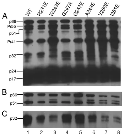
the effects of the six replication-incompetent IN mutants on viral
protein packaging and precursor processing by Western blot
anal-ysis. The G247A IN mutant, which replicates similarly to the WT
(
Fig. 2C
), was included as another positive control. One hundred
nanograms of the p24 equivalent of WT or IN mutant virus was
lysed, separated on a 12% denaturing polyacrylamide gel, and
transferred to nitrocellulose membranes. Blots were probed with
either human anti-HIV serum to detect all packaged viral proteins
(
Fig. 3A
) or with antibodies specific for RT or IN (
Fig. 3B
and
C
).
All IN mutants exhibited proper Gag-Pol packaging and
process-ing at levels comparable to those of the WT control (
Fig. 3A
and
FIG 1Mapping amino acids with significant changes in peak intensity to the IN220 –270 structure. (A) Chemical shift perturbation measurements using 15N-labeled IN
220 –270and unlabeled RT prebound with DNA. A representative region of the HSQC spectra shows free IN220 –270(blue) and IN220 –270mixed with RT-DNA at molar ratios of 10:1 (green), 5:1(red), and 2:1 (yellow). IN residues labeled in red show significant decreases in amide peak intensity upon addition of RT-DNA. (B) HSQC peak intensities as the titration proceeded for residues from the putative RT-DNA-binding surface on IN220 –270, as well as peak intensities from putative DNA-binding residues R262, R263, K264, and K266. Peak intensities for free15N-labeled IN
220 –270and for IN220 –270mixed with unlabeled RT-DNA at molar ratios of 10:1, 5:1, and 2:1 are shown, identified by color as indicated on the figure. Any peak intensities that dropped below the noise threshold of the NMR experiment during the course of the titration are not shown. (C) The apo-RT-binding surface on IN220 –270previously identified is shown in red, and the expanded set of five residues that are newly identified in the current study are shown in blue. Collectively, these 14 residues form the RT-DNA-binding surface on IN220 –270.
on November 7, 2019 by guest
http://jvi.asm.org/
[image:5.585.44.549.65.480.2]B
). The virion incorporation of IN in mutants W243E, V250E, and
I251E was further confirmed by probing with an anti-IN antibody
(
Fig. 3C
). Mutants W243E, V250E, and I251E contained less
in-corporated IN than that found in the WT and less inin-corporated IN
than the amount of incorporated RT. One possible explanation is
that the processed INs of mutants W243E, V250E, and I251E were
subjected to enhanced degradation. However, we found through
direct examination using immunoblotting that purified mutated
INs W243E, V250E, and I251E reacted poorly with the anti-HIV
human serum and the polyclonal anti-IN antibody (data not
shown). The low sensitivity of these particular detection reagents
against the mutated INs during immunoblotting represents at
least a contributing factor to the apparent poor incorporation of
processed IN with these IN mutants.
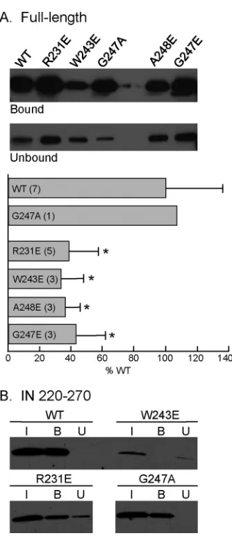
Replication-deficient IN mutants show decreased binding to
RT.
To determine if the replication defect of the IN mutants was
caused by interference with RT-IN complex formation, we
ex-pressed and purified the recombinant WT IN and various
single-amino-acid-substituted IN derivatives and measured their
abili-ties to bind immobilized RT by carrying out a pulldown assay
using RT-conjugated magnetic beads. Based on their WT levels of
viral protein packaging and processing, we selected IN mutants
R231E, W243E, G247E, and A248E for further analyses. A
reac-tion mixture containing 20 pmol of WT IN and magnetic beads
without any conjugated RT served as a negative control to assess
any nonspecific interaction, and IN binding to the beads was not
detected under these conditions (data not shown). When the
re-action mixture contained magnetic beads conjugated with RT,
FIG 2Replication kinetics of WT and IN mutant HIV-1 viral clones. CEM cells were infected with 100 ng of the p24 equivalent of WT or IN mutant viruses, and the culture medium was tested for p24 production at the indicated time points postinfection. Data representative of three replicates are shown. (A) The positions of the six IN substitution mutants on the RT-binding surface of IN220 –270are as labeled. (B) Replication kinetics of the WT (), heat-inactivated WT (HI;Œ), and mock control (no virus added;). (C) Replication kinetics of IN mutants R231E (), W243E (), G247A (Œ), and G247E (). (D) Replication kinetics of IN mutants A248E (), V250A (Œ), V250E (Œ), I251E (), and Q252A (}).
FIG 3Western blot analysis of HIV-1 proteins packaged in WT and IN mu-tant virus particles. Each lane contains 100 ng of the p24 equivalent of the viral lysate. Blots were probed with human HIV-1 serum (A), RT anti-body (B), or anti-IN antianti-body (C). Labels on the left correspond to positions of viral proteins with their molecular masses in kilodaltons. p66 and p51 corre-spond to the subunits of the RT heterodimer, Pr55 and Pr41 represent Gag precursor proteins, p32 corresponds to IN, p24 corresponds to capsid, and p17 corresponds to matrix.
on November 7, 2019 by guest
http://jvi.asm.org/
[image:6.585.114.476.64.341.2] [image:6.585.316.524.419.647.2]incubation with WT IN resulted in 29%
⫾
11% of the IN being
cosedimented (
Fig. 4A
). When the reaction mixture was
incu-bated with IN bearing the G247A substitution, which has no effect
on the replication kinetics (
Fig. 2C
), the amount of IN pulled
down by the beads was 107% of the WT level (
Fig. 4A
). In the
presence of an identical amount of RT-conjugated magnetic
beads, the levels of mutated INs R231E, W243E, G247E, and
A248E cosedimented with the beads were 39%
⫾
19%, 34%
⫾
14%, 36%
⫾
10%, and 43%
⫾
18%, respectively, of the WT IN
level (
Fig. 4A
). The levels of cosedimentation of these four IN
mutants were all significantly less than the WT IN level (
P
ⱕ
0.01).
We have shown previously that the C-terminal domain of IN
(IN
220 –270) is necessary and sufficient in binding RT, and a K258A
substitution in the context of IN220 –270
shows an 8-fold decrease
in binding affinity to RT compared to that of WT IN (
42
). To
further confirm the decreased ability of the mutated INs to bind
RT, we repeated the pulldown assay using purified IN
220 –270con-taining the R231E or W243E substitution (
Fig. 4B
). IN220 –270
bearing the G247A substitution was used as a positive control.
Compared to the WT IN, the cosedimented amounts of IN220 –270
containing the R231E, W243E, and G247A substitutions were
65%, 34%, and 98%, respectively, of the WT IN220 –270
(
Fig. 4B
).
Similar to the observation made earlier for virion incorporated
of IN in different mutants, the recovery (bound plus unbound)
variation among WT and mutated INs, especially W243E, was
likely attributable to their different reactivities to antibodies
dur-ing immunoblottdur-ing (data not shown).
Replication-defective and poor-RT-binding IN mutants are
impaired in early viral cDNA synthesis.
Our analyses thus far
have indicated that several amino acid substitutions on the
pur-ported RT-binding surface of IN results in
replication-incompe-tent viruses and disruption of the RT-IN interaction. To confirm
the biological relevance of the interaction, we examined the ability
of the four replication-defective and poor-RT-binding IN
mu-tants to synthesize viral cDNA. Experiments using mock-infected
cells and cells infected with heat-inactivated WT virus served as
negative controls. Viruses were treated with DNase I prior to
in-fection assays to eliminate contamination from any residual
NL4-3 plasmid DNA carried over from transfection. Total DNA
was extracted from target cells at 16 h postinfection and analyzed
for the early reverse transcription product, minus-strand strong
stop DNA (
⫺
sssDNA), using qPCR as previously described (
34
,
51
). DNA input from each sample was normalized to the level of
the human

-globin gene, which was amplified under identical
conditions. All four replication-defective IN mutants were
signif-icantly impaired for
⫺
sssDNA synthesis, exhibiting activity
rang-ing from 0 to 25% of the WT level (
Fig. 5A
).
To account for the possibility of a defective RT resulting in the
lack of reverse transcription products, we examined endogenous
reverse transcriptase activity in each IN mutant virus. Equal
amounts of detergent-lysed IN mutant virions were supplied with
an exogenous template and radiolabeled TTP as substrates, and
the amount of newly transcribed radioactive transcripts was
mea-sured. All IN mutants packaged RT at levels comparable to the
level of the wild-type virus (
Fig. 3B
). Based on the amount of
[
3H]TTP incorporated into the reverse transcription product, we
observed no significant difference in RT-catalyzed polymerization
activity in any of the mutant IN viruses relative to that of the WT
virus, ruling out the possibility that the observed lack of reverse
transcription products was due to a defective RT (
Fig. 5B
). These
results indicate that IN substitutions at the RT-binding surface
adversely impact reverse transcription, leading to defects in viral
replication without affecting endogenous RT activity.
Replication-defective and poor-RT-binding IN mutants are
competent in viral attachment and entry.
To verify that the
de-fect of the replication-incompetent IN mutants occurs specifically
during the step of reverse transcription, we determined the ability
of the four replication-defective and poor-RT-binding mutant
vi-ruses to attach and enter cells. CEM cells were incubated with
equal amounts of the p24 equivalent of WT virus or each of the
four IN mutant viruses. As a positive control, we included the
replication-incompetent IN C130S mutant, which is normal in
FIG 4Binding of HIV-1 RT-conjugated magnetic beads with the WT or the indicated substitutions within the context of full-length IN or IN220 –270. (A) Full-length IN. Bound and unbound fractions were obtained as described in Materials and Methods. The final volume of the unbound fraction was 500l, and a 40-l aliquot was analyzed by SDS-PAGE and immunoblotting. The level of WT IN binding to RT was normalized to 100%, and the values of mutant INs are expressed as percentages of the binding of the WT IN. Data are expressed as the means⫾standard deviations, and the numbers in parentheses represent the number of experiments. Asterisks denote significant differences from WT (Pⱕ0.01). (B) IN220 –270. The amounts of IN in the input, bound, and unbound fractions are denoted I, B, and U, respectively. The unbound fraction was TCA precipitated, and the pellet was resuspended in 30l of loading buffer before analysis by SDS-PAGE and immunoblotting.
on November 7, 2019 by guest
http://jvi.asm.org/
[image:7.585.80.247.60.446.2]cell attachment and entry and has a specific defect at the step of
reverse transcription (
28
,
34
). Negative controls included both
mock-infected cells and cells infected with heat-inactivated WT
virus. Following a 4-h incubation with virus, cells were either
treated with trypsin to facilitate subsequent detection of
intracel-lular p24 levels in cell lysates or washed and lysed without trypsin
treatment to quantify total amounts of both cell-associated and
intracellular p24. All IN mutants showed amounts of
cell-associ-ated and intracellular p24 comparable to those of the WT virus (
Fig.
6
). These results showed that all four IN mutants deficient in
replica-tion and reverse transcripreplica-tion have no defects in virus attachment
and entry.
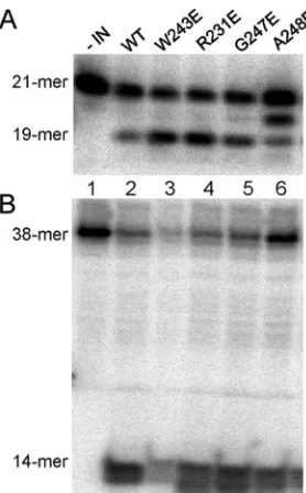
Recombinant mutant INs that bind RT poorly are
catalyti-cally active.
To investigate whether the IN substitutions affected
IN enzymatic activities, we assessed
in vitro
the ability of the
var-ious recombinant INs to catalyze the 3
=
end processing (
Fig. 7A
)
and disintegration reactions (
Fig. 7B
). All replication-defective
mutant INs maintained
in vitro
3
=
-end processing and
disintegra-tion activities similar to those of the WT enzyme (
Fig. 7
). Since the
four substitutions in the IN CTD did not adversely affect catalysis,
we infer that these substitutions do not grossly perturb protein
folding and IN dimerization required for proper 3
=
end processing
and disintegration.
DISCUSSION
In this study, we have mapped the putative RT-binding surface on
HIV-1 IN by NMR and determined the biological relevance of the
IN-RT interaction during viral replication. Using the structural
information as a guide, we have prepared and characterized nine
mutant viruses, with each containing a substitution of an IN
res-idue located within the RT-binding surface. Several IN mutants
are completely replication incompetent with the defect occurring
specifically during synthesis of early reverse transcription
prod-ucts. Purified recombinant HIV-1 INs containing such
substitu-tions, though catalytically active, are deficient in binding RT. The
specific impairment of early reverse transcription in viruses
har-boring IN substitutions at the putative RT-binding surface,
to-gether with the disruption of RT-binding by IN derivatives
con-taining such substitutions, supports our hypothesis that the
RT-IN interaction is biologically significant during reverse
tran-scription.
The mechanistic basis by which IN is specifically required for
reverse transcription during HIV-1 replication is not known.
Re-verse transcription catalyzed by recombinant HIV-1 RT is
ineffi-cient
in vitro
(
66
,
67
). This is partly due to poor processivity and a
propensity for RT to dissociate from the primer-template at pause
sites (
68
). The early events of reverse transcription include two
distinct modes: initiation, which corresponds to the
incorpora-tion of the first five nucleotides, has a primer-specific and
distrib-utive mode of polymerization, while elongation has a nonspecific
and processive mode of DNA synthesis (
69
). Using recombinant
proteins, we previously found that IN stimulates both the
initia-tion and elongainitia-tion modes of reverse transcripinitia-tion by increasing
RT processivity and suppressing formation of pause products
(
27
). Since all four replication-defective IN mutants examined
have a specific defect at the step of early reverse transcription and
FIG 5qPCR analysis of viral cDNA synthesis in cells infected with IN mutant viral clones. Values in the bar graphs represent the means of two replicates. (A) Amounts of early reverse-transcribed viral cDNA present in infected cells were measured 16 h after infection. DNA input of each sample was normalized by the amount of human-globin gene amplified under identical conditions. (B) Endogenous RT activity assay of cell lysates prepared from cells infected by WT or IN mutants. Radioactivity incorporated into reverse transcription products was determined by scintillation counting and expressed in disintegrations per minute (DPM). Viruses are described in the legend toFig. 2.
FIG 6Attachment and entry cell assay analysis of replication-defective IN mutants. CEM cells were infected with 100 ng of the p24 equivalent of WT or IN mutant viruses and analyzed for viral attachment (A) and entry (B) as described in Materials and Methods. Cells exposed to HI virus and a mock control were used as negative controls. Viruses are described in the legend to Fig. 2.
on November 7, 2019 by guest
http://jvi.asm.org/
[image:8.585.338.508.63.325.2] [image:8.585.61.268.64.330.2]since recombinant INs containing the same substitutions are
im-paired in RT binding, we speculate that the IN substitutions
dis-rupt RT-IN complex formation required in the early steps of
re-verse transcription, rendering the virus replication incompetent.
The RT-binding surface on IN has been determined previously
using IN
220 –270and apo-RT alone (
42
), and the effect of the
pres-ence of a DNA substrate on the RT-IN complex was not known.
Binding kinetics between IN and RT are consistent with a
two-state reaction process, suggesting that recognition of IN involves a
conformational change in RT (
42
). Indeed, X-ray crystal
struc-tures of apo-RT and DNA-bound RT (
56
,
57
) and molecular
dy-namics simulations (
70
) indicate that conformational flexibility
and mobility are prominent features of both p66 and p51 subunits
upon substrate recognition and binding. In particular, the p66
subunit adopts an open conformation when bound to nucleic acid
substrate, with the thumb subdomain rotating away from the
fin-gers subdomain to expose a large cleft, while the p66 of apo-RT
assumes a closed conformation, with the thumb domain filling the
cleft (
56
,
71–73
). RT-catalyzed DNA-dependent DNA
polymer-ization studies suggest that IN has greater affinity for RT bound to
a DNA template-primer pair than for RT alone (
44
), implying that
IN interacts with RT in the open conformation in this particular
setting.
To examine the RT-IN interaction under a more physiological
condition, we have performed the same NMR approach to
iden-tify the RT-binding surface on IN
220 –270in the presence of RT that
was prebound with equimolar amounts of a 19-mer/18-mer
du-plex DNA that mimics the primer-template substrate. In addition
to the nine amino acids identified earlier with apo-RT (
42
), the
introduction of the DNA substrate resulted in an expanded
bind-ing surface that included five more IN residues. With the
excep-tion of R231, the residues comprising this expanded binding
sur-face mapped to one side of the IN220 –270
structure (
Fig. 1C
). While
not located precisely at the RT-binding surface, R231 is positioned
within a flexible loop connecting the

1 and

2 strands near the
RT-binding surface (
52
) and can conceivably extend to form RT
contacts with or without conformational adjustment.
We attribute the expanded binding surface to IN
220 –270inter-actions with the RT-DNA complex rather than to any free DNA
species present in our NMR titration experiment. In support of
this interpretation, the previously measured dissociation constant
of
⬃
38 nM for this RT-DNA template-primer complex (
58
)
sug-gests that free DNA levels in our system should be relatively low
(
⬍
9% of the total DNA used in these NMR experiments).
Fur-thermore, we expect that any free DNA in this system would bind
nonspecifically to the positively charged residues R262, R263, and
K264 that are located on the face opposite to the putative
RT-binding surface of IN
220 –270. Previous work with the HIV-2 IN
CTD indicates that R262, R263, and K264 are involved in
nonspe-cific DNA binding (
61
,
62
), and homology modeling of HIV-1 IN
onto the PFV intasome structure, along with cross-linking assays
with DNA and the K266E mutant IN, suggests that HIV-1 IN K266
is involved in forming DNA contacts (
60
). We observed no strong
peak intensity decreases for R262, R263, K264, or K266 during the
course of our NMR titration experiments. Instead, peak
intensi-ties for residues from the putative RT-binding surface drop off
much more quickly than those residues expected to form DNA
contacts (
Fig. 1B
). This result is consistent with a view that the set
of residues showing rapid peak changes form interactions with the
RT-DNA complex and not free DNA, as any free DNA present in
the system should produce peak changes on the opposite side of
the molecule (i.e., at R262, R263, K264, and K266). In all, our data
support a model that includes a specific RT-binding surface on IN
that expands when it interacts with DNA-bound RT. Moreover,
identifying the same protein surface on IN220 –270
in the presence
of the RT-DNA complex or apo-RT alone further validates the
biological relevance of this IN surface in binding RT.
IN mutations can be pleiotropic, affecting various steps in the
virus life cycle (
18
,
19
). Besides integration and reverse
transcrip-tion, other steps include nuclear import of preintegration
com-plexes (
26
,
35–37
), polyprotein processing, assembly, and
matu-ration (
22
,
31
,
38–40
,
65
,
74
). Since IN is synthesized and
packaged into the immature virion as part of the Gag-Pol
poly-protein, many alterations in virion morphology and defects at the
late steps of replication may be caused by the effect of IN
muta-tions on the Gag-Pol precursor protein (
22
,
38
,
40
,
65
,
74
).
How-ever, certain IN mutations, such as His12Ala and Phe185Ala
sub-stitutions and deletion of the C-terminal 22 residues, specifically
impair reverse transcription with no apparent effects on the
Gag-Pol polyprotein and other steps in the life cycle (
29
,
30
,
33
). Also,
many class II mutations involving residues in the IN CTD share a
reverse transcription defect, with no apparent alteration to
poly-protein processing and packaging by the IN substitutions (
22
).
Mutagenesis experiments involving some of the RT-binding IN
residues identified in this study have been reported previously. In
support of our finding that the 14 residues identified by the NMR
represent the RT-binding surface, replacing the conserved Leu
residue at position 241 with Ala (L241A) resulted in a
replication-defective virus at the cDNA synthesis step (
22
). Substituting Glu
FIG 7In vitroIN activity assays for WT and IN mutants. (A) 3=End process-ing. 5=-end32P-labeled 21-mer substrate was incubated with 40 pmol of WT (lane 2) or mutated INs (lanes 3 to 6) to assess dinucleotide cleavage activity, which produces a 19-mer product. (B) Disintegration activity catalyzed by WT or mutated INs using a 5=-end32P-labeled disintegration substrate. The assay measures the ability of IN to resolve the substrate mimicking the integration intermediate into a 14-nucleotide labeled hairpin product (viral DNA) and an unlabeled 24-nucleotide circular molecule (target DNA). In both panels, lane 1 represents a negative control that contained the labeled substrate without IN.
on November 7, 2019 by guest
http://jvi.asm.org/
[image:9.585.92.231.64.288.2]for the important DNA-interacting residue Lys244, which is
among the five additional residues constituting the expanded
RT-binding surface on IN when RT is prebound with DNA, led to a
replication-defective virus with WT levels of late
reverse-tran-scribed products and defective IN enzymatic activity (
75
);
how-ever, a double IN mutant, K240A/K244E, exhibited a significant
decrease in this reverse transcription product (
26
). Also, the
iden-tity of the amino acid substitution plays an important role in the
phenotype of the resulting mutant. In this study, we showed that
replication of the mutant virus G247A is indistinguishable from
that of the WT, whereas the G247E mutant is replication defective
and has impaired RT binding. Similarly, we found that the viral
clone R231E is replication incompetent and defective in binding
RT, whereas an earlier study reported that an R231A mutant is
replication competent (
22
). Additionally, virus harboring Gly247
with a Trp (G247W) substitution is replication incompetent, with
a Gag processing defect caused by activation of an alternative
splice site (
65
). We did not observe any effects on Gag-Pol
pro-cessing when the same amino acid was replaced with either Ala or
Glu (G247A or G247E) (
Fig. 3
).
Of note, the host protein Gemin2/SIP1, a member of the
sur-vival motor neuron complex that mediates the assembly of
spli-ceosomal small nuclear ribonucleoproteins (
76
), has been
re-ported to interact with HIV-1 IN to facilitate reverse transcription
(
77
). Knockdown of Gemin2 with small interfering RNA reduces
HIV-1 infectivity and lowers early reverse transcription products
but has no effect on HIV-1 expression from integrated proviruses
(
77
). Further analyses showed that disrupting the IN-Gemin2
in-teraction abrogates reverse transcription, and Gemin2 functions
to promote reverse transcription by stabilizing IN
multimeriza-tion and enhancing IN-RT assembly on viral RNA (
43
).
There-fore, in addition to direct binding, IN may impact reverse
tran-scription in the context of an IN-Gemin2 complex. Residues
critical for Gemin2 interaction are located within the central core
and C-terminal domains of IN (
43
,
77
). In addition to Gemin2,
several other host proteins have been shown to interact with
HIV-1 IN (for a review, see reference
78
). Some of these host
proteins, such as Ku70 and dynein, can bind to the IN CTD, and
their interactions with IN appear to play a role during reverse
transcription (
79
,
80
). We do not know at present if these
IN-binding host proteins can affect positively the RT-IN interaction
or compete with RT for IN binding. Notably, we have observed
that several IN mutants exhibit a greater defect on replication than
RT binding (compare
Fig. 2
and
4
). It is possible that the
cosedi-mentation assay does not fully recapitulate the RT-IN interaction
in vivo
or that the IN substitutions tested in this study may
addi-tionally affect the binding of IN with other host proteins, which
consequently contribute to the overall defect in viral replication.
Our results showing that substitutions at the RT-binding
sur-face on IN significantly impair viral replication in tissue culture,
specifically at the reverse transcription step, indicate that the
RT-IN interaction is functionally significant during viral
replica-tion. Considering the emergence of viral resistance observed
against many clinically relevant anti-HIV drugs that target the
enzymatic active sites of either RT or IN, the critical interaction
between RT and IN may function as an allosteric target for
design-ing and developdesign-ing new antiviral drugs.
ACKNOWLEDGMENTS
The ELISA for p24 was carried out by the Center for AIDS Research (CFAR) Virology Core Facility of the University of California, Los Angeles (UCLA), which is supported by UCLA CFAR grant 5P30 AI028697 and the UCLA AIDS Institute and by the UCLA Council of Bioscience Re-sources. This work was partly supported by NIH grant GM101416 and an International Collaborative grant (number 2012C24015) from the Science Technology Department of Zhejiang Province, China, to S.A.C. S.S.T. was partly supported by Research Training in Pharmacolog-ical Sciences (NIH T32GM008652) and a UCLA AIDS Institute/CFAR Fellowship Award. J.T.M. and S.F.J.L.G. are supported by the Intramural Research Program of the National Cancer Institute, NIH, Department of Health and Human Services.
REFERENCES
1.Hill M, Tachedjian G, Mak J.2005. The packaging and maturation of the HIV-1 Pol proteins. Curr HIV Res3:73– 85.http://dx.doi.org/10.2174 /1570162052772942.
2.Telesnitsky A, Goff SP.1997. Reverse transcriptase and the generation of retroviral DNA, p 121–160.InCoffin JM, Hughes SH, Varmus HE (ed), Retroviruses. Cold Spring Harbor Laboratory Press, Cold Spring Harbor Laboratory, NY.
3.Hu WS, Hughes SH. 2012. HIV-1 reverse transcription. Cold Spring Harb Perspect Med 2:a006882. http://dx.doi.org/10.1101/cshperspect .a006882.
4.Jacobo-Molina A, Arnold E.1991. HIV reverse transcriptase structure-function relationships. Biochemistry30:6351– 6356.http://dx.doi.org/10 .1021/bi00240a001.
5.Engelman A, Mizuuchi K, Craigie R. 1991. HIV-1 DNA integration: mechanism of viral DNA cleavage and DNA strand transfer. Cell67:1211– 1221.http://dx.doi.org/10.1016/0092-8674(91)90297-C.
6.Li X, Krishnan L, Cherepanov P, Engelman A.2011. Structural biology of retroviral DNA integration. Virology411:194 –205.http://dx.doi.org /10.1016/j.virol.2010.12.008.
7.Chow SA, Vincent KA, Ellison V, Brown PO.1992. Reversal of integra-tion and DNA splicing mediated by integrase of human immunodefi-ciency virus. Science 255:723–726. http://dx.doi.org/10.1126/science .1738845.
8.Hare S, Di Nunzio F, Labeja A, Wang J, Engelman A, Cherepanov P.
2009. Structural basis for functional tetramerization of lentiviral inte-grase. PLoS Pathog 5:e1000515. http://dx.doi.org/10.1371/journal .ppat.1000515.
9.Hare S, Gupta SS, Valkov E, Engelman A, Cherepanov P.2010. Retro-viral intasome assembly and inhibition of DNA strand transfer. Nature
464:232–236.http://dx.doi.org/10.1038/nature08784.
10. Li M, Mizuuchi M, Burke TR, Jr, Craigie R.2006. Retroviral DNA integration: reaction pathway and critical intermediates. EMBO J25:
1295–1304.http://dx.doi.org/10.1038/sj.emboj.7601005.
11. Hare S, Maertens GN, Cherepanov P.2012. 3=-Processing and strand transfer catalysed by retroviral integrasein crystallo. EMBO J31:3020 – 3028.http://dx.doi.org/10.1038/emboj.2012.118.
12. Maertens GN, Hare S, Cherepanov P.2010. The mechanism of retroviral integration from X-ray structures of its key intermediates. Nature468:
326 –329.http://dx.doi.org/10.1038/nature09517.
13. Bera S, Pandey KK, Vora AC, Grandgenett DP.2009. Molecular Inter-actions between HIV-1 integrase and the two viral DNA ends within the synaptic complex that mediates concerted integration. J Mol Biol389:
183–198.http://dx.doi.org/10.1016/j.jmb.2009.04.007.
14. Faure A, Calmels C, Desjobert C, Castroviejo M, Caumont-Sarcos A, Tarrago-Litvak L, Litvak S, Parissi V.2005. HIV-1 integrase crosslinked oligomers are active in vitro. Nucleic Acids Res33:977–986.http://dx.doi .org/10.1093/nar/gki241.
15. Kotova S, Li M, Dimitriadis EK, Craigie R.2010. Nucleoprotein inter-mediates in HIV-1 DNA integration visualized by atomic force micros-copy. J Mol Biol399:491–500.http://dx.doi.org/10.1016/j.jmb.2010.04 .026.
16. Krishnan L, Engelman A.2012. Retroviral integrase proteins and HIV-1 DNA integration. J Biol Chem287:40858 – 40866.http://dx.doi.org/10 .1074/jbc.R112.397760.
17. Wang JY, Ling H, Yang W, Craigie R.2001. Structure of a two-domain fragment of HIV-1 integrase: implications for domain organization in the
on November 7, 2019 by guest
http://jvi.asm.org/
intact protein. EMBO J20:7333–7343.http://dx.doi.org/10.1093/emboj /20.24.7333.
18. Cannon PM, Wilson W, Byles E, Kingsman SM, Kingsman AJ.1994. Human immunodeficiency virus type 1 integrase: effect on viral replica-tion of mutareplica-tions at highly conserved residues. J Virol68:4768 – 4775. 19. Engelman A, Englund G, Orenstein JM, Martin MA, Craigie R.1995.
Multiple effects of mutations in human immunodeficiency virus type 1 integrase on viral replication. J Virol69:2729 –2736.
20. Engelman A.1999. In vivo analysis of retroviral integrase structure and function. Adv Virus Res 52:411– 426. http://dx.doi.org/10.1016/S0065 -3527(08)60309-7.
21. Li X, Koh Y, Engelman A. 2012. Correlation of recombinant integrase activity and functional preintegration complex formation during acute infec-tion by replicainfec-tion-defective integrase mutant human immunodeficiency vi-rus. J Virol86:3861–3879.http://dx.doi.org/10.1128/JVI.06386-11. 22. Lu R, Ghory HZ, Engelman A. 2005. Genetic analyses of conserved
residues in the carboxyl-terminal domain of human immunodeficiency virus type 1 integrase. J Virol79:10356 –10368.http://dx.doi.org/10.1128 /JVI.79.16.10356-10368.2005.
23. Lu R, Limon A, Ghory HZ, Engelman A. 2005. Genetic analyses of DNA-binding mutants in the catalytic core domain of human immuno-deficiency virus type 1 integrase. J Virol79:2493–2505.http://dx.doi.org /10.1128/JVI.79.4.2493-2505.2005.
24. Briones MS, Chow SA.2010. A new functional role of HIV-1 integrase during uncoating of the viral core. Immunol Res48:14 –26.http://dx.doi .org/10.1007/s12026-010-8164-z.
25. Briones MS, Dobard CW, Chow SA.2010. Role of human immunode-ficiency virus type 1 integrase in uncoating of the viral core. J Virol84:
5181–5190.http://dx.doi.org/10.1128/JVI.02382-09.
26. Ao Z, Fowke KR, Cohen EA, Yao X.2005. Contribution of the C-termi-nal tri-lysine regions of human immunodeficiency virus type 1 integrase for efficient reverse transcription and viral DNA nuclear import. Retrovi-rology2:62.http://dx.doi.org/10.1186/1742-4690-2-62.
27. Dobard CW, Briones MS, Chow SA.2007. Molecular mechanisms by which human immunodeficiency virus type 1 integrase stimulates the early steps of reverse transcription. J Virol81:10037–10046.http://dx.doi .org/10.1128/JVI.00519-07.
28. Hehl EA, Joshi P, Kalpana GV, Prasad VR.2004. Interaction between human immunodeficiency virus type 1 reverse transcriptase and integrase proteins. J Virol78:5056 –5067.http://dx.doi.org/10.1128/JVI.78.10.5056 -5067.2004.
29. Leavitt AD, Robles G, Alesandro N, Varmus HE.1996. Human immu-nodeficiency virus type 1 integrase mutants retain in vitro integrase activ-ity yet fail to integrate viral DNA efficiently during infection. J Virol70:
721–728.
30. Masuda T, Planelles V, Krogstad P, Chen ISY.1995. Genetic analysis of human immunodeficiency virus type 1 integrase and the U3attsite: un-usual phenotype of mutants in the zinc finger-like domain. J Virol69:
6687– 6696.
31. Shin C-G, Taddeo B, Haseltine WA, Farnet CM.1994. Genetic analysis of the human immunodeficiency virus type 1 integrase protein. J Virol
68:1633–1642.
32. Tsurutani N, Kubo M, Maeda Y, Ohashi T, Yamamoto N, Kannagi M, Masuda T.2000. Identification of critical amino acid residues in human immunodeficiency virus type 1 IN required for efficient provi-ral DNA formation at steps prior to integration in dividing and non-dividing cells. J Virol74:4795– 4806.http://dx.doi.org/10.1128/JVI.74 .10.4795-4806.2000.
33. Wu X, Liu H, Xiao H, Conway JA, Hehl E, Kalpana GV, Prasad V, Kappes JC.1999. Human immunodeficiency virus type 1 integrase pro-tein promotes reverse transcription through specific interactions with the nucleoprotein reverse transcription complex. J Virol73:2126 –2135. 34. Zhu K, Dobard C, Chow SA.2004. Requirement for integrase during
reverse transcription of human immunodeficiency virus type 1 and the effect of cysteine mutations of integrase on its interactions with reverse transcriptase. J Virol78:5045–5055.http://dx.doi.org/10.1128/JVI.78.10 .5045-5055.2004.
35. Ikeda T, Nishitsuji H, Zhou X, Nara N, Ohashi T, Kannagi M, Masuda T.2004. Evaluation of the functional involvement of human immunode-ficiency virus type 1 integrase in nuclear import of viral cDNA during acute infection. J Virol78:11563–11573.http://dx.doi.org/10.1128/JVI.78 .21.11563-11573.2004.
36. Jayappa KD, Ao Z, Yang M, Wang J, Yao X.2011. Identification of
critical motifs within HIV-1 integrase required for importin␣3 interac-tion and viral cDNA nuclear import. J Mol Biol410:847– 862.http://dx .doi.org/10.1016/j.jmb.2011.04.011.
37. Woodward CL, Prakobwanakit S, Mosessian S, Chow SA.2009. Inte-grase interacts with nucleoporin NUP153 to mediate the nuclear import of human immunodeficiency virus type 1. J Virol83:6522– 6533.http://dx .doi.org/10.1128/JVI.02061-08.
38. Bukovsky A, Gottlinger H.1996. Lack of integrase can markedly affect human immunodeficiency virus type 1 particle production in the presence of an active viral protease. J Virol70:6820 – 6825.
39. Lu R, Limon A, Devroe E, Silver PA, Cherepanov P, Engelman A.2004. Class II integrase mutants with changes in putative nuclear localization signals are primarily blocked at a postnuclear entry step of human immu-nodeficiency virus type 1 replication. J Virol78:12735–12746.http://dx .doi.org/10.1128/JVI.78.23.12735-12746.2004.
40. Quillent C, Borman AM, Paulous S, Dauguet C, Clavel F.1996. Exten-sive regions of pol are required for efficient human immunodeficiency virus polyprotein processing and particle maturation. Virology219:29 – 36.http://dx.doi.org/10.1006/viro.1996.0219.
41. Taddeo B, Haseltine WA, Farnet CM.1994. Integrase mutants of human immunodeficiency virus type 1 with a specific defect in integration. J Virol
68:8401– 8405.
42. Wilkinson TA, Januszyk K, Phillips ML, Tekeste SS, Zhang M, Miller JT, Le Grice SF, Clubb RT, Chow SA.2009. Identifying and character-izing a functional HIV-1 reverse transcriptase-binding site on integrase. J Biol Chem284:7931–7939.http://dx.doi.org/10.1074/jbc.M806241200. 43. Nishitsuji H, Hayashi T, Takahashi T, Miyano M, Kannagi M, Masuda
T.2009. Augmentation of reverse transcription by integrase through an interaction with host factor, SIP1/Gemin2 is critical for HIV-1 infection. PLoS One4:e7825.http://dx.doi.org/10.1371/journal.pone.0007825. 44. Tasara T, Maga G, Hottiger MO, Hubscher U.2001. HIV-1 reverse
transcriptase and integrase enzymes physically interact and inhibit each other. FEBS Lett 507:39 – 44. http://dx.doi.org/10.1016/S0014 -5793(01)02945-3.
45. Adachi A, Gendelman HE, Koenig S, Folks T, Willey R, Rabson A, Martin MA.1986. Production of acquired immunodeficiency syndrome-associated retrovirus in human and nonhuman cells transfected with an infectious molecular clone. J Virol59:284 –291.
46. Fisher CL, Pei GK.1997. Modification of a PCR-based site-directed mutagenesis method. Biotechniques23:570 –574.
47. Shibagaki Y, Chow SA.1997. Central core domain of retroviral integrase is responsible for target site selection. J Biol Chem272:8361– 8369.http: //dx.doi.org/10.1074/jbc.272.13.8361.
48. Le Grice SF, Gruninger-Leitch F.1990. Rapid purification of homodimer and heterodimer HIV-1 reverse transcriptase by metal chelate affinity chromatography. Eur J Biochem187:307–314.http://dx.doi.org/10.1111 /j.1432-1033.1990.tb15306.x.
49. Grandgenett DP, Goodarzi G.1994. Folding of the multidomain human immunodeficiency virus type-I integrase. Protein Sci3:888 – 897. 50. Khan M, Garcia-Barrio M, Powell MD.2001. Restoration of wild-type
infectivity to human immunodeficiency virus type 1 strains lacking nef by intravirion reverse transcription. J Virol75:12081–12087.http://dx.doi .org/10.1128/JVI.75.24.12081-12087.2001.
51. Korin YD, Zack JA.1998. Progression to the G1b phase of the cell cycle is required for completion of human immunodeficiency virus type 1 reverse transcription in T cells. J Virol72:3161–3168.
52. Lodi PJ, Ernst JA, Kuszewski J, Hickman AB, Engelman A, Craigie R, Clore GM, Gronenborn AM.1995. Solution structure of the DNA bind-ing domain of HIV-1 integrase. Biochemistry34:9826 –9833.http://dx.doi .org/10.1021/bi00031a002.
53. Goddard TD, Kneller DG.2008. Sparky 3. University of California, San Francisco, CA.
54. Schrodinger LLC.2012. The PyMOL molecular graphics system, version 1.5.0.5. Schrodinger, LLC, New York, NY.
55. Chow SA.1997. In vitro assays for activities of retroviral integrase. Meth-ods12:306 –317.http://dx.doi.org/10.1006/meth.1997.0484.
56. Ding J, Das K, Hsiou Y, S