Parvovirus Expresses a Small Noncoding
RNA That Plays an Essential Role in Virus
Replication
Zekun Wang,
aWeiran Shen,
aFang Cheng,
aXuefeng Deng,
aJohn F. Engelhardt,
bZiying Yan,
bJianming Qiu
aDepartment of Microbiology, Molecular Genetics and Immunology, University of Kansas Medical Center, Kansas City, Kansas, USAa; Department of Anatomy and Cell Biology, University of Iowa, Iowa City, Iowa, USAb
ABSTRACT
Human bocavirus 1 (HBoV1) belongs to the species
Primate
bocaparvo-virus
of the genus
Bocaparvovirus
of the
Parvoviridae
family. HBoV1 causes acute
re-spiratory tract infections in young children and has a selective tropism for the apical
surface of well-differentiated human airway epithelia (HAE). In this study, we
identi-fied an additional HBoV1 gene, bocavirus-transcribed small noncoding RNA (BocaSR),
within the 3
=
noncoding region (nucleotides [nt] 5199 to 5338) of the viral genome
of positive sense. BocaSR is transcribed by RNA polymerase III (Pol III) from an
intra-genic promoter at levels similar to that of the capsid protein-coding mRNA and is
essential for replication of the viral DNA in both transfected HEK293 and infected
HAE cells. Mechanistically, we showed that BocaSR regulates the expression of
HBoV1-encoded nonstructural proteins NS1, NS2, NS3, and NP1 but not NS4. BocaSR
is similar to the adenovirus-associated type I (VAI) RNA in terms of both nucleotide
sequence and secondary structure but differs from it in that its regulation of viral
protein expression is independent of RNA-activated protein kinase (PKR) regulation.
Notably, BocaSR accumulates in the viral DNA replication centers within the nucleus
and likely plays a direct role in replication of the viral DNA. Our findings reveal
BocaSR to be a novel viral noncoding RNA that coordinates the expression of viral
proteins and regulates replication of viral DNA within the nucleus. Thus, BocaSR may
be a target for antiviral therapies for HBoV and may also have utility in the
produc-tion of recombinant HBoV vectors.
IMPORTANCE
Human bocavirus 1 (HBoV1) is pathogenic to humans, causing acute
respiratory tract infections in young children. In this study, we identified a novel
HBoV1 gene that lies in the 3
=
noncoding region of the viral positive-sense genome
and is transcribed by RNA polymerase III into a noncoding RNA of 140 nt. This
bocavirus-transcribed small RNA (BocaSR) diverges from both adenovirus-associated
(VA) RNAs and Epstein-Barr virus-encoded small RNAs (EBERs) with respect to RNA
sequence, representing a third species of this kind of Pol III-dependent viral
noncod-ing RNA and the first noncodnoncod-ing RNA identified in autonomous parvoviruses. Unlike
the VA RNAs, BocaSR localizes to the viral DNA replication centers of the nucleus
and is essential for expression of viral nonstructural proteins independent of
RNA-activated protein kinase R and replication of HBoV1 genomes. The identification of
BocaSR and its role in virus DNA replication reveals potential avenues for developing
antiviral therapies.
KEYWORDS
bocavirus, DNA replication, noncoding RNA, parvovirus
H
uman bocavirus 1 (HBoV1), which was discovered in 2005 (1), belongs to the
species
Primate bocaparvovirus 1
in the genus
Bocaparvovirus
of the
Parvoviridae
family (2). Increasing evidence suggests that HBoV1 is an etiological pathogen rather
than a bystander in acute respiratory tract infections, especially in children under 5
Received7 December 2016Accepted20 January 2017
Accepted manuscript posted online25 January 2017
CitationWang Z, Shen W, Cheng F, Deng X, Engelhardt JF, Yan Z, Qiu J. 2017. Parvovirus expresses a small noncoding RNA that plays an essential role in virus replication. J Virol 91: e02375-16.https://doi.org/10.1128/JVI.02375-16.
EditorGrant McFadden, The Biodesign Institute, Arizona State University
Copyright© 2017 American Society for Microbiology.All Rights Reserved.
Address correspondence to Jianming Qiu, [email protected].
GENOME REPLICATION AND REGULATION
OF VIRAL GENE EXPRESSION
crossm
on November 7, 2019 by guest
http://jvi.asm.org/
years of age (3). Acute respiratory infections have been clearly linked to HBoV1 infection
as assessed by monodetection, high viral loads (
⬎
10
4viral genomic copies per ml of
respiratory specimen) (4–17), the presence of HBoV1-specific IgM, or a
ⱖ
4-fold increase
in levels of HBoV1-specific IgG antibodies (16, 18–20).
HBoV1 is a nonenveloped icosahedral virus with a linear single-stranded DNA
(ssDNA) genome of 5.5 kb (21). Two terminal palindromic sequences, termed the
left-end hairpin (LEH) and right-end hairpin (REH), correspond to the 3
=
and 5
=
ends,
respectively, of the negative-sense viral genome.
The HBoV1 genome encodes two groups of genes: a set that expresses
nonstruc-tural proteins and another that expresses strucnonstruc-tural (capsid) proteins (VP). One unique
feature of the bocaparvoviruses is the expression of an additional nonstructural protein,
NP1, whose open reading frame (ORF) is located in the middle of the viral genome and
overlaps with the C terminus of the NS1 ORF but is in a different reading frame (22, 23).
NS1, NS2, NS3, and NS4 are of
⬃
100,
⬃
66,
⬃
69, and
⬃
34 kDa, respectively, and share
a C terminus (amino acids [aa] 639 to 781) (24). NS1, which has a putative DNA origin
binding/endonuclease domain (OBD), a helicase activity domain, and a transactivation
domain (TAD) within its N-terminal, middle, and C-terminal regions, respectively, is
essential to replication of the viral DNA (24). NS2 contains the entire OBD and TAD of
the NS1, whereas NS3 contains the helicase domain and TAD of NS1 and NS4 contains
only the TAD. NS2 to -4 are not required for replication of the duplex viral genome
(pIHBoV1) in HEK293 cells; however, NS2 plays an important role during infection of
differentiated human airway epithelial cells (24). The functions of NS3 and NS4 are
currently unknown. NP1, which is comprised of 219 aa, has a molecular mass of 25 kDa.
It plays important roles not only in replication of the viral DNA (21, 23) but also in
processing of the viral mRNA transcripts (25, 26). NP1 is required for the splicing of viral
mRNAs, as well as for read-through from the proximal polyadenylation site (26).
Therefore, NP1 is essential for both the generation of VP-encoding mRNAs and the
production of viral capsid proteins.
The only
in vitro
system in which HBoV1 has been found to be capable of infection
is differentiated (nondividing) epithelial cells of the human airway epithelium (HAE)
cultured at an air-liquid interface (HAE-ALI) (21, 27–30). Neither dividing primary airway
epithelial cells nor monolayer cultures of cell lines derived from the airway epithelium
support significant HBoV1 infection or the replication of pIHBoV1 following transfection
(30). However, HEK293 cells support the replication following transfection of pIHBoV1
plasmid containing the full-length duplex genome, as well as the generation of
infectious progeny virions (21, 24). During infection of nondividing HAE cells (30) and
replication of the duplex HBoV1 genome in dividing HEK293 cells (31), HBoV1
con-scripts the cellular DNA damage and repair machinery to amplify the viral genome,
making HBoV1 unique among autonomous parvoviruses in that its replication is cell
cycle independent (32).
We previously found that replication of the duplex HBoV1 genome in HEK293 cells
requires the presence of a replication origin (Ori) within the REH and the 3
=
end
noncoding region (NCR), as well as the HBoV1 NS1 and NP1 proteins (33). In the current
study, we showed that the 3
=
NCR produces a small nonpolyadenylated RNA of 140
nucleotides (nt) from an intragenic RNA polymerase III (Pol III) promoter. We also
demonstrated that this small RNA is essential for both the productive infection of
nondividing HAE-ALI cultures by HBoV1 and replication of the viral DNA in dividing
HEK293 cells.
RESULTS
HBoV1 expresses a nonpolyadenylated small RNA in the contexts of both
DNA transfection and viral infection.
We previously analyzed the expression
profiles of HBoV1 genes by Northern blotting of mRNA isolated from HEK293 cells
transfected with a plasmid that contains the nonreplicating HBoV1
NS
and
Cap
genes
(pHBoV1NSCap), using
NS
,
Cap
, and
NSCap
probes (34) as shown in Fig. 1A, in which a
negative-sense genome (
⫺
ssDNA) of HBoV1 is diagrammed. We also analyzed
on November 7, 2019 by guest
http://jvi.asm.org/
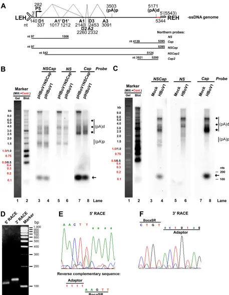
FIG 1HBoV1 encodes a novel nonpolyadenylated small RNA. (A) Schematic diagram of the HBoV1 genome. The negative-sense single-stranded (⫺ssDNA) genome is depicted. Indicated are the locations of the transcription units, promoter P5, splice donors (D1, D1=, D2, and D3), splice acceptors (A1, A1=, A2, and A3), and proximal and distal polyadenylation sites [(pA)p and (pA)d, respectively]. The left- and right-end hairpin (LEH and REH) structures are also shown, as are the 3=noncoding region (NCR, red) and the probes used for Northern blot analysis (nucleotide numbers included). (B and C) Expression of viral transcripts in transfected HEK293 cells and infected HAE-ALI cultures. (B) HEK293 cells were transfected with pHBoV1NSCap or pIHBoV1. (C) HAE-ALI cultures were infected with HBoV1 or mock infected. At 48 h posttransfection, or at 10 days
(Continued on next page)
A Noncoding RNA Is Required for Parvovirus Replication Journal of Virology
on November 7, 2019 by guest
http://jvi.asm.org/
[image:3.585.40.497.72.661.2]encoding mRNA transcripts in total RNA isolated from pCMVNS*Cap-transfected
HEK293 cells, using a
Cap2
probe spanning nt 3921 to 5200 and an
NSCap2
probe
spanning nt 542 to 5124 (26). These analyses revealed that transfection with the HBoV1
duplex genome produces NS-, NP1-, and VP-encoding mRNAs (26, 34) (Fig. 1B).
Notably, the above-described analyses of total RNA isolated from the
pHBoV1NSCap-transfected HEK293 cells detected a small transcript in the range of 100 to 200 nt on
blots by
NSCap
and
Cap
probes (Fig. 1B, lanes 3 and 7, respectively) but not on those
by an
NS
probe (Fig. 1B, lane 5). This small RNA band was also detected in total RNA
isolated from HEK293 cells transfected with pIHBoV1 using the
NSCap
and
Cap
probes
(Fig. 1B, lanes 4 and 8) but not the
NS
probe (Fig. 1B, lane 6). Analysis of the total RNA
isolated from HBoV1-infected HAE cells revealed the same result (Fig. 1C, lanes 4 and
8 versus 6) and that this small RNA is expressed at a level similar to that of the
VP-coding mRNA during infection. These findings suggested that an HBoV1-specifc
small RNA of approximately 150 nt is encoded at the 3
=
end of the duplex HBoV1
genome. We named this small RNA bocavirus-transcribed small RNA (BocaSR).
Given that the
⬃
150-nt BocaSR was detected by probes spanning HBoV1 sequences
that extend through nt 5395 (Fig. 1B and C) but not with those that end at or before
nt 5200 (26), and that it is present only in total RNA and not in mRNA samples (34),
we speculated that this transcript was encoded by HBoV1 sequence encompassed by
the fragment from nt 5124 to 5395 and not a spliced form of the viral polyadenylated
mRNA. To identify the exact ends of the BocaSR, we performed both 5
=
and 3
=
rapid
amplification of cDNA ends (RACE) (Fig. 1D). Subsequent sequencing of the 5
=
RACE
product identified nt 5199 of the HBoV1 genome as the 5
=
end, with the junction
sequence reading 5
=
-tttt/AAGTT-3
=
(capital letter represents viral sequence) (Fig. 1E).
Sequencing of the 3
=
RACE product identified the junction sequence between the
adaptor and HBoV1 sequence as 5
=
-CTGT/actgtag-3
=
(sequences in lowercase represent
the nucleotides on the adaptor), confirming the 3
=
end of the BoaSR to be nt 5338 (Fig.
1F). We concluded that BocaSR spans nt 5199 to 5338.
Collectively, these results define a novel nonpolyadenylated (noncoding) small RNA
of 140 nt transcribed from nt 5199 to nt 5338 of the positive-sense genome of HBoV1,
which encompasses the 3
=
NCR between the VP-coding region and the right-end
hairpin (REH) (Fig. 1A, HBoV1 genome in red).
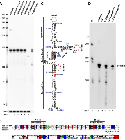
BocaSR is a novel, RNA polymerase III-transcribed intragenic viral small RNA.
To identify the promoter region of BocaSR, we cloned various sequences of the HBoV1
duplex genome that span the BocaSR-transcribing region (nt 5129 to 5360, nt 5165 to
5345, nt 5199 to 5345, nt 5201 to 5345, and nt 5249 to 5360) into the pGEM-3Z vector and
transfected each plasmid into HEK293 cells to test for their expression of BocaSR. The results
of RNase protection assays (RPAs) showed that except for one fragment (nt 5249 to 5360),
all produced BocaSR transcripts of identical length (
⬃
140 nt) and at the level expressed
from pIHBoV1 (Fig. 2A, lanes 1 versus 2 to 6). This RPA result confirmed that HBoV1 nt 5199
to 5345, a sequence that contains the entire BocaSR-transcribing region (nt 5199 to 5338),
is fully capable of transcribing BocaSR, strongly suggesting that BocaSR is transcribed by
an intragenic type II Pol III promoter, as are the adenovirus (Ad) VA RNAs,
Epstein-Barr virus-encoded small RNAs (EBERs), and cellular tRNAs (35, 36).
We speculated that BocaSR shares features with VA RNAs or EBERs. Alignment of
BocaSR with Ad5 VAI RNA revealed that BocaSR shares 51.2% identity in nucleotide
sequence (with particularly high identity in the predicted A-box and B-box, which are
the key elements of a type II Pol III promoter) and has the same transcription
termi-FIG 1Legend (Continued)
postinfection, total RNA was extracted. Five micrograms of each RNA was analyzed by Northern blotting with probes as indicated. (pA)p and (pA)d indicate HBoV1 mRNAs polyadenylated at (pA)p and (pA)d sites, respectively. (pA)d mRNAs indicated by diamonds and stars encode NS and VP, respectively. Arrows indicates BocaSR bands. The Millennium and Century markers were loaded together in one lane for electrophoresis and were visualized with ethidium bromide. An additional lane was loaded with the Millennium markers and blotted with a probe that hybridizes to them, to distinguish them from the Century markers (sizes indicated to left of blots). (D to F) Identification of BocaSR ends by RACE. (D) 5=and 3= RACE-amplified DNA fragments were electrophoresed in a 1.5% agarose gel with DNA markers (sizes indicated at right). (E and F) Sequences of the junction regions between adaptor and HBoV1, for the DNA fragments identified by 5=RACE (E) and 3=RACE (F).
on November 7, 2019 by guest
http://jvi.asm.org/
nation signal (TTTT) (Fig. 2B). We predicted the secondary structure of BocaSR using the
KineFold algorithm (
http://kinefold.curie.fr/
) with constraint parameters (37). Our results
suggest that the RNA has an apical stem, a terminal stem, and a complicated central
domain and that its secondary structure is highly similar to that of the VAI RNA (Fig. 2C)
(38, 39).
FIG 2HBoV1-encoded BocaSR is transcribed from an intragenic Pol III promoter. (A) Identification of the BocaSR-transcribing region by RNase protection assay. HEK293 cells were transfected with pIHBoV1 or pGEM-3Z-based plasmids carrying the indicated coding sequences. At 48 h posttransfection, total RNA was extracted. Ten micrograms of total RNA and 100 ng ofin vitro-transcribed BocaSR (lane 7) were protected with probes pBocaSR and p-actin, respectively. Lane M,32P-labeled RNA markers (64), with sizes shown to the left. (B) CLUSTALW-based sequence alignment. Sequences of BocaSR and Ad5 VAI RNA were aligned using the CLUSTALW algorithm. Identical nucleotides are colored. Consensus sequences of the A- and B-boxes of Pol III are indicated. Mutations used to create the A-box and B-box mutants are shown under the BocaSR sequence. (C) Predicted secondary structure of BocaSR. The BocaSR structure was predicted using the KineFold algorithm, with VAI RNA serving as a reference. Nucleotides of the HBoV1 genome and the BocaSR RNA sequence are shown in blue and black, respectively. Nucleotides that were changed in BocaSR mutants are shown in red. Loop structures are indicated, and the central tetranucleotide pair is shaded in gray. (D) Identification of the A-box and B-box of BocaSR by RNase protection assay. Total RNA was isolated from HEK293 cells transfected with plasmids as indicated, at 48 h posttransfection. Ten micrograms of total RNA was protected using probe pBocaSR or pBocaSRmAB. Lane M,32P-labeled RNA markers, with sizes indicated to the left.
A Noncoding RNA Is Required for Parvovirus Replication Journal of Virology
on November 7, 2019 by guest
http://jvi.asm.org/
[image:5.585.44.474.69.549.2]To verify the predicted A-box and B-box of BocaSR, we mutated two key nucleotides
in the sequence of each (nt 5199 to 5338) and expressed it either without or with the
U6 RNA Pol III promoter (36) (p3Z-BocaSR
mABand p3Z-U6-BocaSR
mAB). Three additional
nucleotides, none of which is located in the predicted A-box or B-box, were mutated to
retain the structure; these were selected using KineFold. Mutations of key nucleotides
in both A-box and B-box abolished the expression of BocaSR (Fig. 2D, lane 5). As
expected, the U6 Pol III transcribed an A-box- and B-box-mutated BocaSR of the
BocaSR
mABmutant (Fig. 2D, lane 6).
Taken together, these results confirmed that BocaSR is transcribed from an
intra-genic Pol III promoter in which the A-box and B-box are present downstream of the
transcription start site (i.e., within the transcribed mature RNA). Based on these results,
we concluded that BocaSR is a typical type II Pol III-derived small noncoding RNA (36).
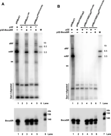
BocaSR is indispensable for replication of the HBoV1 DNA.
Previously, we found
that the presence of the HBoV1 3
=
NCR (nt 5221 to 5291) in
cis
is critical to viral DNA
replication (33). Since the HBoV1 3
=
NCR is encompassed by BocaSR, we hypothesized
that BocaSR is involved in replication of the HBoV1 DNA. This was addressed by
analyzing the ability of BocaSR to complement replication of the replication-deficient
viral DNA in HEK293 cells. We first confirmed that deletion of BocaSR abolished
replication of the HBoV1 genome. Although pIHBoV1
ΔSRwas derived from the
infectious HBoV1 duplex genome clone, the removal of BocaSR sequence abolished
replication of the HBoV1 DNA in transfected HEK293 cells (Fig. 3A, lane 2).
Comple-mentation with wild-type (WT) BocaSR, but not the A-box- and B-box-mutated mutant
(BocaSR
mAB), in
trans
restored replication of pIHBoV1
ΔSR(Fig. 3A, lane 3 versus lane 4).
Notably, U6 Pol III-transcribed BocaSR
mABalso restored replication of the pIHBoV1
ΔSRDNA, albeit not completely (Fig. 3A, lane 5). Expression of BocaSR and BocaSR
mABwas
confirmed by RNase protection assays (Fig. 3D). The lower efficiency of the U6 Pol
III-driven BocaSR
mABin restoring pIHBoV1
ΔSRreplication was partially due to its low
level of expression. This finding suggests that the mutations in the A-box and B-box
disrupted activity of the intragenic Pol III promoter without completely destroying its
ability to facilitate HBoV1 DNA replication. Thus, we confirmed that the expression of
BocaSR is essential to replication of the HBoV1 DNA.
To further characterize the role of BocaSR in replication of the HBoV1 DNA, we
screened point mutants less likely to affect various predicted structures within BocaSR,
or the transcription activity elements of the A- and B-boxes, for function in HBoV1
replication. In principle, we substituted nucleotides located in the loop regions and
introduced parallel changes in the complementary bases in the context of p3Z-BocaSR.
Analysis using the KineFold software suggested that these loop mutants introduce no
changes in structure. Three mutations located in the stem regions were also analyzed,
including within tetranucleotide GGGU/ACCU, that are similar to that of the adenovirus
(Ad) VA RNA (GGGU/ACCC) (40). All of the mutations are highlighted in red in Fig. 2C.
The efficiencies of the BocaSR mutants with respect to complementing replication
of the pIHBoV1
ΔSRDNA were evaluated by Southern blotting. The sequence in loop III
had a greater impact on replication of the viral DNA than did those in the other loops
and in the stem structure. Mutations affecting loop III (85TG/AC86, G86C, and 87GT/
CA88) and the loop III stem (93CC/AA94) were less effective at restoring replication of
the viral DNA than those in loop I (28AG/TC29 and 32AA/TT33) and loop Il (56AT/TA57)
(Fig. 3B, lanes 6 to 9 versus 3 to 5). In contrast, both the mutant affecting loop V
(112CA/GT113) and that affecting the terminal stem of loop V (119TGT/GAC121)
retained full function, as evident from the fact that the levels of both double replicative
form (dRF) and monomer replicative form (mRF) DNAs were the same on Southern
blots (Fig. 3B, lanes 11 and 12 versus lane 2). As expected, the mutation in the
tetranucleotide (C103A) restored DNA replication only partially (
⬃
20% of levels in WT)
(Fig. 3B, lane 10). Of note, the inability of those mutants to restore viral DNA replication
from pIHBoV1
ΔSR-transfected HEK293 cells was not due to loss of the expression of
on November 7, 2019 by guest
http://jvi.asm.org/
BocaSR, as all the tested mutants and control plasmids were capable of transcribing the
140-nt (short) RNA, as confirmed by Northern blotting (Fig. 3E).
We further evaluated the importance of a loop III mutation (85TG/AC86). As shown
in Fig. 3C, pIHBoV1
SR(85TG/AC86)did not replicate at all in transfected HEK293 cells (Fig.
3C, lane 2). As expected, the introduction of WT BocaSR fully restored replication (Fig.
3C, lane 3). Thus, we decided to use the simple 2-nt mutant affecting loop III for further
complementation analysis in HAE-ALI cultures.
Collectively, these findings demonstrated that the expression of BocaSR is essential
to replication of the HBoV1 DNA in HEK293 cells and that loop III contains the element
that is most important to its function in promoting replication of the HBoV1 DNA.
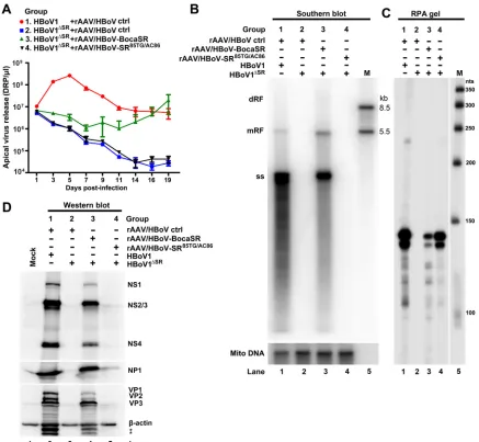
BocaSR is essential for HBoV1 infection of well-differentiated HAE cells.
To
investigate the role of BocaSR in the infection of HAE cells by HBoV1, we performed
functional complementation of BocaSR in HBoV1
ΔSR-infected HAE cells, using the
FIG 3Replication of the HBoV1 DNA can be restored by expressing BocaSR mutants. (A to C) Southern blotting. (A and B) Viral DNA in HEK293 cells transfected with pIHBoV1 and p3Z or pIHBoV1ΔSRand either the indicated p3Z-BocaSR or A-box and B-box mutant (p3Z-BocaSRmAB) (A) or the indicated BocaSR point mutant (B). (C) Viral DNA in HEK293 cells transfected with pIHBoV1 or cotransfected with pIHBoV1SR(85TG/AC86)and vector only or p3Z-BocaSR. In each panel, “M” indicates the marker lane, with sizes shown to right, and the following DNA forms are indicated: monomer replicative form DNA (mRF), double replicative form DNA (dRF), and ssDNA (ss). DpnI-digested bands are indicated. (D) RNase protection assay. Total RNA extracted from HEK293 cells transfected with plasmids as indicated in panel A was protected with probe pBocaSR or pBocaSRmAB. (E) Northern blotting. BocaSR RNA in lanes loaded with 5g of total RNA extracted from transfected HEK293 cells as indicated in panel B, with pBocaSR DNA used as a probe. In lane 13, 500 ng ofin vitro-transcribed BocaSR was used as the marker.
A Noncoding RNA Is Required for Parvovirus Replication Journal of Virology
on November 7, 2019 by guest
http://jvi.asm.org/
[image:7.585.42.475.69.486.2]recombinant adeno-associated virus (rAAV)/HBoV vector to deliver the WT BocaSR or
BocaSR
85TG/AC86mutant in
trans
. A non-BocaSR-expressing rAAV/HBoV vector served as a
negative control. Analyses of apical release of virus from the infected HAE cells revealed that
infection of HBoV1
ΔSRwas productive only in the presence of rAAV/HBoV-BocaSR. As
shown in Fig. 4A, the transduction of rAAV/HBoV-BocaSR led to an increase in apical release
of virus from HBoV1
ΔSR-infected HAE cells starting on day 3. This was not the case for
transduction with either the vector control or rAAV/HBoV-BocaSR
85TG/AC86, in which
cases virus levels gradually decreased to background as in the case of infection with
HBoV1
ΔSRalone (Fig. 4A). At days 16 and 19, rAAV/HBoV-BocaSR completely
comple-mented the function to the HBoV1
ΔSRvirus, with levels comparable to those obtained
by infection with the WT virus (Fig. 4A). Replication of the viral DNA (ssDNA) on day 19
FIG 4Virus replication in HBoV1ΔSRinfected HAE-ALI cultures transduced with BocaSR-expressing rAAV2/HBoV1 vector. Polarized HAE-ALI cultures were coinfected with HBoV1 WT or HBoV1ΔSRmutant purified from transfected HEK293 cells at an MOI of⬃1,000 DRP/cell and rAAV/HBoV vector at an MOI of⬃10,000 DRP/cell (59) in groups as indicated. (A) Virus apical release kinetics. Apical washes were collected from at least three infected HAE-ALI cultures for each virus infection and quantified for DRP by qPCR. Averages and standard deviations are shown. (B) Southern blot analysis of viral DNA. Hirt DNA was extracted from infected cells at 19 days p.i. Levels of viral DNA were assessed by Southern blotting. Mitochondrial DNA (Mito DNA; lower image) was detected by a mitochondrial DNA probe as a control for the recovery of the Hirt DNA (30). The DNA marker (M) is shown with sizes indicated to the right. (C) Analysis of BocaSR expression by RNA protection assay. Total RNA was extracted from infected cells at 19 days p.i. Ten micrograms of the total RNA was protected with the probe pBocaSR or pBocaSR85TG/AC86. The RNA marker (M) is shown, with sizes indicated to the right. (D) Western blot detection of viral proteins in infected HAE-ALI cultures. At 19 days p.i., infected cells were lysed for Western blotting. Samples were initially blotted using anti-NS1C antibody (top) and then reprobed sequentially with anti-NP1 (middle) and anti-VP and anti--actin (bottom). Bands marked with an asterisk may be uncharacterized or degraded forms of VP.
on November 7, 2019 by guest
http://jvi.asm.org/
[image:8.585.40.477.70.473.2]postinfection (p.i.), as assessed by Southern blotting of Hirt DNA extracts from infected
HAE cells, was rescued to nearly WT levels in cells coinfected with rAAV/HBoV-BocaSR
but not rAAV/HBoV-BocaSR
85TG/AC86(Fig. 4B, lane 3 versus lane 4). Expression of BocaSR
and the BocaSR
85TG/AC86mutant from the respective rAAV/HBoV vectors was confirmed
by RNase protection assays (Fig. 4C), although expression of BocaSR from the rAAV/
HBoV-BocaSR vector was relatively poor. Of note, rAAV/HBoV-BocaSR but not rAAV/
HBoV-BocaSR
85TG/AC86restored expression of viral proteins (NS1 to -4, NP1, and VP1 to
-3) in HAE cells (Fig. 4D, lane 4 versus lane 5).
Taken together, these results demonstrated that BocaSR is indispensable for HBoV1
replication and that it functions in
trans
to support replication of the HBoV1 DNA in
well-differentiated HAE-ALI cultures.
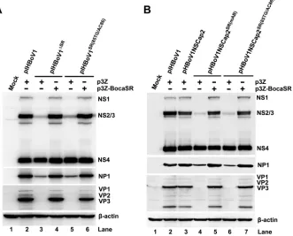
BocaSR regulates the expression of NS1, NS2, NS3, and NP1 but not NS4.
To
investigate the mechanism underlying BocaSR-controlled replication of HBoV1
DNA, we assessed the expression of viral proteins from pIHBoV1 mutants in which
BocaSR was deleted or mutated. Western blotting revealed that neither pIHBoV1
ΔSRnor pIHBoV1
SR(85TG/AC86)supported the expression of NS1, that the expression of NS2/3 was
reduced
⬎
10-fold, and that expression of NP1 was reduced
⬎
3-fold (comparison to WT
pIHBoV1). However, neither the deletion nor the mutation affected the expression of
NS4 significantly (Fig. 5A, lane 2 versus lanes 3 and 5). When BocaSR was provided
in
trans
by cotransfection of p3Z-BocaSR (Fig. 6A, bottom), the expression of NS1, NS2/3,
and NP1 was fully restored (Fig. 5A, lanes 4 and 6 versus lane 2). To rule out the possibility
that this rescue of protein expression was a consequence of replication of the viral DNA
(Fig. 6A, top), we conducted tests in the context of nonreplicating HBoV1 plasmids,
pHBoV1NSCap2
SR(mAB)and pHBoV1NSCap2
SR(85TG/AC86)(Fig. 6B). The protein expression
profiles in cells transfected with pHBoV1NSCap2
SR(mAB)and pHBoV1NSCap2
SR(85TG/AC86)were similar to those in cells transfected with the pIHBoV1-based mutants (Fig. 5B,
FIG 5HBoV1 protein expression analysis of HEK293 cells transfected with pIHBoV1-based and pHBoV1NSCap2-based BocaSR mutants. (A) Expression from replicating pIHBoV1. HEK293 cells were transfected with pIHBoV1, pIHBoV1ΔSR, and pIHBoV1SR(85TG/AC86), with p3Z or p3Z-BocaSR cotransfec-tion. (B) Expression from nonreplicating HBoV1 plasmid. HEK293 cells were transfected with pIHBoV1, pHBoV1NSCap2, pHBoV1NSCap2SR(mAB), and pHBoV1NSCap2SR(85TG/AC86), with p3Z or p3Z-BocaSR cotrans-fection. At 48 h posttransfection, viral proteins were assessed by Western blotting. Blots were probed sequentially with antibodies against HBoV1 NS1C, NP1, and VP and then against-actin.A Noncoding RNA Is Required for Parvovirus Replication Journal of Virology
on November 7, 2019 by guest
http://jvi.asm.org/
[image:9.585.48.379.75.342.2]lanes 4 and 6, versus Fig. 5A, lanes 3 and 5), and expression of WT BocaSR in
trans
(Fig.
6B, bottom) fully restored the expression of NS1, NS2/3, and NP1 (Fig. 5B, lanes 5 and
7 versus lane 3).
These results demonstrated that BocaSR upregulates expression of the viral proteins
NS1, NS2/3, and NP1 but not NS4. As NP1 is required for the expression of VP (26),
BocaSR indirectly regulates VP expression as well (Fig. 5, VP). Since in HEK293 cells
NS2/3 and NS4 are dispensable for replication of the HBoV1 DNA (33), our results
suggest that BocaSR-upregulated expression of NS1 and NP1 is critical to viral DNA
replication in HEK293 cells.
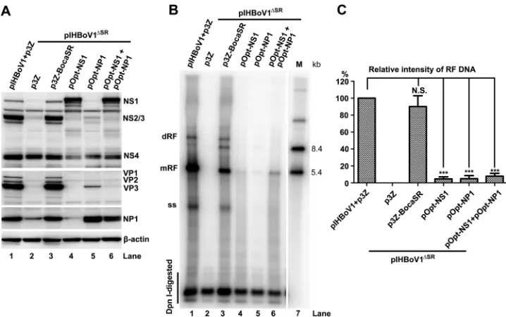
Expression of the nonstructural proteins is not sufficient to fully rescue
repli-cation of the HBoV1 genome in the absence of BocaSR.
We further investigated
FIG 6DNA replication and BocaSR detection of HEK293 cells transfected with pIHBoV1-based and pHBoV1NSCap2-based mutants. (A) Replicating pIHBoV1-pHBoV1NSCap2-based plasmids. HEK293 cells were transfected with pIHBoV1, pIHBoV1ΔSR, and pIHBoV1SR(85TG/AC86)with p3Z or p3Z-BocaSR cotransfection. (B) Nonreplicating pHBoV1NSCap2-based plasmids. HEK293 cells were transfected with pIHBoV1, pHBoV1NSCap2, pHBoV1NSCap2SR(mAB), and pHBoV1NSCap2SR(85TG/AC86), with p3Z-BocaSR or p3Z cotransfection. At 48 h posttransfection, Hirt DNA samples were extracted from the transfected cells and analyzed by Southern blotting with HBoV1NSCapprobe (top). At 48 h posttransfection, total RNA samples were extracted from the transfected cells and analyzed by RNase protection assay with pBocasR, pBocasR85TG/AC86, or pBocasRmABprobe (bottom). Both DNA and RNA markers (M) are shown to the right of the images.on November 7, 2019 by guest
http://jvi.asm.org/
[image:10.585.41.400.73.512.2]whether the reduced expression of NS1 and NP1 accounts fully for the replication
deficiency of the pIHBoV1
ΔSRand pIHBoV1
SR(85TG/AC86)mutants in HEK293 cells. To this
end, we supplemented NS1, NP1, or both in
trans
and tested for complementation of
the lack of BocaSR expression in HEK293 cells transfected with pIHBoV1
ΔSR. Transfected
NS1- and NP1-expressing plasmid produced NS1 and NP1 at levels similar to those in
controls in HEK293 cells transfected with pIHBoV1
ΔSRand p3Z-BocaSR (Fig. 7A).
How-ever, this expression did not lead to a significant increase in viral DNA replication from
pIHBoV1
ΔSR(
⬍
5% increase) relative to that produced by the expression of BocaSR in
trans
(Fig. 7B, lanes 3 versus lanes 4 and 5). However, coexpression NS1 and NP1 had
an additive effect, leading to a 7.8% increase (Fig. 7B, lane 3 versus lane 6, and Fig. 7C).
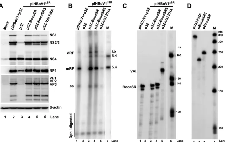
We next tested whether the supplementation of nonstructural proteins in
cis
could
efficiently facilitate viral DNA replication. To this end, we expressed VAI RNA in
trans
with pIHBoV1
ΔSR-transfected HEK293 cells in an attempt to functionally rescue
expres-sion of the nonstructural proteins and investigated its effects on viral DNA replication.
Although VAI RNA restored the expression of NS1 to 3 and NP1 to levels comparable
to those observed when BocaSR was expressed in
trans
(Fig. 8A, lane 6), replication of
viral DNA was only at
⬃
10% of the levels that were observed on complementation with
BocaSR (Fig. 8B, lane 3 versus 5). Thus, in the absence of BocaSR, expression of the
nonstructural protein gene is not sufficient to support effective HBoV1 DNA replication.
Of note, BocaSR3 of HBoV3 fully restored not only the expression of NS1 to -3 and NP1
(Fig. 8A, lane 5) but also the replication of the viral DNA (Fig. 8B, lane 4). The levels of
expression of BocaSR, BocaSR3, and VAI RNA in the above-described complementation
experiments were confirmed to be similar by RNase protection assays (Fig. 8C) using
saturated probes of equivalent activities (Fig. 8D).
Taken together, these results confirmed that BocaSR-upregulated nonstructural
protein expression is necessary but not sufficient to fully support replication of the
HBoV1 DNA, indicating that BocaSR plays additional roles in viral DNA replication.
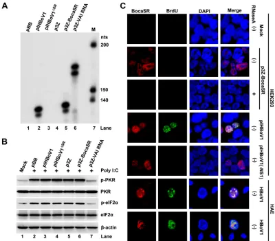
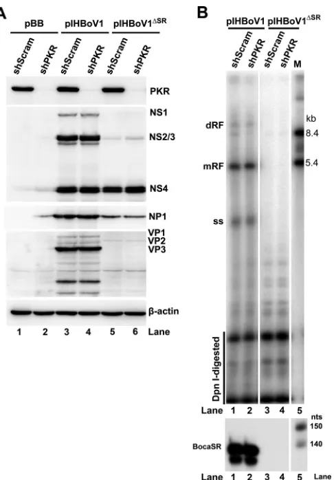
BocaSR-mediated expression of nonstructural proteins is independent of the
protein kinase R pathway.
As the VAI RNA regulates viral protein expression mainly by
FIG 7Overexpressed NS1 and NP1 proteins do not rescue viral DNA replication of pIHBoV1ΔSR. HEK293 cells were cotransfected with pIHBoV1 and p3Z or pIHBoV1ΔSRand the indicated plasmid(s). One microgram of pOpt-NS1 (33) or pOpt-NP1 (26) (supplemented with pCI backbone vector to 2g), or 1g of each, was cotransfected. (A) Expression of viral proteins in transfected cells harvested at 48 h postinfection. Western blots were probed sequentially using antibodies against HBoV1 NS1C, NP, VP, and-actin. (B) Levels of viral DNA in transfected cells harvested at 48 h posttransfection. Hirt DNA sample were assessed by Southern blotting for each form of the HBoV1 DNA. (C) Quantitation of viral mRF DNA in panel B. Averages and standard deviations are shown. Statistics analysis was performed by the Studentttest. N.S., no significance;***,P⬍0.01.A Noncoding RNA Is Required for Parvovirus Replication Journal of Virology
on November 7, 2019 by guest
http://jvi.asm.org/
[image:11.585.41.400.70.295.2]inhibiting the double-stranded RNA-activated protein kinase R (PKR)-
␣
subunit of
eukaryotic initiation factor 2 (eIF2
␣
) pathway (41), we tested BocaSR for an interaction
with PKR. To this end, we first transfected HEK293 cells with various BocaSR-expressing
plasmids and control vectors (p3Z-VAI RNA and pIHBoV1
ΔSR, positive and negative,
respectively). Then the PKR-eIF2
␣
pathway was activated by transfecting the cells with
synthetic double-stranded RNA (dsRNA), poly(I)·poly(C) (P1530; Sigma) (42). The
inhi-bition of PKR activation was assessed by Western blotting for phosphorylated PKR
(p-PKR) and phosphorylated eIF2
␣
(p-eIF2
␣
). The expression of BocaSR following
transfection with either pIHBoV1 or p3Z-BocaSR (Fig. 9A) did not result in an obvious
decrease in the level of p-PKR and p-eIF2
␣
following activation by poly(I)·poly(C) (Fig.
9B, lanes 3 and 6), compared with the level in the context of transfection of the p3Z-VAI
RNA (Fig. 9B, lane 7). This result suggested that in contrast to VAI RNA, BocaSR does not
function as an inhibitor of PKR to reduce phosphorylation of eIF2
␣
in upregulating the
expression of nonstructural proteins.
To more definitively exclude a role for PKR in BocaSR-upregulated expression of
nonstructural proteins and replication of the viral DNA, we established a PKR
knock-down HEK293 cell line (Fig. 10A). We then transfected PKR-null HEK293 cells with
pIHBoV1 and pIHBoV1
ΔSR. HEK293 cells expressing a scrambled short hairpin RNA
(shRNA) control (shScram) were transfected in parallel as a control. Although pIHBoV1
expressed all the viral nonstructural proteins (Fig. 10A, lanes 3 and 4) and replicated in
both PKR-null and shScram-expressing cells (Fig. 10B, lanes 1 and 2), pIHBoV1
ΔSRdid
not (Fig. 10A, lanes 5 and 6, and Fig. 10B, lanes 3 and 4). Expression of BocaSR in
transfected cells was confirmed by RNase protection assays (Fig. 10B). Thus, PKR does
not appear to play a role in BocaSR-facilitated replication of the HBoV1 DNA.
As VAI RNA is exclusively expressed in the cytoplasm (39), where it interacts with PKR
(43, 44), we examined the localization of BocaSR. Indeed, BocaSR is expressed in the
nucleus in HEK293 cells transfected with p3Z-BocaSR, as demonstrated by the RNA
FIG 8Fully restored NS1 and NP1 proteins do not rescue viral DNA replication. HEK293 cells were cotransfected with pIHBoV1 and p3Z or pIHBoV1ΔSRand a complemented BocaSR or VAI RNA-expressing plasmid as indicated in each lane. (A) Western blot analysis of viral proteins. Expression of viral proteins in transfected cells harvested at 48 h postinfection. Western blots were probed with an anti-HBoV1 NS1C antibody and were then reprobed sequentially with anti-NP1, anti-VP, and anti--actin antibodies. (B) Southern blot analysis of viral DNA replication. At 48 h posttransfection, Hirt DNA was extracted, DpnI digested, and assessed by Southern blotting for each form of HBoV1 DNA. (C and D) RNA protection assay of BocaSR and VAI RNA. (C) At 48 h posttransfection, total RNA was extracted. Ten micrograms of the total RNA was protected by probes for BocaSR, BocaSR3, or VAI RNA. (D) Similar levels of RPA probes were used for protection in each sample.
on November 7, 2019 by guest
http://jvi.asm.org/
[image:12.585.44.436.71.319.2]fluorescence
in situ
hybridization-immunofluorescence (FISH-IF) assay [Fig. 9C, RNase
A(
⫺
)/p3Z-BocaSR]. The specificity of detection of BocaSR in this experiment was
confirmed by RNase A treatment of the transfected cells (Fig. 9C, RNase A
⫹
/p3Z-BocaSR). More importantly, in both pItransfected HEK293 cells and
HBoV1-infected HAE cells, BocaSR was located exclusively in the nucleus and within the centers
of viral DNA replication (the autonomous parvovirus-associated replication [APAR]
bodies), which were pulse-labeled with BrdU (Fig. 9C, pIHBoV1 and HBoV1,
respec-tively).
Considering that restoring the expression of viral nonstructural proteins rescues
only a fraction (7.8%) of HBoV1 DNA replication from pIHBoV1
ΔSR(Fig. 7B, lane 6), these
results suggest that BocaSR likely plays a direct role in replication of the viral DNA.
DISCUSSION
In this report, we describe the discovery of a novel Pol III-transcribed viral noncoding
RNA, BocaSR. It is the first of Pol III-transcribed small RNA to be identified in
parvovi-ruses, and it shares a high level of similarity with respect to sequence and secondary
FIG 9BocaSR-facilitated HBoV1 DNA replication in the nucleus does not involve the cytoplasmic PKR pathway. (A and B) Effects of BocaSR on PKR and eIF2␣activation. (A) HEK293 cells in wells of a 12-well plate were transfected with 1g of pIHBoV1 or other indicated plasmids at the same molar amount to a final 1g of DNA supplemented with empty pGEM-3Z. At 24 h posttransfection, the cells were transfected with poly(I)·poly(C) at 400 ng/ml for 4 h using Lipofectamine 2000 (Invitrogen). (A) Total RNA was extracted from the treated cells and was subjected to an RNase protection assay with a single probe, pBocaSR-VAI RNA. (B) The treated cells were analyzed by Western blotting for levels of PKR and eIF2␣proteins and their phosphorylation. Blots were probed with anti-p-eIF2␣and anti-p-PRK antibodies and reprobed with anti-PKR, anti-eIF2␣, and anti--actin, in order. (C) FISH-IF analysis of BocaSR expression. HEK293 cells were transfected with p3Z-BocaSR, pIHBoV1(-NS1), or pIHBoV1. HAE-ALI cultures were infected with HBoV1. Cells were BrdU labeled at 48 h posttransfection or 10 days postinfection and subjected to FISH-IF analysis. DAPI was used to identify nuclei. RNase A-treated transfected HEK293 cells were used as a control. pIHBoV1(-NS1) (21) was transfected as a nonreplication control. Magnification,⫻100.
A Noncoding RNA Is Required for Parvovirus Replication Journal of Virology
on November 7, 2019 by guest
http://jvi.asm.org/
[image:13.585.39.439.70.421.2]structure with Ad VAI RNA but functions differently during virus infection. BocaSR is
indispensable to the replication of viral DNA in both dividing HEK293 cells and
nondividing HAE cells; in its absence, the virus (viral DNA) is not capable of
upregu-lating the expression of NS1 to -3 as well as that of NP1, yet this is not sufficient to
support efficient viral DNA replication. Thus, BocaSR represents a unique viral Pol
III-driven noncoding RNA that plays a direct role in viral DNA replication.
Sequence and structure of BocaSR.
Previous studies revealed a remarkable feature
of the 3
=
NCR of primate bocaparvoviruses: those of HBoV1, HBoV2, and HBoV3 fold into
almost identical secondary structures (45, 46). This feature appears to be unique; it is
not shared by the carnivore bocaparvovirus 1 minute virus of canines (MVC) (23),
bocaparvoviruses from other nonprimate species, or other parvoviruses. The BocaSR of
HBoV1 is the most divergent among these noncoding RNAs of primate
bocaparvovi-ruses. The BocaSR sequences of HBoV2 to -4 and gorilla bocavirus (GBoV1) share 97.8%
identity in the coding region that has been sequenced; whereas HBoV1 shares only 86%
with these of HBoV2 to -4 and GBoV1 (Fig. 11). Importantly, the HBoV1 BocaSR shares
sequence identity ranging from 46.1% to 51.2% with the other four Pol III-driven viral
small RNAs: VAI, VAII, EBER1, and EBER2. The VAI and VAII RNAs share
⬃
60% identity,
EBER1 and EBER2 share
⬃
57% identity, and EBERs and VAs share 37 to 50% identity.
FIG 10Effects of PKR knockdown on expression of HBoV1 protein and replication of the HBoV1 DNA in the pIHBoV1 mutant with a deletion of BocaSR. HEK293 cells subjected to PKR knockdown were transfected with pBB (backbone control), pIHBoV1, or pIHBoV1ΔSR. (A) Western blot analysis. Levels of PKR and viral proteins in HEK293 cells at 48 h posttransfection, as assessed by sequential Western blotting using antibodies against the NS1C, NP1, and VP proteins, are shown. (B) Southern blot analysis and RNA protection assay. Levels of viral DNA in transfected HEK293 cells at 48 h posttransfection, as assessed by Southern blotting for viral DNA, are shown. Extracted total RNA at 48 h posttransfection was protected by the pBocaSR probe. Markers (M) of both DNA and RNA are shown to the right.on November 7, 2019 by guest
http://jvi.asm.org/
[image:14.585.87.330.69.418.2]Therefore, these Pol III-driven viral small RNAs are diverse in their transcribing
se-quences. These findings suggest that BocaSR represents a novel species of Pol III-driven
viral small RNAs.
In the VA RNA genes of 43 human adenoviruses, the tetranucleotide pair (GGGU:
ACCC) in the central domain is highly conserved, with only one nucleotide diverging (to
GGGU:ACCU) in the two groups of type F Ad (40). The tetranucleotide pair in BocaSR is
of the type F Ad VA RNA (Fig. 11). Mutation of a single nucleotide pair within this
sequence drastically decreased the ability of BocaSR to support replication of HBoV1
DNA (Fig. 3), suggesting that a stem in the central domain is critical for the function
(and possibly the structure) of BocaSR.
From the structural perspective, BocaSR more closely resembles the VA RNAs than
the EBERs (47). The predicted structure of BocaSR (Fig. 2C) is comprised of three
structural domains: a terminal stem, a central domain, and an apical stem. Each is highly
conserved in VA RNAs (40). The central domain contains two loop structures that
exhibit key functions of VA RNAs (41). Accordingly, in BocaSR, mutations in loop I, the
central stem, loop III, and the loop III stem, all of which are included in the central
domain, diminish the effectiveness of BocaSR in viral DNA replication, with mutations
in loop III conferring the most severe defects.
BocaSR shares striking similarity with the VAI RNA but functions differently.
During Ad infection, two distinct VA RNAs, VAI and VAII (
⬃
160 nt long), are expressed,
but the VAI RNA (
⬃
10
8copies/cell) is more abundant than VAII RNA (39). Of note,
BocaSR is expressed at a level similar to that of VAI RNA in the context of the VAI gene
only (Fig. 9A). VAI RNA plays a regulatory role during the Ad life cycle, enhancing the
translation of Ad proteins (38, 40). It is essential for efficient virus replication, with its
deletion from Ad5 leading to an
⬃
20-fold decrease in virus production (48).
VAI RNA binds to PKR and functions as a decoy dsRNA, which prevents PKR
phosphorylation and activation (39, 49, 50). In spite of the similarities between VAI and
BocaSR with respect to sequence and structure, it is difficult to imagine that BocaSR
acts by the same mechanism because those in which VAI is involved take place in the
cytoplasm (39) and BocaSR is present mainly in the nucleus. This is supported by the
fact that BocaSR does not inhibit PKR activation (Fig. 9B). More importantly, unlike VAI
RNA, which nonspecifically upregulates translation by inhibiting the phosphorylation of
eIF2
␣
through the PKR-eIF2
␣
pathway (39, 51), BocaSR specifically upregulates the
expression of viral NS1 to -3 and NP1 protein but not that of NS4. We hypothesize that
BocaSR regulates the processing and export of the NS1 to -3 and NP1 mRNAs but not
those of the NS4 transcript.
Despite the finding that BocaSR does not inhibit PKR activation, the fact that VAI
RNA rescued the defects in expression of HBoV1 nonstructural proteins and partially
rescued the replication of viral DNA (
⬃
10%) from pIHBoV1
ΔSRin which BocaSR was
FIG 11BocaSR-encoding sequences of the 3=NCR region of 7 isolates of primate bocaparvovirus, aligned using the CLUSTALW algorithm. Nucleotide identity is indicated by dots. The 3=ends of the BocaSR sequences of HBoV2a, -b, and -c, HBoV4, and GBoV1 are currently not available and are indicated as gray dashes. The GenBank accession number of each isolate is indicated. As noted, the full-length genome sequence is known only in the case of HBoV1. The A-box and B-box are marked, as is the tetranucleotide pair GGGU:ACCU.
A Noncoding RNA Is Required for Parvovirus Replication Journal of Virology
on November 7, 2019 by guest
http://jvi.asm.org/
[image:15.585.42.439.73.199.2]deleted was interesting. It is unlikely that VAI RNA solely functions as an anti-PKR
molecule and that it thereby leads to increased translation of nonstructural proteins
and viral DNA replication.
Like EBERs, BocaSR functions in the nucleus.
EBV synthesizes two abundant small
RNAs, EBER1 and EBER2 (52), that are
⬃
170 nt in size, are highly structured RNA Pol
III-transcribed noncoding RNAs, and are abundant in infected cells (
⬃
10
6/cell) (47). The
EBERs and the VA RNAs share some similarities in function, including the ability to bind
the La protein (52) to substitute for the VAI RNA in lytic infection by Ad5 (53) and to
bind and inhibit PKR
in vitro
(54). However, EBERs reside in the nucleoplasm and do not
undergo nucleocytoplasmic shuttling (55, 56). These observations argue against the
physiological relevance of the binding of the nuclear EBERs to PKR, which is present in
the cytoplasm. In addition, their structures are quite different from that of the VA RNAs
(47, 57). Of note, EBER2 binds to the terminal repeats of the EBV genome and interacts
with transcription factor PAX5, acting in concert with it to regulate the expression of a
subset of EBV latency genes and thereby playing an important role in replication of the
EBV DNA during lytic infection (58).
Since the BocaSR resides exclusively in the nucleus and colocalizes with the
repli-cating viral genomes, we speculate that it likely plays a direct role in replication of the
viral DNA, in addition to contributing to the upregulation of viral nonstructural
pro-teins. In this regard, BocaSR may function like EBV-derived EBER2 in interacting with the
viral genome.
In summary, BocaSR shares much similarity with VAI RNA at the levels of sequence,
secondary structure, and host, yet its nuclear localization and role in the replication of
viral DNA are more like those of EBER2. The ability of the VAI RNA to partially
complement a BocaSR deficiency highlights a novel role for the former in regulating the
expression of viral proteins. Considering that AAV, Ad, and HBoV1 infect the same
natural host tissue—the human airway epithelium—it would not be surprising that VAI
RNA is capable of exerting some BocaSR functions that have not been discovered. It is
likely that both BocaSR and the VA RNAs evolved from cellular tRNA (36) during
coinfection of human airways by Ad, AAV, and HBoV1. Since HBoV1 replicates
auton-omously in human airway epithelia, we believe that BocaSR confers the ability to
replicate autonomously in nondividing airway epithelial cells (30).
MATERIALS AND METHODS
Ethics statement.Primary human airway (tracheobronchial) epithelial cells were isolated from the lungs of healthy human donors at Cell Culture Core of the Center for Gene Therapy, University of Iowa, under Institutional Review Board (IRB) approval by the University of Iowa (IRB no. 9507432). We obtained the well-differentiated (polarized) human airway epithelium (HAE) ALI cultures at the Cell Culture Core without any identification information on them, and therefore, an IRB review was waived.
Cell culture.HEK293 cells (CRL-1573; ATCC, Manassas, VA) were cultured in Dulbecco’s modified Eagle medium (DMEM; HyClone SH30022.01; GE Healthcare BioSciences, Pittsburgh, PA) with 10% fetal calf serum (F0926; Sigma, St. Louis, MO) at 37°C under a 5% CO2atmosphere.
Primary human airway epithelia were generated and cultured at an air-liquid interface (HAE-ALI) in Costar Transwell inserts (3470; Corning, Corning, NY) as previously described (30). HAE-ALI cultures with a transepithelial electrical resistance (TEER) of⬎1,000⍀· cm2were selected for use in this study.
Virus infection.HAE-ALI cultures were infected with HBoV1 at a multiplicity of infection (MOI) of 10 DNase I-resistant particles (DRP) of apical released progeny virion per cell, by following a previously published method (21, 30), unless otherwise indicated.
Construction of plasmids. (i) p3Z-based plasmids.The HBoV1 3=NCR clones p3Z-(nt5129-5360), p3Z-(nt5165-5345), p3Z-(nt5199-5345), p3Z-(nt5201-5345), and p3Z-(nt5249-5360) were constructed by cloning the HBoV1 sequences indicated in parentheses into the pGEM-3Z (p3Z) vector (Promega, Madison, WI) using the EcoRI and HindIII sites. We changed the name for p3Z-(nt5129-5360) to p3Z-BocaSR.
A mutant form of BocaSR in which the A- and B-boxes are disrupted (BocaSRmAB) was synthesized at Integrated DNA Technologies (IDT; Coralville, IA). The following seven mutations were introduced: G17A, G25A, G50A, C53T, G60A, C63T, and C115T. The BocaSRmAB-encoding DNA was cloned it into the pGEM-3Z vector using the EcoRI and HindIII sites, and the resultant clone was designated p3Z-BocaSRmAB. Additional BocaSR mutants were constructed by mutating one or two nucleotides in the parental plasmid, p3Z-BocaSR (p3ZSR). These were p3ZSR28AG/TC29, p3ZSR32AA/TT33, p3ZSR56AT/TA57, p3ZSR85TG/AC86, p3ZSRG86C, p3ZSR87GT/CA88, p3ZSR93CC/AA94, p3ZSRC103A, p3ZSR112CA/TG113, and p3ZSR119TGT/GAC121. In all cases the transcription start site of BocaSR was designated nt 1.
on November 7, 2019 by guest
http://jvi.asm.org/
p3Z-U6-BocaSRmABwere constructed by inserting U6-BocaSRmABsequence into pGEM-3Z using the BamHI and HindIII sites. The U6-BocaSRmABsequence was PCR amplified from pLKO-BocaSRmAB, which was constructed by cloning the BocaSRmAB sequence into a U6 promoter-containing pLKO.1 vector (10878; Addgene, Cambridge, MA).
p3Z-BocaSR3 and p3Z-VAI RNA were constructed as follows. The HBoV3 sequence encompassing nt 5000 to 5205 (GenBank accession no.EU918736) was cloned into pGEM-3Z using the EcoRI and HindIII sites. The VAI gene (nt 10607 to 10769) of adenovirus type 2 (Ad2; GenBank accession no.J01917.1) was cloned into pGEM-3Z using the SacI and HindIII sites.
(ii) pHBoV1NSCap-based plasmids.pHBoV1NSCap2 is a nonreplicating but NS- and VP-expressing plasmid and was constructed by deleting HBoV1 nt 97 to 140 and nt 5344 to 5395 from pHBoV1NSCap (34); this sequence contains the Ori (33). pHBoV1NSCap2SR(mAB) was constructed by substituting BocaSRmABfor BocaSR in pHBoV1NSCap2. pHBoV1NSCap2SR(85TG/AC86)was constructed by substituting BocaSR85TG/AC86for BocaSR in pHBoV1NSCap2.
(iii) pIHBoV1-based plasmids.pIHBoV1, an infectious clone of HBoV1, was reported previously (21). pIHBoV1ΔSRwas constructed by deleting nt 5200 to 5291 of the HBoV1 sequence from pIHBoV1. pIHBoV1SR(85TG/AC86)was constructed by mutating dinucleotide TG at nt 5283/4 of the HBoV1 sequence to AC in pIHBoV1.
(iv) Recombinant adeno-associated virus 2 (rAAV2) gene transfer plasmids. pAVF5tg83luc-CMVmChery(5.4) was constructed by cloning the mCherry expression cassette CMV-mCherry ORF-bGHpA into pAVF5tg83luc(5.4) (59). The HBoV1 DNA fragment of nt 5041 to 5360 that contains the BocaSR coding sequence and the corresponding fragment of the BocaSR85TG/AC86mutant were inserted into pAVF5tg83luc-CMVmChery using the SfiI and NsiI sites, to generate pAVF5tg83luc-CMVmCherry(BocaSR) and the mutant form pAVF5tg83luc-CMVmCherry(BocaSR85TG/AC86), respectively.
(v) pLKO.1-based constructs. The shRNA-expressing constructs were generated as previously described (30). The following shRNA sequences were chosen for targeting of the PKR gene: shPKR, 5=-CCG GGC TGA ACT TCT TCA TGT ATG TCT CGA GAC ATA CAT GAA GAA GTT CAG CTT TTT G-3=, and a scrambled shRNA control (shScram), 5=-CCG GCC TAA GGT TAA GTC GCC CTC GCT CGA GCG AGG GCG ACT TAA CCT TAG GTT TTT G-3=.
(vi) pUC19-based construct.pUC19-T7 HBoV1(nt 5200-5338) was constructed by inserting the HBoV1 DNA from nt 5200 to 5338 and a T7 promoter sequence (5=-TAA TAC GAC TCA CTA TAG GG-3=) adjacent to the 5=terminus of the HBoV1 DNA into the pUC19 vector using the EcoRI and HindIII sites. This vector was used to synthesize BocaSR RNAin vitro.
All the nucleotide numbers of HBoV1 in this study refer to those under GenBank accession number JQ923422unless otherwise specified.
Plasmid DNA transfection.HEK293 cells were seeded in 60-mm plates on the day before transfec-tion unless otherwise described. The cells were transfected with 4g of plasmid DNA at a confluence of ⬃80%, using the LipoD293 reagent (SignaGen, Gaithersburg, MD) and following the manufacturer’s instructions. For complementation of expression of RNA or protein, 2g of each plasmid was cotrans-fected.
Extraction of lower-molecular-weight (Hirt) DNA and Southern blotting.At 48 h posttransfec-tion, Hirt DNA was extracted from transfected cells, the DNA was digested with DpnI, and the samples were subjected to Southern blotting as described previously (60).
Western blot analysis.Transfected HEK293 cells were harvested at 48 h posttransfection. Infected HAE cells were harvested on the days indicated in each relevant figure. The cells were lysed and Western blotting was performed to analyze the lysates as previously described (34), using the specific antibodies that are indicated in each relevant figure.
RNA isolation.RNA samples were prepared using TRIzol reagent (Invitrogen, Carlsbad, CA) according to the manufacturer’s instructions.
Northern blotting on agarose gels. Five micrograms of total RNA was separated in a 1.4% denaturing agarose gel and visualized by staining with ethidium bromide (EB). Northern blot analysis was performed essentially as described previously (23), using32P-labeled DNA probes as diagrammed in Fig. 1A. The Millennium and Century markers (Invitrogen, Carlsbad, CA) were used as size markers.
Northern blotting on polyacrylamide gel.Five-microgram quantities of total RNA samples dena-tured in formamide were electrophoresed in a urea (8%) polyacrylamide gel in 1⫻Tris-borate-EDTA (TBE) buffer (61). The gel was transferred to a nitrocellulose membrane and the immobilized RNA was subjected to hybridization with a32P-radiolabeled DNA probe (nt 5129 to 5360 of HBoV1) and detected by phosphor imaging.
RNase protection assay.An RNase protection assay was carried out as described previously (23, 34). Probes used for RNase protection assay.RNase protection assay probes pBocaSR, pBocaSR3, and pVAI RNA were in vitrotranscribed in the presence of [32P]UTP using the SP6 RNA polymerase and EcoRI-digested p3Z(nt 5129-5360), p3Z-BocaSR3, and p3Z-VAI RNA, respectively. Probe pBocaSR-VAI was
in vitrotranscribed from a synthesized DNA of SP6 promoter sequence, BocaSR (nt 5129 to 5360 of HBoV1 DNA), and VAI RNA (nt 10607 to 10769 of Ad2 DNA), in order.
The human-actin probe (p-actin) was transcribed using EcoRI-linearized pTRI-Actin-human plas-mid (Invitrogen).
Phosphor imaging and quantification.After hybridization, the Southern and Northern blots were exposed to a phosphor screen, which was then scanned on a phosphor imager (Typhoon FLA 9000; GE Healthcare Life Sciences). Densitometry was performed using ImageQuant TL8.1 software (GE Healthcare Life Sciences).
A Noncoding RNA Is Required for Parvovirus Replication Journal of Virology
on November 7, 2019 by guest
http://jvi.asm.org/
Rapid amplification of cDNA ends (RACE). (i) 5=RACE.TRIzol-isolated total RNA from HBoV1-infected HAE cells was reversed transcribed using the SP1RV primer (5=-CAA GGG CTG TCG GCT AGG TTC GAG A-3=; nt 5318 to 5294) and Moloney murine leukemia virus (MMLV) reverse transcriptase (Invitro-gen). The cDNA was purified and incubated with dATP and terminal transferase (TdT; New England BioLabs [NEB], Ipswich, MA) to add multiple adenosines to its the 3=end. The 3=end of the cDNA was then amplified using an anchored oligo(dT) forward primer (5=-GGC CAC GCG TCG ACT AGT ACT TTT TTT TTT TTT TTT TV-3=) and a reverse primer, SP2RV (5=-CGA GAC GGT AAC ACC ACT ACC ATC G-3=; nt 5298 to 5274). The final PCR product was amplified using forward adaptor primer (5=-GGC CAC GCG TCG ACT AGT AC-3=) and SP3RV (5=-CAT CGG GCT GTG GTC TTG AAC CCA T-3=; nt 5278 to 5254).
(ii) 3=RACE.A 5=-adenylated and 3=-blocked oligodeoxyribonucleotide microRNA (miRNA) cloning adaptor (5=rApp CTG TAG GCA CCA TCA AT–NH2 3=; S1315; NEB) was ligated to the 3=OH of the noncoding RNA using T4 RNA ligase 2 (M0351; NEB) by following the manufacturer’s instructions. The cDNA was synthesized using a reverse primer (5=-ATT GAT GGT GCC TAC AG-3=) complementary to the adaptor sequence and MMLV reverse transcriptase (Invitrogen). Then the 5=cDNA end was PCR amplified using forward primer SP4FW (5=-GTG AAG GGT GAC TGT AGT CCT GAG C-3=; nt 5227 to 5251) and the reverse primer.
PCR fragments were electrophoresed on a 1.5% agarose gel. Bands of the expected sizes were excised from the gel and purified for Sanger sequencing at MCLAB, South San Francisco, CA. All primers were synthesized at IDT.
In vitrosynthesis of the BocaSR RNA.The BocaSR RNA wasin vitrotranscribed using a RiboMAX large-scale RNA production kit (Promega) according to the manufacturer’s instructions. Briefly, pUC19-T7 HBoV1 (nt 5200 to 5338) was linearized by digestion with HindIII, and 3g of the linearized DNA was used forin vitrotranscription in a reaction mixture of 50l. The final product was digested with DNase I and purified using an RNeasy minikit (Qiagen, Valencia, CA).
BrdU incorporation and RNA FISH-IF assays.HEK293 cells were transfected with the desired plasmids. At 48 h posttransfection, the cells were incubated with bromodeoxyuridine (BrdU)-containing medium for 30 min as previously described (62). Virus-infected HAE-ALI cultures were treated with 5 mM EDTA for 5 min and then trypsinized (⬃1⫻105cells). The cells were resuspended in 1 ml of ALI medium containing 30M BrdU (Sigma) for 30 min, as previously described (30). The cells were then cytospun onto coverslips for fluorescencein situhybridization-immunofluorescence (FISH-IF) analysis.
The Stellaris RNA FISH kit (Biosearch Technologies, Inc., Novato, CA) was used to perform FISH and IF analysis according to the manufacturer’s protocol for simultaneous FISH and IF, with minor modifi-cations. Briefly, cells on coverslips were fixed with 3.7% paraformaldehyde, followed by permeabilization with 70% ethanol for at least 1 h. Cells were washed once with wash buffer A and incubated with a set of four biotin-labeled antisense oligonucleotide probes in hybridization buffer at 37°C overnight. The four probes were 5=-/5BiosG/TAC AGT CAC CCT TCA CTT T-3=, 5=-/5BiosG/TAA CAC CAC TAC CAT CGG G-3=, 5=-/5BiosG/TGT CGG CTA GGT TCG AGA C-3=, and 5=-/5BiosG/TCC CCC CAC AAT GTA CAA G-3=. They were synthesized at IDT and were used at a final concentration of 125 nM each. After hybridization, the cells on the slides were blocked with 3% bovine serum albumin (BSA) in 4⫻SSC (1⫻SSC is 0.15 M NaCl plus 0.015 M sodium citrate)– 0.2% Tween 20 buffer for 1 h, costained with mouse anti-BrdU (200-301-H50; Rockland, Limerick, PA) and rabbit anti-biotin (A150-109A; Bethyl, Montgomery, TX) antibodies, and then stained with secondary antibodies (Jackson ImmunoResearch Inc., West Grove, PA) for IF analysis. The slides were sequentially washed with wash buffers A and B, followed by staining with 4=,6-diamidino-2-phenylindole (DAPI) to detect the nuclei. Confocal images were taken using an Eclipse C1-Plus confocal microscope (Nikon) controlled by EZ-C1 software.
Production and transduction of lentivirus and establishment of stable cell lines.shPKR- and shScram-expressing lentiviruses were produced and the transduction unit was determined in HEK293 cells as previously described (63). HEK293 cells were transduced at an MOI of⬃5 units per cell. At 48 h posttransduction, the cells were cultured in the presence of puromycin at 3g/ml for three consecutive passages before puromycin was removed.
HBoV1 production.HBoV1 was produced using a method published previously (21). Briefly, HEK293 cells were cultured on 145-mm plates and were transfected with pIHBoV1 or its mutant using polyeth-ylenimine (PEI; Polysciences, Warrington, PA) at a DNA/PEI ratio of 1:3. At 48 h posttransfection, the cells were lysed for virus purification. Both purified virus preparations and virus in medium from the apical side of HAE-ALI cultures were assessed for the number of DRP using quantitative PCR (qPCR) as previously described (21).
rAAV/HBoV vector production.rAAV/HBoV control vector, BocaSR, and rAAV/HBoV-BocaSR85TG/AC86 were produced by cotransfecting HEK293 cells with an rAAV2 proviral vector (pAVF5tg83luc-CMVmChery, pAVF5tg83luc-CMVmChery-BocaSR, or pAVF5tg83luc-CMVmChery-BocaSR85TG/AC86) and the Ad helper plasmid pHelper (Agilent Technologies, Santa Clara, CA), an AAV2 Rep-expressing plasmid (pRep2), and an HBoV1 VP-expressing plasmid (pHBoV1NSCap). Vectors were processed, puri-fied, and quantified by following previously published methods (59).
Antibodies used. Antibodies against the HBoV1 NS1C, NP1, and VP proteins were previously described (24, 34). Anti--actin (A5441; Sigma), anti-PKR (12297; Cell Signaling, Danvers, MA), anti-p-PKR (ab81303; Abcam, Cambridge, MA), anti-eIF2␣(5324; Cell Signaling), and anti-p-eIF2␣(9721; Cell Signal-ing) were purchased.
ACKNOWLEDGMENTS
We thank members of the Qiu lab for valuable discussions.
This study was supported by the following grants: PHS grants AI070723, AI105543,
on November 7, 2019 by guest
http://jvi.asm.org/
and AI112803 from NIAID, NIH, to J.Q.; a subaward of P30 GM103326 from the COBRE
program of the NIGMS, NIH, to J.Q.; and award YAN15XX0 from the Cystic Fibrosis
Foundation to Z.Y.
The funders had no role in study design, data collection and interpretation, or the
decision to submit the work for publication.
REFERENCES
1. Allander T, Tammi MT, Eriksson M, Bjerkner A, Tiveljung-Lindell A, An-dersson B. 2005. Cloning of a human parvovirus by molecular screening of respiratory tract samples. Proc Natl Acad Sci U S A 102:12891–12896. https://doi.org/10.1073/pnas.0504666102.
2. Cotmore SF, Agbandje-McKenna M, Chiorini JA, Mukha DV, Pintel DJ, Qiu J, Söderlund-Venermo M, Tattersall P, Tijssen P, Gatherer D, Davison AJ. 2014. The family Parvoviridae. Arch Virol 159:1239. https://doi.org/ 10.1007/s00705-013-1914-1.
3. Qiu J, Söderlund-Venermo M, Young NS. 2017. Human parvoviruses. Clin Microbiol Rev 30:43–113.https://doi.org/10.1128/CMR.00040-16. 4. Allander T, Jartti T, Gupta S, Niesters HG, Lehtinen P, Osterback R,
Vuorinen T, Waris M, Bjerkner A, Tiveljung-Lindell A, van den Hoogen BG, Hyypiä T, Ruuskanen O. 2007. Human bocavirus and acute wheezing in children. Clin Infect Dis 44:904 –910.https://doi.org/10.1086/512196. 5. Wang K, Wang W, Yan H, Ren P, Zhang J, Shen J, Deubel V. 2010.
Correlation between bocavirus infection and humoral response, and co-infection with other respiratory viruses in children with acute respi-ratory infection. J Clin Virol 47:148 –155. https://doi.org/10.1016/j.jcv .2009.11.015.
6. Christensen A, Nordbø SA, Krokstad S, Rognlien AG, Døllner H. 2010. Human bocavirus in children: mono-detection, high viral load and vi-raemia are associated with respiratory tract infection. J Clin Virol 49: 158 –162.https://doi.org/10.1016/j.jcv.2010.07.016.
7. Deng Y, Gu X, Zhao X, Luo J, Luo Z, Wang L, Fu Z, Yang X, Liu E. 2012. High viral load of human bocavirus correlates with duration of wheezing in children with severe lower respiratory tract infection. PLoS One 7:e34353.https://doi.org/10.1371/journal.pone.0034353.
8. Brieu N, Guyon G, Rodiere M, Segondy M, Foulongne V. 2008. Human bocavirus infection in children with respiratory tract disease. Pediatr Infect Dis J 27:969 –973.https://doi.org/10.1097/INF.0b013e31817acfaa. 9. Zhou L, Zheng S, Xiao Q, Ren L, Xie X, Luo J, Wang L, Huang A, Liu W, Liu E. 2014. Single detection of human bocavirus 1 with a high viral load in severe respiratory tract infections in previously healthy children. BMC Infect Dis 14:424.https://doi.org/10.1186/1471-2334-14-424.
10. Jiang W, Yin F, Zhou W, Yan Y, Ji W. 1 February 2016. Clinical significance of different virus load of human bocavirus in patients with lower respi-ratory tract infection. Sci Rephttps://doi.org/10.1038/srep20246. 11. Ghietto LM, Majul D, Ferreyra SP, Baumeister E, Avaro M, Insfran C,
Mosca L, Camara A, Moreno LB, Adamo MP. 2015. Comorbidity and high viral load linked to clinical presentation of respiratory human bocavirus infection. Arch Virol 160:117–127. https://doi.org/10.1007/s00705-014 -2238-5.
12. Ricart S, Garcia-Garcia JJ, Anton A, Pumarola T, Pons M, Munoz-Almagro C, Marcos MA. 2013. Analysis of human metapneumovirus and human bocavirus viral load. Pediatr Infect Dis J 32:1032–1034.https://doi.org/ 10.1097/INF.0b013e3182932f4f.
13. Edner N, Castillo-Rodas P, Falk L, Hedman K, Soderlund-Venermo M, Allander T. 2012. Life-threatening respiratory tract disease with human bocavirus-1 infection in a four-year-old child. J Clin Microbiol 50: 531–532.https://doi.org/10.1128/JCM.05706-11.
14. Jula A, Waris M, Kantola K, Peltola V, Söderlund-Venerm M, Hedman K, Ruuskanen O. 2013. Primary and secondary human bocavirus 1 infec-tions in a family, Finland. Emerg Infect Dis 19:1328 –1331. https:// doi.org/10.3201/eid1908.130074.
15. Sadeghi M, Kantola K, Finnegan DP, McCaughey C, Hedman L, Söderlund-Venermo M, Hedman K. 2013. Possible involvement of hu-man bocavirus-1 in the death of a middle-aged immunosuppressed patient. J Clin Microbiol 51:3461–3463. https://doi.org/10.1128/JCM .01157-13.
16. Nascimento-Carvalho CM, Cardoso MR, Meriluoto M, Kemppainen K, Kantola K, Ruuskanen O, Hedman K, Söderlund-Venermo M. 2012. Hu-man bocavirus infection diagnosed serologically among children
admit-ted to hospital with community-acquired pneumonia in a tropical re-gion. J Med Virol 84:253–258.https://doi.org/10.1002/jmv.22268. 17. Zhao B, Yu X, Wang C, Teng Z, Wang C, Shen J, Gao Y, Zhu Z, Wang J,
Yuan Z, Wu F, Zhang X, Ghildyal R. 2013. High human bocavirus viral load is associated with disease severity in children under five years of age. PLoS One 8:e62318.https://doi.org/10.1371/journal.pone.0062318. 18. Söderlund-Venermo M, Lahtinen A, Jartti T, Hedman L, Kemppainen K, Lehtinen P, Allander T, Ruuskanen O, Hedman K. 2009. Clinical assess-ment and improved diagnosis of bocavirus-induced wheezing in chil-dren, Finland. Emerg Infect Dis 15:1423–1430.https://doi.org/10.3201/ eid1509.090204.
19. Kantola K, Hedman L, Allander T, Jartti T, Lehtinen P, Ruuskanen O, Hedman K, Söderlund-Venermo M. 2008. Serodiagnosis of human bo-cavirus infection. Clin Infect Dis 46:540 –546.https://doi.org/10.1086/ 526532.
20. Don M, Söderlund-Venermo M, Valent F, Lahtinen A, Hedman L, Canciani M, Hedman K, Korppi M. 2010. Serologically verified human bocavirus pneumonia in children. Pediatr Pulmonol 45:120 –126.https://doi.org/ 10.1002/ppul.21151.
21. Huang Q, Deng X, Yan Z, Cheng F, Luo Y, Shen W, Lei-Butters DC, Chen AY, Li Y, Tang L, Söderlund-Venermo M, Engelhardt JF, Qiu J. 2012. Establishment of a reverse genetics system for studying human bocavi-rus in human airway epithelia. PLoS Pathog 8:e1002899.https://doi.org/ 10.1371/journal.ppat.1002899.
22. Qiu J, Cheng F, Johnson FB, Pintel D. 2007. The transcription profile of the bocavirus bovine parvovirus is unlike those of previously character-ized parvoviruses. J Virol 81:12080 –12085. https://doi.org/10.1128/ JVI.00815-07.
23. Sun Y, Chen AY, Cheng F, Guan W, Johnson FB, Qiu J. 2009. Molecular characterization of infectious clones of the minute virus of canines reveals unique features of bocaviruses. J. Virol 83:3956 –3967.https:// doi.org/10.1128/JVI.02569-08.
24. Shen W, Deng X, Zou W, Cheng F, Engelhardt JF, Yan Z, Qiu J. 2015. Identification and functional analysis of novel non-structural proteins of human bocavirus 1. J Virol 89:10097–10109.https://doi.org/10.1128/ JVI.01374-15.
25. Fasina OO, Dong Y, Pintel DJ. 2016. The bocaparvovirus minute virus of canines NP1 protein controls access to the viral capsid genes via its role in RNA processing. J Virol 90:1718 –1728. https://doi.org/10.1128/ JVI.02618-15.
26. Zou W, Cheng F, Shen W, Engelhardt JF, Yan Z, Qiu J. 2016. Nonstructural protein NP1 of human bocavirus 1 plays a critical role in the expression of viral capsid proteins. J Virol 90:4658 – 4669.https://doi.org/10.1128/ JVI.02964-15.
27. Dijkman R, Koekkoek SM, Molenkamp R, Schildgen O, van der Hoek L. 200