0022-538X/97/$04.00
1
0
Copyright © 1997, American Society for Microbiology
Mutational Analysis of Human Immunodeficiency Virus Type 1
(HIV-1) Accessory Genes: Requirement of a Site in the
nef
Gene for HIV-1 Replication in Activated
CD4
1
T Cells In Vitro and In Vivo
YUJI KAWANO,
1,2YUETSU TANAKA,
3NAOKO MISAWA,
1REIKO TANAKA,
3JUN-ICHI KIRA,
2TOHRU KIMURA,
1MASAYA FUKUSHI,
1KOUICHI SANO,
4TOSHIYUKI GOTO,
4MASUYO NAKAI,
4TAKURO KOBAYASHI,
2NAOKI YAMAMOTO,
1AND
YOSHIO KOYANAGI
1*
Departments of Microbiology and Molecular Virology, Tokyo Medical and Dental University, Tokyo 113,
1Department
of Neurology, Kyushu University, Fukuoka 812,
2Department of Bioscience, School of Science, Kitasato University,
Sagamihara 228,
3and Department of Microbiology, Osaka Medical College, Osaka 569,
4Japan
Received 24 April 1997/Accepted 1 August 1997
Human immunodeficiency virus type 1 (HIV-1) accessory genes including
nef
,
vif
, and
vpr
are important
factors that determine the replication and pathogenesis of HIV-1. The state of activation is also important for
the replication of HIV-1. We evaluated the properties of
nef
-,
vif
-, and
vpr
-minus macrophage-tropic
HIV-1JR-CSFin primary CD4
1Th1- or Th2-like cell cultures which had been activated through CD3 molecules in the
presence of interleukin-2 (IL-2) and IL-12 (Th1-like culture) or IL-4 (Th2-like culture), respectively. In
activated Th1- or Th2-like cultures, replication of
nef
-minus HIV-1JR-CSF
was markedly lower than that of
wild-type HIV-1. Subsequent analysis by site-directed mutagenesis showed that (i) the presence of an acidic
amino acid-rich domain (amino acid residues 72 to 75) in the Nef protein was critical for the enhancement of
viral DNA synthesis, resulting in increased virus growth rate, and (ii) prolines that form part of Src homology
3 binding domain were not essential for viral replication. We also confirmed the importance of sites by using
an HIV-1-infected animal model, the hu-PBL-SCID mouse system, representing HIV-1 replication and
patho-genesis in activated CD4
1T cells in vivo. These results indicate that Nef accelerates viral replication in
activated CD4
1T cells.
The genomes of human immunodeficiency virus type 1
(HIV-1) contain accessory genes such as
nef
,
vif
, and
vpr
that
may be associated with the pathogenesis of HIV-1 infection
(36). Nef is crucial for maintaining a high-titer viral replication
of simian immunodeficiency virus (SIV) in SIV-inoculated
monkeys (28). Analysis of the (SCID)-hu-Thy/Liv mouse, a
mouse strain with severe combined immunodeficiency (SCID)
that is transplanted with human thymus tissue and is
suscepti-ble to HIV-1 infection, showed that Nef of HIV-1 plays a
critical role in high-level viral replication and CD4 depletion in
this animal (3, 26). On the other hand, HIV-1 with truncated
nef
present in some patients with long-term nonprogressive
HIV-1 infection may contribute to the low-level virus
replica-tion and nonpathogenicity (13, 29).
The functional role of the regulatory genes in HIV-1
repli-cation is, however, poorly understood. Whereas Nef is not
required for virus replication in transformed T cell lines and
phytohemagglutinin (PHA)-activated peripheral blood
mono-nuclear cells (PBMC) (4, 9, 17), it seems important for optimal
replication in T cells during cellular induction from a quiescent
to an activated state (37, 61). Several laboratories have recently
reported that Nef enhances the infectivity of the progeny
viri-ons (9, 18, 37, 38) and increases viral DNA synthesis in infected
cells (2, 10, 55). In addition, Nef associates with several cellular
proteins including human hematopoietic cell kinase (Hck)
(50), lymphocyte-specific protein tyrosine kinase (Lck) (11),
mitogen-activated protein kinase (19, 20), protein kinase
C-theta (59), and p21-activated kinase (41) and modulates the
cellular activation state (19, 20, 33, 56). Although several
bio-logical functions of Nef have been reported, the relationship
between these functions and the effect on viral replication,
especially in activated T cells, remains unclear.
The biological properties of other accessory genes,
vif
and
vpr
, have also been reported. Vif protein is necessary for
proper virion formation (6) and reverse transcription (12, 60,
66), and it modulates the postpenetration stability of the viral
nucleoprotein complex (58). However, these effects of Vif are
highly dependent on the cell type (15). Vif is essential in some
cells (H9 and PBMC) but not in others (HeLa and SupT-1).
Vpr protein has many biological functions, including induction
of growth arrest (5, 22, 27) and prevention of the establishment
of chronic infection (47, 49), and it enhances the
preintegra-tion of HIV-1 for effective nuclear transport in nondividing
cells (23).
The biological features of HIV-1 strains vary in tropisms.
T-cell-line-tropic HIV-1 strains, which were used
convention-ally, dominate in some patients who suffer from
immunodefi-ciency syndromes. In contrast, macrophage-tropic HIV-1
strains capable of infecting both CD4
1T cells and
macro-phages dominate in the acute phase of infection (71) and
persistently replicate throughout the entire course of HIV-1
infection (54). Previous studies have shown that the presence
of an activated state in HIV-1-infected cells is essential for the
development of infection (63) and that the persistence of a
macrophage-tropic HIV-1 strain is critical for HIV-1
replica-tion at high levels throughout the entire clinical course (24, 45,
46, 67). Moreover, HIV-1 resides and replicates mainly in
* Corresponding author. Mailing address: Department of
Microbi-ology, Tokyo Medical and Dental University, 1-5-45 Yushima,
Bunkyo-ku, Tokyo 113, Japan. Fax: 3-5803-0124. E-mail: koyanagi
[email protected].
8456
on November 9, 2019 by guest
http://jvi.asm.org/
lymphoid tissue (14, 44), in which CD4
1T cells and
macro-phages are activated in response to various antigens.
Thus, the effects of accessory genes on HIV-1 replication
varies according to the state of activation of the infected cells
and probably according to the viral strain used. Therefore, in
an attempt to investigate the function of accessory genes in a
simple model, we established a primary CD4
1T-cell culture
model of HIV-1 replication and evaluated the properties of
accessory genes by using mutant viruses, lacking
vif
,
nef
, or
vpr
,
that were derived from a macrophage-tropic HIV-1 strain. Our
results showed that Vif was essential for HIV-1 infection in
macrophages and activated T cells, while Nef was essential
only for activated T cells. In addition, our results showed that
an acidic amino acid-rich domain of Nef enhances the viral
growth rate in primary activated CD4
1T cells and in the
hu-PBL-SCID mouse system.
MATERIALS AND METHODS
CD41Th1- or Th2-like cell cultures.CD41Th1- or Th2-like bulk cell cultures were prepared as described previously (64). Briefly, CD41T cells were purified from nonadherent PBMC with Dynabeads M450 (Dynal, Oslo, Norway) coated with monoclonal antibodies to CD4. These CD41T cells were stimulated in RPMI 1640 medium supplemented with 10% fetal calf serum (FCS), interleu-kin-2 (IL-2) (100 U/ml; Shionogi Pharmaceutical Inc., Osaka), and either IL-12 (10 ng/ml; R&D, Minneapolis, Minn.) or IL-4 (10 ng/ml; R&D) with immobi-lized OKT-3 anti-human CD3 monoclonal antibody. These cells were further stimulated twice during the next 8 days (on days 3 and 6). The Th type of bulk T cells was confirmed by assaying the culture supernatants for gamma interferon (IFN-g) and IL-5; the IL-12-pretreated CD41cells produced large amounts of IFN-g(10 to 100 ng/ml) and small amounts of IL-5 (,20 pg/ml), while IL-4-pretreated CD41T cells produced lower levels of IFN-g(,8 ng/ml) but higher levels of IL-5 (.500 pg/ml). These results indicated that IL-12- and IL-4-pre-treated CD41T-cell cultures were of the Th1-like and Th2-like phenotype, respectively.
Monocyte-derived macrophage (MDM) culture. Monocytes were enriched from PBMC of healthy HIV-1-seronegative donors through Ficoll-Hypaque treatment by adherence at 37°C for 24 h to a plastic dish coated with human AB serum in Iscove’s modified Dulbecco’s medium supplemented with 20% FCS. One day later, nonadherent cells were removed, and the adherent cells were cultured for 2 days in Iscove’s modified Dulbecco’s medium supplemented with 15% FCS and 5% giant cell tumor supernatant (IGEN, Rockville, Md.). Adher-ent macrophages were treated with 0.5 mM EDTA in phosphate-buffered saline (PBS) for 10 min to remove the cells from the plates, seeded into 24-well plates at a density of 2.53105cells/well, and used for the infection experiment.
Plasmid construction.Thenef-,vif-, andvpr-minus HIV-1JR-CSF(31) infec-tious plasmid DNAs were generated by a frameshift at theXhoI (nucleotide 8914 of HIV-1JR-CSF),NcoI (nucleotide 5309 of HIV-1JR-CSF), orAflII (nucleotide 5646 of HIV-1JR-CSF) site. Thenef-minus HIV-1NL4-3(1) infectious plasmid DNA, which has also a frameshift atXhoI (nucleotide 8944 of HIV-1NL4-3), was generously provided by A. Adachi (Tokushima University). Thenefmutants of HIV-1JR-CSFinfectious plasmid DNA with mutations in the highly conserved core region of Nef were generated by site-directed mutagenesis by a two-stage PCR procedure (34). First, PCR was performed with an outer-sense primer versus an inner-antisense primer and with an inner-sense primer versus an outer-antisense primer. The inner-sense and inner-antisense primers were com-plementary for 24 bp at the 39end. The inner-sense primer contained an oligo-nucleotide sequence for site-directed mutagenesis. After purification of both PCR products, a second PCR was performed with the outer-sense primer versus the outer-antisense primer, with a mixture of the two PCR products as a tem-plate. The second PCR product contained the full-lengthnefsequence. It was cloned into pGEM-T (Promega, Madison, Wis.), and the mutated bases were confirmed by sequencing. TheXhoI (nucleotide 8914 of HIV-1JR-CSF)-EcoRV (nucleotide 9137 of HIV-1JR-CSF) fragment containing thenefmutation (M1 to M5 [see Fig. 5]) was substituted into the wild-type HIV-1JR-CSF. Oligonucleo-tides used for PCR-based site-directed mutagenesis included outer-sense (59-CG CGGATCCTCCATGGGCTATAAGATGGGTGGCAAG-39), outer-antisense (59-CGCGGATCCTCAGCAGTTCTTGAAGTACTC-39), inner-sense for M1 (59-GATGAGGAAGTGGGTTTTCCAGTCAGAGCTCAGGTAGCTTTAA GA-39), inner-antisense for M1 (59-GACTGGAAAACCCACTTCCTCATC-39), inner-sense for M2 (59-GGACTGGAAGGGCTAATTTACTCACAGTTATTA CAAGATATC-39), inner-antisense for M2 (59-TGAGTAAATTAGCCCTTC CAGTCC-39), inner-sense for M3 (59-GATTGTGCCTGGCTAGAAGCATAT CCTTTAAGACCAATGACT-39), inner-sense for M4 (59-GATTGTGCCTGG CTAGAAGCATATGCGGCTGCGGCAGTGGGTTTTCCA-39), inner-sense for M5 (59-GATTGTGCCTGGCTAGAAGCATATGTGGGTTTTCCAGTCA GACCTCAG-39), and inner-antisense for M3, M4, and M5 (59-ATATGCTTCT AGCCAGGCACAATC-39).
Viruses.COS cells (107cells) were transfected with 40mg of HIV-1 proviral DNA by electroporation at 230 V and 950mF with the Bio-Rad (Richmond, Calif.) gene pulser. The cells were cultured in Dulbecco’s modified Eagle’s medium supplemented with 10% FCS, 100 U of penicillin per ml, and 100mg of streptomycin per ml. One day after transfection, the medium was replaced with fresh RPMI 1640 medium supplemented with 10% FCS. At 2 and 3 days after transfection, the viruses were recovered, filtered through a membrane (pore size, 0.22mm), and assayed for HIV-1 p24gagcontent by enzyme-linked
immunosor-bent assay (ELISA) (Coulter, Hialeah, Fla.). Aliquots of the viral stocks were stored at270°C until use.
The titer of each virus stock was determined by end-point titer determination of threefold limiting dilution in triplicate on PHA-activated PBMC from a single donor. Briefly, human PBMC were obtained from an HIV-1-seronegative normal donor by Ficoll-Hypaque sedimentation, activated for 3 days with 1mg of PHA per ml in RPMI 1640 medium containing 10% FCS, and suspended at 53105 cells/ml in RPMI 1640 medium containing 10% FCS and 100 U of IL-2 per ml. In the next step, 100ml of the cell suspension was mixed with 100ml of threefold serial dilutions of virus solutions in the wells of a flat-bottom microplate and incubated at 37°C. Four days after infection, the medium was replaced with fresh medium, and the mixture was incubated for another 3 days at 37°C. Seven days after infection, the levels of p24gagin the supernatants were measured by a p24gag
ELISA (Coulter). Then the 50% tissue culture infective dose (TCID50) was calculated by the Reed-Muench method (32).
Immunoblot analysis of Nef expression.Cytoplasmic extracts from COS cells transiently transfected with wild-type HIV-1JR-CSFor altered form ofnef infec-tious plasmid DNA were prepared. At the same time, cells were cotransfected with murine leukemia virus long term repeat-driven luciferase expression plas-mid to normalize the transfection efficiency. Aliquots which corresponded to identical levels of luciferase activity prepared from COS cells were denatured in reducing sample buffer, resolved on 12% polyacrylamide gels, and electroblotted onto polyvinylidene difluoride membranes (Immobilon; Millipore Corp.). The membranes were incubated for 1 h in a blocking solution containing TBS-T buffer (150 mM NaCl, 10 mM Tris-HCl [pH 8.0], 0.5% Tween 20) plus 1% bovine serum albumin and 5% nonfat dried milk (Carnation) and then exposed to anti-HIV-1 Nef mouse monoclonal antibody (Advanced Biotechnologies Inc.) in the blocking solution. The membranes were washed three times with TBS-T buffer, incubated for 1 h with horseradish peroxidase-conjugated goat anti-mouse immunoglobulin G antibodies, and developed by using the enhanced chemilu-minescence detection system under the conditions recommended by the manu-facturer (Amersham, Tokyo, Japan).
Electron microscopy of mutant viral particles.COS cells were transfected by electroporation with HIV-1 mutant plasmid DNA and were pelleted 2 days later, prefixed with cold 2% glutaraldehyde in 0.05 M cacodylate buffer (pH 7.2) at 4°C for 1 h, and subsequently fixed in 1% osmium tetroxide in 0.1 M cacodylate buffer at 4°C for 1 h. The fixed pellet was dehydrated through a series of diluted ethanol solutions and embedded in epoxy resin. Ultrathin sections were prepared and stained with uranyl acetate and lead citrate and examined under an electron microscope (Hitachi, H-300 and H-800).
Endogenous reverse transcription reaction.The procedure of endogenous reverse transcription has been described in detail by Masuda et al. (35). Briefly, DNase-treated virus stocks were prepared as described above. Aliquots of virus suspensions containing 20 pg of p24gag
were incubated in 30ml of endogenous reverse transcription reaction mixture (0.01% Triton X-100, 50 mM NaCl, 50 mM Tris-HCl [pH 8.0], 10 mM dithiothreitol, 5 mM MgCl2, 100mM each dATP, dCTP, dGTP, and dTTP). As a negative control, reactions without dTTP were performed in parallel. After 2 h of incubation at 37°C, the reaction was termi-nated by adding 60ml of stop mix (proteinase K, 50mg/ml; tRNA, 20mg/ml; EDTA, 1.5 mM [pH 8.0]). After incubation at 60°C for 1 h, proteinase K was heat inactivated by boiling for 10 min. Finally, 5-ml aliquots of the reaction mixtures were subjected to PCR analysis.
Assays of viral entry.Anti-CD3 activated CD41Th1-like cells (106) were exposed to the virus (100 ng of p24gagantigen) at 37°C for 1 h. The cells were
washed three times with PBS and then incubated for 5 min at 37°C in PBS containing 0.25% trypsin and 0.53 mM EDTA. After trypsinization, the cells were washed three times with PBS and pelleted. The cell-associated p24gag
antigen content was determined by ELISA (Coulter). As a negative control for the entry assay, cells were exposed to the virus in parallel experiments at 0°C and more than 97% of the noninternalized virus was removed by trypsin treatment. We could detect no p24gagin cell extracts infected by heat-inactivated (30 min at
65°C) HIV-1.
In vitro infection. Anti-CD3 activated bulk CD41Th1- or Th2-like cells induced for 8 days with IL-12 or IL-4 were collected and washed twice with PBS. The cells were exposed for 2 h at 37°C to 27 to 184 TCID50per 53105cells. After triplicate washing with the medium to remove residual free virus, the cells were cultured at 37°C in the presence of IL-2 (100 U/ml). MDM were exposed for 2 h at 37°C to 1,000 TCID50of wild-type,nef-minus andvpr-minus HIV-1 or 100 TCID50ofvif-minus HIV-1. After triplicate washing with the medium, MDM were cultured in Iscove’s modified Dulbecco’s medium supplemented with 20% FCS. Virus production in culture supernatants of HIV-1-infected cells was mon-itored by an ELISA specific for the p24gagantigen (Coulter). The total number
of anti-CD3 activated CD41Th1- or Th2-like cells in each culture was counted simultaneously with a hemocytometer.
on November 9, 2019 by guest
http://jvi.asm.org/
Measurement of DNA synthesis in HIV-1-infected CD41T cells.To reduce residual plasmid DNA remaining after transfection, viral stocks were treated with 20mg of DNase per ml in the presence of 10 mM MgCl2at room temper-ature for 30 min. As a negative control for PCR analysis, heat-inactivated viruses were prepared by incubation for 30 min at 65°C. Then 53105cells were infected with HIV-1 mutant strains at a dose of 10 ng of HIV-1 p24gagas described above.
At 24 h after infection, the cells were washed in PBS, lysed in urea lysis buffer (4.7 M urea, 1.3% [wt/vol] sodium dodecyl sulfate, 0.23 M NaCl, 0.67 mM EDTA [pH 8.0], 6.7 mM Tris-HCl [pH 8.0], and then subjected to phenol-chloroform extraction and ethanol precipitation. Total nucleic acids resulting from this purification procedure were used for the PCR analysis.
HIV-1 DNA was quantitated as described before (70). The nucleotide se-quences of the oligonucleotide primers used for HIV-1 DNA detection were M667 at 496 to 516 in the HIV-1JR-CSF(59 -GGCTAACTAGGGAACCCACTG-39, sense) and M668 at 656 to 637 (59-CGCGTCCCTGTTCGGGCGCC-39, antisense). A pair of oligonucleotide primers were complementary to the first
exon of the humanb-globin gene at positions 14 to 33 (59-CACAACTGTGTT CACTAGC-39, sense) and 123 to 104 (59-CAACTTCATCCACGTTCACC-39, antisense). Prior to gene amplification, each sense primer was labeled with32P at the 59end. After gene amplification, the PCR products were subjected to poly-acrylamide gel electrophoresis (8% polypoly-acrylamide) and visualized by direct autoradiography of the dried gels. The densities of the positive bands were analyzed with a Bio-image-analyzer-BAS 2000 (Fuji Film, Tokyo, Japan). HIV-1 orb-globin DNA was quantitated during PCR amplification by analyzing a standard dilution curve of cloned HIV-1JR-CSFDNA or total cellular DNA, respectively. The amount of HIV-1 DNA was normalized to the amount of
b-globin DNA in each sample.
[image:3.612.59.556.84.163.2]In vivo HIV-1 infection.C.B-17-scid/scidmice were purchased from Clea Japan (Kawasaki, Japan) and maintained under specific-pathogen-free condi-tions after microbiological screening. Blood samples from these mice were checked by single radial immunodiffusion (Medical and Biological Laboratory, Nagoya, Japan) to detect serum immunoglobulin M. The mice were 6 weeks old
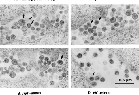
[image:3.612.65.547.368.699.2]FIG. 1. Electron micrographs of thin sections of wild-type and mutant virions of HIV-1JR-CSF(A) Wild type; (B)nefminus; (C)vprminus, (D)vifminus. Samples were prepared as described in Materials and Methods. Note the presence of typical mature particles containing a conical nucleoid with a dense broad end (arrow). The particles of wild-type HIV-1 and most of the particles of mutant HIV-1 had a mature morphology. The morphology was similar in these viruses.
TABLE 1. Summary of the properties of mutant viruses
Virus Amt of p24
gag
(ng/ml)a
Amt of endogenous RTb
(copies/pg of p24gag)a
No. of viral particles (per 30 cells)a
Entry in Th1-like culture (%)a
Viral DNA synthesis after 24-h infection
in Th1-like culture (copies/ng of p24gag)a
TCID50/ng of p24gagin
PHA-activated PBMC
Wild type
35.8
6
1.4
2,680
6
84
276
6
43
0.081
6
0.006
827
6
77
140
nef
minus
55.0
6
1.7
2,860
6
860
353
6
31
0.084
6
0.002
442
6
136
81
vif
minus
47.2
6
1.7
3,700
6
200
188
6
62
0.096
6
0.002
771
6
101
27
vpr
minus
22.8
6
0.5
3,600
6
100
180
6
57
0.081
6
0.004
818
6
137
184
aValues are means6standard deviations. bRT, reverse transcriptase.
on November 9, 2019 by guest
http://jvi.asm.org/
at the time of cell transfer. The experimental protocol was approved by the ethics review committees of the participating institutions. PBMC were isolated from blood samples obtained from healthy HIV-1-seronegative donors by density gradient centrifugation. To deplete NK cells in mice, the animal was treated with anti-IL-2 receptorb-chain monoclonal antibody TMb-1 (1.0 mg/animal) (32) 3 days prior to transfusion. Six days after intraperitoneal inoculation of 13107to 23107human PBMC in 0.5 ml of RPMI 1640 medium containing 10% FCS, 1,000 TCID50of viruses were injected intraperitoneally. The mice were sacrificed 4 or 8 days after infection, and human lymphocytes were recovered from peri-toneal lavages. The collected cells were examined by flow cytometry and PCR analysis as described previously (32). Heparinized blood was collected by car-diocentesis, and mouse plasma was used for the p24gagELISA (Coulter).
Flow cytometric analysis.A total of 0.33106to 1.03106cells were stained with antibodies specific for human cell surface markers (Dako Japan, Kyoto, Japan) for 30 min at 4°C and then washed three times. Fixed cells were analyzed within 24 h by flow cytometry (Coulter Epics Elite flow cytometer). The
anti-bodies used for this analysis were phycoerythrin-conjugated anti-CD4, fluores-cein isothiocyanate-conjugated CD8, and phycoerythrin-conjugated anti-HLA class I.
RESULTS
Properties of
nef
-minus,
vif
-minus, and
vpr
-minus HIV-1.
To
evaluate the role of HIV-1 accessory genes in virus replication,
we generated
nef
-minus,
vif
-minus, and
vpr
-minus HIV-1 by
introducing frameshift mutation and examined a variety of
their properties. We chose a macrophage-tropic HIV-1
JR-CSF [image:4.612.75.545.68.526.2]strain because this isolate can effectively replicate in all systems
by macrophages and activated CD4
1Th1 and Th2 cells. Each
FIG. 2. Kinetics ofnef-,vif-, andvpr-minus HIV-1JR-CSFandnef-minus HIV-1NL4-3in MDM and anti-CD3-activated CD41Th1- and Th2-like cells. Anti-CD3-activated CD41T cells (53105cells) were infected with 27 to 184 TCID
50, and MDM cells (53105cells) were infected with 1,000 TCID50of wild-type,nef-minus, andvpr-minus HIV-1 or 100 TCID50ofvif-minus HIV-1. Experiments with HIV-1NL4-3were performed only in Th2-like CD41T cells because HIV-1NL4-3did not replicate well in Th1-like CD41T cells and MDM cultures. The results of one representative experiment with three different blood donors are shown. The range of
HIV-1 production from culture supernatants in different donors varied from 3-fold (MDM) to 15-fold (Th1- or Th2-like cultures). Symbols:F, wild-type HIV-1JR-CSF; E,nef-minus HIV-1JR-CSF;ƒ,vif-minus HIV-1JR-CSF;‚,vpr-minus HIV-1JR-CSF;■, wild-type HIV-1NL4-3;h,nef-minus HIV-1NL4-3.
on November 9, 2019 by guest
http://jvi.asm.org/
mutant plasmid was transfected, and the virus was recovered
from culture supernatants. The rate of p24
gagrelease of
vpr
-minus HIV-1 was approximately 70% of that of the control
virus, and that of
nef
-minus HIV-1 and
vif
-minus HIV-1 was
approximately 150 and 130%, respectively, of that of the
wild-type virus (Table 1). The level of endogenous reverse
tran-scriptase RT activity of mutant HIV-1 particles was similar to
that of the wild type. Although the morphology of each mutant
particle was indistinguishable from the wild-type virus on
elec-tron microscopy, the number of viral particles per 30 cells
varied (Fig. 1; Table 1).
nef
deletion increased the number of
viral particles, whereas
vif
or
vpr
deletion resulted in a decrease
in the number viral particles. We recently showed that
mac-rophage-tropic HIV-1 isolates could efficiently replicate in
both CD4
1Th1-like cultures and CD4
1Th2-like cultures and
induce lytic infection particularly in CD4
1Th1-like cells (64).
Using this culture system, we examined the influence of Nef,
Vif, and Vpr. Although we observed a similar efficiency of viral
entry into primary CD4
1Th1-like (Table 1) and Th2-like (data
not shown) cultures, viral DNA synthesis in
nef
-minus virus in
CD4
1cultures was consistently approximately 50% of that
observed in the wild type. Furthermore, the infectivity of
vif
-minus or
nef
-minus mutant virus in PHA-activated PBMC was
about 20 and 60% of that of the wild type, respectively (Table
1).
Kinetics of
nef
-minus,
vif
-minus, and
vpr
-minus HIV-1
pro-duction in primary cultures.
As shown in Fig. 2, replication of
nef
-minus virus in anti-CD3-activated CD4
1Th1- and Th2-like
cell cultures was 10 to 100 times less efficient, respectively, 14
days after infection, than that in the wild-type virus. In
con-trast, replication of
nef
-minus virus in PHA-activated PBMC
was similar to that of the wild-type virus (data not shown),
confirming the results of previous reports (37, 61). The effect of
nef
on virus replication in anti-CD3-activated CD4
1Th1- or
Th2-like cells was not specific to HIV-1
JR-CSFisolate.
nef
-minus HIV-1
NL4-3, a prototype T-cell-line-tropic virus, also
replicated less efficiently than the parental wild-type strain in
CD4
1Th2-like cell culture (Fig. 2). We could not evaluate the
replication of HIV-1
NL4-3in CD4
1Th1-like cell culture
be-cause this isolate did not replicate sufficiently in Th1-like cells.
In MDM cultures,
nef
-minus HIV-1 showed reduced virus
pro-duction in one donor (Fig. 2), whereas a small change or no
difference in virus production was observed in MDM cultures
from two other donors (data not shown). Thus, the effect of
nef
on virus replication in MDM culture varied from one donor to
another. Production of the
vif
-minus virus was always lower
than that of the wild-type virus in all primary cultures including
MDM and activated CD4
1Th1- and Th2-like cell cultures,
while
vpr
-minus virus replicated well in MDM and
anti-CD3-activated CD4
1Th1- and Th2-like cell cultures.
[image:5.612.61.294.69.253.2]Since HIV-1 replication is highly dependent on cell
prolif-eration (63), we counted the number of activated CD4
1T cells
infected with each mutant virus or mock virus to exclude a
possible cytotoxic effect by mutations in
nef
or
vif
. As shown in
Fig. 3A, the cell growth kinetics of CD4
1Th1-like cell cultures
infected with either
nef
or
vif
mutated virus were similar to
those of mock-infected T cells. In contrast, reduced cell growth
in wild-type virus-infected culture was apparent 1 week after
infection, which could have resulted from high levels of HIV-1
replication (Fig. 3B). Thus, cytotoxicity paralleled HIV-1
pro-duction, and the reduced replication efficiency of
vif
-minus and
nef
-minus HIV-1 was not due to increased cell death in this
culture system. Since no culture system is yet available to
evaluate Nef function, especially of macrophage-tropic HIV-1
in activated T cells, our results indicate that the
anti-CD3-activated CD4
1Th1- or Th2-like cell culture system used in
this study is particularly useful to investigate the effect of
nef
of
HIV-1 on viral replication.
In the next step, we measured the exponential slope defining
HIV-1 production for the wild-type and
nef
-minus HIV-1 in
anti-CD3-activated Th1-like cell cultures before initiation of
loss of T cells (Fig. 4). The ratio of wild-type HIV-1 to
nef
-minus HIV-1 growth rate was 2.53
6
0.46 in two experiments
with CD4
1Th1-like cells from a single donor. Repeated
ex-periments with two other donors showed similar results (data
not shown). The exponential slope of HIV-1 production was
probably multiplicity of infection independent, since a 100-fold
increase in
nef
-minus HIV-1 load did not significantly change
the value of the exponential slope of HIV-1 production (Fig.
4). These data suggest that
nef
is crucial for the high viral load
in activated CD4
1Th1 or Th2 cells.
Since macrophage-tropic HIV-1 vigorously replicates in
CD4
1Th1-like cells, the use of anti-CD3 activated CD4
1Th1-like cell culture should be an adequate evaluation system
FIG. 3. Relationship between viral production and cell growth. (A) Total viable cells in each culture; (B) total HIV-1 p24gag
production in culture super-natants.
FIG. 4. Kinetics of nef-minus, 100-fold-increased (1003 nef-minus), and wild-type HIV-1JR-CSFin anti-CD3-activated CD41Th1-like cell culture. Anti-CD3 activated CD41Th1-like cells (53105cells) were infected with 81 TCID
50 ofnef-minus, 8,100 TCID50ofnef-minus, or 140 TCID50of wild-type HIV-1. Lines represent the least-squares linear regression lines for data points between days 4 and 18.
on November 9, 2019 by guest
http://jvi.asm.org/
[image:5.612.319.551.518.673.2]for HIV-1 production. Such a system can be used to examine
the effects of Nef on HIV-1 replication as well as pathogenicity.
In the next series of experiments, we examined Nef function in
more detail with this culture system.
Functional domains of Nef that induce efficient HIV-1
pro-duction in activated CD4
1T cells.
To determine the domains
of Nef responsible for efficient HIV-1 replication in activated
CD4
1Th1-like cells, we targeted three regions in the central
core region of Nef for site-directed mutagenesis: a glutamic
acid-rich segment (amino acids 72 to 75) (57), a proline-rich
motif that can interact with the SH3 domain containing cellular
proteins (amino acids 82 and 85) (50, 57), and conserved Lys/
Arg-Arg residues (amino acids 115 and 116) essential for
mul-tiple function including the binding of Nef to cellular serine
kinases, CD4 downregulation, and the effect on CD3 signaling
in T cells (25, 52, 69). Figure 5 shows the sites of mutation
introduced in Nef of HIV-1
JR-CSF. In the first step, we
ana-lyzed the steady-state levels of protein expression of each Nef
mutant by immunoblotting. COS cells were transfected with
each
nef
-mutated proviral DNA, and similar levels of p24
gagrelease were observed (data not shown). Nef-specific antibody
failed to detect signals from cell extracts in
nef
-minus HIV-1
DNA-transfected cells but could detect similar levels of
mu-tated Nef expression (M1, M3, M4, and M5) and wild-type
expression except for one mutant (M2). An approximately
10-fold reduction was observed in cells transfected with M2. In
the next step, we compared the kinetics of HIV-1 production in
cells infected by
nef
-mutant viruses with the kinetics in cells
infected with the parental wild-type HIV-1
JR-CSF(Fig. 6).
HIV-1 with point mutations in residues 115 and 116 (M2) and
deletion of the acidic amino acid-rich domain (residues 72 to
84 [M3] or residues 72 to 75 [M4]) replicated two- or threefold
less efficiently during 18 days of infection and showed less
cytopathicity than did wild-type virus. However, changing the
acidic amino acids (residues 72 to 75) to a neutral amino acid
(M5) and mutation at the proline-rich motif (M1) resulted in
no apparent change in HIV-1 production and cytopathic
prop-erties compared with the wild-type virus.
We also investigated the efficiency of viral DNA synthesis
after 24 h of infection of anti-CD3-activated CD4
1Th1-like
cells by each mutant virus (Fig. 7). The efficiency of viral DNA
synthesis of both M3 and M4 was two- to fourfold lower than
for the wild type, compared with the efficiency for complete
nef
-minus virus. M1 and M5, which grew in anti-CD3-activated
CD4
1Th1-like cells as efficiently as the wild-type virus did,
showed a rate of viral DNA synthesis in anti-CD3-activated
CD4
1Th1-like cells similar to that of the wild type. Although
the rate of M2 replication in anti-CD3-activated CD4
1Th1-like cells was always lower than that of the wild type, its rate of
viral DNA synthesis in anti-CD3-activated CD4
1Th1-like cells
was less impaired.
In vivo infection.
To evaluate Nef function in vivo, we used
a human/mouse chimera model consisting of SCID mice
en-grafted with human peripheral blood leukocytes
(hu-PBL-SCID) (21, 31, 39, 40). Recent studies have shown that a
human immune response and initial activation of CD4
1T cells
occur in hu-PBL-SCID mice within the first 2 to 3 weeks
following reconstitution (48, 65). Therefore, we injected HIV-1
into hu-PBL-SCID mice 6 days after reconstitution and
ana-lyzed the viral load and CD4
1T-cell depletion 4 and 8 days
later. In this modified hu-PBL-SCID mice model, a severe
CD4
1T-cell depletion was observed 8 days after infection with
wild-type HIV-1
JR-CSFbut not 4 days after infection (Table 2).
We also examined the effects of four Nef mutants on CD4
1FIG. 5. Mutations of the central core region of Nef and immunoblot analysis. (A) Amino acid sequences of a set of mutant Nef are aligned with that of the central core region of wild-type HIV-1JR-CSFNef. Dots indicate amino acid identity with the wild-type protein, and letters identify amino acid substitutions in the single-letter code. The nomenclature of the mutant Nef and corresponding mutations are defined on the left. (B) Immunoblot analysis of Nef expression was performed with anti-Nef monoclonal antibody from transiently transfected COS cells by wild-type HIV-1JR-CSFor an altered form ofnefinfectious provirus plasmid DNA. Wild-type and site-directed altered mutants (M1, M2, and M5) expressed 27-kDa Nef, and deletion mutants (M3 and M4) expressed smaller Nef. The Nef-minus mutant has no signal.
on November 9, 2019 by guest
http://jvi.asm.org/
T-cell killing in the hu-PBL-SCID mouse system. The mean
percentages of human CD4
1T cells based upon HLA
1cells in
the peritoneal cavity 8 days after infection are shown in Table
2. CD4
1T-cell depletion was less pronounced in
nef
-minus
virus-infected mice and only mild in M2 and M4 virus-infected
mice. In contrast, depletion was severe in M1 virus-infected
mice and wild-type virus-infected mice.
The amount of viral DNA in peritoneal cells from
nef
-mi-nus-, M2-, and M4-infected mice was significantly smaller than
in M1- or wild-type virus-infected mice 4 days after infection.
The mean levels of HIV-1 DNA (per 10
5human cells) in
peritoneal cells from infected mice 4 days after infection are
shown in Table 2. The levels of viral DNA in mice infected with
nef
-minus, M2, and M4 HIV-1 were about 70- to 700-fold
lower than in mice infected with M1 or wild-type virus. In
contrast, 8 days after infection, almost equal levels of HIV-1
DNA were found in peritoneal cells in all infected mice (Table
2). It seems that the level of viral DNA in mice after 8 days of
infection reached a peak during the course of HIV-1 infection
and no more permissive cells may reside in the chimera mice.
The level of p24
gagantigen in the plasma 4 days after infection
showed a pattern similar to the level of DNA, and the level 8
days after infection was almost identical (data not shown). We
concluded that the presence of residues 72 to 75 is important
for the induction of HIV-1 replication and subsequent CD4
killing in vivo.
DISCUSSION
Our data clearly showed a crucial role for Nef and Vif of
HIV-1 in activated T cells and demonstrated that Nef
acceler-ated viral replication in activacceler-ated T cells both in an in vitro
primary culture system and in an in vivo system, hu-PBL-SCID
mice. We found that residues 72 to 75 of Nef are important for
viral replication in vitro and in vivo whereas the prolines that
form part of the SH3 binding surface of Nef are not important
for viral replication in activated T cells.
The process of HIV-1 replication in patients has been
de-scribed as dynamic, involving continuous cycles of de novo
virus infection (45) with an estimated average total HIV-1
production of 10.3
3
10
9virions per day, a mean half-life of
plasma virions of 0.24 day, a mean half-life of productively
infected cells of 1.6 days, and an average HIV-1 generation
time of 2.6 days. Furthermore, HIV-1 replicates mainly in the
lymphoid organs (14, 44), where T cells are activated
contin-uously by several antigens, and virus replication is enhanced
after vaccination against influenza virus or IL-2 injection into
HIV-1-infected individuals (30, 42, 62), suggesting the
pres-ence of a close relationship between T-cell activation and
HIV-1 replication. Previous studies have demonstrated that
nef
of SIV or HIV significantly contributed to the high level of
viral replication and pathogenesis in vivo (3, 26, 28) and that
Nef played a positive role in the establishment of viral infection
in quiescent T cells in vitro (37, 61). However, its role in
primary activated T cells has not been demonstrated by in vitro
conventional methods such as PHA-activated blast culture.
Several investigators have observed the replication of
nef
-mi-nus HIV-1 in PHA blast cultures at a rate similar to that of the
wild type (61). To establish a suitable system of T-cell
activa-tion to study the funcactiva-tion of Nef in activated T cells, we
stim-ulated CD4
1T cells with anti-CD3 in the presence of IL-2 in
addition to either IL-12 or IL-4 and established an HIV-1
infection system with CD4
1Th1- and Th2-like cell cultures.
Our results showed a positive effect of Nef on HIV-1
replica-tion in these cells. Using this culture system, we first examined
the rate of virus growth. A high level of viral replication in in
vitro culture may cause the loss of CD4
1T cells followed by
limitation of viral spreading. To exclude these possibilities, we
ignored the peak levels of p24
gagproduction and compared the
growth rate of each mutant virus before the commencement of
T-cell loss. By estimating the slope defining HIV-1 production,
we were able to demonstrate that the growth-enhancing effects
of Nef might be attained during a single cycle of viral
replica-tion. This finding is similar to those described by other
inves-tigators (37). It is conceivable that a reduction in the
replica-tion ability of Nef-mutated HIV-1 will result in a significant
change after multiple rounds of infection, because a high rate
of de novo infection and replication of HIV-1 in infected
individuals may be crucial in the development of AIDS.
We also examined the effect of Nef in hu-PBL-SCID mice.
Previous studies with SCID-hu-Thy/Liv mice showed that
nef
-minus HIV-1 replicates less efficiently and causes less
deple-tion of CD4
1T cells with a T-cell line-tropic HIV-1
NL4-3strain
[image:7.612.62.296.70.391.2](3, 26). Although it was shown that a macrophage-tropic
HIV-1 strain did not induce a severe cytokilling effect in the
SCID-hu-Thy/Liv model, we were able to demonstrate
aug-mentation of HIV-1 replication and severe CD4
1T-cell
de-pletion with Nef by using hu-PBL-SCID mice. Mosier and
coworkers reported that macrophage-tropic HIV-1 induced
more severe pathogenic changes than did T-cell line-tropic
HIV-1 (40) and that
nef
-minus HIV-1 was less pathogenic in
hu-PBL-SCID mice (21). It seems that the cell condition of
transferred human T cells differs between SCID-hu Thy/Liv
FIG. 6. HIV-1 production and cytopathicity on HIV-1JR-CSF-infected or each nefmutant-infected Th1-like culture. Data are from two blood donors, donor M (A and B) and donor I (C and D). (A and C) Total production of HIV-1 p24gag
into culture supernatants; (B and D) total viable cells in each culture. Symbols: E, wild type;F,nefminus;‚, M1;■, M2;Œ, M3;}, M4;{, M5;v, mock.
on November 9, 2019 by guest
http://jvi.asm.org/
and hu-PBL-SCID mice. At 14 days after engrafting human
PBMC into SCID mice, all the human T cells in the peritoneal
cavity became CD45RO positive memory T-cell phenotype
and responded to anti-CD3 stimulation when the affected cells
were observed under in vitro conditions (64a). On the other
hand, 50% of recovered T cells from the thymus tissue in
SCID-hu-Thy/Liv expressed CD45RO molecules and the rest
were CD45RA-positive naive T cells. These results suggest that
the cells susceptible to HIV-1 infection in our hu-PBL-SCID
mice system are fully activated memory T cells. Therefore, the
present results confirm that Nef is also necessary for viral
replication in vivo in activated T cells.
[image:8.612.139.483.67.420.2]Based on our finding that anti-CD3-stimulated CD4
1T cells
were useful for examination of the effect of Nef, we also
in-vestigated the effect of Vif and Vpr on viral replication by
using the same in vitro system. Consistent with previous results
obtained with the SCID-hu-Thy/Liv system (3) or by the
con-ventional in vitro method (15), our results suggested that Vif is
FIG. 7. PCR analysis of humanb-globin DNA and HIV-1 DNA in cells infected with wild-type ornefmutant viruses at 24 h after infection. (A) Detection of human
b-globin DNA and HIV-1 DNA. “Standards” indicates the number of proviral DNA or humanb-globin DNA per each reaction. (B) Quantitation of HIV-1 DNA in infected cells at 24 h after infection withnefmutated viruses. Specific signals of humanb-globin DNA and HIV-1 DNA were measured from panel A and calculated from a standard curve of serially diluted HIV DNA or human DNA. The level of HIV-1 DNA synthesis after entry was expressed per nanogram of input p24gagantigen.
Each bar represents the mean6standard deviation (n52) from a representative experiment.
TABLE 2. Results for the hu-PBL-SCID-mice infected with HIV-1
aVirus
Day 4 Day 8
HIV-1 DNA/105
human cells % CD4-positive cells
HIV-1 DNA/105
human cells % CD4-positive cells
Mock
ND
b36.5
6
6.1
ND
34.6
6
11.4
Wild type
3,877
6
2,135
35.3
6
11.9
127,000
6
25,000
1.7
6
0.9
nef
minus
5.3
6
2.6
29.2
6
3.2
179,000
6
114,000
27.7
6
10.4
M1
1,463
6
1,300
35.4
6
0.9
73,700
6
27,400
1.2
6
0.4
M2
54
6
5
32.7
6
4.6
192,000
6
49,200
4.1
6
1.1
M4
32
6
12
35.3
6
5.2
217,000
6
125,000
28.4
6
18.7
aResults of three representative experiment for one blood donor are shown. Values are means6standard deviations. bND, not detected.
on November 9, 2019 by guest
http://jvi.asm.org/
[image:8.612.66.555.611.709.2]essential in all primary T-cell cultures. In contrast, we observed
no significant difference in the replication of
vpr
-minus HIV-1
in the activated CD4
1T-cell culture system compared with
that of wild-type virus. These results do not necessarily exclude
a potential role for Vpr in HIV-1-mediated pathogenesis of
infected individuals, although Vpr may have no direct effect on
virus production in activated T cells in vivo.
Several studies have shown that Nef enhances virus
infectiv-ity, as determined by the efficiency of viral DNA synthesis
rather than the level of binding or entry into target cells (2, 10,
55). Our study also confirmed these observations. Goldsmith et
al. (18) demonstrated that deletion of 10 amino acids,
corre-sponding to the M3 mutant in our present study and including
a part of the proline-rich domain, abrogated infectivity. While
M1 had little effect on viral DNA synthesis in the present
study, only the 4-amino-acid-deleted mutant (M4) showed
re-duced viral DNA synthesis in infected cells. Thus, these amino
acids must play an important role in Nef-induced HIV-1
aug-mentation. Although we cannot explain how Nef enhances
viral DNA synthesis and the lack of effect of the site-directed
mutant (M5) on viral DNA synthesis, future studies should
clarify these issues. Reduced infectivity of
nef
-minus HIV can
be complemented by expression of Nef on virus producer cells
(2, 38, 43). Nef is incorporated into the virion (8, 43, 68) and is
cleaved specifically by the viral protease (8, 68). The specific
cleavage site of Nef is contiguous with the acidic domain (16,
53). Cyclophilin A-deficient HIV-1 also shows an impaired
viral life cycle at the initiation of viral DNA synthesis (7).
Hence, we assumed that the acidic domain of Nef may be
important for Nef incorporation into the virion or for
proteo-lytic processing of Nef by the viral protease and that cleaved
Nef itself could act at the stage of virus uncoating as a
molec-ular chaperone that stabilizes the viral RNA-reverse
transcrip-tase complex. Alternatively, other unknown cellular factors,
e.g., a member of the chaperonin group, could associate with
Nef and may be incorporated into virion together with Nef.
Several groups of investigators have shown that
enhance-ment of infectivity and viral growth are both dependent on an
intact proline-rich motif (18, 50, 69), findings different from
those of the present study. These differences may be due to
different experimental procedures used in the two studies. In
the above studies, the HeLa-CD4-LTR/
b
-galactosidase cell
system was used for the infectivity assay, and Goldsmith et al.
(18) measured the expressed
b
-galactosidase by ELISA.
Therefore, the intact proline-rich motif may be required for
HIV-1 infection in HeLa-CD4 cells. Other results reported by
Saksela et al. (50) showed that the proline-rich motif should be
significant for the induction of virus replication from a latent
state within resting T cells. Therefore, the phenomenon that
the proline-rich motif in Nef may not be necessary for virus
replication may be characteristic in activated normal T cells.
Although previous reports suggested that the Nef protein is
associated with a cellular serine kinase through the conserved
Lys/Arg-Arg residues (residues 115 and 116) (19, 51, 52, 69),
the functional importance of this protein with regard to viral
replication has remained elusive. Recently, mutation in
resi-dues 115 and 116 was shown to disrupt several independent
functions of Nef including CD4 downregulation and the effect
of Nef on signal transduction in T cells, in addition to
disrupt-ing the association of Nef with serine kinase activity (25).
These phenomena seem to be explained by the stability of Nef,
because the Nef mutant with mutation in this 115 and 116
residues was unstable (25) (Fig. 5). The reduced viral
replica-tion of M2 in vitro and in vivo in the present study may also
result from the instability of mutant Nef. Unstable expression
of the Nef mutant (M2) might also influence the results of the
long-term experiment. The rate of viral DNA synthesis in our
in vitro T-cell culture was less impaired, although the rate of
viral production was lower than in the wild type. The rate of
viral DNA synthesis was measured at 24 h postinfection,
whereas viral production was monitored for 18 days after
in-fection.
In summary, our study demonstrated the significant role of
the
nef
gene in HIV-1 replication and its role in the
pathogen-esis of primary CD4
1T cells stimulated through the T-cell
receptor in the presence of IL-12 or IL-4. We also identified
the critical domain of Nef in the expression of its effects. These
data will be helpful for the development of both specific
ther-apeutic agents targeting Nef and safe live attenuated vaccines.
ACKNOWLEDGMENTS
We thank F. G. Issa for reading and editing the manuscript, Y.
Takahashi for technical support, J. Zack for providing
nef
-minus
HIV-1
JR-CSFplasmid, and M. Miyasaka for providing of TM
b
-1 monoclonal
antibody.
This work was supported by grants from the Ministry of Public
Health and Welfare and the Ministry of Biotechnology and Science in
Japan. Y.K., Y.T., and N.Y. were sponsored by the Japan Health
Sciences Foundation. N.Y. and Y.T. were also supported by Priority
Areas from the Ministry of Education, Sports and Culture, and
CREST (Core Research for Evolutional Science and Technology) of
Japan Science and Technology Corporation (JST). N.Y. was also
sup-ported by the Program for Promotion of Fundamental Studies in
Health Sciences of the Organization for Drug ADR Relief, R&D
Promotion and Product Review of Japan.
REFERENCES
1.Adachi, A., H. E. Gendelman, S. Koenig, T. Folks, R. Willey, A. Rabson, and M. A. Martin.1986. Production of acquired immunodeficiency syndrome-associated retrovirus in human and nonhuman cells transfected with an infectious molecular clone. J. Virol.59:284–291.
2.Aiken, C., and D. Trono.1995. Nef stimulates human immunodeficiency virus type 1 proviral DNA synthesis. J. Virol.69:5048–5056.
3.Aldrovandi, G. M., and J. A. Zack.1996. Replication and pathogenicity of human immunodeficiency virus type 1 accessory gene mutants in SCID-hu mice. J. Virol.70:1505–1511.
4.Balliet, J. W., D. L. Kolson, G. Eiger, F. M. Kim, K. A. McGann, A. Srini-vasan, and R. Collman.1994. Distinct effects in primary macrophages and lymphocytes of the human immunodeficiency virus type 1 accessory genes vpr,vpu, andnef: mutational analysis of a primary HIV-1 isolate. Virology
200:623–631.
5.Bartz, S. R., M. E. Rogel, and M. Emerman.1996. Human immunodeficiency virus type 1 cell cycle control: Vpr is cytostatic and mediates G2 accumula-tion by a mechanism which differs from DNA damage checkpoint control. J. Virol.70:2324–2331.
6.Borman, A. M., C. Quillent, P. Charneau, C. Dauguet, and F. Clavel.1995. Human immunodeficiency virus type 1 Vif2mutant particles from restrictive cells: role of Vif in correct particle assembly and infectivity. J. Virol.69:
2058–2067.
7.Braaten, D., E. K. Franke, and J. Luban.1996. Cyclophilin A is required for an early step in the life cycle of human immunodeficiency virus type 1 before the initiation of reverse transcription. J. Virol.70:3551–3560.
8.Bukovsky, A. A., D. Tatyana, A. Weimann, and G. G. Heinrich.1997. Nef association with human immunodeficiency virus type 1 virions and cleavage by the viral protease. J. Virol.71:1013–1018.
9.Chowers, M. Y., C. A. Spina, T. J. Kwoh, N. J. Fitch, D. D. Richman, and J. C. Guatelli.1994. Optimal infectivity in vitro of human immunodeficiency virus type 1 requires an intactnefgene. J. Virol.68:2906–2914.
10. Chowers, M. Y., M. W. Pandori, C. A. Spina, D. D. Richman, and J. C. Guatelli.1995. The growth advantage conferred by HIV-1nefis determined at the level of viral DNA formation and is independent of CD4 downregu-lation. Virology212:451–457.
11. Collette, Y., H. Dutartre, A. Benziane, F. Ramos-Morales, R. Benarous, M. Harris, and D. Olive.1996. Physical and functional interaction of Nef with Lck. J. Biol. Chem.271:6333–6341.
12. Courcoul, M., C. Patience, F. Rey, D. Blanc, A. Harmache, J. Sire, R. Vigne, and B. Spire.1995. Peripheral blood mononuclear cells produce normal amounts of defective Vif2human immunodeficiency virus type 1 particles which are restricted for the preretrotranscription steps. J. Virol.69:2068– 2074.
on November 9, 2019 by guest
http://jvi.asm.org/
13. Deacon, N. J., A. Tsykin, A. Solomon, K. Smith, M. Ludford-Menting, D. J. Hooker, D. A. McPhee, A. L. Greenway, A. Ellett, C. Chatfield, V. A. Lawson, S. Crowe, A. Maerz, S. Sonza, J. Learmont, J. S. Sullivan, A. Cunningham, D. Dwyer, D. Dowton, and J. Mills.1995. Genomic structure of an attenuated quasi species of HIV-1 from a blood transfusion donor and recipients. Science270:988–991.
14. Embretson, J., M. Zupancic, J. L. Ribas, A. Burke, P. Racz, K. Tenner-Racz, and A. T. Haase.1993. Massive covert infection of helper T lymphocytes and macrophages by HIV during the incubation period of AIDS. Nature (Lon-don)362:359–362.
15. Gabuzda, D. H., K. Lawrence, E. Langhoff, E. Terwilliger, T. Dorfman, W. A. Haseltine, and J. Sodroski.1992. Role ofvifin replication of human immu-nodeficiency virus type 1 in CD41T lymphocytes. J. Virol.66:6489–6495. 16. Gawdigk-Nitschko, K., A. Scho¨n, G. Wachinger. V. Erfle, and B. Kohleisen.
1995. Cleavage of recombinant and cell derived human immunodeficiency virus 1 (HIV-1) Nef protein by HIV-1 protease. FEBS Lett.357:275–278. 17. Gibbs, J. S., D. A. Regier, and R. C. Desrosiers.1994. Construction and in
vitro properties of HIV-1 mutants with deletion in “nonessential” genes. AIDS Res. Hum. Retroviruses10:343–350.
18. Goldsmith, M. A., M. T. Warmerdam, R. E. Atchison, M. D. Miller, and W. C. Greene.1995. Dissociation of the CD4 downregulation and viral infectivity enhancement functions of human immunodeficiency virus type 1 Nef. J. Virol.69:4112–4121.
19. Greenway, A., A. Azad, and D. McPhee.1995. Human immunodeficiency virus type 1 Nef protein inhibits activation pathways in peripheral blood mononuclear cells and T-cell lines. J. Virol.69:1842–1850.
20. Greenway, A., A. Azad, J. Mills, and D. McPhee.1996. Human immunode-ficiency virus type 1 Nef binds directly to Lck and mitogen-activated protein kinase, inhibiting kinase activity. J. Virol.70:6701–6708.
21. Gulizia, R. J., R. G. Collman, J. A. Levy, D. Trono, and D. E. Mosier.1997. Deletion ofnefshows but does not prevent CD4-positive T-cell depletion in human immunodeficiency virus type 1-infected human-PBL-SCID mice. J. Virol.71:4161–4164.
22. He, J., S. Choe, R. Walker, P. Di Marzio, D. O. Morgan, and N. R. Landau.
1995. Human immunodeficiency virus type 1 viral protein R (Vpr) arrests cells in the G2phase of the cell cycle by inhibiting p34cdc2activity. J. Virol.
69:6705–6711.
23. Heinzinger, N. K., M. I. Bukinsky, S. A. Haggerty, A. M. Ragland, V. Kewal-ramani, M. A. Lee, H. E. Gendelman, L. Ratner. M. Stevenson, and M. Emerman.1994. The Vpr protein of human immunodeficiency virus type 1 influences nuclear localization of viral nucleic acids in nondividing host cells. Proc. Natl. Acad. Sci. USA91:7311–7315.
24. Ho, D. D., A. U. Neumann, A. S. Perelson, W. Chen, J. M. Leonard, and M. Markowitz.1995. Rapid turnover of plasma virions and CD4 lymphocytes in HIV-1 infection. Nature (London)373:123–126.
25. Iafrate, A. J., S. Bronson, and J. Skowronski.1997. Separable functions of Nef disrupt two aspects of T cell receptor machinery—CD4 expression and CD3 signaling. EMBO J.16:673–684.
26. Jamieson, B. D., G. M. Aldrovandi, V. Planelles, J. B. Jowett, L. Gao, L. M. Bloch, I. S. Chen, and J. A. Zack.1994. Requirement of human immuno-deficiency virus type 1neffor in vivo replication and pathogenicity. J. Virol.
68:3478–3485.
27. Jowett, J. B., V. Planelles, B. Poon, N. P. Shah, M. L. Chen, and I. S. Chen.
1995. The human immunodeficiency virus type 1vprgene arrests infected T cells in the G21M phase of the cell cycle. J. Virol.69:6304–6313. 28. Kestler, H. W., III, D. J. Ringler, M. Kazuyasu, D. L. Panicali, P. K. Sehgal,
M. D. Daniel, and R. C. Desrosiers.1991. Importance of thenefgene for maintenance of high virus loads and for development of AIDS. Cell65:651– 662.
29. Kirchhoff, R., T. C. Greenough, D. B. Brettler, J. L. Sullivan, and R. C. Desrosiers.1995. Absence of intactnefsequence in a long-term survivor with nonprogressive HIV-1 infection. N. Engl. J. Med.332:228–232.
30. Kovacs, J. A., M. Baseler, R. J. Dewar, S. Vogel, R. T. Davey, Jr., J. Falloon, M. A. Polis, R. E. Walker, R. Stevens, N. P. Salzman, J. A. Metcalf, H. Masur, and H. G. Lane.1995. Increases in CD4 T lymphocytes with inter-mittent courses of interleukin-2 in patients with human immunodeficiency virus infection. A preliminary study. N. Engl. J. Med.332:567–575. 31. Koyanagi, Y., S. Miles, R. T. Mitsuyasu, J. E. Merrill, H. V. Vinters, and
I. S. Y. Chen.1987. Dual infection of the central nervous system by AIDS viruses with distinct cellular tropisms. Science236:819–822.
32. Koyanagi, Y., Y. Tanaka, J. Kira, M. Ito, K. Hioki, N. Misawa, Y. Kawano, K. Yamasaki, R. Tanaka, Y. Suzuki, Y. Ueyama, E. Terada, T. Tanaka, M. Miyasaka, T. Kobayashi, Y. Kumazawa, and N. Yamamoto.1997. Primary human immunodeficiency virus type 1 viremia and central nervous system invasion in a novel hu-PBL-immunodeficiency mouse strain. J. Virol.71:
2417–2424.
33. Luria, S., I. Chambers, and P. Berg.1991. Expression of the type 1 human immunodeficiency virus Nef protein in T cells prevents antigen receptor-mediated induction of interleukin 2 mRNA. Proc. Natl. Acad. Sci. USA
88:5326–5330.
34. Maruta, H., J. Holden, A. Sizeland, and G. D’Abaco.1991. The residues of
Ras and rap proteins that determine their GAP specificities. J. Biol. Chem.
266:11661–11668.
35. Masuda, T., V. Planelles, P. Krogstad, and I. S. Y. Chen.1995. Genetic analysis of human immunodeficiency virus type 1 integrase and the U3 att site: unusual phenotype of mutants in the zinc finger-like domain. J. Virol.
69:6687–6696.
36. Michael, N. L., G. Chang, L. A. d’Arcy, P. K. Ehrenberg, R. Mariani. M. P. Busch, D. L. Birx, and D. H. Schwartz.1995. Defective accessory genes in a human immunodeficiency virus type 1-infected long-term survivor lacking recoverable virus. J. Virol.69:4228–4236.
37. Miller, M. D., M. T. Warmerdam, I. Gaston, W. C. Greene, and M. B. Feinberg.1994. The human immunodeficiency virus-1nefgene product: a positive factor for viral infection and replication in primary lymphocytes and macrophages. J. Exp. Med.179:101–113.
38. Miller, M. D., M. T. Warmerdam, K. A. Page, M. B. Feinberg, and W. C. Greene.1995. Expression of the human immunodeficiency virus type 1 (HIV-1)nefgene during HIV-1 production increases progeny particle infec-tivity independently of gp160 or viral entry. J. Virol.69:579–584. 39. Mosier, D. E., R. J. Gulizia, S. M. Baird, D. B. Wilson, D. H. Spector, and
S. A. Spector.1991. Human immunodeficiency virus infection of human-PBL-SCID mice. Science251:791–794.
40. Mosier, D. E., R. J. Gulizia, P. D. MacIsaac, B. E. Torbett, and J. A. Levy.
1993. Rapid loss of CD41T cells in human-PBL-Scid-mice by noncytopathic HIV isolates. Science260:689–692.
41. Nunn, M. F., and J. W. Marsh.1996. Human immunodeficiency virus type 1 Nef associates with a member of the p21-activated kinase family. J. Virol.
70:6157–6161.
42. O’Brien, W. A., K. Govit-Ferbas, A. Namazi, S. Ovcak-Derzic, H. J. Wang, J. Park, C. Yeramian, S. H. Mao, and J. A. Zack.1995. Human immunodefi-ciency virus-type 1 replication can be increased in peripheral blood of sero-positive patients after influenza vaccination. Blood.86:1082–1089. 43. Pandori, M. W., N. J. Fitch, H. M. Craig, D. D. Richman, C. A. Spina, and
J. C. Guatelli.1996. Producer-cell modification of human immunodeficiency virus type 1: Nef is a virion protein. J. Virol.70:4283–4290.
44. Pantaleo, G., C. Graziosi, J. F. Demarest, L. Butini, M. Montroni, C. H. Fox, J. M. Orenstein, D. P. Kotler, and A. S. Fauci.1993. HIV infection is active and progressive in lymphoid tissue during the clinically latent stage of dis-ease. Nature (London)362:355–358.
45. Perelson, A. S., A. U. Neumann, M. Markowitz, J. M. Leonard, and D. D. Ho.
1996. HIV-1 dynamics in vivo: virion clearance rate, infected cell life-span, and viral generation time. Science271:1582–1586.
46. Piatak, M., Jr., M. S. Saag, L. C. Yang, S. J. Clark, J. C. Kappes, K. C. Luk, B. H. Hahn, G. M. Shaw, and J. D. Lifson.1993. High levels of HIV-1 in plasma during all stages of infection determined by competitive PCR. Sci-ence259:1749–1754.
47. Planelles, V., F. Bachelerie, J. B. Jowett, A. Haislip, Y. Xie, P. Banooni, T. Masuda, and I. S. Chen.1995. Fate of the human immunodeficiency virus type 1 provirus in infected cells: a role forvpr. J. Virol.69:5883–5889. 48. Rizza, P., S. M. Santini, M. Logozzi, C. Lapenta, P. Sestili, G. Gherardi, R.
Lande, M. Spada, S. Parlato, F. Belarelli, and S. Fais.1996. T-cell dysfunc-tions in hu-PBL-SCID mice infected with human immunodeficiency virus (HIV) shortly after reconstitution: in vivo effects of HIV on highly activated human immune cells. J. Virol.70:7958–7964.
49. Rogel, M. E., L. I. Wu, and M. Emerman.1995. The human immunodefi-ciency virus type 1vprgene prevents cell proliferation during chronic infec-tion. J. Virol.69:882–888.
50. Saksela, K., G. Cheng, and D. Baltimore.1995. Proline-rich (PxxP) motifs in HIV-1 Nef bind to SH3 domains of a subset of Src kinases and are required for the enhanced growth of Nef1viruses but not for down-regulation of CD4. EMBO J.14:484–491.
51. Sawai, E. T., A. S. Baur, B. M. Peterlin, J. A. Levy, and C. Cheng-Mayer.
1994. Human immunodeficiency virus type 1 Nef associates with a cellular serine kinase in T lymphocytes. Proc. Natl. Acad. Sci. USA91:1539–1543. 52. Sawai, E. T., A. S. Baur, B. M. Peterlin, J. A. Levy, and C. Cheng-Mayer.
1995. A conserved domain and membrane targeting of Nef from HIV and SIV are required for association with a cellular serine kinase activity. J. Biol. Chem.270:15307–15314.
53. Schorr, J., R. Kellner, O. Fackler, J. Freund, J. Konvalinka, N. Kienzle, H. G. Kra¨usslich, N. Mueller-Lantzsch, and H. R. Kalbitzer.1996. Specific cleavage sites of Nef proteins from human immunodeficiency virus types 1 and 2 for the viral proteases. J. Virol.70:9051–9054.
54. Schuitemaker, H., N. A. Kootstra, R. E. de Goede, F. de Wolf, F. Miedema, and M. Tersmette.1991. Monocytotropic human immunodeficiency virus type 1 (HIV-1) variants detectable in all stages of HIV-1 infection lack T-cell line tropism and syncytium-inducing ability in primary T-cell culture. J. Vi-rol.65:356–363.
55. Schwartz, O., V. Marechal, O. Danos, and J. M. Heard.1995. Human immunodeficiency virus type 1 Nef increases the efficiency of reverse tran-scription in the infected cell. J. Virol.69:4053–4059.
56. Skowronski, J., D. Parks, and R. Mariani.1993. Altered T cell activation and development in transgenic mice expressing the HIV-1nefgene. EMBO J.
12:703–713.
on November 9, 2019 by guest
http://jvi.asm.org/
57. Shugars, D. C., M. S. Smith, D. H. Glueck, P. V. Nantermet, F. Seillier-Moiseiwitsch, and R. Swanstrom.1993. Analysis of human immunodefi-ciency virus type 1nefgene sequences present in vivo. J. Virol.67:4639–4650. 58. Simon, J. H. M., R. A. M. Fouchier, and M. H. Malim.1996. The human immunodeficiency virus type 1 Vif protein modulates the postpenetration stability of viral nucleoprotein complexes. J. Virol.70:5297–5305. 59. Smith, B. L., B. W. Krushelnycky, D. Mochly-Rosen, and P. Berg.1996. The
HIVnefprotein associates with protein kinase C theta. J. Biol. Chem.
271:16753–16757.
60. Sova, P., and D. J. Volsky.1993. Efficiency of viral DNA synthesis during infection of permissive and nonpermissive cells withvif-negative human immunodeficiency virus type 1. J. Virol.67:6322–6326.
61. Spina, C. A., T. J. Kwoh, M. Y. Chowers, J. C. Guatelli, and D. D. Richman.
1994. The importance ofnefin the induction of human immunodeficiency virus type 1 replication from primary quiescent CD4 lymphocytes. J. Exp. Med.179:115–123.
62. Staprans, S. I., B. L. Hamilton, S. E. Follansbee, T. Elbeik, P. Barbosa, R. M. Grant, and M. B. Feinberg.1995. Activation of virus replication after vac-cination of HIV-1-infected individuals. J. Exp. Med.182:1727–1737. 63. Stevenson, M., T. L. Stanwick, M. P. Dempsey, and C. A. Lamonica.1990.
HIV-1 replication is controlled at the level of T cell activation and proviral integration. EMBO J.9:1551–1560.
64. Tanaka, Y., Y. Koyanagi, R. Tanaka, Y. Kumazawa, T. Nishimura, and N. Yamamoto.1997. Productive and lytic infection of CD41type 1 helper T
cells with macrophage-tropic human immunodeficiency virus type 1. J. Virol.
71:465–470.
64a.Tanaka, Y., et al.Unpublished data.
65. Tary-Lehmann, M., A. Saxon, and P. V. Lehmann.1995. The human immune system in hu-PBL-SCID mice. Immunol. Today16:529–533.
66. von Scherdler, U., J. Song, C. Aiken, and D. Trono.1993.vifis critical for human immunodeficiency virus type 1 proviral DNA synthesis in infected cells. J. Virol.67:4945–4955.
67. Wei, X., S. K. Ghosh, M. E. Taylor, V. A. Johnson, E. A. Emini, P. Deutsch, J. D. Lifson, S. Bonhoeffer, M. A. Nowak, B. H. Hahn, M. S. Saag, and G. M. Shaw.1995. Viral dynamics in human immunodeficiency virus type 1 infec-tion. Nature (London).373:117–122.
68. Welker, R., H. Kottler, H. R. Kalbitzer, and H. G. Kra¨usslich.1996. Human immunodeficiency virus type 1 Nef protein is incorporated into virus parti-cles and specifically cleaved by the viral proteinase. Virology219:228–236. 69. Wiskerchen, M., and C. Cheng-Mayer.1996. HIV-1 Nef association with
cellular serine kinase correlates with enhanced virion infectivity and efficient proviral DNA synthesis. Virology224:292–301.
70. Zack, J. A., S. J. Arrigo, S. R. Weitsman, A. S. Go, A. Haislip, and I. S. Y. Chen.1990. HIV-1 entry into quiescent primary lymphocytes: molecular analysis reveals a labile, latent viral structure. Cell61:213–222.
71. Zhu, T., H. Mo, N. Wang, D. S. Nam, Y. Cao, R. A. Koup, and D. D. Ho.1993. Genotypic and phenotypic characterization of HIV-1 patients with primary infection. Science261:1179–1181.