Identification of a Key Target Sequence To Block Human
Immunodeficiency Virus Type 1 Replication
within the
gag-pol
Transframe Domain
SHIZUKO SEI,
1* QUAN-EN YANG,
1DENNIS O’NEILL,
1KAZUHISA YOSHIMURA,
2†
KUNIO NAGASHIMA,
3ANDHIROAKI MITSUYA
1,2,4HIV Clinical Interface Laboratory
1and Laboratory of Cell and Molecular Structure,
3SAIC-Frederick, NCI-Frederick
Cancer Research and Development Center, Frederick, Maryland 21702; Experimental Retrovirology Section,
National Cancer Institute, Bethesda, Maryland 20892
2; and Department of Internal Medicine II,
Kumamoto University School of Medicine, Kumamoto, Japan
4Received 10 November 1999/Accepted 17 February 2000
Although the full sequence of the human immunodeficiency virus type 1 (HIV-1) genome has been known for
more than a decade, effective genetic antivirals have yet to be developed. Here we show that, of 22 regions
examined, one highly conserved sequence (ACTCTTTGGCAACGA) near the 3
ⴕ
end of the HIV-1
gag-pol
trans-frame region, encoding viral protease residues 4 to 8 and a C-terminal Vpr-binding motif of p6
Gagprotein in
two different reading frames, can be successfully targeted by an antisense peptide nucleic acid oligomer named
PNA
PR2. A disrupted translation of
gag-pol
mRNA induced at the PNA
PR2-annealing site resulted in a
de-creased synthesis of Pr160
Gag-Polpolyprotein, hence the viral protease, a predominant expression of Pr55
Gagdevoid of a fully functional p6
Gagprotein, and the excessive intracellular cleavage of Gag precursor proteins,
hindering the processes of virion assembly. Treatment with PNA
PR2abolished virion production by up to 99%
in chronically HIV-1-infected H9 cells and in peripheral blood mononuclear cells infected with clinical HIV-1
isolates with the multidrug-resistant phenotype. This particular segment of the
gag-pol
transframe gene
appears to offer a distinctive advantage over other regions in invading viral structural genes and restraining
HIV-1 replication in infected cells and may potentially be exploited as a novel antiviral genetic target.
The persistence of integrated proviruses in host cells
pre-sents formidable challenges to the treatment of human
im-munodeficiency virus type 1 (HIV-1) infection.
Replication-competent HIV-1 can be recovered from resting CD4
⫹T
lymphocytes even from individuals receiving potent
combina-tion antiretroviral therapy, whose plasma virus levels have
re-mained below the detection limit for a prolonged period (19,
33, 36, 147, 153). The discovery of a long-lived viral reservoir,
which is established early in the infection (18, 33, 147), has
suggested that HIV-1 cannot be easily eradicated from
in-fected individuals with the current treatments. Another
imped-iment to controlling HIV-1 infection is the emergence of
drug-resistant viral strains (21, 47, 50, 61, 62, 67, 78, 90, 107, 110,
111, 130). HIV-1 protease, in particular, seemingly tolerates
extensive sequence variations (8, 76, 79), contributing to a
rapid emergence of protease inhibitor-resistant strains, which
can be cross-resistant to multiple protease inhibitors (21, 47,
90, 107, 110, 111). Indeed, multidrug-resistant (MDR) HIV-1
strains are isolated increasingly from patients who have been
extensively treated with various antiretroviral agents of similar
classes (151), and the spread of these MDR strains may
be-come a serious threat to the containment of the AIDS
epi-demic in the future. The identification of novel viral targets is
clearly needed to empower anti-HIV-1 therapeutic strategies.
In recent years, viral coreceptors (25, 26, 39, 99, 105, 132),
integrase (7, 29, 31, 93, 101, 115, 154, 155), and the viral
nucleocapsid protein zinc finger motif (94, 117–120, 137) have
emerged as novel antiviral targets. However, substantial
progress has yet to be made before any of the candidate
com-pounds can be brought to practical applications.
Another possible antiviral target pursued over the years is
the HIV-1 genome itself. Antisense reagents, in particular,
have been extensively investigated primarily in the form of
nuclease-resistant phosphorothioate oligodeoxynucleotides
(PsODN). However, these previous attempts targeting various
regions of the HIV-1 genome with antisense PsODN have
produced inconsistent results in chronically infected cells (2,
4, 5, 64, 66, 83–86, 92, 141, 148). The reasons for a lack of
consistency may vary. Selected target sequences may have been
less critical for virion production in chronically HIV-1-infected
cells or less accessible to the PsODN molecules because of the
RNA-binding proteins or intracellular folding of the target
RNA. It is also possible that antisense PsODN molecules were
simply ineffective in abating ribosome elongation (9, 14, 139)
or that available antisense molecule numbers were insufficient
to overcome the enormous amount of viral transcripts
ex-pressed in the chronically infected cells (140).
Selection of optimal genetic targets and the use of potent
gene-intervening reagents are equally critical elements of a
successful antigene or antisense strategy. Peptide nucleic acid
(PNA) (27, 45, 103, 145), initially developed as a reagent for
strand invasion of the duplex DNA, is a DNA mimic, consisting
of a peptide backbone of
N
-(2-aminoethyl)glycine units in
place of a deoxyribose backbone. Although unmodified PNA
has a relatively poor cellular uptake compared to that of ODN
(104, 144), it has unique molecular characteristics which may
enhance its utility as a genome-intervening tool, such as
resis-* Corresponding author. Mailing address: HIV Clinical Interface
Laboratory, SAIC Frederick, NCI-Frederick Cancer Research and
Development Center, Bldg. 322, Rm. 27B, P.O. Box B, Frederick, MD
21702. Phone: (301) 846-1780. Fax: (301) 846-6067. E-mail: SEI
@dtpax2.ncifcrf.gov.
† Present address: Division of Clinical Retrovirology and Infectious
Diseases, Center for AIDS Research, Kumamoto University,
Kuma-moto, Japan.
4621
on November 9, 2019 by guest
http://jvi.asm.org/
tance to nucleases and proteases (24) and sequence-specific
hybridization to DNA or RNA targets using Watson-Crick
base pair formation (27) with much higher thermal stability
than that of ODN (27, 28, 51).
Using a PNA oligomer as a prototype molecular tool, we
explored viral sequences that were susceptible to
PNA-medi-ated inhibition of gene expression and asked whether such
sequences might be considered potential anti-HIV-1 genetic
targets in the current study. Target sequences were selected
from the previously less explored
gag-pol
gene, with a
partic-ular focus on the
pol
gene, as few studies have tried to directly
block the expression of viral enzymes. In particular, we were
interested in the protease-encoding sequence that begins
up-stream of the 3
⬘
end of the
gag-pol
transframe gene. Despite its
extensive sequence variations, a short segment of sequence
toward the 5
⬘
end of the viral protease-encoding gene, ACTC
TTTGGCAACGA, which also encodes the C-terminal
Vpr-binding motif of p6
Gagprotein, LXXLFG, by a different
read-ing frame (17, 68, 88), is highly conserved among various
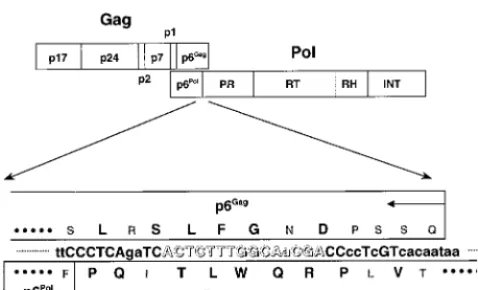
HIV-1 subtypes (Fig. 1). We hypothesized that nucleotide
se-quences of certain segments of the transframe domain must be
invariably conserved if a single transcript has to encode critical
amino acid sequences of two different proteins by a ribosomal
frameshift, thus reducing the probability of escape mutants,
and considered this particular sequence as one of the prime
targets.
MATERIALS AND METHODS
PNA oligomers.Because of the propensity of PNA molecules to be confined within the cytoplasm (104) (also see below), we decided to focus on the antisense intervention, primarily targeting the translation of viral RNA, and designed 22 PNA oligomers, 14- to 15-mers in length, complementary to the site of our in-terest within the gag-pol transframe domain or other highly conserved re-gions within thegagandpolgenes for the initial screening (Table 1). PNA oligomers were synthesized on the Expedite Nucleic Acid Synthesis System (PerSeptive Biosystems, Inc., Framingham, Mass.) by Research Genetics, Inc., Huntsville, Ala. Although all these PNA oligomers were easily dissolved in water, some oligomers, including PNAGAG1, PNAGAG2, PNAPR1, PNAPR2,
PNART1, PNART5, PNART8, and PNAINT3, formed fine precipitates when added
to the culture medium at higher concentrations, as has previously been reported (104). There was no discernible relationship between the precipitate formation and effects on cell growth or HIV-1 production (see below).
Evaluation of cellular uptake of PNA oligomers.H9 cells chronically infected with HIV-1LAI (H9LAI) were incubated with fluorescein-tagged PNAPR2,
PNAPR4, or PNART2at 10, 30, and 100 M in fresh RPMI 1640 complete
medium supplemented with 13% fetal bovine serum (HyClone, Logan, Utah), 4 mML-glutamine, 100 U of penicillin per ml, and 100g of streptomycin per ml
at 37°C in 5% CO2-containing humidified air overnight. The cells were fixed with
2% paraformaldehyde and examined by fluorescence microscopy or by fluores-cence-activated cell sorter analysis.
Assessment of effects of PNA oligomers on HIV-1 infection in various cell culture systems.H9LAIcells or H9 cells chronically infected with another HIV-1
strain, HIV-1RF(H9RF), were extensively washed to remove previously produced
virions and incubated in a 96-well culture plate at 104cells/well in 200l of
RPMI 1640 complete medium in the absence or presence of PNA oligomers at various concentrations. After 4 days in culture, supernatant was collected from each well and the level of p24 Gag antigen was determined by radioimmunoassay (RIA) (HIV-1 p24 RIA kit; NEN Life Science Products, Boston, Mass.). To compare the antiviral effects of PNAPR2and PNA oligomers targeting the
se-quences proximate to the PNAPR2-annealing site, H9LAIcells were cultured as
described above in the absence or presence of 100M PNAPR2or other PNA
oligomers tested. The relative antiviral effects were computed from the following formula: (% p24 antigen suppression by a given PNA oligomer)/(% p24 antigen suppression by PNAPR2). Each PNA oligomer was tested in triplicate. H9LAIand
H9RFcells were also cultured in a six-well culture plate at 105cells/ml in the
absence or presence of 30 to 100M PNAPR2. After 4 days, cells were counted
by the trypan blue dye exclusion method and lysed at 106cells/ml in
phosphate-buffered saline (PBS) containing 0.5% Triton X-100. The amounts of p24 antigen in the culture supernatant and cell lysate were measured by RIA. The experi-ments were repeated at least three times.
Phytohemagglutinin (PHA)-activated peripheral blood mononuclear cells (PBMC) obtained from healthy blood bank donors (2⫻106cells/ml) were
infected with seven MDR HIV-1 clinical isolates (151) in RPMI 1640 complete medium supplemented with 2.5 ng of recombinant human interleukin-2 (R&D Systems, Minneapolis, Minn.) per ml and cultured for at least 2 weeks or until the p24 antigen levels in the culture medium persistently exceeded 30 ng/ml. The PNAPR2-targeting nucleotide sequence of the transframe domain of each isolate
was consistent with the consensus B sequence. Freshly PHA-stimulated PBMC from healthy donors (2⫻106cells/ml) were added to each culture flask every 3
to 4 days. PBMC (106cells/ml) infected with each MDR isolate were vigorously
washed and cocultured with PHA-stimulated PBMC (106cells/ml) in 200l of
recombinant human interleukin-2-containing complete medium in the absence or presence of 10 to 60M PNAPR2. The number of infectious virions released
after 7 days in culture was determined by an assay with multinuclear activation of a galactosidase indicator (MAGI assay) (see below) using an indicator cell line, MAGI-CCR5 cells (16) (AIDS Research and Reference Reagent Program, Division of AIDS, National Institute of Allergy and Infectious Diseases, contrib-uted by Julie Overbaugh). All assays were performed in triplicate.
MT2 cells (104cells/ml) were exposed to 64 50% tissue culture infectious doses
of HIV-1NL4-3at 37°C for 1 h. After a vigorous virus washout, the cells were
cultured in 200l of RPMI 1640 complete medium at 37°C in 5% CO2
[image:2.612.55.294.70.215.2]-contain-ing humidified air in the absence or presence of various PNA oligomers at 100
FIG. 1. Schematic representation of thegagandpolregions of HIV-1 (top) and the amino acid sequences of portions of p6Gagprotein and viral protease encoded by two different reading frames (bottom). The amino acid residues conserved in⬎98% of the majority HIV-1 substrains (group M), which include subtypes A, B, C, D, F, G, H, and J and circulating recombinant forms (AE, AG, AGI, and AB) (HIV Sequence Database [http://hiv-web.lanl.gov]), are shown in large capital letters. The B subtype consensus sequences are shown in small capital letters. The nucleotide sequence of our interest is shown in open letters. PR, protease; RH, RNase H; INT, integrase.
TABLE 1. Sequences of PNA oligomers and locations of targeted
genes relative to the HXB2 numbering system
PNA oligomer Sequence (5⬘-3⬘) HIV-1 genomePosition in
PNA
Gag1CGCTCTCGCACCCAT
790–804
PNA
Gag2TCCCTGCTTGCCCAT
894–908
PNA
Gag3TCATCATTTCTTCT
1818–1831
PNA
PR1CCAAAGAGTGATTTT
2256–2270
PNA
PR2TCGTTGCCAAAGAGT
2262–2276
PNA
PR3ATCATCTGCTCCTGT
2328–2342
PNA
PR4CCCCCTATCATTTTT
2384–2398
PNA
RT1TTTTACTGGTACAGT
2568–2582
PNA
RT2CTGTCAATGGCCATT
2617–2631
PNA
RT3ATTTTCAGGCCCAA
2698–2711
PNA
RT4CTGTCTTTTTTCTTT
2738–2752
PNA
RT5ATTGTACTGATATCT
2976–2990
PNA
RT6AATCATCCATGTATT
3094–3108
PNA
RT7TGCTGCCCTATTTCT
3128–3142
PNA
RT8ATTGACAGTCCAGCT
3300–3314
PNA
RH1TGTGCTGGTACCCAT
4151–4165
PNA
INT1TAGCCATTGCTCTCC
4285–4299
PNA
INT2TCTACTTGTCCATGC
4379–4393
PNA
INT3CTTCCTGCTAATTTT
4535–4549
PNA
INT4CCCCCAATCCCCCCT
4793–4807
PNA
INT5AATTTTGAATTTTT
4883–4896
PNA
INT6TTGCTGGTCCTTTCC
4933–4947
on November 9, 2019 by guest
http://jvi.asm.org/
[image:2.612.313.549.499.728.2]M. The amount of p24 antigen produced by the MT2 cells was determined by RIA on day 7. All assays were performed in triplicate.
COS-7 cells were transfected with 1g of pNL4-3 by FuGENE 6 transfection reagent (Boehringer Mannheim, Indianapolis, Ind.) in a six-well culture plate (35-mm diameter) for 5 h, immediately followed by incubation in Dulbecco’s modified Eagle medium (DMEM) supplemented with 13% fetal bovine serum (HyClone), 4 mM L-glutamine, 100 U of penicillin per ml, and 100g of
streptomycin per ml in the absence or presence of 100M PNAPR2. After 48 h,
cells and supernatants were harvested from each sample and evaluated for viral protein expression and virion production. HLtat cells (AIDS Research and Reference Reagent Program; contributed by Barbara K. Felber and George Pavlakis) were transfected with 1g of p55M1-10 (kindly provided by George Pavlakis, National Cancer Institute-Frederick Cancer Research and Develop-ment Center) by FuGENE 6 transfection reagent (Boehringer Mannheim) for 5 h, immediately followed by incubation in complete DMEM in the absence or presence of 100M PNAPR2. After 24 h, HLtat cells were lysed for Western blot
analysis.
MAGI assay.The MAGI assay was employed to determine the number of newly produced virion particles in the culture supernatant of HIV-1-infected cells as described previously (65). Briefly, the HeLa–CD4–long terminal repeat– -galactosidase indicator cells were plated in a 96-well tissue culture plate at 104
cells per well, each well containing 125l of complete DMEM, 24 h prior to the assay. On the following day, the cells were generally 20 to 30% confluent. The cells were washed with 200l of Opti-MEM (Life Technologies, Inc., Rockville, Md.) twice and then exposed to serially diluted infectious culture supernatants in a total volume of 30l per well in the presence of 20g of DEAE-dextran (Sigma, St. Louis, Mo.) per ml. The infectious titers of the supernatants from PHA-PBMC infected with MDR isolates were examined with the MAGI-CCR5 indicator cell line (16), using 30 or 60l of inoculum. After the plates were incubated at 37°C in 5% CO2-containing humidified air for 2 h, 140l of
complete DMEM was added to each well. The plates were incubated for another 46 h, followed by fixation at room temperature with 1% formaldehyde and 0.2% glutaraldehyde in PBS for 5 min. The cells were then washed with PBS and incubated in 100l (per well) of staining solution containing 4 mM potassium ferrocyanide, 4 mM potassium ferricyanide, 2 mM MgCl2, and 0.4 mg of X-Gal
(5-bromo-4-chloro-3-indolyl--D-galactopyranoside) per ml. The blue cells were
counted under a microscope.
Electron microscopy. Electron microscopic examination was performed as previously described (38). Briefly, the harvested cells were centrifuged at 1,500⫻ gfor 5 min. The cell pellets were fixed in 1.25% glutaraldehyde and then in 1% osmium, dehydrated in graded alcohol, and embedded in pure epoxy resins. Thin sections (60 nm) were stained with uranyl acetate and lead citrate and stabilized by carbon evaporation for an examination.
RNA analysis.The culture supernatant was subjected to microcentrifugation at 32,800⫻gfor 2 h to pellet virions (35, 142). Pelleted virion particles were subjected to RNA extraction as previously described (6), followed by reverse transcriptase PCR (RT-PCR) with a primer pair, SK38-SK39 (128, 129), to estimate the amount of virion-derived RNA. The harvested cells were subjected to RNA extraction as previously described (129) followed by RT-PCR using two primer pairs, SK38-SK39 (128, 129) and BSS-KPNA (102, 124), in order to evaluate the levels of unspliced and singly spliced HIV-1 RNA, respectively.
Western blot analysis.The virion particles pelleted from the culture superna-tant (see above) were lysed in viral lysis buffer (10 mM Tris [pH 7.4], 1M EDTA, 0.02% NP-40). The harvested cells were washed in PBS and lysed in cell lysis buffer (10 mM Tris [pH 7.4], 50 mM NaCl, 100 mM KCl, 1 mM EDTA, 1% NP-40, 1 mM phenylmethanesulfonyl fluoride) at 2⫻107cells/ml at 4°C for
30 min, followed by centrifugation at 13,800⫻gto remove cell debris. Protein concentrations of the cell lysates were determined with the bicinchoninic acid protein assay kit (Pierce, Rockford, Ill.). Virion-associated protein derived from 100 l of supernatant and 2 to 25g of cellular protein were resolved by electrophoresis on sodium dodecyl sulfate–4 to 12% polyacrylamide gradient gels (Novex, San Diego, Calif.) under reducing conditions, followed by electrob-lotting onto a polyvinylidene difluoride membrane (Novex). The HIV-1 Gag proteins were visualized by a chemiluminescence detection system (Amersham Pharmacia Biotech, Inc., Piscataway, N.J.) using anti-p24Gagantiserum
(Ad-vanced Biotechnologies, Inc., Columbia, Md.) (2g of cellular protein per lane loaded for this antibody), anti-p17Gagmonoclonal antibody (Advanced
Biotech-nologies, Inc.) (2g of protein per lane), and anti-p6Gagantiserum (kindly
provided by Louis E. Henderson, National Cancer Institute-Frederick Cancer Research and Development Center) (5g of protein per lane). For the evalu-ations ofpolgene products, antiprotease antiserum (kindly provided by Louis E. Henderson), (25g of protein loaded per lane) and anti-p66RTmonoclonal
antibody (AIDS Research and Reference Reagent Program; contributed by Paul Yoshihara) (15g of protein per lane) were employed. For a resolution of anti-p66RTantibody-reactive proteins, sodium dodecyl sulfate–10%
polyacryl-amide gel electrophoresis was used instead of a 4 to 12% gradient gel. The anti-p66RT antibody is reactive with the p66RTbut not the p51RTband by
Western blot analysis (NIH AIDS Research and Reference Reagent Program Catalog). Anti-gp120 antiserum (AIDS Research and Reference Reagent Pro-gram; contributed by Margarita Quiroga) (10g of protein loaded per lane) was used to evaluate the amount ofenvgene product expressed in HIV-1-infected cells.
Peptide ELISA.The affinity of rabbit anti-p6Gagantiserum (kindly provided by
Louis E. Henderson) for a series of HIV-1 HXB2 (subtype B) Gag synthetic peptides, 441–458 (YKGRPGNFLQSRPEPTAP), 451–470 (SRPEPTAPPEESF RSGVETT), 459–479 (PEESFRSGVETTTPPQKQEPI), 470–489 (TTPPQKQ EPIDKELYPLTSL), and 480–500 (DKELYPLTSLRSLFGNDPSSQ) (obtained through AIDS Research and Reference Reagent Program), which encompassed the entire p6Gagdomain, was determined by enzyme-linked immunosorbent
assay (peptide ELISA). Briefly, 96-well plates were coated with serially diluted Gag peptides (9.8 to 2,500 ng/well) in triplicate. Anti-p6Gagantiserum diluted
1:500 was incubated in the peptide-coated plates for 90 min at room tempera-ture. After washing, horseradish peroxidase-labeled rabbit secondary anti-body was added to each well, followed by the peroxidase substrate. The antianti-body affinity for each peptide was determined by the optical density (OD) at 405 nm.
RESULTS
Cellular uptake of PNA and its intracellular localization.
First, in order to determine concentrations of PNAs required
to achieve a sufficient cellular delivery in tissue culture, H9LAI
cells were incubated with fluorescein-tagged PNA at 10 to 100
M. We elected to deliver PNA by simply adding it to the
culture medium, thus relying on endocytosis (42, 104, 131)
rather than the microinjection technique commonly adopted in
the previous studies (10, 45, 104). After an overnight
incuba-tion, H9LAI
cells were examined under a fluorescence
micro-scope. A clear fluorescent signal was demonstrated in the
ma-jority of cells incubated with PNA at
ⱖ
30
M compared to
untreated H9LAI
cells (Fig. 2A to C), albeit the signal was
virtually confined to the cytoplasm, as has previously been
reported (104) (Fig. 2D). The fluorescence intensity increased
in a dose-dependent manner as demonstrated by
fluorescence-activated cell sorter analysis regardless of the sequences tested
(Fig. 2E).
PNA
PR2, targeting the 3
ⴕ
end of the transframe domain,
decreases extracellular virion production while increasing
in-tracellular concentration of Gag protein in HIV-1-infected
cells.
Preliminary effects of various PNA oligomers were
eval-uated in chronically HIV-1-infected H9LAI
cells. While the
majority of PNA oligomers, all tested at 100
M, exhibited
virtually no antiviral effect, PNAPR2
reduced the HIV-1 p24
antigen production in the culture supernatant by up to 98.4%
(Fig. 3A). This p24-inhibitory effect of PNAPR2
appeared to be
dose dependent in H9LAI
as well as in H9RF
cells (Fig. 3B) and
specific to the PNAPR2-targeting region, as shifting the target
sequence by two or more nucleotides upstream or downstream
resulted in a more than 50% decrease in the inhibitory effect
(Fig. 3C). Significant inhibition of p24 by PNAPR2
was also
observed in MT2 cells acutely infected with HIV-1NL4-3
(inhi-bition of [99.6
⫾
0.4] % [mean
⫾
standard deviation (SD) of
triplicate values]) compared to untreated HIV-1-infected MT2
cells. Similarly, PNAPR2
reduced infectious virion production
from PBMC infected with clinical MDR isolates, originally
isolated from extensively pretreated patients (151), by 97.3 to
99.4% (Table 2). These data suggested a potential antiviral
effect of PNAPR2
against a broad spectrum of HIV-1 strains.
In contrast to a marked decrease in the supernatant p24
antigen levels, the amount of intracellular p24 antigen was
increased in H9LAI
cells treated with 30 or 60
M PNAPR2
(Fig. 3D) with moderately reduced cell numbers at the time of
harvest. Comparable results were obtained in other
experi-ments, in which PNAPR2
was tested up to 100
M. Percent cell
growth of H9LAI
cells incubated with 60 to 100
M PNAPR2
ranged from 68.3 to 78.1% (mean
⫾
SD, [73.9
⫾
5.1]%).
Similar degrees of impaired cell growth were observed in H9RF
as well as uninfected H9 cells incubated with up to 100
M
PNAPR2, while other PNA oligomers had no effect on cell
proliferation in H9LAI, H9RF, or uninfected H9 cells (data not
shown). These findings suggested that the impaired cell growth
on November 9, 2019 by guest
http://jvi.asm.org/
was specific to PNAPR2-targeted sequence intervention and
may have resulted from an accumulation of p24 Gag protein
within the cytoplasm and/or a down-modulation of certain host
proteins that have yet to be identified, rather than from general
toxicity of the PNA molecules.
Next, the processes of virion morphogenesis and virion
re-lease from the cell surface were examined by electron
micros-copy. Treatment with PNAPR2
appeared to hinder the steps of
virion assembly, unlike protease inhibitors, which halt the
pro-cess of virion morphological maturation. For 100 cells surveyed
within one 60-nm thin section, the estimated average number
of HIV-1 virions formed and released from the cells was
re-duced to less than two per sectioned cell (62 cells with no virion
and 38 cells with less than five virions per cell) in
PNAPR2-treated H9LAI
cells compared to 10 to 15 virions per sectioned
cell in untreated H9LAI
cells, while there were no apparent
changes in virion morphology (Fig. 4).
PNA
PR2blocks production of Pr160
Gag-Polpolyprotein and
induces excessive intracellular cleavage of Pr55
Gag.
To
de-termine the mechanism(s) of virion assembly inhibition by
PNAPR2, H9LAI
cells cultured in the presence of 30 to 100
M
PNAPR2
were evaluated for viral protein expression and virion
production. Consistent with the preliminary results, the level of
p24 antigen production and the amount of pelletable virions
were significantly reduced in the supernatant of
PNAPR2-treated cells compared to the unPNAPR2-treated control (Fig. 5A). The
amounts of intracellular HIV-1 unspliced and singly spliced
RNA transcripts were comparable between PNAPR2-treated
and untreated cells (Fig. 5A), indicating that PNAPR2
did not
interfere with the transcription of the HIV-1 genome.
Strik-ingly, Western blot analysis with anti-p24
Gagantibody
demon-strated that PNAPR2-treated H9LAI
cells contained
predomi-nantly p24 Gag protein with Gag precursor Pr55
Gagmarkedly
reduced and Pr160
Gag-Polvirtually undetectable within the cells
(Fig. 5A).
In order to further examine the expression and subsequent
proteolytic processing of Gag protein in PNAPR2-treated cells,
H9LAI
cells were cultured with an HIV-1 protease inhibitor,
indinavir, in addition to PNAPR2. The combination of indinavir
and PNAPR2
resulted in a reduction of intracellular p24 and
p17 Gag proteins and the reappearance of Pr55
Gagprotein, but
not Pr160
Gag-Pol(Fig. 5B). Because the PNAPR2-targeting site
was near the 3
⬘
end of the p6
Gag-encoding region, the
proteo-lytic processing of Pr55
Gag, also a precursor of p6
Gagprotein,
was examined by Western blot analysis with anti-p6
Gaganti-body. The peptide ELISA demonstrated that the anti-p6
Gagantibody was most reactive to the peptide 480–500, which
cor-responds to the C-terminal domain of p6
Gagprotein, with the
ODs linearly increasing from 0.303
⫾
0.095 to 1.468
⫾
0.052
(mean
⫾
SD of triplicate values) in detecting 9.8 to 625 ng of
the peptide per well, followed by the peptide 451–470, with
ODs ranging from 0.212
⫾
0.064 to 0.595
⫾
0.067. The
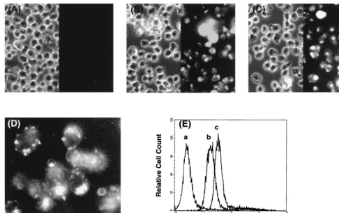
affin-FIG. 2. Cellular uptake of PNA. (A to C) Chronically HIV-1-infected H9LAIcells were incubated with fluorescein-tagged PNA at various concentrations overnight,
fixed with 3.7% formaldehyde for 20 min, washed and resuspended in PBS, and examined by fluorescence microscopy. Shown are the cells incubated with 0 (A), 30 (B), and 60 (C)M fluorescein-tagged PNAPR2. Note that the cells treated with fluorescein-tagged PNAPR2were resuspended in a slightly larger volume of PBS than
was the untreated control so that each cell could be clearly visualized. The panel on the right-hand side is the fluorescence image, and that on the left is the phase-contrast image of the same field. The discernible fluorescent signal was seen in the majority of cells at 30M PNA or greater. (D) Shown at a higher magnification are the cells incubated with 30M PNAPR2. Note that the fluorescent signal is virtually confined to the cytoplasm. (E) H9LAIcells incubated with fluorescein-tagged
PNA were also evaluated by fluorescence-activated cell sorter analysis. Curves a, b, and c represent H9LAIcells incubated with 0, 30, and 60M fluorescein-tagged
PNAPR2, respectively. Similar results were obtained with fluorescein-tagged PNAPR4and PNART2.
on November 9, 2019 by guest
http://jvi.asm.org/
[image:4.612.55.542.70.376.2]ities to the other three peptides were negligible. Western blot
analysis with the anti-p6
Gagantibody could detect p6
Gagpro-tein in the HIV-1 virions (data not shown and reference 106)
as well as its precursor (Pr55
Gag) in the cell lysate of untreated
H9LAI
cells (Fig. 5B). This p6
Gagantibody also demonstrated
the p6
Gag-containing intermediate (p15) in the lysate of
un-treated H9RF
cells (data not shown). Notably, the cell lysate of
H9LAI
treated with 60
M PNAPR2
contained virtually no
Pr55
Gagprotein which could be detected by the anti-p6
Gagantibody, even with the addition of a protease inhibitor,
indi-navir, while the anti-p24 antibody readily recognized Pr55
Gagin the same lysate (Fig. 5B), indicating that the Pr55
Gagex-pressed in PNAPR2-treated cells had insufficient binding
deter-minants for the anti-p6
Gagantibody. We then examined the
effects of PNAPR2
on the expression of Pr55
Gagprotein in
HLtat cells (32, 127) transfected with the Gag expression
plas-mid p55M1-10, in which Pr55
Gagwas not expected to be
cleaved by the viral protease, as p55M1-10 contained solely the
gag
reading frame (126). While Western blot analysis with
anti-p24
Gagantibody demonstrated Pr55
Gagprotein in both
untreated and PNAPR2-treated cells, the band intensity of
anti-p6
Gagantibody-reactive Pr55
Gagwas significantly diminished in
PNAPR2-treated HLtat cells (Fig. 5C).
These data suggested that PNAPR2
treatment could induce
the truncation of Pr55
Gagtoward the C terminus of p6
Gag,
[image:5.612.79.523.75.376.2]presumably by disrupting a translation of the
gag
reading frame
at its site of binding to the target mRNA in HIV-1-infected
cells. It is also possible that PNAPR2
effectively blocked the
translation of the
gag-pol
reading frame, resulting in
genera-tion of truncated Pr160
Gag-Pol, namely, Pr55
Gagwith p6
Polwhich could not be detected by the anti-p6
Gagantibody. Thus,
the intracellular Gag precursor protein in PNAPR2-treated
TABLE 2. HIV-1 virion production from PHA-PBMC infected with
clinical MDR isolates and cultured in the absence
or presence of 60
M PNA
PR2MDR isolate no. Result of MAGI-CCR5 assay
a
No PNAPR2 60M PNAPR2
1
467.7
⫾
138.6
12.7
⫾
4.2 (97.3)
2
486.7
⫾
112.0
4.7
⫾
5.5 (99.0)
3
210.0
⫾
16.10
1.3
⫾
0.6 (99.4)
4
589.7
⫾
32.10
7.7
⫾
4.2 (98.7)
5
214.0
⫾
142.6
5.7
⫾
1.5 (97.4)
6
219.7
⫾
25.20
3.7
⫾
1.5 (98.3)
7
208.3
⫾
96.70
2.0
⫾
1.0 (99.0)
aResults are shown as the mean numbers⫾SDs of blue cells per well. Values
[image:5.612.312.551.611.710.2]in parentheses are the mean percent reductions.
FIG. 3. Effects of PNA oligomers on HIV-1 production in chronically HIV-1-infected cells. (A) Levels of p24 antigen in the supernatants of H9LAIcells cultured in the absence or presence of various PNA oligomers used at a 100M concentration. All compounds were tested in triplicate, and the values shown are means with SDs. The p24 antigen level in a supernatant of H9LAItreated with an HIV-1 protease inhibitor, indinavir, is shown as a reference. (B) Levels of p24 antigen in the supernatants of H9LAIand H9RFcells cultured in the absence or presence of various concentrations of PNAPR2. Each dose was tested in triplicate. The values shown are means with SDs. (C) The degrees of inhibition of p24 antigen production in H9LAIcells cultured with 100M PNAPR2or other PNA oligomers targeting the sequences proximate to the PNAPR2-annealing site were compared. Each PNA oligomer was tested in triplicate. Each bar represents the target sequence shifted upstream (⫺) or downstream (⫹) by one to six nucleotides away from the PNAPR2-annealing site. The values shown are relative antiviral effects (means⫾SDs). (D) Amounts of p24 antigen in the supernatant (solid bars) and cell lysate (crosshatched bars) of H9LAIcells cultured in the absence or presence of 30 and 60M PNAPR2. The cell numbers are indicated by circles. The experimental results shown are representative of three separate experiments.
on November 9, 2019 by guest
http://jvi.asm.org/
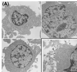
FIG. 4. Electron microscopy of H9LAIcells cultured in the absence (A) or presence (B) of 60M PNAPR2. Shown are representatives of 100 cells surveyed in each
sample (magnification,⫻13,500). Virions are shown at a higher magnification (⫻54,000) in each inset on the right-hand side at the bottom.
on November 9, 2019 by guest
http://jvi.asm.org/
cells appeared to be predominantly Pr55
Gaglacking the
full-size p6
Gagprotein, which was excessively cleaved to the final
products by a certain amount of viral protease.
PNA
PR2reduces synthesis of viral protease as well as
triggering its premature activation.
We then asked whether
preexisting viral protease at the beginning of H9LAI
cell
culture subsequently influenced the proteolytic processing
of the viral protein. To this end, nonpermissive COS-7 cells
were transfected with an infectious HIV-1 molecular clone,
pNL4-3, immediately followed by incubation with 100
M
PNAPR2
in tissue culture. Virion production was significantly
suppressed in PNAPR2-treated COS-7 cells as demonstrated by
the reduced p24 antigen level and the decreased number of
infectious virion particles in the culture supernatant
deter-mined by the MAGI assay (65). The intracellular Gag protein
was predominantly p24 and p17 with significantly decreased
amounts of Pr55
Gagand virtually undetectable Pr160
Gag-Polas
observed in chronically HIV-1-infected H9 cells (Fig. 6A).
Consistent with the decreased quantity of full-size Pr160
Gag-Pol, the amount of intracellular viral protease was reduced in
PNAPR2-treated cells compared to untreated control (Fig. 6B).
It should be noted that the antiprotease antibody that we
employed in the current study detected a monomeric form of
viral protease, not a homodimer, an active form of viral
pro-tease (112, 146). The latter is known to continually undergo
autodegradation as it is being activated (123, 135). Thus, the
amount of intracellular monomeric viral protease may not
nec-essarily reflect the total amount of active viral protease within
the cytoplasm but may simply correlate with the amount of
precursor polyprotein. These results strongly suggested that
the disrupted translation of
gag-pol
mRNA induced by
PNAPR2
subsequently led to a premature activation of viral
protease, albeit synthesized in probably a much smaller
quan-tity than untreated control, resulting in an excessive
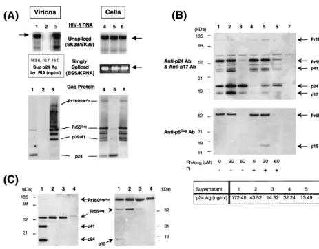
intracel-FIG. 5. Effects of PNAPR2on Gag-Pol expression and virion production in HIV-1-infected cells (A and B) and on Gag protein expression in HLtat cells transfected
with the Gag expression plasmid p55M1-10 (C). (A) The amounts of viral RNA in the culture supernatant (pelleted virion derived) (lanes 1 to 3) and in the cells (lanes 4 to 6) were evaluated by RT-PCR (top). Arrows indicate the pertinent PCR products for each primer pair. The virion-derived Gag protein in the culture supernatant (lanes 1 to 3) and intracellular Gag protein processing (lanes 4 to 6) were evaluated by Western blot analysis using anti-p24Gagantibody (bottom). Shown are the
positions of Pr160Gag-Pol, Pr55Gag, p41, and p24 Gag proteins. Lanes of each gel represent H9
LAIcells cultured alone (lanes 1 and 4) or incubated with 100M PNAPR2
(lanes 2 and 5) and 2M indinavir (lanes 3 and 6). (B) Western blot analyses with anti-p24Gagantibody and anti-p17Gagantibody (top) and with anti-p6Gagantibody
(bottom) of cell lysates processed from H9LAIcells cultured alone (lane 1) or in the presence of 30M PNAPR2(lane 2), 60M PNAPR2(lane 3), 2M indinavir
alone (lane 4), 30M PNAPR2plus 2M indinavir (lane 5), or 60M PNAPR2plus 2M indinavir (lane 6). Cell lysate of uninfected H9 cells is shown in lane 7. PI⫺
or⫹represents the absence or presence of a protease inhibitor, indinavir, respectively. The level of p24 antigen in each culture supernatant is indicated at the bottom. (C) Western blot analyses with anti-p24Gagantibody (left) and anti-p6Gagantibody (right) of cell lysates processed from HLtat cells transfected with pHXB2-based
plasmid pSUM9 (136) (lanes 1) or with p55M1-10 and cultured alone (lanes 2) or in the presence of 100M PNARR2(lanes 3). The cell lysate of mock-transfected
HLtat cells is included as a reference (lanes 4). Sup, supernatant; Ag, antigen; Ab, antibody.
on November 9, 2019 by guest
http://jvi.asm.org/
[image:7.612.70.534.70.432.2]lular cleavage of the Gag protein and an impairment of HIV-1
virion morphogenesis.
Interestingly, the amount of another
pol
gene product, RT,
appeared to be disproportionately high compared to the
low-level expression of its precursor, Pr160
Gag-Polpolyprotein, in
PNAPR2-treated cells. Western blot analysis using a
monoclo-nal antibody against p66
RT, which also recognizes the
precur-sor (p160) and several intermediates, but not p51
RT(NIH
AIDS Research and Reference Reagent Program Catalog,
1998), clearly demonstrated p66
RTin both untreated and
PNAPR2-treated cells (Fig. 6B). However, the cell lysate of
PNAPR2-treated COS-7 cells contained fewer precursor
inter-mediates, smaller than 100 kDa in size (Fig. 6B). These data
suggested that, in PNAPR2-treated cells, the translation of
gag-pol
mRNA may have been reinitiated at the appropriate AUG
codon located downstream of the PNAPR2-targeting site by the
mechanism described previously as ribosomal leaky scanning
(71, 74, 75). A consensus sequence for the efficient initiation of
translation by eukaryotic ribosomes has been reported as
xx-PuxxAUGG with a purine (Pu) in the
⫺
3 position (three
nu-cleotides upstream of the AUG codon) having a dominant
effect (70, 72, 73). The potential AUG codons downstream of
the PNAPR2-annealing site were identified at various positions,
including 2358 to 2360 (GAAAUGA), 2388 to 2390 (AAAA
UGA), 2595 to 2597 (GGAAUGG), and 2670 to 2672 (GAG
AUGG) of the HIV-1 HXB2 genome, preserving the amino
acid sequence of the remaining Pol. If the translation is
inter-nally initiated at the sites listed above, resulting Pol proteins
will be approximately 103.7, 102.5, 95.2, and 92.3 kDa,
respec-tively. These Pol proteins may be cleaved by viral protease
expressed in
trans
or by host proteases (34, 87). A similar
pattern of p66
RTexpression was also observed in chronically
infected H9 cells, and the addition of a viral protease inhibitor,
indinavir, to PNAPR2
did not restore detection of the full-size
precursor p160 (data not shown). Expression of HIV-1 Env
was not affected by PNAPR2
in either COS-7 cells transfected
with pNL4-3 (Fig. 6C) or chronically infected H9 cells (data
not shown).
DISCUSSION
The introduction of protease inhibitors (55–57, 63, 77, 121)
to conventional RT inhibitor-based antiretroviral therapy (23,
46, 95–97, 149, 150) has induced unprecedented degrees of
viral load reduction and a recovery of CD4
⫹T-lymphocyte
counts even in patients with advanced HIV-1 disease (20, 22,
43, 44, 91, 125). Substantial declines in AIDS incidence and
death observed in the United States and other industrialized
countries for the past 2 years have also been attributed to this
improved treatment regimen (1, 15), often referred to as highly
active antiretroviral therapy. While RT inhibitors, which block
de novo HIV-1 infection of uninfected cells, have no control
over production of virions from HIV-1-infected cells, protease
inhibitors actively exert their effect in infected cells, driving
them to release noninfectious immature virions and thereby
preventing the spread of HIV-1. Dramatic effects of highly
active antiretroviral therapy on individual cases of HIV-1
in-fection as well as on the global AIDS epidemic, although they
may possibly be short-lived, underscore the importance of
tar-geting the existing HIV-1 reservoir (HIV-1-infected cells) to
better control HIV-1 disease, rather than simply providing
protective shields for uninfected cells.
In the current study, we attempted to identify the viral gene
sequences that were not only critical for the virion production
but also accessible to gene-intervening molecules in chronically
HIV-1-infected, provirus-laden cells. We showed that PNAPR2,
which is complementary to a region of the viral
gag-pol
mRNA
sequence near the 3
⬘
end of the transframe domain, encoding
protease residues 4 to 8 and the C-terminal Vpr binding motif
of p6
Gagprotein, can effectively interrupt a translation of
gag-pol
mRNA. The disrupted translation of
gag-pol
mRNA at the
PNAPR2-annealing site gave rise to Pr55
Gagas a predominant
Gag precursor, which lacked a full-length p6
Gagprotein due to
a C-terminal truncation of p6
Gagand/or production of Pr55
Gagcontaining p6
Polprotein. The arrest of
gag-pol
mRNA
transla-tion by PNAPR2
was not complete, thereby permitting a
syn-thesis of a small quantity of full-size Pr160
Gag-Polpolyprotein,
which was presumably processed to viral protease.
[image:8.612.56.293.71.436.2]Despite the small amount of viral protease synthesized in
PNAPR2-treated cells, Gag precursors were almost entirely
cleaved to the final products of p24 and p17 within the
cyto-plasm. These seemingly counterintuitive findings prompted us
to examine the intracellular expression profiles of viral
pro-tease. The Western blot analysis of cell lysate with antiprotease
FIG. 6. Effects of PNAPR2ongag,pol, andenvgene expression and virion
production in COS-7 cells transfected with an infectious HIV-1 molecular clone, pNL4-3. Shown are results of Western blot analyses with anti-p24Gagantibody
and anti-p17Gagantibody (A), antiprotease antibody and anti-p66RTantibody
(B), and anti-gp120 antibody (C) of pNL4-3-transfected COS-7 cells cultured with no drug (lanes 1), 100M PNAPR2 (lanes 2), 1M saquinavir (also a
protease inhibitor) (lanes 3), or 3M indinavir (lanes 4). The cell lysate of mock-transfected COS-7 cells is shown in lanes 5. Viral lysate from pelleted virion particles (vp) is included in the Western blot analyses for thepolgene products as a reference. The levels of p24 antigen in supernatant and the infectious titers determined by MAGI assay are shown at the bottom. Ab, antibody; Ag, antigen.
on November 9, 2019 by guest
http://jvi.asm.org/
antibody demonstrated a significantly decreased amount of
monomeric viral protease in PNAPR2-treated COS-7 cells
trans-fected with an HIV-1 molecular clone compared to untreated
control, whereas an active form of homodimeric viral protease
(112, 146) could not be visualized in either PNAPR2-treated or
untreated cells, probably because the homodimer had
under-gone autolysis as it was being activated (123, 135). It has
pre-viously been reported that the level of viral protease activity
required to properly process the precursor proteins may be
re-duced by 4- to 50-fold, below which a limited amount of
pro-teolytic processing can still be demonstrated (122). Therefore,
it would not have been surprising to see some degree of Gag
precursor processing in PNAPR2-treated cells even if the actual
quantity of active viral protease had been significantly
de-creased. However, the extent of intracellular Gag precursor
cleavage observed in PNAPR2-treated cells notably exceeded
that in the untreated control, resulting in a significant
accumu-lation of p24 and p17 within the cells. These data strongly
sug-gested that the rate or timing of protease activation was
mark-edly accelerated within the cytoplasm of PNAPR2-treated cells.
The impact of PNAPR2-mediated translation arrest of
gag-pol
mRNA seems to be severalfold. Not only did it impede the
synthesis of full-size Pr160
Gag-Pol, leading to decreased
produc-tion of viral protease, but it also appeared to trigger the
pre-mature activation of viral protease, resulting in excessive and
untimely intracellular cleavage of Gag proteins and
signifi-cantly reduced virion production. In HIV-1, approximately 5 to
10% of
gag-pol
mRNA translational events are mediated by
⫺
1
ribosomal frameshifting to produce Pr160
Gag-Polprecursor
poly-protein (52, 143), which is eventually incorporated into viral
particles to provide viral protease, RT, and integrase. This
delicately balanced synthesis of Pr55
Gagand Pr160
Gag-Poland
their intermolecular association are believed to play a critical
role in coordinating sequential events of virion assembly and
release. The final stages of virion morphogenesis begin with
the interaction of Pr55
Gagand Pr160
Gag-Polpolyprotein (80, 81,
108, 133), which then attach to the plasma membrane of host
cells (13, 41, 116). Viral protease embedded in Pr160
Gag-Polremains inactive until Gag and Gag-Pol precursors reach the
plasma membrane, where precursor protein processing and
virion assembly are initiated upon activation of the viral
pro-tease, followed by the budding and completion of virion
mat-uration (58). Although viral protease-mediated precursor
cleavage may take place within the cytoplasm (13, 41, 59),
the membrane-associated precursor processing and
assem-bly events must occur in a regulated sequence in order for the
infectious virions to be released (58).
Excessive intracellular processing of Gag precursor proteins
without concomitant extracellular production of mature
viri-ons as induced by PNAPR2
has also been observed in a number
of other conditions, including (i) sole expression of
Pr160
Gag-Polencoded in a single reading frame and thus
lack-ing p6
Gag(60, 109), (ii) p6
Gag-deletion or -truncation HIV-1
molecular clones (40, 49, 152), and (iii) HIV-1 molecular
clones containing p6
Gagwith mutated Vpr-binding motifs (49,
152). Notably, inactivation of the viral protease induced by
mutational changes could restore the production of virions,
albeit immature virions, in the p6
Gag-deletion HIV-1
provirus-containing cells (49). These data suggest that the majority of
viral protease may be prematurely released from the
precur-sors and activated in the absence of fully functional p6
Gag.
How p6
Gagprotein exerts such a regulatory effect on viral
protease has yet to be determined. It is possible that p6
Gagmay
actively participate in the process of intermolecular association
of Pr55
Gagand Pr160
Gag, in addition to the major homology
domain of capsid protein (48, 134), such that the release of
viral protease occurs in a coordinated fashion at the plasma
membrane. Whether the binding of Vpr to p6
Gagplays any role
in regulating the processes of Gag and Gag-Pol precursor
association and subsequent protease activation is unclear, since
some studies have shown that a deletion of Vpr had no effect
on viral infectivity or replication (3, 37).
In the current study, we could not determine whether the
Pr55
Gagsynthesized in the PNAPR2-treated cells was the
prod-uct of the
gag
reading frame and thus contained C-terminally
truncated p6
Gagor was predominantly the product of the
gag-pol
reading frame and had p6
Polprotein. It is conceivable that
the antisense PNA oligomer targeted downstream of the
stem-loop structure within the
gag-pol
overlap region may have
en-hanced the ribosomal frameshifting, as has been demonstrated
with the use of antisense oligonucleotides (138), resulting in
increased synthesis of
gag-pol
gene products, most of which
were nonetheless truncated toward the N terminus of the viral
protease in PNAPR2-treated cells. Regardless, the lack of fully
functional p6
Gagprotein was evidently as important a factor as
the reduced synthesis of viral protease, both resulting from the
PNAPR2-mediated interference with the translation of
gag-pol
mRNA, in blocking virion production from HIV-1-infected
cells. In addition to these posttranscriptional events, it is also
possible that the reverse transcription may be inhibited by the
PNA oligomers tightly bound to the viral RNA target (69),
thus preventing the integration of viral DNA in uninfected
cells. This may potentially augment the overall antiviral effect
of PNAPR2
in the setting of de novo HIV-1 infection.
One of the most challenging aspects of the development of
antisense or antigene strategies is, in general, to identify the
ge-netic targets the expression of which is crucial to the pathological
process of disease. Targeting various regulatory genes of the
HIV-1 genome has not always been effective against diverse
HIV-1 strains (2, 64, 66, 83–85, 92, 141). The structural
gag
gene
has also been targeted by antisense PsODN at a
gag
translation
initiation site or various regions of capsid-encoding domain (4, 5,
66, 86, 141, 148). Such an anti-
gag
antisense strategy, however,
exhibited only a modest, at most, antiviral effect in tissue culture.
These previous studies have clearly demonstrated that a complete
disruption of
gag
gene expression is difficult to achieve and that
the moderate suppression of Gag protein production may not be
substantially detrimental to HIV-1. Furthermore, even if the
anti-gag
antisense could block the translation of full-length Gag
pro-tein, eukaryotic ribosomes would most likely scan the
down-stream
gag
or
gag-pol
mRNA and identify the optimal initiation
codon by leaky scanning as discussed above. The resulting
trun-cated Gag and Gag-Pol may still be sufficient for virion particle
assembly and release (11), or compensatory downstream
muta-tions may arise to negate deletional effects and restore replication
competency, as has been demonstrated for similar deletion
mu-tants (82). The viral gene sequence identified by the current study
appeared to be sufficiently accessible to the PNA oligomers within
the living cells and presented a narrow window of opportunity to
invade HIV-1 structural genes, as shifting the target by a few
nucleotides upstream or downstream resulted in a significant loss
of antiviral activity. This sequence, therefore, can be considered a
vulnerable spot of the HIV-1 genome. Such genetically vulnerable
spots may also be identified in other viruses that adopt a
frame-shift for the translation of critical proteins (12, 30, 53, 54, 89, 98,
100) and possibly exploited to develop pathogen-specific antiviral
genetic intervention.
Development of optimal genome-blocking molecules with an
appropriate delivery system is another critical step toward a
suc-cessful antigene-antisense intervention. Antisense PsODN
target-ing the same sequence as PNAPR2
(PsODNPR2) did not show a
substantial antiviral effect in chronically infected H9LAI
or
on November 9, 2019 by guest
http://jvi.asm.org/
H9RF
cells in our laboratory (data not shown). The antiviral
effects of antisense PsODN are exhibited through several
mech-anisms, including sequence-specific suppression of
transcrip-tion or translatranscrip-tion and a sequence-independent inhibitranscrip-tion of
viral adsorption (83, 85, 148). Moreover, the translational
sup-pression induced by antisense PsODN is predominantly
medi-ated by degradation of target RNA by RNase H (9, 14, 139),
unlike PNA, which blocks ribosomal elongation in an RNase
H-independent manner (45). Thus, PsODN may not be the
best possible agent to efficiently block genes in cells that are
already infected with HIV-1, and this may explain the
discrep-ancy between the antiviral effects of PNAPR2
and PsODNPR2
observed in our laboratory. Although PNA exhibits seemingly
superior properties as a DNA-RNA-blocking tool compared
with other oligonucleotide analogs, the current unmodified
form of PNA cannot immediately be utilized as a therapeutic
agent. Because of the poor cellular uptake of PNA, relatively
higher concentrations of PNA molecules in tissue culture were
required to sufficiently block the expression of the target
se-quence in our study. A conjugation of certain transporter
mol-ecules to the PNA may facilitate its cellular uptake (113, 114)
and may prove useful if the gene-blocking ability of such a
modified PNA construct is maintained. Innovative and optimal
modifications to the existing PNA molecules or development
of novel gene-intervening agents will clearly advance the
ef-forts to develop potent antiviral therapeutics targeting viral
genes.
ACKNOWLEDGMENTS
We thank Robert Wittes, John Erickson, and Yoshitatsu Sei for
critical reviews of the manuscript; Louis E. Henderson for kindly
providing anti-p6
Gagand antiprotease antisera and George Pavlakis for
kindly providing p55M1-10; Douglas Ferris, Eiichi Kodama, and
Mut-suko Kumagai for helpful discussions; Elena Afonina for help with
fluorescence microscopy; and Kathleen Noer for
fluorescence-acti-vated cell sorting analysis.
This work was supported, in part, by federal funds from the National
Cancer Institute, National Institutes of Health, under contract no.
NO1-CO-56000.
REFERENCES
1.Aalen, O. O., V. T. Farewell, D. De Angelis, N. E. Day, and O. N. Gill.1999. New therapy explains the fall in AIDS incidence with a substantial rise in number of persons on treatment expected. AIDS13:103–108.
2.Agrawal, S., T. Ikeuchi, D. Sun, P. S. Sarin, A. Konopka, J. Maizel, and P. C. Zamecnik.1989. Inhibition of human immunodeficiency virus in early infected and chronically infected cells by antisense oligodeoxynucleotides and their phosphorothioate analogues. Proc. Natl. Acad. Sci. USA86: 7790–7794.
3.Aldrovandi, G. M., and J. A. Zack.1996. Replication and pathogenicity of human immunodeficiency virus type 1 accessory gene mutants in SCID-hu mice. J. Virol.70:1505–1511.
4.Anazodo, M. I., E. Duta, A. D. Friesen, and J. A. Wright.1996. Relative levels of inhibition of p24 gene expression by different 20-mer antisense oligonucleotide sequences targeting nucleotides⫹1129 to⫹1268 of the HIV-1 gag genome: an analysis of mechanism. Biochem. Biophys. Res. Commun.229:305–309.
5.Anazodo, M. I., M. A. Wainberg, A. D. Friesen, and J. A. Wright.1995. Sequence-specific inhibition of gene expression by a novel antisense oli-godeoxynucleotide phosphorothioate directed against a nonregulatory re-gion of the human immunodeficiency virus type 1 genome. J. Virol.69: 1794–1801.
6.Aoki-Sei, S., R. Yarchoan, S. Kageyama, D. Hoekzema, J. M. Pluda, K. M. Wyvill, S. Broder, and H. Mitsuya.1992. Plasma HIV-1 viremia in HIV-1 infected individuals assessed by polymerase chain reaction. AIDS Res. Hum. Retrovir.8:1263–1270.
7.Artico, M., R. Di Santo, R. Costi, E. Novellino, G. Greco, S. Massa, E. Tramontano, M. E. Marongiu, A. De Montis, and P. La Colla.1998. Geo-metrically and conformationally restrained cinnamoyl compounds as inhib-itors of HIV-1 integrase: synthesis, biological evaluation, and molecular modeling. J. Med. Chem.41:3948–3960.
8.Barrie, K. A., E. E. Perez, S. L. Lamers, W. G. Farmerie, B. M. Dunn, J. W. Sleasman, and M. M. Goodenow.1996. Natural variation in HIV-1
pro-tease, Gag p7 and p6, and protease cleavage sites within gag/pol polypro-teins: amino acid substitutions in the absence of protease inhibitors in mothers and children infected by human immunodeficiency virus type 1. Virology219:407–416.
9.Boiziau, C., N. T. Thuong, and J. J. Toulme.1992. Mechanisms of the inhibition of reverse transcription by antisense oligonucleotides. Proc. Natl. Acad. Sci. USA89:768–772.
10.Bonham, M. A., S. Brown, A. L. Boyd, P. H. Brown, D. A. Bruckenstein, J. C. Hanvey, S. A. Thomson, A. Pipe, F. Hassman, J. E. Bisi, et al.1995. An assessment of the antisense properties of RNase H-competent and steric-blocking oligomers. Nucleic Acids Res.23:1197–1203.
11.Borsetti, A., A. Ohagen, and H. G. Gottlinger.1998. The C-terminal half of the human immunodeficiency virus type 1 Gag precursor is sufficient for efficient particle assembly. J. Virol.72:9313–9317.
12.Brierley, I., M. E. Boursnell, M. M. Binns, B. Bilimoria, V. C. Blok, T. D. Brown, and S. C. Inglis.1987. An efficient ribosomal frame-shifting signal in the polymerase-encoding region of the coronavirus IBV. EMBO J.6: 3779–3785.
13.Bryant, M., and L. Ratner.1990. Myristoylation-dependent replication and assembly of human immunodeficiency virus 1. Proc. Natl. Acad. Sci. USA 87:523–527.
14.Cazenave, C., C. A. Stein, N. Loreau, N. T. Thuong, L. M. Neckers, C. Subasinghe, C. Helene, J. S. Cohen, and J. J. Toulme.1989. Comparative inhibition of rabbit globin mRNA translation by modified antisense oli-godeoxynucleotides. Nucleic Acids Res.17:4255–4273.
15.Centers for Disease Control and Prevention.1998. HIV/AIDS surveillance report, midyear ed. vol. 10. Centers for Disease Control and Prevention, Public Health Service, Department of Health and Human Services, Atlanta, Ga.
16.Chackerian, B., E. M. Long, P. A. Luciw, and J. Overbaugh.1997. Human immunodeficiency virus type 1 coreceptors participate in postentry stages in the virus replication cycle and function in simian immunodeficiency virus infection. J. Virol.71:3932–3939.
17.Checroune, F., X. J. Yao, H. G. Gottlinger, D. Bergeron, and E. A. Cohen. 1995. Incorporation of Vpr into human immunodeficiency virus type 1: role of conserved regions within the P6 domain of Pr55gag. J. Acquir. Immune Defic. Syndr. Hum. Retrovirol.10:1–7.
18.Chun, T. W., D. Engel, M. M. Berrey, T. Shea, L. Corey, and A. S. Fauci. 1998. Early establishment of a pool of latently infected, resting CD4(⫹) T cells during primary HIV-1 infection. Proc. Natl. Acad. Sci. USA95:8869– 8873.
19.Chun, T. W., L. Stuyver, S. B. Mizell, L. A. Ehler, J. A. Mican, M. Baseler, A. L. Lloyd, M. A. Nowak, and A. S. Fauci.1997. Presence of an inducible HIV-1 latent reservoir during highly active antiretroviral therapy. Proc. Natl. Acad. Sci. USA94:13193–13197.
20.Collier, A. C., R. W. Coombs, D. A. Schoenfeld, R. L. Bassett, J. Timpone, A. Baruch, M. Jones, K. Facey, C. Whitacre, V. J. McAuliffe, H. M. Fried-man, T. C. Merigan, R. C. ReichFried-man, C. Hooper, and L. Corey.1996. Treatment of human immunodeficiency virus infection with saquinavir, zidovudine, and zalcitabine. AIDS Clinical Trials Group. N. Engl. J. Med. 334:1011–1017.
21.Condra, J. H., W. A. Schleif, O. M. Blahy, L. J. Gabryelski, D. J. Graham, J. C. Quintero, A. Rhodes, H. L. Robbins, E. Roth, M. Shivaprakash, et al. 1995. In vivo emergence of HIV-1 variants resistant to multiple protease inhibitors. Nature374:569–571.
22.Danner, S. A., A. Carr, J. M. Leonard, L. M. Lehman, F. Gudiol, J. Gonzales, A. Raventos, R. Rubio, E. Bouza, V. Pintado, et al.1995. A short-term study of the safety, pharmacokinetics, and efficacy of ritonavir, an inhibitor of HIV-1 protease. European-Australian Collaborative Ritona-vir Study Group. N. Engl. J. Med.333:1528–1533.
23.De Clercq, E.1992. HIV inhibitors targeted at the reverse transcriptase. AIDS Res. Hum. Retrovir.8:119–134.
24.Demidov, V. V., V. N. Potaman, M. D. Frank-Kamenetskii, M. Egholm, O. Buchard, S. H. Sonnichsen, and P. E. Nielsen.1994. Stability of peptide nucleic acids in human serum and cellular extracts. Biochem. Pharmacol. 48:1310–1313.
25.Donzella, G. A., D. Schols, S. W. Lin, J. A. Este, K. A. Nagashima, P. J. Maddon, G. P. Allaway, T. P. Sakmar, G. Henson, E. De Clercq, and J. P. Moore.1998. AMD3100, a small molecule inhibitor of HIV-1 entry via the CXCR4 co-receptor. Nat. Med.4:72–77.
26.Doranz, B. J., K. Grovit-Ferbas, M. P. Sharron, S. H. Mao, M. B. Goetz, E. S. Daar, R. W. Doms, and W. A. O’Brien.1997. A small-molecule inhibitor directed against the chemokine receptor CXCR4 prevents its use as an HIV-1 coreceptor. J. Exp. Med.186:1395–1400.
27.Egholm, M., O. Buchardt, L. Christensen, C. Behrens, S. M. Freier, D. A. Driver, R. H. Berg, S. K. Kim, B. Norden, and P. E. Nielsen.1993. PNA hybridizes to complementary oligonucleotides obeying the Watson-Crick hydrogen-bonding rules. Nature365:566–568.
28.Egholm, M., O. Buchardt, P. E. Nielsen, and R. H. Berg.1992. Peptide nucleic acids (PNA). Oligonucleotide analogues with an achiral peptide backbone. J. Am. Chem. Soc.114:1895–1897.
29.Eich, E., H. Pertz, M. Kaloga, J. Schulz, M. R. Fesen, A. Mazumder, and Y.
on November 9, 2019 by guest
http://jvi.asm.org/
Pommier.1996. (⫺)-Arctigenin as a lead structure for inhibitors of human immunodeficiency virus type-1 integrase. J. Med. Chem.39:86–95. 30.Falk, H., N. Mador, R. Udi, A. Panet, and A. Honigman.1993. Twocis
-acting signals control ribosomal frameshift between human T-cell leukemia virus type IIgagandprogenes. J. Virol.67:6273–6277.
31.Farnet, C. M., B. Wang, M. Hansen, J. R. Lipford, L. Zalkow, W. E. Robinson, Jr., J. Siegel, and F. Bushman.1998. Human immunodeficiency virus type 1 cDNA integration: new aromatic hydroxylated inhibitors and studies of the inhibition mechanism. Antimicrob. Agents Chemother.42: 2245–2253.
32.Felber, B. K., C. M. Drysdale, and G. N. Pavlakis.1990. Feedback regula-tion of human immunodeficiency virus type 1 expression by the Rev protein. J. Virol.64:3734–3741.
33.Finzi, D., M. Hermankova, T. Pierson, L. M. Carruth, C. Buck, R. E. Chaisson, T. C. Quinn, K. Chadwick, J. Margolick, R. Brookmeyer, J. Gallant, M. Markowitz, D. D. Ho, D. D. Richman, and R. F. Siliciano.1997. Identification of a reservoir for HIV-1 in patients on highly active antiret-roviral therapy. Science278:1295–1300.
34.Flexner, C., S. S. Broyles, P. Earl, S. Chakrabarti, and B. Moss.1988. Characterization of human immunodeficiency virus gag/pol gene products expressed by recombinant vaccinia viruses. Virology166:339–349. 35.Freed, E. O., J. M. Orenstein, A. J. Buckler-White, and M. A. Martin.1994.
Single amino acid changes in the human immunodeficiency virus type 1 matrix protein block virus particle production. J. Virol.68:5311–5320. 36.Furtado, M. R., D. S. Callaway, J. P. Phair, K. J. Kunstman, J. L. Stanton,
C. A. Macken, A. S. Perelson, and S. M. Wolinsky.1999. Persistence of HIV-1 transcription in peripheral-blood mononuclear cells in patients re-ceiving potent antiretroviral therapy. N. Engl. J. Med.340:1614–1622. 37.Gibbs, J. S., D. A. Regier, and R. C. Desrosiers.1994. Construction and in
vitro properties of HIV-1 mutants with deletions in “nonessential” genes. AIDS Res. Hum. Retrovir.10:343–350.
38.Gonda, M. A., F. Wong-Staal, R. C. Gallo, J. E. Clements, O. Narayan, and R. V. Gilden. 1985. Sequence homology and morphologic similarity of HTLV-III and visna virus, a pathogenic lentivirus. Science227:173–177. 39.Gong, W., O. M. Howard, J. A. Turpin, M. C. Grimm, H. Ueda, P. W. Gray,
C. J. Raport, J. J. Oppenheim, and J. M. Wang.1998. Monocyte chemo-tactic protein-2 activates CCR5 and blocks CD4/CCR5-mediated HIV-1 entry/replication. J. Biol. Chem.273:4289–4292.
40.Gottlinger, H. G., T. Dorfman, J. G. Sodroski, and W. A. Haseltine.1991. Effect of mutations affecting the p6 gag protein on human immunodefi-ciency virus particle release. Proc. Natl. Acad. Sci. USA88:3195–3199. 41.Gottlinger, H. G., J. G. Sodroski, and W. A. Haseltine.1989. Role of capsid
precursor processing and myristoylation in morphogenesis and infectivity of human immunodeficiency virus type 1. Proc. Natl. Acad. Sci. USA86: 5781–5785.
42.Gray, G. D., S. Basu, and E. Wickstrom.1997. Transformed and immor-talized cellular uptake of oligodeoxynucleoside phosphorothioates, 3⬘ -alky-lamino oligodeoxynucleotides, 2⬘-O-methyl oligoribonucleotides, oligode-oxynucleoside methylphosphonates, and peptide nucleic acids. Biochem. Pharmacol.53:1465–1476.
43.Gulick, R. M., J. W. Mellors, D. Havlir, J. J. Eron, C. Gonzalez, D. Mc-Mahon, D. D. Richman, F. T. Valentine, L. Jonas, A. Meibohm, E. A. Emini, and J. A. Chodakewitz.1997. Treatment with indinavir, zidovudine, and lamivudine in adults with human immunodeficiency virus infection and prior antiretroviral therapy. N. Engl. J. Med.337:734–739.
44.Hammer, S. M., K. E. Squires, M. D. Hughes, J. M. Grimes, L. M. Demeter, J. S. Currier, J. J. Eron, Jr., J. E. Feinberg, H. H. Balfour, Jr., L. R. Deyton, J. A. Chodakewitz, and M. A. Fischl.1997. A controlled trial of two nucle-oside analogues plus indinavir in persons with human immunodeficiency virus infection and CD4 cell counts of 200 per cubic millimeter or less. AIDS Clinical Trials Group 320 Study Team. N. Engl. J. Med.337:725–733. 45.Hanvey, J. C., N. J. Peffer, J. E. Bisi, S. A. Thomson, R. Cadilla, J. A. Josey, D. J. Ricca, C. F. Hassman, M. A. Bonham, K. G. Au, et al.1992. Antisense and antigene properties of peptide nucleic acids. Science258:1481–1485. 46.Hargrave, K. D., J. R. Proudfoot, K. G. Grozinger, E. Cullen, S. R. Kapadia,
U. R. Patel, V. U. Fuchs, S. C. Mauldin, J. Vitous, M. L. Behnke, et al.1991. Novel non-nucleoside inhibitors of HIV-1 reverse transcriptase. 1. Tricyclic pyridobenzo- and dipyridodiazepinones. J. Med. Chem.34:2231–2241. 47.Ho, D. D., T. Toyoshima, H. Mo, D. J. Kempf, D. Norbeck, C. M. Chen,
N. E. Wideburg, S. K. Burt, J. W. Erickson, and M. K. Singh. 1994. Characterization of human immunodeficiency virus type 1 variants with in-creased resistance to a C2-symmetric protease inhibitor. J. Virol.68:2016–2020.
48.Huang, M., and M. A. Martin.1997. Incorporation of Pr160gag-polinto virus
particles requires the presence of both the major homology region and adjacent C-terminal capsid sequences within the Gag-Pol polyprotein. J. Vi-rol.71:4472–4478.
49.Huang, M., J. M. Orenstein, M. A. Martin, and E. O. Freed.1995. p6Gag is required for particle production from full-length human immunodefi-ciency virus type 1 molecular clones expressing protease. J. Virol.69: 6810–6818.
50.Husson, R. N., T. Shirasaka, K. M. Butler, P. A. Pizzo, and H. Mitsuya. 1993. High-level resistance to zidovudine but not to zalcitabine or
di-danosine in human immunodeficiency virus from children receiving anti-retroviral therapy. J. Pediatr.123:9–16.
51.Igloi, G. L.1998. Variability in the stability of DNA-peptide nucleic acid (PNA) single-base mismatched duplexes: real-time hybridization during affinity electrophoresis in PNA-containing gels. Proc. Natl. Acad. Sci. USA 95:8562–8567.
52.Jacks, T., M. D. Power, F. R. Masiarz, P. A. Luciw, P. J. Barr, and H. E. Varmus.1988. Characterization of ribosomal frameshifting in HIV-1 gag-pol expression. Nature331:280–283.
53.Jacks, T., K. Townsley, H. E. Varmus, and J. Majors.1987. Two efficient ribosomal frameshifting events are required for synthesis of mouse mam-mary tumor virus gag-related polyproteins. Proc. Natl. Acad. Sci. USA84: 4298–4302.
54.Jacks, T., and H. E. Varmus.1985. Expression of the Rous sarcoma virus pol gene by ribosomal frameshifting. Science230:1237–1242.
55.Johnson, V. A., D. P. Merrill, T. C. Chou, and M. S. Hirsch.1992. Human immunodeficiency virus type 1 (HIV-1) inhibitory interactions between protease inhibitor Ro 31-8959 and zidovudine, 2⬘,3⬘-dideoxycytidine, or recombinant interferon-alpha A against zidovudine-sensitive or -resistant HIV-1 in vitro. J. Infect. Dis.166:1143–1146.
56.Kageyama, S., T. Mimoto, Y. Murakawa, M. Nomizu, H. Ford, Jr., T. Shirasaka, S. Gulnik, J. Erickson, K. Takada, H. Hayashi, S. Broder, Y. Kiso, and H. Mitsuya.1993. In vitro anti-human immunodeficiency virus (HIV) activities of transition state mimetic HIV protease inhibitors con-taining allophenylnorstatine. Antimicrob. Agents Chemother.37:810–817. 57.Kageyama, S., J. N. Weinstein, T. Shirasaka, D. J. Kempf, D. W. Norbeck, J. J. Plattner, J. Erickson, and H. Mitsuya.1992. In vitro inhibition of human immunodeficiency virus (HIV) type 1 replication byC2
symmetry-based HIV protease inhibitors as single agents or in combinations. Anti-microb. Agents Chemother.36:926–933.
58.Kaplan, A. H., M. Manchester, and R. Swanstrom.1994. The activity of the protease of human immunodeficiency virus type 1 is initiated at the mem-brane of infected cells before the release of viral proteins and is required for release to occur with maximum efficiency. J. Virol.68:6782–6786. 59.Kaplan, A. H., and R. Swanstrom.1991. Human immunodeficiency virus
type 1 Gag proteins are processed in two cellular compartments. Proc. Natl. Acad. Sci. USA88:4528–4532.