0022-538X/95/$04.0010
Copyrightq1995, American Society for Microbiology
Complete Replication of Poliovirus In Vitro: Preinitiation RNA
Replication Complexes Require Soluble Cellular Factors
for the Synthesis of VPg-Linked RNA
DAVID J. BARTON, E. PENELOPE BLACK,ANDJAMES B. FLANEGAN*
Department of Molecular Genetics and Microbiology, College of Medicine, University of Florida, Gainesville, Florida 32610-0266
Received 23 January 1995/Accepted 12 May 1995
Translation of poliovirion RNA in HeLa S10 extracts resulted in the formation of RNA replication complexes which catalyzed the asymmetric replication of poliovirus RNA. Synthesis of poliovirus RNA was detected in unfractionated HeLa S10 translation reactions and in RNA replication complexes isolated from HeLa S10
translation reactions by pulse-labeling with [32
P]CTP. The RNA replication complexes formed in vitro con-tained replicative-intermediate RNA and were enriched in viral protein 3CD and the membrane-associated viral proteins 2C, 2BC, and 3AB. Genome-length poliovirus RNA covalently linked to VPg was synthesized in large amounts by the replication complexes. RNA replication was highly asymmetric, with predominantly positive-polarity RNA products. Both anti-VPg antibody and guanidine HCl inhibited RNA replication and virus formation in the HeLa S10 translation reactions without affecting viral protein synthesis. The inhibition of RNA synthesis by guanidine was reversible. The reversible nature of guanidine inhibition was used to demonstrate the formation of preinitiation RNA replication complexes in reaction mixes containing 2 mM
guanidine HCl. Preinitiation complexes sedimented upon centrifugation at 15,000 3 g and initiated RNA
replication upon their resuspension in reaction mixes lacking guanidine. Initiation of RNA synthesis by preinitiation complexes did not require active protein synthesis or the addition of soluble viral proteins. Initiation of RNA synthesis by preinitiation complexes, however, was absolutely dependent on soluble HeLa cytoplasmic factors. Preinitiation complexes also catalyzed the formation of infectious virus in reaction mixes
containing exogenously added capsid proteins. The titer of infectious virus produced in suchtrans
-encapsida-tion reac-encapsida-tions reached 43 107
PFU/ml. The HeLa S10 translation-RNA replication reactions represent an efficient in vitro system for authentic poliovirus replication, including protein synthesis, polyprotein process-ing, RNA replication, and virus assembly.
Poliovirus is a member of the Picornaviridae family of posi-tive-stranded RNA viruses. Poliovirion RNA, 7,500
nucleo-tides long, contains a 59-terminal covalently linked protein,
VPg (18, 33), and a 39-terminal poly(A) sequence. RNA
rep-lication initiates with the synthesis of full-length negative-strand RNA, which subsequently serves as the template during the synthesis of progeny positive-strand RNA. Overall, viral RNA replication is highly asymmetric, with the ratio of posi-tive-strand to negaposi-tive-strand RNA synthesis being greater than 30:1 (22, 43).
Poliovirus RNA synthesis occurs in replication complexes associated with smooth membranes in the cytoplasm of fected cells (15). The complexes contain RNA replication in-termediates (8) and the viral and cellular proteins required for RNA replication (11–13, 60). The virus encodes a
template-and primer-dependent RNA polymerase, 3Dpol, which is
re-sponsible for the elongation of nascent RNA chains (17, 20, 61). A number of viral proteins are required for RNA
repli-cation in addition to 3Dpol. Protein 2C contains a
nucleotide-binding motif found in RNA helicases (23–25) that is required for infectivity (37). Purified 2C (36) and a maltose-binding protein-2C fusion protein (51) possess ATPase and GTPase activity but have not yet been shown to possess helicase activ-ity. Low concentrations (2 mM) of guanidine HCl inhibit po-liovirus RNA replication in cell culture (50). All guanidine-resistant and guanidine-requiring mutations map near the
nucleotide-binding domain of protein 2C (7, 46–48, 59), yet guanidine does not inhibit the nucleoside triphosphatase (NTPase) activity of 2C in vitro (51). Thus, the function of 2C in the replication of poliovirus RNA is still unknown. 2B (or its precursor, 2BC) is another viral protein required for RNA replication. RNA-negative 2B mutants are noncomplement-able, which suggests that 2B may be required in cis during replication complex formation (16, 31). Protein 3CD is the protease responsible for the processing of the capsid precursor
protein, P1 (67). 3CD also binds near the 59end of poliovirion
RNA in conjunction with a 36-kDa cellular protein (3, 4, 26), and it has been hypothesized that 3CD may play a direct role in the initiation of positive-strand RNA synthesis (3, 4, 26). Finally, VPg is required for viral RNA replication (49), and the membrane-associated VPg precursor protein, 3AB, may be used for the delivery of VPg during RNA replication (22). Thus, several viral and perhaps one or more cellular proteins are required for the complete replication of the viral genome. All newly synthesized viral RNA isolated from infected cells is covalently linked to VPg (18, 41, 44). Only viral mRNA isolated from polyribosomes does not contain VPg (30, 42, 45). In this case, VPg is removed by a cellular enzyme which cleaves
the phosphodiester bond between the 59-terminal phosphate in
the RNA and the tyrosine residue in VPg (1, 2). Although the linkage of VPg to the RNA is a fundamental aspect of viral RNA replication, the molecular details of this process have not yet been determined. Several models have been proposed (5, 6, 19, 21, 53, 55, 56, 58, 66), but definitive studies on the mech-anisms involved in the initiation of viral RNA synthesis and the
* Corresponding author. Phone: (904) 0688. Fax: (904) 392-3133.
5516
on November 9, 2019 by guest
http://jvi.asm.org/
linkage of VPg to the RNA have been hampered by the lack of an experimental system to study the complete replication of poliovirus RNA in vitro. Until recently, biochemical studies were limited to crude replication complexes formed in vivo and subsequently fractionated from infected cells (53–56) or to
reconstitution experiments with purified 3Dpoland RNA
tem-plates (9, 20, 34, 40, 61, 62, 66). Neither of these methods supported the complete replication of viral RNA. What is needed is a complete and efficient in vitro system like those recently described for cucumber mosaic virus RNA (27) and flockhouse virus RNA (64).
The exciting finding by Molla et al. (38) that infectious poliovirus is formed during the in vitro translation of polio-virion RNA provides a new approach to study poliovirus rep-lication. Their results suggested that newly synthesized viral RNA was encapsidated in the translation reactions, since gua-nidine HCl blocked virus formation and negative-strand viral RNA was detected by PCR amplification (38). We character-ized similar reactions and showed that the translation of po-liovirus RNA resulted in the synthesis of biochemically active polymerase, which was followed by the formation of infectious virus (10). Polymerase activity was assayed by adding small amounts of HeLa S10 translation products to standard poly-merase reaction mixes containing exogenous poliovirion RNA (vRNA) (10). The product RNAs synthesized in such reac-tions, however, were not linked to VPg (10). Because our objective was to study the synthesis of authentic VPg-linked RNA products in vitro, we sought to determine through bio-chemical investigations if functional RNA replication com-plexes which synthesized VPg-linked product RNAs could be recovered from HeLa S10 translation reactions.
In this study, we report direct evidence for the formation of poliovirus RNA replication complexes in HeLa S10 in vitro translation reactions. The replication complexes supported the authentic replication of poliovirus RNA, synthesizing full-length positive-polarity RNA covalently linked to VPg. Both anti-VPg antibody and guanidine HCl inhibited RNA replica-tion in these reacreplica-tions without affecting protein synthesis. In reaction mixes containing guanidine, preinitiation RNA cation complexes which were capable of initiating RNA repli-cation upon the removal of guanidine were generated. We demonstrate that soluble HeLa cell cytoplasmic factors and the guanidine-inhibited function of viral protein 2C are required for the initiation of viral RNA synthesis by exploiting the reversible nature of guanidine inhibition.
MATERIALS AND METHODS
Cells and virus.Suspension cultures of HeLa S3 cells were maintained at 378C by daily passage at 23105to 43105cells per ml in Joklik’s modified Eagle’s medium (ICN Flow, Costa Mesa, Calif.) supplemented with 5% calf serum and 2% fetal calf serum. HeLa cells were pelleted and resuspended in fresh medium at 23105cells per ml 18 to 20 h before HeLa S10 preparations. Cells were infected with poliovirus type 1 (Mahoney strain) as described before (63).
RNA preparation.vRNA was isolated from CsCl-banded virus by phenol-chloroform-isoamyl alcohol (25:24:1) extraction and ethanol precipitation. vRNA was quantitated by determining its A260. To remove VPg, vRNA was treated twice with 300mg of proteinase K per ml, extracted with phenol-chloroform-isoamyl alcohol, and precipitated with ethanol as described before (10). This treatment with proteinase K prevents immunoprecipitation of the vRNA by anti-VPg antibody (65).
HeLa S10 translation-replication reaction.HeLa S10 extracts and HeLa cell translation initiation factors were prepared as previously described (10) except that the HeLa cell pellet was resuspended in 1.5 volumes rather than 2 volumes of hypotonic buffer before Dounce homogenization in the preparation of HeLa S10 extracts. The final concentrations of the exogenously added components in the HeLa S10 extracts were as follows: 40 mM HEPES (N-2-hydroxyethylpipera-zine-N9-2-ethanesulfonic acid)-KOH (pH 7.4), 120 mM KCH3CO2, 5.5 mM Mg(CH3CO2)2, 6 mM dithiothreitol (DTT), 10 mM KCl, 1 mM CaCl2, and 2 mM EGTA (ethylene glycol tetraacetic acid). In addition, HeLa S10 extract
contained the endogenous salts and components obtained from the HeLa cells. HeLa cell initiation factors were dialyzed against a solution containing 5 mM Tris HCl (pH 7.5), 100 mM KCl, 0.05 mM EDTA, 1 mM DTT, and 5% glycerol prior to storage at2708C.
HeLa S10 translation-replication reaction mixes (50ml, final volume) con-tained 25ml of HeLa S10 extract, 10ml of HeLa cell translation initiation factors, 5ml of a 103nucleotide reaction mix (10 mM ATP, 2.5 mM GTP, 2.5 mM CTP, 2.5 mM UTP, 600 mM KCH3CO2, 300 mM creatine phosphate, 4 mg of creatine kinase per ml, and 155 mM HEPES-KOH [pH 7.4]), and 25mg of vRNA per ml. RNasin (400 U/ml) and amino acids (20mM) were added to some reactions but were subsequently found to be unnecessary. Reaction mixes were incubated at 30 or 348C for the times indicated.
The final contents of the HeLa S10 translation-replication reaction mixes which contained components from the HeLa S10 extract, HeLa cell translation initiation factors, and the 103nucleotide reaction mix were 35.5 mM HEPES-KOH (pH 7.4), 120 mM KCH3CO2, 2.75 mM Mg(CH3CO2)2, 25 mM KCl, 3.2 mM DTT, 30 mM creatine phosphate, 400mg of creatine kinase per ml, 0.5 mM CaCl2, 1 mM EGTA, 0.01 mM EDTA, 1 mM Tris-HCl (pH 7.5), 1% glycerol, 1 mM ATP, 250mM GTP, 250mM CTP, and 250mM UTP. These buffer and salt conditions do not account for the undetermined contribution of endogenous salts and components derived from the HeLa cells.
Protein synthesis.HeLa S10 translation-replication reaction mixes containing 1.2 mCi of [35S]methionine per ml (1,200 Ci/mmol; Amersham) were incubated as indicated.35S-labeled proteins were solubilized in protein sample buffer (2% sodium dodecyl sulfate [SDS; Sigma], 62.5 mM Tris HCl [pH 6.8], 0.5% 2-mer-captoethanol, 0.1% bromophenol blue, 20% glycerol) and analyzed by SDS-polyacrylamide gel electrophoresis (PAGE) in 10 or 13.5% SDS-polyacrylamide gels (29:1, acrylamide-bisacrylamide) containing 0.1% SDS and 187.5 mM Tris-HCl (pH 8.8). Gels were fixed in 50% trichloroacetic acid, fluorographed with 22% 2,5-diphenyloxazole in dimethylsulfoxide, dried, and exposed to XAR-5 film (Kodak) at2708C.
RNA replication.RNA synthesis was measured by three different methods: (i) labeling of viral RNA synthesized directly in unfractionated HeLa S10 transla-tion-replication reactions; (ii) labeling of viral RNA synthesized in preformed RNA replication complexes isolated from HeLa S10 translation-replication re-actions; and (iii) labeling of viral RNA synthesized in preinitiation RNA repli-cation complexes isolated by the guanidine reversal procedure.
(i) Method 1: unfractionated HeLa S10 translation-replication reactions.
RNA synthesis was measured in 50ml of HeLa S10 translation-replication reac-tion mixes by adding 5ml of a nucleotide mixture (25mCi of [a-32
P]CTP [410 Ci/mmol; Amersham], 4.5 mM ATP, 4.5 mM GTP, 4.5 mM UTP, 6 mM MgCl2) to the translation-replication reaction mixes at the indicated times and then continuing their incubation. The addition of nucleotides resulted in a theoretical final concentration of 1.3 mM ATP, 0.64 mM GTP, 0.64 mM UTP, and 0.23 mM CTP in the final assay buffer; however, the HeLa S10 extract contained endog-enous NTPs (10) as well as nucleotide phosphatase activity. Thus, it was not possible to accurately determine the exact concentration of each NTP in these assays or the specific activity of the radiolabeled substrate.
(ii) Method 2: preformed RNA replication complexes.RNA synthesis was measured in reaction mixes containing preformed viral RNA replication com-plexes that were fractionated from the HeLa S10 translation-replication reaction mixes by centrifugation at 15,0003g for 15 min in a microcentrifuge at 48C. The supernatant (S15) was removed with a pipette, and the pelleted replication complexes were resuspended in 50-ml reaction mixes containing 50 mM HEPES (pH 8.0), 3 mM MgCl2, 10 mM DTT, 0.5 mM each unlabeled NTP (ATP, GTP, and UTP), and 25mCi of [a-32P]CTP (410 Ci/mmol) adjusted to a final concen-tration of 5mM.
(iii) Method 3: guanidine reversal.HeLa S10 translation-replication reaction mixes containing 2 mM guanidine HCl were incubated at 308C for 5 h. The reaction mixes were centrifuged at 15,0003g for 15 min at 48C. The supernatant (S15) containing soluble proteins and guanidine HCl was removed with a pipette and discarded. In the standard reaction, the pellet (P15) containing the preini-tiation complexes was resuspended in 50-ml reaction mixes containing 25ml of HeLa S10, 10ml of HeLa cell translation initiation factors, 5ml of 103 nucle-otide reaction mix lacking CTP (10 mM ATP, 2.5 mM GTP, 2.5 mM UTP, 600 mM KCH3CO2, 300 mM creatine phosphate, 4 mg of creatine kinase per ml, 155 mM HEPES-KOH [pH 7.4]), and 10mCi of [a-32P]CTP. Alternatives to these standard conditions are described in the legends to Fig. 9 and 10.
For all three methods, the reaction mixes were pulse-labeled by incubation at 308C for the indicated period of time. The reactions were terminated by the addition of 250ml of 0.5% SDS buffer (0.5% SDS, 10 mM Tris-HCl [pH 7.5], 1 mM EDTA, 100 mM NaCl), and the labeled product RNAs were extracted with phenol-chloroform-isoamyl alcohol (25:24:1) and precipitated with ethanol. The RNAs were analyzed by denaturing CH3HgOH–1% agarose gel electrophoresis as previously described (66) or in a nondenaturing 1% agarose gel in TBE (Tris-borate-EDTA) buffer. An RNA molecular weight ladder (Bethesda Re-search Laboratories) was used to determine the sizes of labeled products. The gels were stained with ethidium bromide and photographed under UV light to visualize the RNA in each lane. The gels were then dried, and the labeled RNA was detected by autoradiography.
One-dimensional RNase T1fingerprint analysis.Viral RNA was translated in
a 100-ml HeLa S10 translation-replication reaction as described above except
on November 9, 2019 by guest
http://jvi.asm.org/
that exogenous CTP was not added. At 6 h, 14ml of a nucleotide mixture containing 100mCi of [a-32P]CTP (410 Ci/mmol), 1.4 mM ATP, 1.4 mM GTP, 1.4 mM UTP, and 6 mM MgCl2was added to the reaction mix, and the reaction mix was incubated for 2 h at 308C. The32P-labeled viral RNA remained associ-ated with replication complexes, which were isolassoci-ated by centrifugation at 15,000
3g. The pelleted replication complexes (containing the radiolabeled viral RNA) were solubilized in 400ml of 0.5% SDS buffer, and the32P-labeled viral RNA was extracted three times with phenol-chloroform-isoamyl alcohol and ethanol pre-cipitated. The labeled viral RNA (25,000 cpm) was dissolved in 9ml of a solution of 10 mM Tris (pH 7.5), 1 mM EDTA, and 1ml of RNase T1(3 U/ml in 50 mM Tris [pH 7.5], 50% glycerol; Calbiochem Corporation) was added. The labeled viral RNA-RNase T1reaction mixture was incubated at 378C for 30 min; then 10
ml of 23urea gel-loading buffer (18 M urea, 8.9 mM Tris base, 8.9 mM boric acid [pH 8.3], 0.2 mM EDTA, 20% [wt/vol] sucrose, 0.05% [wt/vol] bromophenol blue, 0.05% [wt/vol] xylene cyanol) was added, and the reaction mixture was heated to 1008C for 3 min. The RNase T1-digested RNA was then analyzed by electrophoresis at 25 W for 4.5 h in a 0.4-mm-thick 20% polyacrylamide–7 M urea gel in TBE buffer (89 mM Tris base, 89 mM boric acid [pH 8.3], 2 mM EDTA). The labeled oligonucleotides were detected by autoradiography. Oligo-nucleotide markers specific for either positive- or negative-strand poliovirus RNA were prepared by RNase T1digestion of [
32
P]CMP-labeled (25,000 cpm) full-length transcripts of poliovirus cDNA clones.
Immunoprecipitation.32
P-labeled RNAs synthesized in vitro were treated with 0.5% SDS buffer, extracted six times with 2 volumes of phenol-chloroform-isoamyl alcohol to remove noncovalently bound VPg, and then ethanol precip-itated. Control experiments with synthetic VPg mixed with radiolabeled protein-ase K-treated vRNA showed that all the VPg noncovalently bound to RNA was removed by this procedure. The RNA was then solubilized in 20ml of water and 20ml of IP buffer (1% Triton X-100, 0.5% Nonidet P-40, 0.5% SDS in phos-phate-buffered saline [135 mM NaCl, 2.5 mM KCl, 1.5 mM KH2PO4, 8 mM Na2HPO4, pH 7.4]). Affinity-purified anti-VPg antibody (1ml, 0.35 mg/ml) (65) or preimmune serum was added to the reaction mixes, and they were incubated at 238C for 35 min. Then, 75ml of a 10% suspension of Pansorbin cells (Calbio-chem, La Jolla, Calif.) in IP buffer was added, and the reaction mixes were incubated on ice for 45 min. The Pansorbin cells were collected by centrifugation at 10,000 rpm for 1 min in a microcentrifuge and washed twice with 300ml of IP buffer, and the immunoprecipitated material was released from the Pansorbin cells by suspension in 2% SDS–62.5 mM Tris-HCl (pH 6.8). The Pansorbin cells were removed by centrifugation, a portion of the immunoprecipitate was pre-cipitated in trichloroacetic acid and filtered, and radioactivity was quantitated by scintillation counting. The remainder of the immunoprecipitate was ethanol precipitated and analyzed by CH3HgOH–1% agarose gel electrophoresis. The immunoprecipitated viral RNA was visualized by autoradiography of the dried gel. The efficiency of immunoprecipitation of32P-labeled vRNA depended on the integrity of the vRNA. We typically immunoprecipitated between 15 and 50% of radiolabeled vRNA.
RESULTS
Replication of poliovirus RNA in HeLa S10
translation-replication reactions.Most of the infectious virus synthesized
in HeLa S10 in vitro translation-replication reactions is
pro-duced between 6 and 12 h at 308C (10). Pulse-labeling with
[32P]CTP at 0, 2, 4, 6, 8, and 10 h showed that viral RNA
replication started at about 4 h and reached a peak value at 6 to 8 h (Fig. 1, left panel). In contrast, labeled viral RNA was
not detected in continuous-labeling experiments when [32P]
CTP was added at the beginning of these reactions (data not shown). This apparently resulted from the breakdown of the labeled substrate in the HeLa S10 extracts before the initiation of viral RNA replication at 4 h. From previous studies, which showed that viral RNA was synthesized in membrane-associ-ated replication complexes (12, 13, 15), it seemed likely that such complexes formed de novo in the HeLa S10 translation-replication reactions. To determine if active RNA translation-replication complexes could be isolated by sedimentation, we centrifuged
the HeLa S10 reaction mixes in a microcentrifuge at 15,0003
g for 15 min at the indicated times. We removed the
superna-tant fractions (S15) and resuspended the pellets (P15) in RNA replication assay buffer (see Materials and Methods). Full-length viral RNA was synthesized in reaction mixes containing the resuspended replication complexes in the P15 (Fig. 1, right panel). The time course of RNA replication activity with the isolated replication complexes (Fig. 1, right panel) was essen-tially the same as that observed in identical unfractionated
HeLa S10 reactions (Fig. 1, left panel). The increased amount of labeled viral RNA synthesized in the assays containing the resuspended replication complexes primarily resulted from the
increased specific radioactivity of the [32P]CTP in these
reac-tions. These results showed that RNA replication complexes
that sedimented at 15,0003g were formed in the HeLa S10
translation-replication reactions by 4 h and that viral RNA synthesis reached a maximum rate at approximately 6 h.
Protein and RNA components of RNA replication
com-plexes.Poliovirus proteins were labeled with [35S]methionine
from 0 to 8 h, and RNA replication complexes were isolated by centrifugation (see Materials and Methods). Analysis of the labeled viral proteins by SDS-PAGE showed that, compared with the S15, the RNA replication complexes in the P15 were enriched in the viral membrane-associated proteins 2BC and 2C (Fig. 2A). Small viral protein components in the replication complexes were analyzed on a higher-percentage polyacryl-amide gel. We found that most of the membrane-associated VPg precursor protein, 3AB, also fractionated with the
repli-cation complexes (Fig. 2B). In contrast, most of the 3Dpol
remained in the S15 (Fig. 2). From the polymerase-elongating activity in Fig. 1, however, we concluded that at least small (i.e.,
catalytic) amounts of 3Dpolfractionated with the isolated
rep-lication complexes. Most of the other viral proteins also re-mained in the S15, although a significant amount of 3CD was present in the replication complex (Fig. 2). Thus, functional RNA replication complexes, which contained the membrane-associated proteins 2C, 2BC, and 3AB and the soluble proteins
3CD and 3Dpol, were formed by 8 h at 308C in the HeLa S10
translation-replication reactions.
Resuspended RNA replication complexes were
pulse-la-beled with [32P]CTP for increasing amounts of time, and the
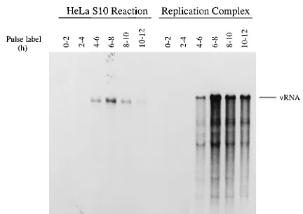
[image:3.612.326.542.68.221.2]labeled RNA products were characterized on a nondenaturing agarose gel. A heterogeneous high-molecular-weight RNA characteristic of poliovirus replicative-intermediate (RI) RNA was labeled within the first minute of the reaction (Fig. 3, left panel). The amount of labeled RI RNA increased for about 15 min and then reached a steady-state level. Labeled full-length
FIG. 1. Time course of viral RNA replication in HeLa S10 lication reactions. Poliovirus RNA was translated in HeLa S10 translation-rep-lication reactions. The viral RNA replicating in unfractionated reactions (HeLa S10 reaction lanes) was pulse labeled at the indicated times with [a-32P]CTP as described under method 1 in the RNA replication section of Materials and Methods. RNA replication complexes were isolated by centrifugation at 15,000
3g from identical translation reaction mixes incubated for 0, 2, 4, 6, 8, and 10 h, as indicated. The isolated complexes (replication complex lanes) were resus-pended and pulse labeled for 2 h with [a-32P]CTP as described under method 2 in the RNA replication section of Materials and Methods. The labeled viral RNAs were analyzed by CH3HgOH-agarose gel electrophoresis and autoradiog-raphy.
on November 9, 2019 by guest
http://jvi.asm.org/
single-stranded viral RNA was not detected until 5 min and then continued to accumulate throughout the remainder of the reaction (Fig. 3, left panel). When the RNA products were treated with RNase A, the single-stranded viral RNA disap-peared and the RI RNA was converted into double-stranded or replicative form (RF)-like molecules (Fig. 3, right panel). These results showed that the RNA replication complexes contained RI, RF, and single-stranded viral RNAs and that they were the site of viral RNA replication in these reactions. Asymmetric synthesis of VPg-linked positive-strand viral
RNA.The formation of RI RNA indicated that both
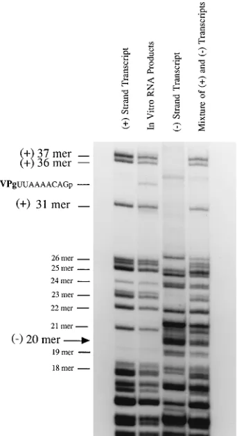
positive-and negative-strpositive-and RNAs were synthesized in the HeLa S10 translation-replication reactions. To characterize the polarity of newly synthesized viral RNA, we used a one-dimensional
RNase T1fingerprint analysis. The large RNase T1
oligonucle-otides derived from32P-labeled poliovirus positive- and
nega-tive-strand RNA transcripts migrated to their characteristic positions in a 20% polyacrylamide–7 M urea gel (Fig. 4).
Di-gests of RNA pulse labeled with [32P]CTP in the HeLa S10
translation-replication reactions from 6 to 8 h contained all of the oligonucleotides characteristic of positive-strand viral RNA (Fig. 4). In contrast, negative-strand-specific oligonucle-otides were not detected in the products of the RNA radiola-beled in the S10 reaction (Fig. 4). This result confirmed that the labeled RNA synthesized in the HeLa S10 reactions from 6 to 8 h was positive-strand poliovirus RNA. Labeled RNA oligonucleotides throughout the positive strand were detected, indicating that the full-length viral RNAs were not simply end
labeled. Furthermore, a unique T1oligonucleotide which
[image:4.612.107.246.71.691.2]mi-FIG. 2. Viral protein composition of RNA replication complexes formed in HeLa S10 translation-replication reactions. Poliovirus RNA was translated for 8 h at 308C in a 50-ml HeLa S10 translation-replication reaction mix containing [35S]methionine. A sample from the unfractionated reaction mix (HeLa S10
FIG. 3. RI RNA is present in RNA replication complexes formed in HeLa S10 translation-replication reactions. A 1-ml HeLa S10 translation-replication reaction mix was incubated at 308C for 6 h and then centrifuged at 15,0003g to isolate the RNA replication complexes. The RNA replication complexes were resuspended in a 100-ml reaction mix containing 100mCi of [a-32
P]CTP and incubated at 308C as described for method 2. Aliquots (20ml) were removed into SDS buffer after incubation for 1, 5, 15, 30, and 60 min, as indicated. The product RNAs were phenol-chloroform-isoamyl alcohol extracted and ethanol precipi-tated. One-quarter of the products of each time point were untreated (total RNA lanes), and 75% of the products of each reaction were digested for 15 min at 238C with 0.2mg of RNase A per ml in 0.3 M NaCl (RNase A-resistant RNA lanes). The RNAs were separated by gel electrophoresis in a nondenaturing 1% agarose–TBE gel.
reaction lanes) was solubilized in protein sample buffer. The remainder of the reaction mix was fractionated by centrifugation at 15,0003g at 48C for 15 min. Samples of the supernatant and resuspended pellet (replication complex lanes) were solubilized in protein sample buffer, and the radiolabeled viral proteins were analyzed by SDS-PAGE in a 10% (A) and a 13.5% (B) polyacrylamide gel. A longer exposure time was used in panel B to visualize 3AB. The 3AB band was identified by immunoprecipitation with anti-VPg antibody in a separate experiment.
on November 9, 2019 by guest
http://jvi.asm.org/
[image:4.612.330.546.73.221.2]grated between the positive-strand 31 and the positive-strand 36 oligomers was recovered from the labeled viral RNA syn-thesized in the HeLa S10 reactions (compare in vitro products with mixture of positive and negative transcript RNAs in Fig.
4). This oligonucleotide was most likely the VPg-linked 59
-terminal T1 oligonucleotide (i.e., VPg-pUUAAAACAGp),
since it migrated to the expected position for the VPg-T1
oligonucleotide (53) and was not present in the RNase T1
digests of the transcript RNAs (Fig. 4). These results show that viral RNA replication in HeLa S10 translation-replication re-actions is highly asymmetric, as it is in vivo (22, 43), and that VPg-linked positive-strand RNA was the predominant product synthesized during the peak of RNA replication in the HeLa S10 translation-replication reactions.
Immunoprecipitation experiments with an affinity-purified anti-VPg antibody were used to confirm that VPg-linked viral RNA was synthesized in the HeLa S10 translation-replication reactions (Fig. 5). Viral RNA synthesized in reaction mixes containing isolated RNA replication complexes was pulse
la-beled with [32P]CTP and then treated with SDS and phenol
extracted to remove any noncovalently bound VPg (see
Mate-rials and Methods). The32P-labeled viral RNA recovered from
this reaction mix was specifically immunoprecipitated with an-ti-VPg antibody (Fig. 5). Similar results were obtained with the
32P-labeled viral RNA synthesized in unfractionated HeLa S10
translation-replication reactions (Fig. 5). As expected, vRNA
end labeled with [32P]pCp was also specifically
immunoprecipi-tated with anti-VPg antibody. From these results, we con-cluded that the viral RNA synthesized in the RNA replication complexes present in the HeLa S10 reactions was covalently linked to VPg.
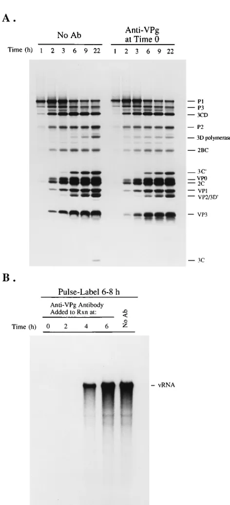
Anti-VPg antibody inhibition of viral replication.To further
investigate the role of VPg in poliovirus replication in the HeLa S10 translation-replication reactions, we investigated how the addition of affinity-purified anti-VPg antibody affected viral protein synthesis, virus formation, and viral RNA repli-cation. Viral protein synthesis and processing were not inhib-ited by anti-VPg antibody (Fig. 6A). Some minor effects on polyprotein processing were observed, which included the
ap-pearance of 3Dpolat earlier times and the accumulation of less
3Dpoland 3Cproat 22 h (Fig. 6A). Adding anti-VPg antibody to
the HeLa S10 reactions totally blocked virus formation (Table 1). This inhibitory effect was mediated by a specific interaction of the antibody, since adding excess synthetic VPg to the re-action mixes reversed the antibody-induced inhibition of virus formation (Table 1). Synthetic VPg by itself did not affect virus production when added to the in vitro translation reaction mixes lacking antibody (Table 1). Viral RNA replication was
measured by pulse labeling with [32P]CTP from 6 to 8 h in
[image:5.612.91.262.71.385.2]unfractionated HeLa S10 translation-replication reaction mixes to which anti-VPg antibody was added at either 0, 2, 4, or 6 h. Addition of anti-VPg antibody at 0 or 2 h totally inhibited viral RNA synthesis (Fig. 6B). When added at 4 h,
FIG. 4. RNase T1fingerprint of viral RNA replicating in a HeLa S10 trans-lation-replication reaction. Full-length poliovirus RNA transcripts of positive polarity [(1) strand transcript lane] and negative polarity [(2) strand transcript lane] were synthesized from cDNA clones in the presence of [a-32P]CTP with T7 polymerase. Poliovirus RNA was pulse labeled in an unfractionated HeLa S10 translation-replication reaction (in vitro RNA products lane) from 6 to 8 h at 308C as described for one-dimensional RNase T1fingerprint analysis in Materials and Methods. The radiolabeled RNAs were digested with RNase T1, and the RNase T1-resistant oligonucleotides were fractionated by electrophoresis in a 20% polyacrylamide–7 M urea gel containing TBE buffer. The fourth lane, a marker lane, contains the positive- and negative-polarity T1oligonucleotides which were mixed after digestion of equal molar amounts of each transcript RNA [mixture of (1) and (2) transcripts lane] Oligonucleotides were located by autoradiography.
FIG. 5. Immunoprecipitation of the viral RNA replicating in HeLa S10 translation-replication reactions. Proteinase K-treated vRNA was translated in HeLa S10 translation-replication reactions as described in Materials and Meth-ods. RNA replicating in a 50-ml HeLa S10 translation-replication reaction mix (HeLa S10 reaction lanes) was pulse labeled from 6 to 8 h at 308C by adding a nucleotide mixture containing 50mCi of [a-32P]CTP to the translation-replica-tion reactranslation-replica-tion mix at 6 h as described in method 1. Replicatranslation-replica-tion complexes (rep-lication complex lanes) were isolated from a translation-rep(rep-lication reaction mix by centrifugation at 6 h and resuspended in a 50-ml reaction mix containing 50
mCi of [a-32P]CTP and incubated at 308C for 2 h as described in method 2. The reactions were treated with SDS, phenol extracted, and ethanol precipitated. 32
P-labeled vRNA (vRNA lanes) was used as a control for immunoprecipitation. The radiolabeled RNAs were divided into equal aliquots and immunoprecipi-tated with anti-VPg antibody or preimmune serum, as indicated. The immuno-precipitated RNAs were fractionated in a denaturing CH3HgOH–1% agarose gel and visualized by autoradiography.
on November 9, 2019 by guest
http://jvi.asm.org/
[image:5.612.368.507.72.233.2]RNA synthesis was inhibited by about 50%, whereas adding the antibody at 6 h had no measurable effect (Fig. 6B). Thus, anti-VPg antibody blocked RNA synthesis when added early in the reaction, but had little or no effect once active replication complexes were formed by 6 h.
Guanidine HCl inhibition of viral replication.At low
con-centrations (2 mM), guanidine HCl inhibits a required func-tion of viral protein 2C and inhibits poliovirus RNA replicafunc-tion in infected cells (32). To further characterize viral replication in the HeLa S10 translation-replication reactions, we deter-mined how the addition of guanidine affected protein synthe-sis, virus formation, and viral RNA replication. As expected, guanidine had no effect on viral protein synthesis or polypro-tein processing (Fig. 7A) (38), and the addition of 2 mM guanidine at the beginning of the reaction totally blocked the formation of all infectious virus (Table 1). Viral RNA
replica-tion was measured by pulse labeling with [32P]CTP from 6 to 8
h in reaction mixes to which 2 mM guanidine was added at 0, 2, 4, and 6 h. Guanidine blocked the synthesis of all labeled viral RNA when added from 0 to 4 h (Fig. 7B). Viral RNA synthesis was inhibited by about 50% when guanidine was added at 6 h (Fig. 7B). Thus, in contrast to anti-VPg antibody, guanidine was able to significantly inhibit viral RNA synthesis in preformed replication complexes. The sensitivity of virus formation and viral RNA replication to guanidine indicated that authentic poliovirus RNA replication was being measured in these in vitro reactions.
Isolation of preinitiation RNA replication complexes.
Be-cause guanidine completely blocked viral RNA replication (Fig. 7B) without affecting viral protein synthesis and process-ing (Fig. 7A), we expected replication complexes which had not yet initiated RNA synthesis (i.e., preinitiation RNA repli-cation complexes, or PI complexes) to form in these reactions. In addition, since guanidine is a reversible inhibitor of polio-virus RNA replication in infected cells (32), RNA synthesis should initiate in the PI complexes in a synchronous manner once guanidine is removed. To test these predictions, we used the procedure shown in Fig. 8 to reverse the inhibition of viral RNA replication by guanidine. The ability to isolate RNA replication complexes formed in the HeLa S10 reactions by centrifugation was used to facilitate the removal of guanidine from PI complexes formed in reaction mixes containing gua-nidine (Fig. 8). The PI complexes were isolated by
centrifuga-tion at 15,0003g from HeLa S10 translation-replication
re-action mixes incubated for 5 h in the presence of guanidine. The isolated PI complexes were resuspended in a supernatant fraction (viral S15) prepared from an identical HeLa S10 trans-lation-replication reaction mix not containing guanidine and
were assayed for RNA replication activity by adding [32P]CTP
[image:6.612.57.293.78.595.2](Fig. 9, viral S15). Resuspension of the PI complexes in the
FIG. 6. Anti-VPg antibody (Ab) inhibited RNA replication in HeLa S10 translation-replication reactions. (A) Proteinase K-treated virion RNA was translated in 50-ml HeLa S10 translation-replication reactions in the absence (no Ab lanes) or presence (anti-VPg Ab at time 0 lanes) of 2ml of affinity-purified anti-VPg antibody (65). The reactions were performed in the presence of [35S]methionine, and 1-ml samples were solubilized in protein sample buffer at the indicated times. The samples were analyzed by SDS–10% PAGE and flu-orography. (B) Proteinase K-treated virion RNA was translated in HeLa S10 translation-replication reactions, and anti-VPg antibody was added at the indi-cated times. At 6 h, the HeLa S10 translation-replication reaction mixes were pulse labeled with [32P]CTP for 2 h as described in method 1. The reaction products were analyzed by CH3HgOH-agarose gel electrophoresis and autora-diography.
TABLE 1. Virus production in HeLa S10 translation-replication reactionsa
Addition to reaction mix Titer
(PFU/ml)
None ... 1.43106
Anti-VPg antibody ... 0 Anti-VPg antibody1VPg (100mg/ml)... 2.53105
VPg (100mg/ml) ... 1.73106
Guanidine HCl (2 mM) ... 0
aHeLa S10 translation-replication reaction mixes (50ml) were incubated at
348C for 15 h in the presence of 1ml of anti-VPg antibody (0.35 mg/ml), synthetic VPg, or guanidine HCl, as indicated. The reaction mixes were treated with RNases T1and A to remove infectious vRNA, and infectious virus was counted by plaque assay on BSC40 cells.
on November 9, 2019 by guest
http://jvi.asm.org/
[image:6.612.318.556.92.163.2]viral S15 fraction resulted in the synthesis of32P-labeled viral
RNA (Fig. 9, viral S15). As expected, the continued presence of guanidine in this reaction mix totally blocked RNA replica-tion (Fig. 9, viral S15). Hence, the inhibireplica-tion of viral RNA replication by guanidine was completely reversible. Resuspen-sion of the PI complexes in the viral S15 fraction in essence reconstituted the original conditions of the HeLa S10 transla-tion-replication reaction and resulted in the initiation of viral RNA replication and the synthesis of full-length viral RNA molecules.
To determine if soluble viral proteins in the viral S15 frac-tion were required for viral RNA synthesis, PI complexes were resuspended in a mock S15 fraction or a fresh HeLa S10 translation-replication reaction mix lacking vRNA. Similar amounts of labeled viral RNA were synthesized in both of these reactions and in the viral S15 reaction with the soluble viral proteins (Fig. 9). This indicated that the soluble viral proteins in the viral S15 fraction (Fig. 2) were not required to initiate viral RNA replication in the PI complexes. Because the PI complexes contained input virion RNA from the initial translation reaction and were being resuspended in fresh HeLa S10 translation-replication reactions, we used puromycin to inhibit the potential synthesis of any new viral proteins in these reactions. The addition of puromycin had no inhibitory effect on the synthesis of labeled viral RNA in this reaction relative to the other reactions without puromycin (Fig. 9). Instead, we observed a small increase in the amount of labeled viral RNA (Fig. 9). The increased amount of labeled viral RNA synthe-sized in the presence of puromycin was observed in repeated experiments. Therefore, soluble viral proteins or newly synthe-sized viral proteins were not required for viral RNA replication in the guanidine reversal reactions. The viral proteins that were already a part of the PI complex were the only viral proteins required for the initiation and continued synthesis of viral RNA.
Soluble cellular factors required to initiate viral RNA
rep-lication.To determine if soluble cellular factors were required
to initiate viral RNA replication, we measured viral RNA synthesis in PI complexes resuspended in reaction mixtures with and without an added S15 fraction that was prepared from cytoplasmic extracts of uninfected HeLa cells (Fig. 10). La-beled viral RNA was synthesized in the reaction mix which contained the HeLa S15 fraction. Similar amounts of labeled RNA were synthesized in a reaction mix containing a HeLa S200 fraction (Fig. 10, HeLa S200). Viral RNA was not syn-thesized in the reaction mix lacking HeLa extract (Fig. 10, buffer). PI complexes sometimes exhibited activity when resus-pended in reaction mixes lacking HeLa extract, in contrast to the results above. This activity, however, was no longer present after the PI complexes were washed with an S10 salts-wash
buffer [40 mM HEPES (pH 7.4), 120 mM KCH3CO2, 10 mM
KCl, 5.5 mM Mg(CH3CO2)2, 6 mM DTT, 1 mM CaCl2, 2 mM
EGTA] (data not shown). Washed PI complexes still retained activity when resuspended in HeLa S15 as described above (data not shown). These results indicated that one or more soluble cellular factors were required for the initiation of po-liovirus RNA replication in isolated PI complexes.
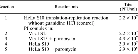
Encapsidation of progeny viral RNA to form infectious
vi-rus.The final step in the viral replication cycle is the formation
of infectious virus. We used the guanidine reversal procedure described above to isolate PI complexes and study the encap-sidation of newly synthesized virion RNA. PI complexes were resuspended in reaction mixes containing a viral S15 fraction in the absence or presence of added puromycin. In the absence and presence of puromycin, the virus yield was essentially the same as in the control HeLa S10 translation-replication
reac-FIG. 7. Guanidine HCl inhibition of viral RNA replication in HeLa S10 translation-replication reactions. (A) vRNA was translated in 50-ml HeLa S10 translation-replication reactions in the absence (no guanidine lanes) or presence (guanidine at time 0 lanes) of 2 mM guanidine HCl. The reactions were per-formed in the presence of [35
S]methionine, and 1-ml samples were solubilized in protein sample buffer at the indicated times. The samples were analyzed by SDS–10% PAGE and fluorography. (B) vRNA was translated in 50-ml HeLa S10 translation-replication reactions. At the indicated times, 50 mM guanidine (2ml) was added to the reaction mixes, resulting in a final concentration of 2 mM. At 6 h, the reaction mixes were pulse labeled for 2 h with [32P]CTP as described in method 1. The reaction products were analyzed by electrophoresis in a CH3 HgOH-agarose gel, and the dried gel was visualized by autoradiography.
on November 9, 2019 by guest
http://jvi.asm.org/
tion (Table 2, reactions 1, 2, and 3). These results indicated that efficient encapsidation of newly synthesized progeny RNA occurred in the absence of ongoing viral protein synthesis. To determine the amount of virus formed in the absence of pre-formed soluble capsid proteins, we resuspended PI complexes
in fresh HeLa S10 translation-replication reaction mixes lack-ing vRNA in the presence or absence of puromycin. In these reactions, the inhibition of protein synthesis decreased the virus yield by 100-fold (Table 2, reactions 4 and 5). This indi-cated that newly synthesized capsid proteins were required for efficient virus formation when soluble capsid proteins were not provided (Table 2, compare reaction 3 with reactions 4 and 5). A small amount of capsid proteins fractionated with the PI complexes, and this allowed a small amount of virus produc-tion in the absence of exogenous capsid proteins or the syn-thesis of new capsid proteins (Table 2, reaction 5). The com-position of viral proteins in PI complexes (data not shown) was
[image:8.612.148.472.73.258.2]FIG. 8. Reversal of guanidine HCl inhibition of poliovirus RNA synthesis. The time line diagrams the method used in the guanidine reversal experiments. PI complexes form in HeLa S10 translation-replication reaction mixes which contain 2 mM guanidine HCl. After translation for 5 h at 308C in the presence of 2 mM guanidine HCl, the PI complexes are pelleted from the reaction mixes by centrifugation at 15,0003g. The viral S15, containing the soluble viral proteins (see Fig. 2, supernatant), and the guanidine HCl are removed with a pipette and discarded. The PI complexes are then resuspended in reaction mixes containing [32P]CTP as described in method 3. The reaction mixes are incubated for an additional 90 min, and the reactions are terminated by solubilization in 0.5% SDS buffer. The RNA products are then phenol-chloroform-isoamyl alcohol extracted, ethanol precipitated, and analyzed by CH3HgOH-agarose gel electrophoresis.
FIG. 9. Guanidine HCl inhibition of RNA replication is reversible. PI com-plexes were obtained as described in the legend to Fig. 8 and Materials and Methods. The PI complexes were resuspended in 50-ml reaction mixes as de-scribed in method 3. The reactions were performed with and without 2 mM guanidine HCl, as indicated. The conditions in the viral S15 and mock S15 reactions were modified as follows. Viral S15 (see Fig. 2, supernatant) was obtained from a HeLa S10 translation-replication reaction mix containing vRNA after incubation for 5 h at 308C and centrifugation at 48C for 15 min at 15,0003 g. Mock S15 was obtained similarly from a HeLa S10 translation-replication reaction mix lacking vRNA. Then, 50ml of the supernatant fractions (viral S15 and mock S15) was used to resuspend the PI complexes. Lanes HeLa S10, standard reaction described in method 3. Lanes HeLa S101puromycin, stan-dard reaction described in method 3 with puromycin (50mg/ml). All the reaction mixes contained 10mCi of [a-32
P]CTP. The reaction mixes were incubated for 90 min at 308C, and the products were analyzed by CH3HgOH-agarose gel electro-phoresis and autoradiography.
FIG. 10. Soluble cellular cytoplasmic factors are required for the initiation of viral RNA synthesis. PI complexes were prepared by the guanidine reversal procedure as described in the legend to Fig. 8. The PI complexes were resus-pended in 50-ml reaction mixes as described in method 3 except that 25ml of HeLa S15, HeLa S200, or buffer [40 mM HEPES-KOH (pH 7.4), 120 mM KCH3CO2, 5.5 mM Mg(CH3CO2)2, 6 mM DTT, 10 mM KCl, 1 mM CaCl2, 2 mM EGTA] was used in place of the HeLa S10 extract. HeLa cell translation initiation factors were not used in these reactions. HeLa S15 and S200 were obtained by centrifugation of HeLa S10 extract at 15,0003g and 200,0003g, respectively. All the reaction mixes contained 10mCi of [a-32
P]CTP. The reac-tion mixes were incubated at 308C for 90 min, and the labeled RNA products were separated by CH3HgOH-agarose gel electrophoresis. Radiolabeled RNA was detected by autoradiography.
on November 9, 2019 by guest
http://jvi.asm.org/
[image:8.612.73.279.410.594.2]indistinguishable from the composition of viral proteins in normal RNA replication complexes (Fig. 2). Because only a small amount of capsid proteins fractionated with the PI com-plexes, the capsid proteins used for the encapsidation of virion RNA synthesized by PI complexes (after the reversal of gua-nidine inhibition) must be either newly synthesized in the re-actions containing the PI complexes or provided in trans from a second in vitro translation reaction.
DISCUSSION
From the results of this study, we concluded that the HeLa S10 translation-replication reactions supported the synthesis of the poliovirus proteins, the formation of RNA replication com-plexes, the asymmetric replication of viral RNA, and the pro-duction of infectious virus. All aspects of viral RNA replication in the in vitro reactions were authentic relative to poliovirus replication in infected cells. In addition, we showed that preini-tiation RNA replication complexes (PI complexes) were formed in reaction mixes containing 2 mM guanidine HCl. The PI complexes contained all of the viral proteins and the tem-plate RNA (input vRNA) required for viral RNA replication and initiated the synthesis of normal amounts of viral RNA and infectious virus upon the removal of guanidine HCl. Fi-nally, by using resuspended PI complexes to assay for viral RNA synthesis, we concluded that a soluble cellular factor(s) was required for the initiation of poliovirus RNA replication. RNA replication was detected in the HeLa S10 translation-replication reactions by pulse-labeling the viral RNA as it replicated directly in the unfractionated reaction. Alterna-tively, RNA replication was detected in RNA replication com-plexes isolated from the HeLa S10 translation-replication re-actions. The poliovirus proteins that were found associated with the RNA replication complexes (e.g., 2C, 2BC, 3AB, and 3CD) are known to fractionate with membrane-bound RNA replication complexes isolated from poliovirus-infected cells (12, 13). Although 3CD is not a membrane-associated protein,
3CD binds near the 59end of poliovirus positive-strand RNA
and may be involved in the initiation of positive-strand RNA replication (3, 4, 26). Thus, we might expect that 3CD, in
addition to catalytic amounts of 3Dpol, would be part of an
active RNA replication complex. We found that Triton X-100 prevented RNA synthesis in the HeLa S10 reactions (data not shown), consistent with the association of RNA replication complexes with cellular membranes (15) and the requirement for cellular membranes for normal RNA replication (39, 53, 55). The RNA replication complexes formed in the HeLa S10
translation-replication reactions therefore appeared to be sim-ilar in viral protein composition to RNA replication complexes in infected cells and to be associated with membranous vesi-cles.
Analysis of the RNA synthesized by the RNA replication complexes indicated that RI RNA was present in the replica-tion complexes. Short pulse-labeling of isolated replicareplica-tion complexes resulted in the incorporation of radiolabel into RI RNA preceding the accumulation of radiolabel into single-stranded viral RNA, consistent with the precursor-product re-lationship of nascent molecules of the RI RNA and single-stranded viral RNA (8). RI RNA was also predicted to be present from the detection of minus-strand RNA in HeLa S10 translation reactions (38). The presence of RI RNA also indi-cated that poliovirus positive-strand RNA was actively
initiat-ing and elongatinitiat-ing. RNase T1 fingerprint analysis indicated
that the RNA replication occurring in the in vitro translation reactions was indeed highly asymmetric and that predomi-nantly positive-strand RNA was synthesized from 6 to 8 h. This is consistent with the highly asymmetric replication of
poliovi-rus RNA in infected cells (22, 43). The complete set of T1
oligonucleotides also indicated that the RNA radiolabeled in the HeLa S10 translation-replication reactions was not simply end labeled but labeled throughout the length of the molecule, consistent with replication.
Immunoprecipitation with anti-VPg antibody indicated that the viral RNA synthesized in these reactions was covalently linked to VPg. This was in marked contrast to the quantitative removal of VPg from the input virion RNA by the VPg-un-linking activity which is present in HeLa S10 extracts (1, 2). Thus, it appeared that newly synthesized poliovirus RNA was protected from the VPg-unlinking enzyme. It may be that the RNA synthesized by the replication complexes was protected by the membranes found associated with RNA replication complexes (60). Alternatively, this result may be explained by the association of this RNA with the poliovirus capsid proteins present in these reactions. This possibility is consistent with the rapid increase in the virus titer which begins at about 6 h (10). In addition, if it is assumed that only VPg-linked RNA can be encapsidated, then only newly synthesized viral RNA should be packaged in virions synthesized in these reactions. This hypothesis would explain why the presence or absence of VPg on the input RNA has no effect on the yield of infectious virus (38). It is also consistent with the requirement for RNA rep-lication to precede virus production. In future experiments, it will be important to determine if exogenous VPg-linked polio-virion RNA which is added to these reactions at late reaction times (e.g., 6 to 10 h) can be encapsidated or if there is an obligatory linkage between RNA replication and virion forma-tion. Likewise, it will be interesting to determine whether RNA replication and packaging of the replicated RNA can be tem-porally separated, as protein synthesis has been temtem-porally separated from RNA replication in the guanidine reversal pro-cedure (see below).
[image:9.612.62.297.92.185.2]To investigate the role of VPg in viral RNA replication and virus formation, we added anti-VPg antibody to the in vitro translation-RNA replication reaction mixes. When present at the beginning of the reaction, the antibody blocked the initia-tion of viral RNA replicainitia-tion and virus formainitia-tion. The insen-sitivity to anti-VPg antibody inhibition observed at 6 h may be explained by the inability of the antibody to enter preformed replication complexes and to bind to VPg and/or VPg precur-sor proteins. Thus, anti-VPg antibody inhibits the initiation of viral RNA replication and the formation of active RNA rep-lication complexes when added at early reaction times, but appears to have little effect on preformed complexes. These
TABLE 2. Virus production by preinitiation RNA replication complexesa
Reaction Reaction mix Titer
(PFU/ml)
1 HeLa S10 translation-replication reaction without guanidine HCl (control)
2.23107
PI complex in:
2 Viral S15 2.23107
3 Viral S151puromycin 4.33107
4 HeLa S10 3.93107
5 HeLa S101puromycin 2.93105
a
The control reaction without guanidine HCl was incubated at 308C for 5 h and then shifted to 348C for 12 h. PI complexes were resuspended in reaction mixes as described in the legend to Fig. 9 and under method 3 in the RNA replication section in Materials and Methods and incubated at 348C for 12 h. The reaction products were digested with RNases T1and A, and infectious virus was counted by plaque assay on BSC40 cells.
on November 9, 2019 by guest
http://jvi.asm.org/
results support a model of poliovirus RNA replication in which VPg (or a VPg precursor protein) is required at the initiation step of poliovirus RNA replication.
Guanidine HCl was also found to inhibit virus formation (Table 1) (38) and viral RNA replication in the HeLa S10 translation-replication reactions. Previously, it has been sug-gested that guanidine HCl specifically inhibits the initiation of poliovirus RNA replication without affecting the polymerase elongation reaction (32, 57). The results of this study support this conclusion. The inhibition of RNA replication by guani-dine HCl was reversible in the HeLa S10 in vitro translation-replication reactions. The reversal was facilitated by the ability to isolate RNA replication complexes by a short centrifugation step in which the supernatant containing the guanidine HCl could be discarded. Indeed, when PI complexes were resus-pended in HeLa S10 reactions lacking guanidine HCl, RNA replication occurred at normal levels. Apparently, RNA repli-cation complexes were formed in the HeLa S10 in vitro trans-lation-replication reactions in the presence of guanidine HCl despite the lack of RNA synthesis. We describe these com-plexes as PI RNA replication comcom-plexes because guanidine HCl completely blocked RNA synthesis during their forma-tion. The PI complexes would be expected to contain input vRNA and the normal complement of viral and cellular pro-teins in a functional complex. The results suggest that PI RNA replication complexes synthesize negative-strand RNA first and then asymmetric quantities of positive-strand RNA once the inhibition of protein 2C by guanidine is reversed.
The PI complexes required one or more soluble cellular factors to initiate viral RNA replication. Reconstitution exper-iments showed that soluble viral proteins were not necessary for the initiation of RNA synthesis and that continued viral protein synthesis was not required. In contrast, either soluble viral capsid proteins present in viral S15 or newly synthesized capsid proteins were required for the production of infectious virus. Therefore, the formation of infectious virus upon the reversal of guanidine inhibition was dependent on a source of capsid proteins. In the case of the guanidine reversal reaction with puromycin, the capsid proteins were provided in trans from the viral S15 used to resuspend the PI complex, as new capsid proteins could not be synthesized in the presence of puromycin. This in vitro trans-encapsidation reaction should be very useful in future studies on the molecular details of viral RNA packaging.
The guanidine reversal procedure with PI complexes is unique in that viral protein synthesis and viral RNA replication are completely temporally separated. That is, PI complexes are formed in the presence of an inhibitor of RNA replication and are then resuspended in the presence of an inhibitor of protein synthesis. Protein synthesis and polyprotein processing occur normally in the complete absence of RNA replication, and RNA replication occurs in the complete absence of continued protein synthesis. This is different from what is observed in vivo, when protein synthesis is coupled to RNA replication and vice versa. In infected cells, protein synthesis is dependent on the synthesis of additional viral positive-strand RNA, which serves as mRNA for protein synthesis. Likewise, RNA repli-cation requires protein synthesis to provide the viral proteins that are required to form additional RNA replication com-plexes. Our results indicate, however, that once RNA replica-tion complexes are formed, addireplica-tional viral protein synthesis is not required. In the guanidine reversal procedure described in this report, optimal amounts of input virion RNA are provided at the beginning of the translation-replication reaction, and thus maximum levels of protein synthesis are reached without multiple rounds of replication. This allows the uncoupling of
RNA replication and protein synthesis. In addition, the mature viral proteins present in the PI complexes appear to be suffi-cient for normal RNA replication. It will be interesting to pursue experimentally the potential requirement for continued polyprotein processing in RNA replication.
An important extension of this work will be to use the gua-nidine reversal experiment in conjunction with molecular frac-tionation to identify the cellular factor(s) necessary for the guanidine reversal and thus identify the role of the cellular factor(s) in the initiation of poliovirus RNA synthesis. An expanding number of cellular proteins are being identified as participants in poliovirus translation and RNA replication. The identification and characterization of cellular factors will facil-itate our understanding of the pathogenesis and tissue tro-pisms exhibited by poliovirus. The cap-independent translation of poliovirus RNA requires the La autoantigen for the correct initiation of protein synthesis (35, 52). Cellular p57, a pyrimi-dine tract-binding protein, also binds to the poliovirus internal ribosome entry site to facilitate the initiation of protein syn-thesis (28, 29). A 36-kDa protein, identified as a proteolytic
fragment of eEF-1a, was shown to bind to the 59stem-loop of
poliovirus positive-strand RNA (26) and may be required for the initiation of poliovirus positive-strand RNA synthesis.
eEF-1ais the eukaryotic homolog of bacterial EF-Tu, which is
required for polypeptide chain elongation and as a host factor
for the initiation of RNA synthesis by bacteriophage Qb
rep-licase (14). The guanidine reversal assay developed in this study, which requires soluble cellular factors for RNA synthe-sis, should facilitate the functional analysis of cellular proteins putatively described as being required for poliovirus RNA rep-lication.
The HeLa S10 translation-replication reactions described in this study are a complete in vitro system for studying poliovirus replication (i.e., translation, polyprotein processing, RNA rep-lication, and virus assembly). These reactions are efficient,
since virus titers of 4 3 107 PFU/ml can be achieved under
optimal conditions. In addition, this system also appears to reflect the molecular events occurring during viral replication in vivo, but with the advantages of an in vitro experimental system. Our ability to form authentic, biochemically functional RNA replication complexes and infectious virus in vitro pro-vides us with direct access to these replicative processes, which are normally shielded from experimental manipulation in in-fected cells by the cell membrane. The absence of the cell membrane barrier allows antibody studies, as demonstrated herein with anti-VPg antibody. This in vitro system also solves the difficult problem of studying poliovirus RNA replication in vitro, when it appears that RNA replication may be directly linked to the translation of some viral proteins in a mechanistic and functional way (e.g., cis-acting viral RNA replication pro-teins). Finally, the PI complexes described in this study should greatly facilitate the analysis of initiation of RNA synthesis.
ACKNOWLEDGMENTS
We thank Brian O’Donnell and Joan Morasco for excellent techni-cal assistance.
This work was supported by Public Health Service grant AI 15539 from the National Institute of Allergy and Infectious Diseases. David J. Barton was supported by Public Health Service training grant AI 07110 from the National Institute of Allergy and Infectious Diseases.
REFERENCES
1. Ambros, V., and D. Baltimore. 1980. Purification and properties of a HeLa cell enzyme able to remove the 59-terminal protein from poliovirus RNA. J. Biol. Chem. 255:6739–6744.
2. Ambros, V., R. F. Pettersson, and D. Baltimore. 1978. An enzymatic activity in uninfected cells that cleaves the linkage between poliovirion RNA and the
on November 9, 2019 by guest
http://jvi.asm.org/
59terminal protein. Cell 15:1439–1446.
3. Andino, R., G. E. Rieckhof, P. L. Achacoso, and D. Baltimore. 1993. Polio-virus RNA synthesis utilizes an RNP complex formed around the 59-end of viral RNA. EMBO J. 12:3587–3598.
4. Andino, R., G. E. Rieckhof, and D. Baltimore. 1990. A functional ribonu-cleoprotein complex forms around the 59end of poliovirus RNA. Cell 63: 369–380.
5. Andrews, N. C., and D. Baltimore. 1986. Purification of a terminal uridylyl-transferase that acts as host factor in the in vitro poliovirus replicase reac-tion. Proc. Natl. Acad. Sci. USA 83:221–25.
6. Andrews, N. C., D. Levin, and D. Baltimore. 1985. Poliovirus replicase stimulation by terminal uridylyl transferase. J. Biol. Chem. 260:7628–7635. 7. Baltera, R. F., Jr., and D. R. Tershak. 1989. Guanidine-resistant mutants of
poliovirus have distinct mutations in peptide 2C. J. Virol. 63:4441–4444. 8. Baltimore, D. 1968. Structure of the poliovirus replicative intermediate
RNA. J. Mol. Biol. 32:359–368.
9. Baron, M. H., and D. Baltimore. 1982. In vitro copying of viral positive strand RNA by poliovirus replicase: characterization of the reaction and its products. J. Biol. Chem. 257:12359–12366.
10. Barton, D. J., and J. B. Flanegan. 1993. Coupled translation and replication of poliovirus RNA in vitro: synthesis of functional 3D polymerase and infectious virus. J. Virol. 67:822–831.
11. Bienz, K., D. Egger, and L. Pasamontes. 1987. Association of polioviral proteins of the P2 genomic region with the viral replication complex and virus-induced membrane synthesis as visualized by electron microscopic im-munocytochemistry and autoradiography. Virology 160:220–226. 12. Bienz, K., D. Egger, T. Pfister, and M. Troxler. 1992. Structural and
func-tional characterization of the poliovirus replication complex. J. Virol. 66: 2740–2747.
13. Bienz, K., D. Egger, M. Troxler, and L. Pasamontes. 1990. Structural orga-nization of poliovirus RNA replication is mediated by viral proteins of the P2 genomic region. J. Virol. 64:1156–1163.
14. Blumenthal, T., T. A. Landers, and K. Weber. 1972. Bacteriophage Qb replicase contains the protein biosynthesis elongation factors EF-Tu and EF-Ts. Proc. Natl. Acad. Sci. USA 69:1313–1317.
15. Caliguiri, L. A., and I. Tamm. 1970. Characterization of poliovirus-specific structures associated with cytoplasmic membranes. Virology 42:112–122. 16. Collis, P. S., B. J. O’Donnell, D. J. Barton, J. A. Rogers, and J. B. Flanegan.
1992. Replication of poliovirus RNA and subgenomic RNA transcripts in transfected cells. J. Virol. 66:6480–6488.
17. Flanegan, J. B., and D. Baltimore. 1979. Poliovirus polyuridylic acid poly-merase and RNA replicase have the same viral polypeptide. J. Virol. 29: 352–360.
18. Flanegan, J. B., R. F. Petterson, V. Ambros, N. J. Hewlett, and D. Baltimore. 1977. Covalent linkage of a protein to a defined nucleotide sequence at the 59-terminus of virion and replicative intermediate RNAs of poliovirus. Proc. Natl. Acad. Sci. USA 74:961–965.
19. Flanegan, J. B., G. J. Tobin, M. S. Oberste, B. J. Morasco, D. C. Young, and
P. S. Collis.1990. Mechanism of poliovirus negative-strand RNA synthesis and the self-catalyzed covalent linkage of VPg to RNA, p. 55–60. In M. Brinton and F. X. Heinz (ed.), New aspects of positive-strand RNA viruses. American Society for Microbiology, Washington, D.C.
20. Flanegan, J. B., and T. A. Van Dyke. 1979. Isolation of a soluble and template-dependent poliovirus RNA polymerase that copies virion RNA in vitro. J. Virol. 32:155–161.
21. Flanegan, J. B., D. C. Young, G. J. Tobin, M. A. Stokes, C. D. Murphy, and
S. M. Oberste.1987. Mechanism of RNA replication by the poliovirus RNA polymerase, HeLa cell host factor, and VPg. UCLA Symp. Mol. Cell. Biol. New Ser. 54:273–284.
22. Giachetti, C., and B. L. Semler. 1991. Role of a viral membrane polypeptide in strand-specific initiation of poliovirus RNA synthesis. J. Virol. 65:2647– 2654.
23. Gorbalenya, A. E., V. M. Blinov, A. P. Donchenko, and E. Koonin. 1989. An NTP-binding motif is the most conserved sequence in a highly divergent monophyletic group of proteins involved in positive strand RNA viral rep-lication. J. Mol. Evol. 28:256–268.
24. Gorbalenya, A. E., and E. V. Koonin. 1989. Viral proteins containing the purine NTP-binding sequence pattern. Nucleic Acids Res. 17:8413–8440. 25. Gorbalenya, A. E., E. V. Koonin, A. P. Donchenko, and V. M. Blinov. 1988.
A conserved NTP-motif in putative helicases. Nature (London) 333:22. 26. Harris, K. S., W. Xiang, L. Alexander, W. S. Lane, A. V. Paul, and E.
Wimmer.1994. Interaction of poliovirus polypeptide 3CDpro
with the 59and 39 termini of the poliovirus genome: identification of viral and cellular cofactors needed for efficient binding. J. Biol. Chem. 269:27004–27014. 27. Hayes, R. J., and K. W. Buck. 1990. Complete replication of a eukaryotic
virus RNA in vitro by a purified RNA-dependent RNA polymerase. Cell
63:363–368.
28. Hellen, C. U. T., T. V. Pestova, M. Litterst, and E. Wimmer. 1994. The cellular polypeptide p57 (pyrimidine tract-binding protein) binds to multiple sites in the poliovirus 59nontranslated region. J. Virol. 68:941–950. 29. Hellen, C. U. T., G. W. Witherell, M. Schmid, S. H. Shin, T. V. Pestova, A.
Gil, and E. Wimmer. 1993. A cytoplasmic 57 kDa protein (p57) that is
required for translation of picornavirus RNA by internal ribosome entry is identical to the nuclear pyrimidine tract-binding protein. Proc. Natl. Acad. Sci. USA 90:7642–7646.
30. Hewlett, M. J., J. K. Rose, and D. Baltimore. 1976. 59-terminal structure of poliovirus polyribosomal RNA is pUp. Proc. Natl. Acad. Sci. USA 73:327– 330.
31. Johnson, K. L., and P. Sarnow. 1991. Three poliovirus 2B mutants exhibit noncomplementable defects in viral RNA amplification and display dosage-dependent dominance over wild-type poliovirus. J. Virol. 65:4341–4349. 32. Koch, F., and G. Koch. 1985. The molecular biology of poliovirus, p. 372–
420. Springer-Verlag, New York.
33. Lee, Y. F., A. Nomoto, B. M. Detjen, and E. Wimmer. 1977. A protein covalently linked to poliovirus genome RNA. Proc. Natl. Acad. Sci. USA
74:59–63.
34. Lubinski, J. M., L. J. Ransone, and A. Dasgupta. 1987. Primer-dependent synthesis of covalently linked dimeric RNA molecules by poliovirus repli-case. J. Virol. 61:2997–3003.
35. Meerovitch, K., Y. V. Svitkin, H. S. Lee, F. Lejbkowicz, D. J. Kenan, E. K. L.
Chan, V. I. Agol, J. D. Keene, and N. Sonenberg.1993. La autoantigen enhances and corrects aberrant translation of poliovirus RNA in reticulocyte lysate. J. Virol. 67:3798–3807.
36. Mirzayan, C., and E. Wimmer. 1994. Biochemical studies on poliovirus polypeptide 2C: evidence for ATPase activity. Virology 199:176–187. 37. Mirzayan, C., and E. Wimmer. 1992. Genetic analysis of an NTP-binding
motif in poliovirus polypeptide 2C. Virology 189:547–555.
38. Molla, A., A. V. Paul, and E. Wimmer. 1991. Cell-free, de novo synthesis of poliovirus. Science 254:1647–1651.
39. Molla, A., A. V. Paul, and E. Wimmer. 1993. Effects of temperature and lipophilic agents on poliovirus formation and RNA synthesis in a cell-free system. J. Virol. 67:5932–5938.
40. Neufeld, K. L., O. C. Richards, and E. Ehrenfeld. 1991. Purification, char-acterization, and comparison of poliovirus RNA polymerase from native and recombinant sources. J. Biol. Chem. 266:24212–24219.
41. Nomoto, A., B. Detjen, R. Pozzatti, and E. Wimmer. 1977. The location of the polio genome protein in viral RNAs and its implication for RNA synthesis. Nature (London) 268:208–213.
42. Nomoto, A., N. Kitamura, F. Golini, and E. Wimmer. 1977. The 59-terminal structures of poliovirion RNA and poliovirus mRNA differ only in the genome-linked protein VPg. Proc. Natl. Acad. Sci. USA 74:5345–5349. 43. Novak, J. E., and K. Kirkegaard. 1991. Improved method for detecting
poliovirus negative strands used to demonstrate specificity of positive-strand encapsidation and the ratio of positive to negative strands in infected cells. J. Virol. 65:3384–3387.
44. Pettersson, R. F., V. Ambros, and D. Baltimore. 1978. Identification of a protein linked to nascent poliovirus RNA and to the polyuridylic acid of negative-strand RNA. J. Virol. 27:357–365.
45. Pettersson, R. F., J. B. Flanegan, J. K. Rose, and D. Baltimore. 1977. 59-Terminal nucleotide sequences of polio virus polyribosomal RNA and virion RNA are identical. Nature (London) 268:270–272.
46. Pincus, S. E., D. C. Diamond, E. A. Emini, and E. Wimmer. 1986. Guanidine-selected mutants of poliovirus: mapping of point mutations to polypeptide 2C. J. Virol. 57:638–646.
47. Pincus, S. E., H. Rohl, and E. Wimmer. 1987. Guanidine-dependent mutants of poliovirus: identification of three classes with different growth require-ments. Virology. 157:83–88.
48. Pincus, S. E., and E. Wimmer. 1986. Production of guanidine-resistant and -dependent poliovirus mutants from cloned cDNA: mutations in polypeptide 2C are directly responsible for altered guanidine sensitivity. J. Virol. 60:793– 796.
49. Reuer, Q., R. J. Kuhn, and E. Wimmer. 1990. Characterization of poliovirus clones containing lethal and nonlethal mutations in the genome-linked pro-tein VPg. J. Virol. 64:2967–2975.
50. Rightsel, W. A., J. R. Dice, R. J. McAlpine, E. A. Timm, I. W. McLean, Jr.,
G. L. Dixon, and F. M. Schabel, Jr.1961. Antiviral effect of guanidine. Science 134:558–559.
51. Rodriguez, P. L., and L. Carrasco. 1993. Poliovirus protein 2C has ATPase and GTPase activities. J. Biol. Chem. 268:8105–8110.
52. Svitkin, Y. V., K. Meerovitch, H. S. Lee, J. N. Dholakia, D. J. Kenan, V. I.
Agol, and N. Sonenberg.1994. Internal translation initiation on poliovirus RNA: further characterization of La function in poliovirus translation in vitro. J. Virol. 68:1544–1550.
53. Takeda, N., R. J. Kuhn, C. F. Yang, T. Takegami, and E. Wimmer. 1986. Initiation of poliovirus plus-strand RNA synthesis in a membrane complex of infected HeLa cells. J. Virol. 60:43–53.
54. Takeda, N., C. F. Yang, R. J. Kuhn, and E. Wimmer. 1987. Uridylylation of the genome-linked protein of poliovirus in vitro is dependent upon an en-dogenous RNA template. Virus Res. 8:193–204.
55. Takegami, T., R. J. Kuhn, C. W. Anderson, and E. Wimmer. 1983. Mem-brane-dependent uridylylation of the genome-linked protein VPg of polio-virus. Proc. Natl. Acad. Sci. USA 80:7447–7451.
56. Takegami, T., B. L. Semler, C. W. Anderson, and E. Wimmer. 1983. Mem-brane fractions active in poliovirus RNA replication contain VPg precursor