JOURNAL OFVIROLOGY, Feb. 1996, p. 992–998 Vol. 70, No. 2 0022-538X/96/$04.0010
Copyrightq1996, American Society for Microbiology
Identification of an Essential Region for Internal Initiation of
Translation in the Aphthovirus Internal Ribosome Entry
Site and Implications for Viral Evolution
ENCARNACIO
´ N MARTI´NEZ-SALAS, M. PAZ REGALADO,†
ANDESTEBAN DOMINGO*
Centro de Biologı´a Molecular ‘‘Severo Ochoa,’’ Consejo Superior de Investigaciones Cientı´ficas-Universidad
Auto
´noma de Madrid, 28049 Madrid, Spain
Received 24 July 1995/Accepted 30 October 1995
Translation of aphthovirus RNA is initiated at an internal ribosome entry site (IRES) element, preceding the
first functional AUG initiation codon. The effect of mutations at the base of domain 3 of the aphthovirus IRES
on translation activity has been analyzed by site-directed mutagenesis and expression of bicistronic RNAs in
transfected cells. The results have shown that the enhanced IRES activity associated with a single pyrimidine
transition fixed in a persistent aphthovirus variant (E. Martı´nez-Salas, J. C. Sa
´iz, M. Da
´vila, G. J. Belsham,
and E. Domingo, J. Virol. 67:3748–3755, 1993) is base specific. Mutations predicted to destabilize the base of
domain 3 were detrimental to IRES function, but subsequent restoration of the RNA structure gave rise to fully
competent IRES. In contrast, single or multiple mutations that did not affect predicted helical structures
modified the relative efficiency of translation by at most 10-fold, suggesting that primary sequence also plays
a role in IRES activity. A correlation between the energy of stabilization of the IRES structure and the efficiency
of translation has been noted. None of the 15 mutations studied reached a level of initiation of translation
comparable to that of the IRES from the persistent variant. The results indicate a critical participation of the
base of domain 3 in the activity of the aphthovirus IRES, with a strong effect of secondary or higher-order
structures and minor effects of primary structure.
The long 5
9
untranslated region of picornavirus RNAs
con-tains a sequence of 400 to 500 nucleotides that directs internal
initiation of translation (32). This region, termed the internal
ribosome entry site (IRES), is predicted to form extensive
secondary structures and contains several unused AUG codons
(reviewed in references 18, 20, and 43). On the basis of
phy-logenetic comparisons and biochemical probing of secondary
structures, two types of picornavirus IRES have been defined.
Type 1 includes IRES of enteroviruses and rhinoviruses, and
type 2 includes those of aphthoviruses and cardioviruses. They
differ in sequence, structure, and the location of conserved
regions across the element (20). However, both IRES types
include one pyrimidine tract located upstream of the initiator
AUG within the protein synthesis starting window (18, 36).
The secondary structure and probably also the tertiary
struc-ture (23) play a critical role in IRES activity. RNA-RNA
in-teractions have been suggested on the basis of observed trans
complementation between poliovirus and foot-and-mouth
dis-ease virus (FMDV) RNA IRES deletion mutants (9, 40). In
addition, RNA-protein interactions are involved in the
mech-anism of cap-independent internal initiation of translation (7,
17, 28, 44) and, in FMDV, p57/pTB (polypyrimidine
tract-binding protein) and eukaryotic initiation factor 4B (eIF-4B)
form a complex involving several IRES stem-loop structures
(24, 29).
Most mutations in the IRES, either from natural variants or
produced by site-directed mutagenesis, either were neutral or
led to an impairment of translation initiation (reviewed in
reference 43). For example, of a total of 27 reported mutations
in the polypyrimidine tract of several picornaviruses, 8 were
neutral and 19 were detrimental (21, 22, 31, 34, 37). A similar
negative effect was observed with most mutations affecting
predicted domain structures (15, 31). Only in a few cases did
nucleotide substitutions or insertions result in an increase of
initiation of translation (13, 14). One such case was observed in
an FMDV variant isolated after prolonged persistence in
BHK-21 cells (26). Persistence of FMDV (clone C-S8c1) in cell
culture is characterized by a coevolution of the host cells and
the resident virus whereby the cells become increasingly
resis-tant to the virus and the virus acquires a hypervirulent
pheno-type for the parental BHK-21 cells (5, 6). Hypervirulent variant
* Corresponding author. Mailing address: Centro de Biologı´a Mo-lecular ‘‘Severo Ochoa’’ (CSIC. UAM), Universidad Auto´noma de Madrid, 28049 Madrid, Spain. Phone: 3978485. Fax: 34-1-3974799.
[image:1.612.315.554.530.660.2]† Present address: Instituto Cajal, 28002 Madrid, Spain.
TABLE 1. Primers used in the site-directed mutagenesis
Primera Sequence (59–39)b Template Positionc
NR4 CACGAGCTCAGCAGGTTTCC 2470
ND6 CCGAGCTCAGGGTCATTAATTG 21
ND9 GAGCCAAATGCAGTACAAAGTG pBIR 2366
ND10 GAGCCAAATGTAGTACAAAGTG pBIR 2366
ND11 CCAAAGGCAGTACAAAGTGTTACC pBIR 2363
ND12 CCAAAGGAANTACAAAGTGTTACC pBIR 2363
ND13 GGAGCCAAACGCAGTACAAAG pBIC 2367
ND14 GTCACCAACATTTGGGTACCAG pBIR 2156
ND15 GTCACCAACATTTGGGTACCAG pND12 2156
ND16 GGAGCCAAACTCAGTACAAAG pBIR 2367
ND17 GGAGCCAAACCCAGTACAAAG pBIR 2367
ND18 GTCACCAGCGTGTGGGTACCAG pBIR 2156
a
Oligonucleotides were synthesized by Isogen Bioscience bv, Amsterdam, The Netherlands.
b
Boldface letters indicate nucleotides not present in the FMDV sequence, added to create a SacI restriction site (underlined). N indicates any nucleotide at the corresponding position. The sequences of the different mutant IRES ob-tained are given in Fig. 1A.
c
Nucleotide position at the 59end of the primer relative to the initiator AUG in the FMDV C-S8c1 IRES sequence (10), where A is given as position11.
992
on November 9, 2019 by guest
http://jvi.asm.org/
R100 included two point mutations in the IRES element, a
U-to-C transition at position
2
376 and an A-to-G transition at
position
2
15 (counting as
1
1 the adenosine of the first
func-tional AUG [see Fig. 1]). Expression studies with bicistronic
constructs indicated that the pyrimidine transition at position
2
376, which affected the base of domain 3 (see Fig. 1), was
responsible for a 1.5- to 5-fold increase in the IRES activity of
FMDV R100 relative to its parental FMDV C-S8c1 element
(26). Given the rarity of naturally occurring mutations which
result in enhanced initiation of translation and previous
evi-dence that domain 3 is essential for IRES activity of FMDV
(22), it was of interest to explore the effect of additional
sub-stitutions at the base of domain 3 of the IRES of FMDV
C-S8c1 with two main objectives: first, to explore the possible
role of the primary sequence and of the stability of this domain
in initiation of translation, and second, to assess the tolerance
to nucleotide substitutions of a regulatory domain of FMDV
RNA. Here we report results of a site-directed mutagenesis
analysis of the aphthovirus IRES.
MATERIALS AND METHODS
Plasmid constructions.The IRES region of bicistronic constructs pBIC and pBIR (26) was subjected to site-specific mutagenesis by PCR amplification ba-sically as described by Perrin and Gilliland (33) with minor modifications. Briefly, 1 to 10 pg of linearized template was PCR amplified with the desired mutagenic oligonucleotide and primer NR4 (Table 1) in PCR buffer (10 mM Tris-HCl [pH 8.3], 50 mM KCl, 1.5 mM MgCl2, 0.01% gelatin, 100mM each deoxynucleotide).
Either equal molar amounts of primers or a 1:100 molar ratio was used in the PCR and subjected to 30 cycles of 948C for 30 s, 378C for 30 s, and 728C for 1 min with 2.5 U of AmpliTaq (Cetus). The PCR product was diluted in 2 ml of distilled water and transferred to a Centricon 30 microconcentrator (Amicon) to remove excess primers prior to its use in a second PCR as the 59-flanking primer, with an equal molar amount of primer ND6 as the partner in the reaction. The annealing temperature in the second PCR was increased to 558C, and no new addition of template was required. However, when the first PCR was carried out with equal molar amounts of primers, linearized plasmid template was added at a 1:30 molar ratio to the megaprimer molecule. The products of the second PCR were digested with SacI, purified by agarose gel electrophoresis, and ligated to the large SacI fragment of pBIR, upstream of the luciferase gene, to produce the desired constructs (given the same name as the corresponding mutagenic oligo-nucleotide described in Table 1 with the prefix p). In some cases, the incorpo-ration of the mutagenic nucleotide was screened by differential hybridization with32
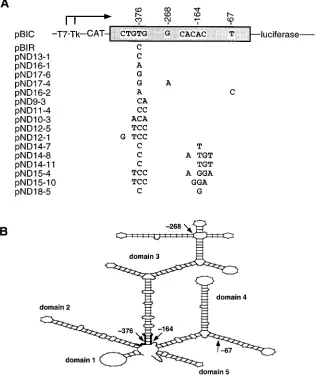
P-labeled oligonucleotide (39) at two temperatures, one permissive for all FIG. 1. (A) Diagram of the plasmids used in this work. The name of the plasmids and their relevant features are indicated. Symbols:™™™™3, transcription start sites; T7, Tk, CAT, luciferase, promoters and marker genes used in the constructs (26). The box between CAT and luciferase depicts the IRES element; letters within the box indicate the nucleotides which differ between C-S8c1 and the mutants tested. Numbers indicate relevant positions within the IRES. Only the differences between each mutant (plasmid series pND) and the C-S8c1 IRES (pBIC) are indicated. The steps followed in the construction of each plasmid are detailed in Materials and Methods. (B) Scheme of the predicted secondary structure (M-Fold program) of the IRES of FMDV C-S8c1, with the location (thick tracing) of the mutations which distinguish the IRES from C-S8c1 and from mutants described in this work. Domain numbering is that proposed by Pilipenko et al. (35).
on November 9, 2019 by guest
http://jvi.asm.org/
[image:2.612.156.474.76.451.2]inserted molecules and one restrictive for nonmutagenized molecules. The ori-entation of the mutant IRES elements was determined by NcoI-HindIII diges-tion. Prior to expression analysis, the nucleotide sequence of the entire length of each IRES under study was obtained by primer extension and dideoxy chain termination with T7 DNA polymerase. Chloramphenicol acetyltransferase (CAT) and luciferase expression from bicistronic mRNAs transcribed either from the T7 or the Tk promoter (see Fig. 1A) was quantitated as a relative measure of IRES activity (26).
Transient-expression assays.The cell lines used have been described previ-ously (26). When transcription from the T7 promoter was desired, BHK-21 or BSC40 monolayers were infected with the vaccinia virus recombinant vTF7-3 (11) 1 h before transfection with the relevant plasmids. Lipofectin (Bethesda Research Laboratories)-mediated transfection was carried out as specified by the supplier. Usually, 0.25 mg of the appropriate plasmid DNA(s) was used to transfect monolayers of about 23105
cells. Fetal calf serum (5%) was added to the monolayer 5 to 6 h later. Cell extracts were prepared 18 and 24 h posttrans-fection in 200ml of 0.5% Nonidet P40–120 mM NaCl–50 mM Tris-HCl (pH 7.8). Experiments were performed on triplicate plates, and each experiment was repeated at least four times.
Luciferase assays were carried out as described by Martı´nez-Salas et al. (25), with a model 2010 luminometer (Analytical Luminescence). CAT activity was determined as described previously (30), except that the reaction was adjusted to 0.08 M sodium borate (pH 9) and 4 M NaCl at the end of the incubation period to decrease the background. Appropriate dilutions of the extracts were chosen to be in the linear range of the assay. The results are presented as luciferase activity relative to CAT activity, measured in the same extract. In the experiment mea-suring expression from the Tk promoter, the enzyme activities were further normalized to the amount of protein measured in the cell extracts by the Lowry method.
RESULTS
Specificity of the point mutation which causes an increase in
IRES activity.
A U-to-C transition at position
2
376 of the
IRES of FMDV C-S8c1 is responsible for the increased
inter-nal initiation of translation in the persistent variant R100 (26).
To test whether the enhancement of IRES activity was base
specific, a series of mutants with substitutions at the base of
domain 3 of the aphthovirus IRES were generated by
site-directed mutagenesis (Fig. 1). Most of the mutants were
de-rived from pBIR (Table 1), the plasmid with the IRES element
of the persistent FMDV R100. Mutant pND13-1 was
con-structed from pBIC to reproduce by site-directed mutagenesis
the pyrimidine transition at position
2
376, which is also
present in pBIR (26). Comparison of CAT and luciferase
ac-tivities expressed from the bicistronic constructs corresponding
to mutants pND13-1, pND16-1, pND17-6, pBIC, and pBIR
indicates that only the U-to-C transition, not U-to-A or U-to-G
transversions, at position
2
376 conferred increased IRES
ac-tivity (Fig. 2A). The presence of additional point substitutions
at position
2
67 (mutant pND16-2) or
2
268 (mutant
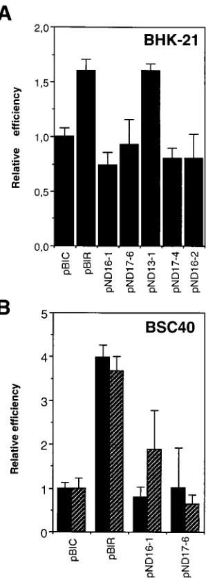
pND17-4) had no significant effect (compare Fig. 1 and 2). The
spec-ificity of the pyrimidine transition was seen in both BHK-21
and BSC40 cells, although the effect was more pronounced in
the latter, as previously noted in the comparison between pBIC
and pBIR (26). No difference in relative activity was observed
when expression was directed by the Tk promoter or by the T7
promoter in concert with recombinant vaccinia virus infection
(Fig. 2B). Expression from the T7 promoter is about 10
3-fold
higher than from the Tk promoter (26). Thus, the change at
position
2
376 leading to enhanced IRES activity in FMDV
R100 is base specific.
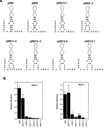
Destabilization of the base of domain 3 leads to defective
IRES.
To test whether destabilization of the base of domain 3
could affect IRES activity, the series of mutants pND9-3,
pND11-4, pND10-3, and pND12-5 were constructed (Fig. 1A).
In these mutants, the base of domain 3 is expected to be
progressively open, because of two or three noncanonical or
unfavorable C-C, C-A, and C-U pairings (Fig. 3A). The IRES
activity of these mutants was severely impaired relative to that
of pBIC both in BHK-21 and in BSC40 cells (Fig. 3B). The
adverse effect on translation efficiency seems to correlate with
perturbation of secondary structure rather than with primary
sequence. In particular, the presence of a G-U pair between
residues
2
374 and
2
163 in mutant pND14-7 resulted in a
translation efficiency comparable to that of pBIC (Fig. 3B).
Furthermore, very low IRES activity, which means at least a
3-log-unit decrease in activity relative to pBIC, was observed
with mutant pND12-1. This mutant, in addition to maintaining
the substitutions of pND12-5 at the base of domain 3, includes
a C-to-G transversion at position
2
373 (Fig. 3). This
transver-sion is expected to affect the stability of the base of domain 2
(compare Fig. 1B and 3), which also contributes to IRES
func-tion (22). Thus, the aphthovirus IRES region at the base of
FIG. 2. Relative efficiency of translation of luciferase by FMDV IRES point mutations at position2376. (A) The indicated plasmids (constructs depicted in Fig. 1) were transfected into BHK-21 cells previously infected with vTF7-3. Cell extracts were prepared 18 and 24 h after serum addition, as detailed in Materials and Methods. The luciferase activity produced by 13104
to 23104
transfected cells was normalized to the CAT activity present in the same extract. (B) Lucif-erase activity produced in BSC40 cells from either the T7 promoter (black rectangles) or the Tk promoter (striped rectangles). Assays were carried out as described in Materials and Methods. Error bars correspond to the standard error of the mean. ‘‘Relative efficiency’’ refers to the activity produced by pBIC.
994 MARTI´NEZ-SALAS ET AL. J. VIROL.
on November 9, 2019 by guest
http://jvi.asm.org/
[image:3.612.361.510.70.488.2]domain 3 is critical for function and very intolerant to
substi-tutions which tend to perturb the structure of this domain.
Neutral mutants maintain the predicted structure.
A series
of mutations designed to restore the predicted structure at the
base of domain 3 in defective mutants led to a functional IRES
(Fig. 4). Relative to the activity of pBIC, pND14-8, pND14-11,
pND15-10, pND15-4, and pND18-5 showed no significant or
very modest differences in activity. The comparisons
empha-size again the contribution of the secondary structure of this
region to IRES activity. In particular, mutant pND12-5 showed
a 100-fold-lower activity than its derivatives pND15-4 or
pND15-10 (compare Fig. 3 and 4); both mutants had the
pre-dicted helical regions at the base of domain 3. Mutants
pND15-4 and pND15-10 differed in seven and six mutations,
respectively, from pBIC, with no significant consequence with
regard to their ability to initiate translation. None of the
mu-tants tested evidenced any increase in IRES activity over the
activity displayed by pBIR, the construct which includes the
IRES sequence found in persistent FMDV R100.
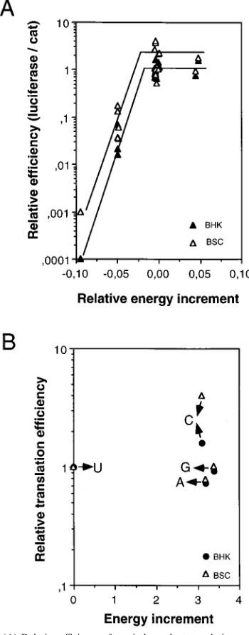
A correlation between predicted IRES stability and
trans-lation efficiency.
The entire IRES RNA (nucleotides
2
458 to
2
1) was folded by using the M-fold program of the Genetics
Computer Group package (45). The energy of stabilization of
the entire IRES of C-S8c1 (in pBIC) was calculated by using
the program M-fold. The value obtained was
D
G
5 2
1,147.8
kcal mol
21(
2
4,802.4 kJ mol
21). Then the same calculation
was carried out for each of the mutants studied. Values ranged
from
D
G
5 2
1,040.4 kcal mol
21(
2
4,353.0 kJ mol
21) to
2
1,202.7 kcal mol
21(
2
5,032.1 kJ mol
21). The difference
be-tween the
D
G of each mutant and that of pBIC, divided by the
D
G of pBIC, was then plotted against the relative translation
efficiency (Fig. 5A). In all the mutants, the base of domain 3
was the only part of the IRES affected in terms of predicted
secondary structure, except for pND12-1, pND17-4, and
FIG. 3. (A) Predicted secondary structure at the base of domain 3 of mutant IRES, obtained with the M-Fold program. Outlined symbols depict mutated nucleotides relative to pBIC. (B) Analysis of the effect of point mutations in the FMDV IRES on the efficiency of translation of bicistronic mRNAs in BHK-21 and BSC40 cells. vTF7-3-infected cells transfected with the plasmids indicated (depicted in Fig. 1) were processed as described in the legend to Fig. 2.
on November 9, 2019 by guest
http://jvi.asm.org/
[image:4.612.132.483.76.506.2]pND16-2, in which additional changes were observed, as
indi-cated above. The results show three main groups of sequences
corresponding to different levels of IRES activity. Decreased
stability of the base of domain 3 was clearly associated with low
activity. Within each group, however, up to 10-fold differences,
which could be partly assigned to the cell type (the values in
BSC40 cells are systematically slightly larger than in BHK-21
cells [Fig. 5A]) or to the nucleotide sequence of each particular
mutant (Fig. 5), were observed. The comparison emphasizes
that translation efficiency forms a plateau at relative energy
increments above zero (Fig. 5A). The translation efficiencies of
FMDV C-S8c1 or R100 are associated with domain 3
se-quences which are close to the maximum translation efficiency,
at least among the range of substitutions analyzed.
DISCUSSION
The results of site-directed mutagenesis have shown that the
region around nucleotide
2
376 of the aphthovirus IRES,
lo-cated at the predicted base of domain 3 (Fig. 1B), plays a
critical role in internal initiation of translation. A strong
de-crease in IRES activity was seen in mutants with nucleotide
substitutions predicted to destabilize the helical region at the
base of domain 3 (Fig. 3). The need for a helical structure at
this domain is supported by a strong correlation between the
efficiency of translation and the energy of stabilization
pre-dicted for the corresponding mutant IRES (Fig. 5).
Restora-tion of a helical structure resulted in recovery of IRES activity;
thus, the decrease in activity of 100-fold seen in mutant
pND12-5 relative to pBIC was converted to a one- to twofold
increase in pND15-10 (Fig. 4).
Additional, albeit less pronounced, effects were also noted
among mutants which maintained a similar predicted structure
but differed in nucleotide sequence (Fig. 3, compare mutants
pND11-4 and pND14-7). In particular, the increase in IRES
activity in the persistent FMDV variant R100 could be
achieved only by the U-to-C substitution but not by the U-to-A
or U-to-G substitution at residue
2
376 (Fig. 2), in spite of the
very similar effects of the three substitutions on predicted
sta-bility (Fig. 5B). Enhanced functional activities associated with
noncanonical Watson-Crick base pairs have been documented
in other biological systems (1, 3, 12, 38). Within any of the
energy levels, differences in translation efficiency have been
observed (Fig. 5A); these must presumably arise from effects of
the primary structure. These comparisons suggest very subtle
effects of position
2
376 on IRES activity. However, in the
absence of knowledge about the tertiary structure of IRES
elements, it is difficult to define in molecular terms the
struc-tural perturbations triggered by single or multiple mutations
and their possible effect on RNA-RNA and RNA-protein
in-teractions (2, 14, 17, 42). Furthermore, synergy between loops
for binding of cell factors has been recently documented for
the poliovirus IRES (16). Such a synergy could contribute to
the very low IRES activity of mutant pND12-1, a variant with
a potentially important alteration at the base of domain 2 in
addition to domain 3 (Fig. 3).
The comparative results presented here for 17 variant
se-quences suggest a very limited tolerance of the IRES region
analyzed to accept point mutations that disrupt the predicted
structure and remain functional. A high proportion of
delete-rious mutations have recently been reported for the helical J-K
FIG. 4. (A) Predicted secondary structure at the base of domain 3 of mutant IRES, according the M-Fold program. Outlined symbols depict mutated nucleotides relative to pBIC. (B) Analysis of the effect of mutations on the efficiency of translation of bicistronic mRNAs. BHK-21 and BSC40 cells, previously infected with vTF7-3, were transfected with the indicated plasmids (Fig. 1). Cell extracts were processed as described in the legend to Fig. 2.
996 MARTI´NEZ-SALAS ET AL. J. VIROL.
on November 9, 2019 by guest
http://jvi.asm.org/
[image:5.612.87.526.77.369.2]junction of the encephalomyocarditis virus IRES (19).
Previ-ous studies with poliovirus revertants selected in vivo suggested
that extensive sequence rearrangements can lead to restoration
of IRES function (14, 15, 37). Point mutations and block
re-arrangements may occur at high frequency during RNA virus
replication, but they will not be noticeable in progeny RNA
unless the parental genome shows an important selective
dis-advantage relative to the rearranged genome (8). A second
important consideration in the mutations observed is viral
pop-ulation size. The substitution at position
2
376 fixed in this
region in the persistent FMDV R100 was probably one of the
very few compatible with high IRES activity. If such a single
mutation occurred at the expected frequency of about 10
24, its
fixation would be compatible with the viral population size
involved in persistence, which was variable but ranged from 10
4to 10
6PFU per cell passage (4). In contrast, double or
qua-druple substitutions, the minimum number needed to restore a
functional domain (Fig. 4), are expected to occur at
frequen-cies of 10
28and as low as 10
216, respectively, which are too low
for these types of variants to be present and become dominant
during persistent infections of FMDV in cell culture. The
re-sults of site-directed mutagenesis of the IRES reported here
extend to a regulatory region the evidence for strong
limita-tions of variation previously documented for the capsid-coding
region of FMDV (27, 41). Such limitations must operate in
both coding and noncoding regions of the picornavirus
ge-nomes, with many mutations leading to profound losses of
fitness (8, 43). However, the results also illustrate the high
connectivity of sequence space with respect to fitness gain. A
single compensatory mutation is often sufficient to rescue a
viral genome from near defectiveness to full functionality.
ACKNOWLEDGMENTS
Work at CBMSO was supported by CICYT PB91-0051-C02-01 PB94-0034-C02-01 and Fundacio´n Ramo´n Areces.
REFERENCES
1. Bartel, D. P., M. L. Zapp, M. R. Green, and J. W. Szostak. 1991. HIV-1 Rev regulation involves recognition of non-Watson-Crick base pairs in viral RNA. Cell 67:529–536.
2. Borovjagin, A. V., M. V. Ezrokhi, V. M. Rostapshov, T. Y. Ugarova, T. F.
Bystrova, and I. N. Shatsky.1990. RNA-protein interactions within the internal translation initiation region of encephalomyocarditis virus RNA. Nucleic Acids Res. 19:4999–5005.
3. Costa, M., and F. Michel. 1995. Frequent use of the same tertiary motif by self-folding RNAs. EMBO J. 14:1276–1285.
4. de la Torre, J. C., M. Da´vila, F. Sobrino, J. Ortı´n, and E. Domingo.1985. Establishment of cell lines persistently infected with foot-and-mouth disease virus. Virology 145:24–35.
5. de la Torre, J. C., E. Martı´nez-Salas, J. Dı´ez, A. Villaverde, F. Sobrino, E.
Rocha, M. Da´vila, and E. Domingo.1988. Coevolution of cells and viruses in a persistent infection of foot-and-mouth disease virus in cell culture. J. Virol.
62:2050–2058.
6. Dı´ez, J., M. Hofner, E. Domingo, and A. I. Donaldson. 1991. Foot-and-mouth disease virus strains isolated from persistently infected cell cultures are attenuated for mice and cattle. Virus Res. 18:3–8.
7. Dildine, S. L., and B. L. Semler. 1992. Conservation of RNA-protein inter-actions among picornaviruses. J. Virol. 66:4364–4376.
8. Domingo, E., and J. J. Holland. 1994. Mutation rates and rapid evolution of RNA viruses, p. 161–184. In S. S. Morse (ed.), Evolutionary biology of viruses. Raven Press, New York.
9. Drew, J., and G. J. Belsham. 1994. trans complementation by RNA of defective foot-and-mouth disease virus internal ribosome entry site ele-ments. J. Virol. 68:697–703.
10. Escarmı´s, C., M. Toja, M. Medina, and E. Domingo. 1992. Modifications of the 59untranslated region of foot-and-mouth disease virus after prolonged persistence in cell culture. Virus Res. 26:113–125.
11. Fuerst, T. R., E. G. Niles, F. W. Studier, and B. Moss. 1986. Eukaryotic transient expression system based on recombinant vaccinia virus that syn-thesizes bacteriophage T7 RNA polymerase. Proc. Natl. Acad. Sci. USA 83: 8122–8126.
12. Giver, L., D. Bartel, M. Zapp, A. Pawul, M. Green, and A. D. Ellington. 1993. Selective optimization of the Rev-binding element of HIV-1. Nucleic Acids Res. 21:5509–5516.
13. Glass, M. J., X. Jia, and D. F. Summers. 1993. Identification of the hepatitis A virus internal ribosome entry site: in vivo and in vitro analysis of bicistronic RNAs containing the HAV 59noncoding region. Virology 193:842–852. 14. Gmyl, A. P., E. V. Pilipenko, S. V. Maslova, G. A. Belov, and V. I. Agol. 1993.
Functional and genetic plasticities of the poliovirus genome: quasi-infectious RNAs modified in the 59-untranslated region yield a variety of pseudorever-tants. J. Virol. 67:6309–6316.
15. Haller, A. A., and B. L. Semler. 1992. Linker-scanning mutagenesis of the internal ribosome entry site of poliovirus RNA. J. Virol. 66:5075–5086. 16. Haller, A. A., and B. L. Semler. 1995. Stem-loop structure synergy in binding
cellular proteins to the noncoding region of poliovirus RNA. Virology 206: 923–934.
FIG. 5. (A) Relative efficiency of cap-independent translation as a function of the relative increment of free energy of each mutant IRES. Mean values of luciferase activity were corrected for CAT activity and are given relative to pBIC for each experiment. The value of energy of the optimal entire IRES structure (DG378Cin kilocalories per mole) produced by the M-fold program was used to
calculate the relative increment of energy as the difference between the energy of each mutant RNA and that of the control pBIC divided by the energy of pBIC as detailed in the text. (B) Influence of the energy increment associated with each of the four possible nucleotide substitutions at position2376 on the relative translation efficiency of the corresponding IRES.
on November 9, 2019 by guest
http://jvi.asm.org/
[image:6.612.91.266.75.520.2]17. Hellen, C. U. T., T. V. Pestova, M. Litterst, and E. Wimmer. 1994. The cellular polypeptide p57 (pyrimidine tract-binding protein) binds to multiple sites in the poliovirus 59nontranslated region. J. Virol. 68:941–950. 18. Hellen, C. U. T., and E. Wimmer. 1995. Enterovirus genetics, p. 25–72. In
H. A. Rotbart (ed.), Human enterovirus infections. ASM Press, Washington, D.C.
19. Hoffman, M. A., and A. Palmenberg. 1995. Mutational analysis of the J-K stem-loop region of the encephalomyocarditis virus IRES. J. Virol. 69:4399– 4406.
20. Jackson, R., M. T. Howell, and A. Kaminski. 1990. The novel mechanism of initiation of picornavirus RNA translation. Trends Biochem. Sci. 15:477–483. 21. Kaminski, A., G. J. Belsham, and R. J. Jackson. 1994. Translation of en-cephalomyocarditis virus RNA: parameters influencing the selection of the internal initiation site. EMBO J. 13:1673–1681.
22. Kuhn, R., N. Luz, and E. Beck. 1990. Functional analysis of the internal translation initiation site of foot-and-mouth disease virus. J. Virol. 64:4625– 4631.
23. Le, S., J. Chen, N. Sonenberg, and J. V. Maizel. 1992. Conserved tertiary structure elements in the 59untranslated region of human enteroviruses and rhinoviruses. Virology 191:858–866.
24. Luz, N., and E. Beck. 1991. Interaction of a cellular 57-kilodalton protein with the internal translation initiation site of foot-and-mouth disease virus. J. Virol. 65:6486–6494.
25. Martı´nez-Salas, E., E. Linney, J. Hassell, and M. L. DePamphilis. 1989. The need for enhancers in gene expression first appears during mouse develop-ment with formation of the zygotic nucleus. Genes Dev. 3:1493–1506. 26. Martı´nez-Salas, E., J. C. Sa´iz, M. Da´vila, G. J. Belsham, and E. Domingo.
1993. A single nucleotide substitution in the internal ribosome entry site of foot-and-mouth disease virus leads to enhanced cap-independent translation in vivo. J. Virol. 67:3748–3755.
27. Mateu, M. G., J. Herna´ndez, M. A. Martı´nez, D. Feigelstock, S. Lea, J. J. Pe´rez, E. Giralt, S. Stuart, E. L. Palma, and E. Domingo.1994. Antigenic heterogeneity of foot-and-mouth disease virus serotype in the field mediated by very limited sequence variation at several antigenic sites. J. Virol. 68: 1407–1417.
28. Meerovitch, K., Y. V. Svitkin, H. S. Lee, F. Lejbkowicz, D. J. Kenan, E. K. L.
Chan, V. I. Agol, J. K. Keene, and N. Sonenberg.1993. La autoantigen enhances and corrects aberrant translation of poliovirus RNA in reticulocyte lysate. J. Virol. 67:3798–3807.
29. Meyer, K., A. Petersen, M. Niepman, and E. Beck. 1995. Interaction of eukaryotic initiation factor eIF-4B with a picornavirus internal translation initiation site. J. Virol. 69:2819–2824.
30. Neumann, J. R., C. A. Morency, and K. O. Russian. 1987. A novel rapid assay for chloramphenicol acetyl transferase gene expression. BioTechniques 5: 444–447.
31. Nicholson, R., J. Pelletier, S. Le, and N. Sonenberg. 1991. Structural and functional analysis of the ribosome landing pad of poliovirus type 2: in vivo translation studies. J. Virol. 65:5886–5894.
32. Pelletier, J., and N. Sonenberg. 1988. Internal initiation of translation of eukaryotic mRNA directed by a sequence derived from poliovirus RNA. Nature (London) 334:320–325.
33. Perrin, S., and G. Gilliland. 1990. Site-specific mutagenesis using asymmet-ric polymerase chain reaction and a single mutant primer. Nucleic Acids Res.
18:7433–7438.
34. Pestova, T. V., C. U. T. Hellen, and E. Wimmer. 1991. Translation of polio-virus RNA: role of an essential cis-acting oligopyrimidine element within the 59nontranslated region and involvement of a cellular 57-kilodalton protein. J. Virol. 65:6194–6204.
35. Pilipenko, E. V., V. M. Blinov, B. K. Chernov, T. M. Dimitrieva, and V. I.
Agol.1989. Conservation of the secondary structure elements of the 59 -untranslated region of cardio and aphthovirus RNAs. Nucleic Acids Res. 17: 5701–5711.
36. Pilipenko, E. V., A. P. Gmyl, S. V. Maslova, G. A. Belov, A. N. Sinyakov, M.
Huang, T. D. K. Brown, and V. I. Agol.1994. Starting window, a distinct element in the cap-independent internal initiation of translation on Picor-naviridae RNA. J. Mol. Biol. 241:398–414.
37. Pilipenko, E. V., A. P. Gmyl, S. V. Maslova, Y. V. Svitkin, A. N. Sinyakov, and
V. I. Agol.1992. Prokaryotic-like cis elements in the cap-independent inter-nal initiation of translation on picornavirus RNA. Cell 68:119–131. 38. Pley, H. W., K. M. Flaherty, and D. B. McKay. 1994. Three-dimensional
structure of a hammerhead ribozyme. Nature (London) 372:68–74. 39. Sambrook, J., E. F. Fritsch, and T. Maniatis. 1989. Molecular cloning: a
laboratory manual, 2nd ed. Cold Spring Harbor Laboratory, Cold Spring Harbor, N.Y.
40. Stone, D. M., J. W. Almond, J. K. Brangwyn, and G. J. Belsham. 1993. trans complementation of cap-independent translation directed by poliovirus 59 noncoding region deletion mutants: evidence for RNA-RNA interactions. J. Virol. 67:6215–6223.
41. Verdaguer, N., M. G. Mateu, D. Andreu, E. Giralt, E. Domingo, and I. Fita. 1995. Structure of the major antigenic loop of foot-and-mouth disease virus complexed with a neutralizing antibody: direct involvement of the Arg-Gly-Asp motif in the interaction. EMBO J. 14:1690–1696.
42. Weeks, K. M., and D. M. Crothers. 1993. Major groove accessibility of RNA. Science 261:1574–1577.
43. Wimmer, E., C. U. T. Hellen, and X. Cao. 1993. Genetics of poliovirus. Annu. Rev. Genet. 27:353–436.
44. Witherell, G. W., and E. Wimmer. 1994. Encephalomyocarditis virus internal ribosome entry site RNA-protein interactions. J. Virol. 68:3183–3192. 45. Zuker, M. 1989. On finding all suboptimal foldings of an RNA molecule.
Science 244:48–52.
998 MARTI´NEZ-SALAS ET AL. J. VIROL.