Bethesda, Maryland,
and Division of Infectious Diseases, Children’s Hospital Research Foundation,
Department of Pediatrics, University of Cincinnati College of Medicine, Cincinnati, Ohio
2Received 8 September 1995/Accepted 30 November 1995
The role of putative promoter or activator sequences downstream of the herpes simplex virus type 2
latency-associated transcript (LAT) promoter and upstream of the LAT intron was investigated in vivo by
constructing and evaluating mutant viruses with deletions in this region. The deletion of LAT promoter
sequences upstream of the primary LAT transcript reduced levels of LAT expression during productive
infections, compared with the LAT expression level of wild-type virus, and abolished LAT expression during
latency. The deletion of the putative downstream regulatory elements reduced but did not eliminate LAT
expression during productive and latent infections. The deletion of both regions almost completely eliminated
acute LAT transcription, although additional acute LAT-region transcription directed by sequences upstream
of either region was detected by reverse transcriptase PCR. The deletion of the downstream elements did not
influence the ability of the virus to reactivate from latently infected guinea pigs relative to the ability of the
wild-type virus to reactivate; thus, decreased LAT expression did not affect the frequency of recurrence. The
deletion of both regions did not affect the ability of the virus to establish latency. We conclude that downstream
regulatory elements are necessary for maximal acute LAT expression but do not constitute an independent
promoter during latency and do not play an obvious role in the establishment of or reactivation from latency.
After replication at a site of inoculation, herpes simplex
virus (HSV) establishes latency in the sensory ganglia
inner-vating that site. Periodically, the virus may reactivate to cause
symptomatic or asymptomatic recurrences (4, 20). Of the 80 or
so described genes of HSV, only the latency-associated
tran-script (LAT) is uniformly transcribed during latency (6, 8, 35).
Mutations in sequences directing HSV type 1 (HSV-1) (2, 14,
21, 28, 34, 36) and HSV-2 (17) LAT transcription impair virus
reactivation but do not influence the establishment of latency
in rabbits (15) or guinea pigs (17). The role of these sequences
in the establishment of virus latency in mice is unclear (28, 34).
During HSV-1 latency, abundant LAT introns of 2.0 and 1.4
to 1.5 kb (18, 30, 38, 40) are spliced (12) from a less abundant
primary LAT transcript (37, 41) (approximately 8 to 9 kb in
size). During productive infections, only the 2.0-kb LAT intron
and the primary LAT have been detected. While transcription
during HSV-2 latency has not been as thoroughly investigated,
a single 2.2-kb LAT intron is also readily detected both during
acute and latent infection (5, 19, 24).
Promoter sequences which appear to direct transcription of
the HSV-1 primary LAT are present approximately 600 to 700
bp upstream of the LAT intron (1, 9, 10, 42). This promoter
contains a TATA box, a cyclic AMP-responsive-element
bind-ing (CREB) site (27), Sp1 sites, and a bindbind-ing site for a protein
denoted the LAT promoter binding factor (43). The deletion
of these promoter sequences abolishes latent HSV-1 LAT
transcription but permits continued detection of the HSV-1
LAT intron during acute infections in tissue culture (26). This
observation, together with the identification of promoter
ac-tivity in transient transfection and infection assays in sequences
downstream of the primary LAT 5
9
end but upstream of the
LAT intron (13), led to the hypothesis that acute HSV-1 LAT
transcription may be regulated by two independent promoters.
The traditional promoter upstream of the primary LAT has
been referred to as LAP1, and the putative downstream
reg-ulatory sequences between LAP1 and the LAT intron have
been denoted LAP2. The LAP2 sequences contain potential
AP2 and Sp1 binding sites in a high-GC-content region,
to-gether with a CT-rich element which is characteristic of several
TATA-less constitutive eukaryotic promoters (16). In a
re-cently published experiment, a StyI-StyI fragment upstream of
the CT-rich element contained within the HSV-1 LAP2 region
was removed from wild-type HSV-1, and this deletion did not
affect acute or latent HSV-1 LAT expression or the HSV-1
reactivation phenotype in explanted mouse trigeminal ganglia
(23).
HSV-2 LAT promoter sequences homologous to the HSV-1
LAP1 region containing TATA, CREB, and Sp1-AP2
homol-ogies have also been described (19, 25). More-detailed
func-tional analyses of this region revealed multiple AP2 binding
sites, an AP1 binding site, the CREB site, and four additional
unidentified DNA binding footprints (39). The deletion of
sequences homologous to HSV-1 LAP1 from HSV-2 abolishes
latent LAT transcription and dramatically reduces
spontane-ous recurrences from latently infected guinea pigs but permits
acute HSV-2 LAT transcription (17). HSV-2 also contains
sequence elements homologous to HSV-1 LAP2 between the
5
9
ends of the primary LAT and the LAT intron, including
Sp1-AP2 homologies and CT-rich elements (Fig. 1B). To
ex-plain acute LAT transcription in LAP1
2mutants, as in HSV-1,
we hypothesized that HSV-2 LAP2 may contain important
* Corresponding author. Mailing address: HFM-457, Bldg. 29A, Rm. 1C16, MSC4555, 29 Lincoln Dr., Bethesda, MD 20892-4555.
1535
on November 9, 2019 by guest
http://jvi.asm.org/
regulatory elements, possibly including an additional
pro-moter. The roles of these sequences in LAT transcription and
in latency establishment and reactivation phenotypes have not
yet been investigated.
To delineate the role of downstream sequences in HSV-2
LAT transcription and latency, we deleted putative HSV-2
LAP2 sequences from a mutant virus already lacking the
HSV-2 LAP1 promoter and from a virus with an HSV-2 333
wild-type genotype. We then characterized productive and
la-tent infections of wild-type HSV-2 and of strains with LAP1,
LAP2, or LAP1 and LAP2 deletions. Latent infections and
reactivation patterns were observed in guinea pigs, which
pro-vide the only animal model of HSV latency in which the virus
spontaneously reactivates to cause recurrent lesions, as it does
in humans (31–33).
MATERIALS AND METHODS
Cells and tissue culture experiments.Vero and SK-N-SH (human neuroblas-toma) cells were obtained from the American Type Culture Collection (Rock-ville, Md.), and maintained in 1:1 minimum essential medium-medium 199, with 1% GASP (a mixture of glutamine, aureomycin, streptomycin, and penicillin) and 10% heat-inactivated fetal bovine serum (all from Quality Biologicals, Gaithersburg, Md.). To determine one-step growth characteristics of parental virus and mutant viruses, confluent Vero cells in 6-well dishes were inoculated in duplicate at time zero with 105
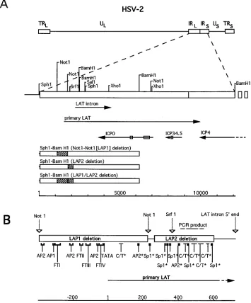
[image:2.612.126.481.75.503.2]PFU (at a multiplicity of infection [MOI] of approximately 0.1 PFU per cell) of each virus. For the 3- and 16-h time points, virus was allowed to adsorb for 60 min before the addition of medium. Cells were FIG. 1. Map of HSV-2 LAT and constructs used to generate mutations. (A) Restriction endonuclease sites are shown relative to the positions of the HSV-2 major and primary LATs, ICP0, ICP34.5, and ICP4. Introns are denoted within each transcript by a box. Regions corresponding to the LAP1 and LAP2 deletions are shaded within boxes corresponding to the SphI-BamHI fragment, which was the insert in the plasmid vectors used to construct the mutant viruses. Distance in base pairs is shown relative to the first SphI site, within the unique long region. Abbreviations: UL, unique long; TRL, terminal repeat, long; IRL, internal repeat, long; IRS, internal
repeat, short; US, unique short; TRS, terminal repeat, short. (B) Transcription factor binding sites within LAP1 and LAP2 regions are shown. Upstream of the primary
LAT 59end, these sites were mapped by functional assays (39). In addition, a CREB site overlies FTIV. Downstream of the primary LAT 59end, the sites marked with an asterisk are based only on sequence analyses. Consensus sequences used were Sp1 (GGGCGG), AP2 (CCGCCCGCG), and CT (more than 20 bases exceeding 90% CT content). The expected product of the reverse transcriptase PCR experiments is denoted with a dashed line. Distance in base pairs is shown relative to the 59end of the primary LAT, as mapped in vitro (39).
on November 9, 2019 by guest
http://jvi.asm.org/
333WT for convenience), which has a wild-type genotype and phenotype, has been described elsewhere (17). LAP2 sequences were deleted (via homologous recombination of deleted plasmids) from 333pLAT2(renamed 333LAP12for convenience) and 333pLATRto construct 333LAP1&22and 333LAP22,
respec-tively.
In order to make the LAP2 deletion mutant and the LAP1-LAP2 double promoter deletion mutant, appropriate plasmid DNA was cotransfected into Vero cells with purified 333pLATR viral DNA and 333pLAT2viral DNA,
respectively, with lipofectin (Life Technologies). The resultant viruses were plated in serial dilutions on 6-well plates, and individual plaques were selected, grown, and screened by Southern hybridization as previously described (17). After identification of mutant viruses, plaque purifications were performed until Southern hybridization identified no evidence of contamination with the parents. At this point, two additional plaque purifications were performed to yield stocks of each mutant. Additional Southern hybridizations were carried out on purified viral DNA digested with the enzyme SphI, BamHI, SrfI, NotI, or NotI plus XhoI. Blots were probed with SphI-BamHI plasmid or intact virion DNA that was radiolabeled with32
P with a random primer labeling kit (Amersham, Arlington Heights, Ill.).
Guinea pig studies.Animal studies were approved by the Children’s Hospital Research Foundation Institutional Animal Care and Use Committee. Female Hartley guinea pigs (Charles River Breeding Laboratories, Wilmington, Mass.) weighing 400 to 500 g were inoculated with approximately 105.7
PFU of 333WT, 333LAP12, 333LAP22, or 333LAP1&22on day 0 by rupture of the vaginal closure membrane with a moistened calcium alginate-tipped swab and instilla-tion of 0.1 ml of virus. Lesion severity was scored daily (on a scale from 0 to 4) until resolution of the acute infection (32). All observations of the guinea pigs were performed by investigators who were unaware of the virus inoculum. Guinea pigs that were not evaluable for the entire observation period were excluded from analysis. Animals were housed in American Association for the Accreditation of Laboratory Animal Care-approved facilities and cared for in accordance with institutional guidelines.
Northern (RNA) hybridization.Vero cells and SK-N-SH cells were infected with virus at an MOI of 1.0 PFU per cell. Sixteen hours after infection, RNA was extracted from the infected cells. RNA was also extracted from latently infected guinea pig ganglia. Five micrograms of each RNA was subjected to electrophore-sis on 1.5% agarose-formaldehyde denaturing gels, transferred to nitrocellulose membranes (Schleicher & Schuell, Keene, N.H.), and hybridized according to the instructions of the manufacturer. The blots were probed with32
P-radiola-beled gel-pure SalI-XhoI double-stranded DNA restriction fragments. A radio-labeled human glyceraldehyde-3-phosphate dehydrogenase (G3PDH) cDNA probe (Clontech, Palo Alto, Calif.) was used to control for RNA quantity in some experiments.
PCR amplification of viral RNA.Vero cells were infected at an MOI of 5.0 PFU per cell. RNA was extracted from the infected cells 10 h after infection. Five micrograms of each extracted total RNA sample was treated with 80 U of RNase-free DNase I (Boehringer Mannheim) for 1 h and then denatured at 948C for 15 min. Each sample was then incubated at 398C for 1 h in a 30-ml solution containing Moloney murine leukemia virus reverse transcriptase (Life Technol-ogies) and the downstream primer. Reverse-transcribed samples and RNA sam-ples without reverse transcription were used in standard 100-ml PCR mixtures with both primers (upstream primer, AGAGCGAGACAGACACACGCAG; downstream primer, CGTTCCGTTTCTTCTCCCTCC).
Amplification was carried out in a DNA thermal cycler (model 480; Perkin-Elmer Cetus, Norwalk, Conn.) under the following cycle conditions: 948C for 1 min; and then 2 cycles at 988C for 1 min, 558C for 1 min, and 728C for 2 min; and 28 cycles at 958C for 1 min, 558C for 2 min, and 728C for 2 min. After completion of amplification, 10ml of the reaction mixture was loaded on a 1.5% NuSieve GTG agarose gel (FMC BioProducts, Rockland, Maine). The products were electrophoresed and subjected to Southern hybridization with32
P-radiolabeled gel-pure NotI-SalI fragments from a plasmid containing the HSV-2 SphI-BamHI fragment.
Semiquantitative PCR.Semiquantitative PCRs to assess the quantity of viral DNA in latently infected ganglia were performed as previously described (17). Briefly, 100 ng of genomic DNA (quantified spectrophotometrically) extracted from pooled S1 to S3 ganglia of guinea pigs killed 27 days after inoculation with
333WT or 333LAP1&22was subjected to PCR (948C for 1.5 min, 528C for 3 min, 728C for 3 min) for 35 cycles, simultaneously using the primers CACCTGCCAG TCGAACGACCTCAT and AGCCGCCACCCCCCTCCCCTGCGT (which are specific for a 500-bp product in the HSV-2 morphologic transforming region, located in the HSV-2 BglII N fragment), and AGTCCATTTCTTGTCTGTCTC TCT and CTGGGGAACAAAGTAAGAGTCAAC (which are specific for a 500-bp product in guinea pig lactalbumin). The products were electrophoresed and subjected to Southern hybridization with32P-radiolabeled gel-pure internal
amplified fragments from a plasmid containing the HSV-2 BglII N fragment and with strain 2 fetal guinea pig DNA as a probe. Membranes were exposed to XAR film (Eastman Kodak, Rochester, N.Y.) for 6 h, and bands were quantified by densitometry on a digital image system (model IS-1000; Alpha Innotech Corp., San Leandro, Calif.). For each sample, the ratio of HSV-2 DNA signal to the guinea pig lactalbumin signal was calculated.
RESULTS
Mutant virus characterization.
To examine the role of
LAP2 sequences during acute and latent infection, we
con-structed and evaluated mutants containing deletions in the
LAP1 region (333LAP1
2), the LAP2 region (333LAP2
2), or
both the LAP1 and LAP2 regions (333LAP1&2
2). Plasmid
inserts used to construct these mutant viruses by homologous
recombination with parent virus are shown in Fig. 1A. Binding
sites for potential enhancer elements in this region are shown
in Fig. 1B, mapped either by DNase footprinting and
methyl-ation interference (39) (upstream of the primary LAT start
site) or on the basis of sequence analysis.
Appropriate construction of each mutant virus was
demon-strated by Southern hybridization of DNA extracted from
pro-ductively infected Vero cells (Fig. 2). The
32P-labeled
double-stranded DNA probes used in these analyses spanned the
SphI-BamHI fragment used in the construction of the mutants.
After digestion with BamHI, each virus DNA yielded the
expected 3.9-kb fragment between the two BamHI sites
inter-FIG. 2. Southern hybridizations of DNAs extracted from 333WT, 333LAP12, 333LAP22, and 333LAP1&22. Virus DNAs were digested with the denoted enzymes, subjected to electrophoresis, and hybridized with an SphI-BamHI probe. Marker sizes (in kilobase pairs) are shown to the left of each panel.on November 9, 2019 by guest
http://jvi.asm.org/
[image:3.612.318.553.68.337.2]nal to the probe. The two other bands represented fragments
originating in each unique long segment and ending at the
BamHI site within the probe. Relative to the sizes of the band
pair obtained with 333WT DNA, each pair of bands from the
mutants was appropriately smaller, depending on the size of
the deleted segment (624 bp for LAP1, 301 bp for LAP2, 925
bp for both LAP1 and LAP2).
Digestion of 333LAP1
2and 333WT with SrfI yielded a pair
of
;
6-kb bands corresponding to fragments between the SrfI
site internal to the probe and another site contained within the
short repeats close to the junction of the inverted long repeat
and inverted short repeat or to the 5
9
end of the genome.
Another band of similar size, corresponding to the SrfI
frag-ment between the unique long segfrag-ment on one side and the
probe, was also superimposed on this band. The reduction in
size of one of the three comigrating bands by the 624-bp LAP1
deletion in 333LAP1
2was not resolved on this gel. The
;
3.3-kb band (in 333WT), corresponding to a SrfI fragment
spanning the unique long repeat junction on the other side,
was appropriately diminished to about 2.7 kb in 333LAP1
2. In
333LAP2
2, the SrfI site internal to the probe was deleted, so
on one side of the unique long repeat the SrfI fragment
spanned approximately 11 to 12 kb and on the other side it was
approximately 8 to 9 kb. These bands appeared to be
appro-priately smaller in 333LAP1&2
2, although resolution of bands
of this size is difficult.
The digestion of each LAP1-containing virus with NotI
yielded the 624-bp NotI fragment and 3.4- and 2.3-kb
frag-ments corresponding to sequences originating in the unique
long
segment.
LAP1
deletions
in
333LAP1
2and
in
333LAP1&2
2were reflected by the loss of the 624-bp NotI
fragment, and LAP2 deletions were reflected by changes in the
size of the largest (
;
5-kb) NotI fragment. These latter changes
were more easily identified when XhoI was added to the
di-gestion; this band became sufficiently small to readily identify
the absence of the LAP2 sequences from the appropriate
mu-tants.
We examined the effect of each deletion on virus infections
in one-step growth experiments in tissue culture (Fig. 3).
Con-fluent Vero cells were infected with each virus at an MOI of 0.1
PFU per cell and harvested 3 and 16 h after infection. The
virus yield at each time point was determined by plaque assay
titration. Each virus displayed similar one-step growth
charac-teristics, with similarly reduced titers of virus at 3 h after
inoculation, corresponding to virus entry into cells, and with
increased titers after 16 h.
Characterization of LAT expression in productive
infec-tions.
To determine the effect of each mutation on LAT
ex-pression during productive infections, Vero cells were infected
with each virus at an MOI of 1.0 PFU per cell. Total cellular
RNA was extracted from infected cells 16 h after infection and
subjected to Northern hybridization with a radiolabeled
SalI-XhoI double-stranded DNA probe, which is homologous both
to the 2.2-kb LAT intron and to ICP0.
In this experiment (Fig. 4), 333WT produced the largest
amount of the 2.2-kb LAT, while 333LAP1
2and 333LAP2
2each produced smaller (reduced by 60 to 70%, on the basis of
comparison of the signal intensities) but detectable quantities
of LAT. No 2.2-kb LAT was detectable by Northern
hybrid-ization from cells infected with 333LAP1&2
2. Each virus
pro-duced comparable quantities of the 2.6-kb ICP0 transcript.
Equivalency of the amounts of RNA loaded onto each lane
was demonstrated by subsequent hybridization of these RNAs
with a radiolabeled G3PDH probe (data not shown). In similar
experiments, in which SK-N-SH human neuroblastoma cells
were infected with each virus, the relative intensities of the
LAT bands with each virus were similar to those observed in
infected Vero cells (data not shown).
Identification of acute transcription unrelated to either
LAP1 or LAP2.
To determine whether the residual expression
of LAT observed during productive infection by the 333LAP1
2virus could be completely attributed to sequences downstream
of LAP1, including LAP2, we performed reverse transcriptase
PCRs on RNAs extracted from Vero cells infected with each
virus. We used a primer pair near the 5
9
end of LAP2 in these
experiments. If all residual LAT synthesized by 333LAP1
2were attributable to LAP2, one would expect to detect no
primary LAT transcription in this region from 333LAP1
2.
[image:4.612.59.298.70.238.2]The results of this experiment are displayed in Fig. 5. The
expected 134-bp PCR product is identified in cells productively
infected with 333LAP1
2and 333WT. RNA extracted from
cells infected with 333LAP2
2did not contain sequences
ho-mologous with these primers and was negative, as was RNA
from uninfected Vero cells and from a reaction mixture
con-taining no template. In each case in which a band was
ob-served, omission of reverse transcriptase from the reaction
gave no product, supporting the conclusion that the PCR
prod-ucts result from the amplification of reverse-transcribed RNA
FIG. 3. One-step growth of 333WT, 333LAP12, 333LAP22, and333LAP1&22in tissue culture. Vero cells (106
) were inoculated with each virus at an MOI of approximately 0.1 at time zero. The plaque titers of cultures were determined 0, 3, and 16 h after inoculation.
FIG. 4. Expression of LAT by each virus during productive infections. RNA extracted from Vero cells productively infected with each virus was subjected to electrophoresis and hybridized with a double-stranded, gel-pure SalI-XhoI probe. Positions of size markers (in kilobases) of 18S and 28S rRNAs (denoted with asterisks) and of the 2.2-kb LAT and 2.6-kb ICP0 transcripts are shown.
on November 9, 2019 by guest
http://jvi.asm.org/
[image:4.612.385.486.74.202.2]rather than from DNA contamination of the samples.
There-fore, regulatory sequences upstream of either LAP1 or LAP2
also direct some LAT-region transcription.
Characterization of LAT expression in latent infections.
The
ability of each virus to produce LAT during ganglionic latency
was determined by Northern hybridization of RNAs extracted
from sacral dorsal root ganglia 27 days after intravaginal
inoc-ulation of guinea pigs with each virus (Fig. 6). Figure 6 shows
results obtained by hybridization of RNAs extracted from the
sacral dorsal root ganglia of two different guinea pigs latently
infected with each virus. A
32P-labeled, double-stranded,
gel-pure SalI-XhoI DNA probe was also used in this experiment.
As we observed previously, the ganglia of guinea pigs latently
infected with 333LAP1
2contained no demonstrable LAT (17).
The wild-type virus 333WT synthesized the greatest amount of
LAT in the ganglia, 333LAP2
2produced less (but detectable)
LAT, and the combined deletion mutant 333LAP1&2
2pro-duced no LAT. Appropriately, ICP0 was not detected during
latency. A comparison of the intensities of the LAT bands with
those obtained by hybridization with a G3PDH probe showed
that differences in levels of LAT expression observed with each
virus strain could not be attributed to differences in the
amounts of RNA on each blot.
Characteristics of acute and recurrent infections of guinea
pigs.
There were no discernible differences in levels of
viru-lence among the viruses during the productive infection of the
guinea pigs (Table 1). The abilities to produce acute disease, as
measured by lesion scores, were comparable for each virus.
Acute spinal cord and dorsal root ganglion virus titers were
comparable for 333WT and 333LAP1&2
2, suggesting that
LAT did not influence the abilities of these viruses to reach the
central nervous system.
While the deletion of LAP2 influenced acute and latent
LAT expression, it did not abolish it. To determine whether
this reduction in levels of LAT expression influenced virus
reactivation, we observed guinea pigs infected with 333WT and
333LAP2
2from days 21 to 63 after intravaginal inoculation
with each virus and tabulated spontaneous recurrences.
In this experiment, guinea pigs infected with 333WT
expe-rienced spontaneous recurrences a mean
6
standard error
(SE) of 3.7
6
0.7 (median
5
3) times over the 42-day
obser-vation period, compared with a mean
6
SE of 4.0
6
1.3
(median
5
4) times for 333LAP2
2. Thus, the deletion of LAP2
sequences from HSV-2 333 did not influence recurrence
fre-quency. Appropriately, 333LAP1&2
2recurred much less
fre-quently, i.e., a mean
6
SE of 0.5
6
0.5 (median
5
0) time per
animal. This experiment demonstrated significant differences
between the recurrence frequencies of 333LAP1&2
2and each
of the other two viruses (P
,
0.05 by the Wilcoxon rank sum
test [7]) but not between 333WT and 333LAP2
2(P
.
0.5).
Establishment of latency.
No studies to date have addressed
with each virus or with uninfected cells (Uninf.) and subjected either to reverse [image:5.612.104.245.74.174.2]transcription (RT) (1) or no reverse transcription (2). Additional samples included DNA extracted from cells infected with 333WT (333WT DNA) and PCR reagents without any template (w/o template). PCR was carried out with the primers indicated in Fig. 1B, which give an expected product of 134 bp.
FIG. 6. LAT expression by each virus during latent infections. RNA ex-tracted from guinea pig ganglia latently infected with each virus was subjected to electrophoresis and hybridized with a double-stranded, gel-pure SalI-XhoI probe and with a probe to detect cellular G3PDH. Positions of size markers (in kilo-bases) of 18S and 28S rRNAs (denoted with asterisks) and of the 2.2-kb LAT and 1.5-kb G3PDH transcripts are shown.
TABLE 1. Primary, recurrent, and latent HSV-2 infection in guinea pigs
Virus Severity of primary infectiona
Virus titerb
in:
Recurrencec
Latent DNAd
DRG SC
333WT 6.260.9 (11) 3.160.1 (3) 4.360.4 (3) 3.760.7 (3) 1.360.4
333LAP22 6.661.2 (10) NDe ND 4.061.3 (5) ND
333LAP1&22 6.260.5 (16) 3.060.2 (3) 5.160.2 (5) 0.560.5 (4) 1.560.3
a
Severity is defined as the area under the lesion score-per-day curve; data are the means of two experiments6SEs. The numbers of animals tested are shown in parentheses.
b
Virus titer in spinal cord (SC) or sacral dorsal root ganglia (DRG) 5 days after intravaginal HSV-2 inoculation. Values are the mean log10PFU per gram of tissue
6SEs. The numbers of animals tested are in parentheses.
c
Recurrence is defined as the median number of days animals experienced recurrent infections from days 21 to 63 after HSV-2 inoculation. The numbers of animals tested are in parentheses.
d
Latent virus DNA values were determined by semiquantitative PCR of DNA extracted from four latently infected animals and are reported as means6SEs.
e
ND, not done.
on November 9, 2019 by guest
http://jvi.asm.org/
[image:5.612.359.512.76.270.2] [image:5.612.54.557.600.662.2]the influence that HSV-2 LAT expression might have on the
establishment of latency. To determine whether HSV-2 LAT
RNA expression influences the establishment of latency, we
used a semiquantitative PCR assay to determine the latent
virus DNA burdens in guinea pig ganglia 27 days after
inocu-lation with 333WT and 333LAP1&2
2(Table 1). Relative to
levels of guinea pig lactalbumin, the mean adjusted relative
HSV DNA content for four guinea pigs infected with 333WT
was 1.3, while the mean value for four guinea pigs infected with
333LAP1&2
2was 1.5. This difference falls within the expected
error of this assay and is indicative of no substantial differences
in the abilities of these viruses to establish latency in guinea
pigs.
DISCUSSION
The deletion of both LAP2 and LAP1 eliminated our ability
to detect LAT in acute and latent infections. The deletion of
LAP2 alone diminished levels of LAT expression in both acute
and latent infections. Thus, sequences important for
wild-type-level LAT transcription during productive infections and
dur-ing latency appear to reside within LAP2. LAP2 does not
contain promoter elements sufficient to direct latent LAT
ex-pression autonomously, as evidenced by the inability of LAP1
deletion mutants to transcribe LAT during latency.
In previously reported experiments, the quantities of LAT
expressed in different ganglia latently infected with the same
virus varied from animal to animal. Such experimental error
could have influenced the LAT quantities we detected in some
of the tested guinea pig ganglia. In this study, a consistent
effect of the LAP2 deletion on LAT expression was observed in
every experiment we performed, including those performed
during the productive infection of Vero and human
neuroblas-toma cells and with the latently infected ganglia. We therefore
conclude that LAP2 sequences are necessary for
maximum-level LAT expression.
It is unclear whether LAP2 behaves as a completely
inde-pendent promoter during productive infection. Reverse
tran-scriptase PCR of RNA extracted from cells infected with the
LAP1 deletion mutant showed small amounts of RNA
tran-scription in the direction of the LAT intron, between LAP1
and LAP2. This small amount of transcription could represent
a modified primary LAT RNA, directed by residual elements
of the LAP1 promoter which were not completely eliminated
by the NotI-NotI deletion or by run-on transcription from
RNAs further upstream. It is possible that LAP2 sequences
merely enhance or activate this residual upstream
transcrip-tion. Recently, two polyadenylated gamma (late) transcripts
have been identified upstream of HSV-1 LAP1 (29). HSV-2
could transcribe similar RNAs during productive infection.
Experiments in which the HSV-1 LAP1 promoter linked to
b
-galactosidase sequences was inserted into the gC locus did
not identify long-term expression of the reporter except when
LAT promoter sequences (from LAP1) were juxtaposed with
long terminal repeat promoter sequences from Maloney
mu-rine leukemia virus (22). It has been suggested that LAP2
sequences may supply such enhancer or activator function to
LAP1, contributing to sustained LAT expression in neurons.
Our experiments showed that the deleted LAP2 sequences
were not required for long-term expression in neurons,
sug-gesting that any required enhancer sequences for long-term
expression by LAP1 are not contained in the LAP2 region.
While it is established that LAP1 is required for efficient
reactivation of HSV-2 (17), the deletion of LAP2 did not
influence the recurrence phenotype of this virus in guinea pigs.
This observation is consistent with previous reports that the
quantity of HSV-1 or HSV-2 LAT in ganglia or the level of
acute HSV-2 LAT transcribed does not influence the
fre-quency of recurrence in viruses that are capable of LAT
tran-scription (3, 11). This finding also makes it less likely that
primary LAT sequences upstream of the LAT intron
contrib-ute to a virus’ reactivation phenotype.
While LAP1 deletion from HSV-2 was shown previously not
to influence the establishment of latency (17), these
experi-ments did not fully exclude a role for the LAT in the
estab-lishment of latency because the mutation did not totally
elim-inate acute LAT transcription. Any role which LAT might play
in establishing latency is more likely to be related to acute than
to latent LAT transcription. In our study of guinea pig
infec-tion, we identified no difference in the abilities of the 333WT
and 333LAT1&2
2strains to spread to ganglia, as reflected by
disease severity and virus titers in neural tissues during acute
infections. Moreover, a semiquantitative PCR assay did not
discern any differences in the relative concentrations of latent
viral DNA attained by wild-type virus and 333LAP1&2
2.
While a small effect cannot be excluded by an experiment of
this size using this technique, these results allow us to conclude
that HSV-2 LAT plays no major role in the establishment of
latency in guinea pigs, as has previously been concluded for the
HSV-1 LAT in rabbits (15).
Although significant similarities with respect to LAT
tran-scription patterns and LAP1 regulatory elements exist between
HSV-1 and HSV-2, the present findings may not necessarily
extend to HSV-1. It is important to recall that HSV-1 and
HSV-2 exhibit distinct reactivation phenotypes (each virus has
a preferred site for reactivation [20]) and that the RNA
se-quences of the HSV-1 and HSV-2 LAT introns are quite
dif-ferent (19). The deletion of HSV-1 LAP1 did not influence
acute LAT transcription (26), although deletions in this region
affected the ability of this promoter to direct acute
transcrip-tion when the gene for
b
-galactosidase was inserted into the
virus as a reporter (10). While the LAP1 regions of HSV-1 and
HSV-2 are fairly similar, the LAP2 regions exhibit considerably
less overall homology, although major elements (AP2, Sp1,
and CT-rich sites) are conserved. Recently reported
experi-ments with an HSV-1 mutant from which sequences between
two StyI sites within HSV-1 LAP2 were deleted showed no
impairment of either acute or latent LAT transcription or of
the reactivation phenotype from explanted mouse ganglia (23).
Our finding that LAP2 influences acute and latent HSV-2
transcription may be attributable to a more complete deletion
of LAP2 sequences, as the related mutant of HSV-1 did not
eliminate the putative CT-rich element within LAP2, or to
other inherent differences between HSV-1 and HSV-2.
To achieve maximum-level LAT expression in productively
and latently infected neurons, HSV-2 requires both the LAP1
and LAP2 regions. Our experiments definitely assign a role to
LAP2 in LAT expression, in the context of the whole virus.
Despite this role, LAP2 does not appear to be important for
virus reactivation or establishment of latency. LAP2 does not
constitute a promoter that can function autonomously during
latency, and it is not required for latent transcription. Further
study of these and other mutant viruses may help to elucidate
the molecular mechanisms of HSV-2 reactivation from latently
infected ganglia.
ACKNOWLEDGMENTS
This work was supported in part by grant AI22667 from the National Institutes of Health.
We acknowledge the helpful comments of Stephen E. Straus, Barry Falgout, and Jerry Weir.
on November 9, 2019 by guest
http://jvi.asm.org/
human sacral ganglia and in cell culture. J. Infect. Dis. 163:23–28. 6. Croen, K. D., J. M. Ostrove, L. J. Dragovic, J. E. Smialek, and S. E. Straus.
1987. Latent herpes simplex virus in human trigeminal ganglia. N. Engl. J. Med. 314:1427–1432.
7. Dawson-Saunders, B., and R. G. Trapp. 1994. Basic and clinical biostatistics, 2nd ed., p. 117–119. Appleton and Lange, Norwalk, Conn.
8. Deatly, A. M., J. G. Spivack, E. Lavi, and N. W. Fraser. 1987. RNA from an immediate early region of the HSV-1 genome is present in the trigeminal ganglia of latently infected mice. Proc. Natl. Acad. Sci. USA 84:3204–3208. 9. Dobson, A. T., T. P. Margolis, W. A. Gomes, and L. T. Feldman. 1995. In vivo deletion analysis of the herpes simplex virus type 1 latency-associated tran-script promoter. J. Virol. 69:2264–2270.
10. Dobson, A. T., F. Sederati, G. Devi-Rao, M. W. Flanagan, M. J. Farrell, J. G.
Stevens, E. K. Wagner, and L. T. Feldman. 1989. Identification of the latency-associated transcript promoter by expression of rabbit beta-globin mRNA in mouse sensory ganglia latently infected with a recombinant herpes simplex virus. J. Virol. 63:3844–3851.
11. Ecob-Prince, M. S., F. J. Rixon, C. M. Preston, K. Hassan, and P. G.
Kennedy.1993. Reactivation in vivo and in vitro of herpes simplex virus from mouse dorsal root ganglia which contain different levels of latency-associated transcripts. J. Gen. Virol. 74:995–1002.
12. Farrell, M. J., A. T. Dobson, and L. T. Feldman. 1991. Herpes simplex virus latency-associated transcript is a stable intron. Proc. Natl. Acad. Sci. USA
88:790–794.
13. Goins, W. F., L. R. Sternberg, K. D. Croen, P. R. Krause, R. L. Hendricks,
D. J. Fink, S. E. Straus, M. Levine, and J. C. Glorioso.1994. A novel latency-active promoter is contained within the herpes simplex virus type 1 ULflanking repeats. J. Virol. 68:2239–2252.
14. Hill, J. M., F. Sedarati, R. T. Javier, E. K. Wagner, and J. G. Stevens. 1990. Herpes simplex virus latent phase facilitates in vivo reactivation. Virology
174:117–125.
15. Javier, R. T., J. G. Stevens, V. B. Dissette, and E. K. Wagner. 1988. A herpes simplex virus transcript abundant in latently infected neurons is dispensable for establishment of the latent state. Virology 166:254–257.
16. Kolluri, R., T. A. Torrey, and A. J. Kinniburgh. 1992. A CT promoter element binding protein: definition of a double-strand and a novel-strand DNA binding motif. Nucleic Acids Res. 20:111–116.
17. Krause, P. R., N. Bourne, B. Connelly, J. F. Kurawadwala, A. Patel, L. R.
Stanberry, and S. E. Straus.1995. Expression of the herpes simplex virus type 2 latency-associated transcript enhances spontaneous reactivation of genital herpes in latently infected guinea pigs. J. Exp. Med. 181:297–306. 18. Krause, P. R., K. D. Croen, J. M. Ostrove, and S. E. Straus. 1988. Detection
and preliminary characterization of herpes simplex virus type 1 transcripts in latently infected human trigeminal ganglia. J. Virol. 62:4819–4823. 19. Krause, P. R., J. M. Ostrove, and S. E. Straus. 1991. The nucleotide
se-quence, 59end, promoter domain, and kinetics of expression of the gene encoding the herpes simplex virus type 2 latency-associated transcript. J. Virol. 65:5619–5623.
20. Lafferty, W. E., R. W. Coombs, J. Benedetti, C. Critchlow, and L. Corey. 1987. Recurrences after oral and genital herpes simplex virus infection: influence of site of infection and viral type. N. Engl. J. Med. 316:1444–1449. 21. Leib, D. A., C. L. Bogard, M. Kosz-Vnenchak, K. A. Hicks, D. M. Coen, D. M.
Knipe, and P. A. Schaffer.1989. A deletion mutant of the latency-associated transcript of herpes simplex virus type 1 reactivates from the latent state with reduced frequency. J. Virol. 63:2893–2900.
22. Lokensgard, J. R., D. C. Bloom, A. T. Dobson, and L. T. Feldman. 1994. Long-term promoter activity during herpes simplex virus latency. J. Virol.
68:7148–7158.
Leib.1993. In vivo characterization of site-directed mutations in the pro-moter of the herpes simplex virus type 1 latency-associated transcripts. J. Gen. Virol. 74:1859–1869.
28. Sawtell, N. M., and R. L. Thompson. 1992. Herpes simplex virus type 1 latency-associated transcription unit promotes anatomical site-dependent establishment and reactivation from latency. J. Virol. 66:2157–2169. 29. Singh, J., and E. K. Wagner. 1993. Transcriptional analysis of the herpes
simplex virus type 1 region containing the TRL/UL junction. Virology 196: 220–231.
30. Spivack, J. G., and N. W. Fraser. 1988. Detection of herpes simplex virus type 1 transcripts during latent infection in mice. J. Virol. 61:3841–3847. 31. Stanberry, L. R. 1994. Animal models and HSV latency. Semin. Virol.
5:213–219.
32. Stanberry, L. R., E. R. Kern, J. T. Richards, T. M. Abbott, and J. C. Overall,
Jr.1984. Genital herpes in guinea pigs: pathogenesis of the primary infection and the description of recurrent disease. J. Infect. Dis. 146:391–404. 33. Stanberry, L. R., E. R. Kern, J. T. Richards, and J. C. Overall, Jr. 1985.
Recurrent genital herpes simplex virus infection in guinea pigs. Intervirology
24:226–231.
34. Steiner, I., J. G. Spivack, R. P. Lirette, S. M. Brown, A. R. MacLean, J.
Subak Sharpe, and N. W. Fraser.1989. Herpes simplex virus latency-asso-ciated transcripts are evidently not essential for latent infection. EMBO J.
8:505–511.
35. Stevens, J. G., E. K. Wagner, G. Devi-Rao, M. Cook, and L. T. Feldman. 1987. RNA complementary to a herpesvirus alpha gene mRNA is prominent in latently infected mice. Science 235:1056–1059.
36. Trousdale, M. D., I. Steiner, J. G. Spivack, S. L. Deshmane, S. M. Brown,
A. R. MacLean, J. H. Subak-Sharpe, and N. W. Fraser.1991. In vivo and in vitro reactivation impairment of a herpes simplex virus type 1 latency-asso-ciated transcript variant in a rabbit eye model. J. Virol. 65:6989–6993. 37. Wagner, E. K., G. Devi-Rao, L. T. Feldman, A. T. Dobson, Y. F. Zhang, J. M.
Hill, W. M. Flanagan, and J. G. Stevens.1988. Physical characterization of the herpes simplex virus latency-associated transcript in neurons. J. Virol.
62:1194–1202.
38. Wagner, E. K., W. M. Flanagan, G. Devi Rao, Y.-F. Zhang, J. M. Hill, K. P.
Anderson, and J. G. Stevens.1988. The herpes simplex virus latency-associ-ated transcript is spliced during the latent phase of infection. J. Virol.
62:4577–4585.
39. Wang, K., P. R. Krause, and S. E. Straus. 1995. Analysis of the promoter and cis-acting elements regulating expression of herpes simplex virus type 2 latency-associated transcripts. J. Virol. 69:2873–2880.
40. Wechsler, S. L., A. B. Nesburn, R. Watson, S. Slanina, and H. Ghiasi. 1988. Fine mapping of the major latency-related gene of herpes simplex virus type 1 in humans. J. Gen. Virol. 69:3101–3106.
41. Wechsler, S. L., A. B. Nesburn, R. Watson, S. M. Slanina, and H. Ghiasi. 1988. Fine mapping of the latency-related gene of herpes simplex virus type 1: alternative splicing produces distinct latency-related open reading frames. J. Virol. 62:4051–4058.
42. Zwaagstra, J., H. Ghiasi, S. M. Slanina, A. B. Nesburn, S. C. Wheatley, K.
Lillycrop, J. Wood, D. S. Latchman, K. Patel, and S. L. Wechsler.1990. Activity of herpes simplex virus type 1 latency associated transcript promoter in neuron-derived cells: evidence for neuron specificity and for a large LAT transcript. J. Virol. 64:5019–5028.
43. Zwaagstra, J. C., H. Ghiasi, A. B. Nesburn, and S. L. Wechsler. 1991. Identification of a major regulatory sequence in the latency associated tran-script (LAT) promoter of herpes simplex virus type 1 (HSV-1). Virology
182:287–297.