0022-538X/96/$04.0010
Copyrightq1996, American Society for Microbiology
Characterization of Nuclear Structures in Cells Infected with Herpes
Simplex Virus Type 1 in the Absence of Viral DNA Replication
CHRISTOPHER J. LUKONIS
ANDSANDRA K. WELLER*
Department of Microbiology, University of Connecticut Health Center, Farmington, Connecticut 06030
Received 7 September 1995/Accepted 30 November 1995
Herpes simplex virus type 1 DNA replication occurs in nuclear domains termed replication compartments,
which are areas of viral single-stranded DNA-binding protein (UL29) localization (M. P. Quinlan, L. B. Chen,
and D. M. Knipe, Cell 36:857–868, 1984). In the presence of herpesvirus-specific polymerase inhibitors, UL29
localizes to punctate nuclear foci called prereplicative sites. Using versions of the helicase-primase complex
proteins containing short peptide epitopes which can be detected in an immunofluorescence assay, we have
found that the helicase-primase complex localizes to prereplicative sites and replication compartments. To
determine if prereplicative site formation is dependent upon these and other essential viral replication
pro-teins, we have studied UL29 localization in cells infected with replication-defective viruses. Cells infected with
viruses that fail to express one of the three helicase-primase subunits or the origin-binding protein show a
dif-fuse nuclear staining for UL29. However, in the presence of polymerase inhibitors, mutant-infected cells
con-tain UL29 in prereplicative sites. Replication-defective viruses concon-taining subtle mutations in the helicase or
origin-binding proteins behaved identically to their null mutant counterparts. In contrast, cells infected with
viral mutants which fail to express the polymerase protein contain prereplicative sites in the absence and
presence of polymerase inhibitors. We propose that active viral polymerase prevents the formation of
prerep-licative sites. Models of the requirement of essential viral replication proteins in the assembly of
prereplica-tive sites are presented.
Cellular DNA replication in eukaryotic nuclei occurs in
dis-crete domains called replicons (26, 27). The large number of
cellular gene products involved in DNA replication suggests
that replicon formation may be complex. Because its genes can
be manipulated, herpes simplex virus type 1 (HSV-1) may be a
useful model for studying the nuclear organization of a DNA
replication apparatus. Genetic analysis has resulted in the
identification of replication-defective mutants representing
seven complementation groups (reviewed in reference 33).
These seven viral proteins are required for the amplification of
plasmids containing an HSV-1 origin of replication in a
tran-sient cotransfection assay (4, 37). The essential HSV-1
repli-cation proteins have been identified as a heterotrimeric
heli-case-primase complex (UL5, UL8, and UL52), a DNA
polymerase (UL30) and its accessory subunit (UL42), an
ori-gin-binding protein (UL9), and a single-stranded
DNA-bind-ing protein (UL29) (reviewed in references 29 and 33). Many
interactions between these proteins have been described. For
example, UL5 (helicase), UL8, and UL52 (primase) interact to
form a helicase-primase complex, and the polymerase and
polymerase accessory protein interact to form a heterodimeric
holoenzyme (6, 9, 18). Physical interactions between UL9 and
UL29 and between UL9 and UL8 have been reported (1, 25).
Finally, two lines of evidence suggest an interaction between
the viral polymerase and UL29. First, the virus sensitivity to
polymerase inhibitors can be altered by mutations which map
within the UL29 gene (5). Second, the action of UL30 on a
singly primed single-stranded DNA circle is stimulated by the
addition of UL29 (28, 32).
HSV-1 DNA replication occurs in nuclear domains termed
‘‘replication compartments,’’ initially identified on the basis of
UL29 staining patterns in an immunofluorescence assay (31);
these compartments may represent the viral equivalent of
cel-lular replicons. A subset of the essential viral replication
pro-teins have been shown to localize to replication compartments,
specifically the origin-binding and polymerase complex
pro-teins (14, 30). In contrast, UL5, UL8, and UL52 were reported
to be present in a diffuse staining pattern in infected nuclei
(30). However, as noted by the authors, the polyclonal antisera
used may not have been sensitive enough to detect the
sub-nuclear localization of these nonabundant proteins. We now
show that the helicase-primase complex localizes to replication
compartments in infected cells by using epitope-tagged
ver-sions of these proteins in an immunofluorescence assay.
When viral replication is blocked by HSV-specific
poly-merase inhibitors, such as phosphonoacetic acid (PAA) or
acyclovir (ACG), UL29 localizes to numerous punctate
struc-tures in the nucleus termed ‘‘prereplicative sites’’ (7, 31).
Pre-replicative sites colabel with bromodeoxyuridine (BrdU) and
likely represent areas of cellular DNA synthesis (7). The
lo-calization of UL30 and UL42 has been examined in infected
cells treated with PAA; UL30 localizes weakly to prereplicative
sites, and UL42 is found in a diffuse nuclear pattern (2, 16). We
now report that the members of the helicase-primase complex
colocalize with UL29 in prereplicative sites.
It has been proposed that prereplicative sites represent
in-termediates in the assembly of the viral replication apparatus
on the basis of the observation that, early in infection, UL29
localizes to structures in the nucleus which resemble
prerepli-cative sites (31). If prerepliprerepli-cative sites are subassemblies of
viral replication proteins, one would expect that other essential
viral replication proteins would be necessary for their
forma-tion. Indeed, UL29 plays a key role in prereplicative site
for-mation. Cells infected with UL29 mutants fail to form
prerep-licative sites in the presence and absence of polymerase
inhibitors (2, 8, 9). In contrast, UL30 is not necessary for
prereplicative site formation; UL29 localizes to prereplicative
* Corresponding author. Mailing address: Department ofMicrobi-ology, University of Connecticut Health Center, 263 Farmington Ave., Farmington, CT 06030. Phone: (203) 679-2310. Fax: (203) 679-1239. Electronic mail address: [email protected].
1751
on November 9, 2019 by guest
http://jvi.asm.org/
sites in cells infected with viruses that fail to express UL30 (2).
To determine whether the helicase-primase complex and UL9
are essential for prereplicative site formation, we have
exam-ined prereplicative site formation in cells infected with viruses
that fail to express UL5, UL8, UL52, or UL9 or with viruses
that contain subtle mutations in UL5 or UL9.
MATERIALS AND METHODS
Cells and viruses.African green monkey kidney fibroblasts (Vero cells; Amer-ican Type Culture Collection) were propagated and maintained as described previously (35). The KOS strain of HSV-1 was used as the wild-type virus. The following mutant viruses were used in this study (Table 1) and have been de-scribed previously: hr94 and hr27 (21); hr99 (39); hr99G102V, hr99K103A, and
hr99R345K (38); hr80 (3); hr114 (13); and HP66, which was kindly provided by
Donald M. Coen (Harvard Medical School, Boston, Mass. [22]).
Reagents and antibodies. Glycerol gelatin, 1,4-diazobicyclo-[2.2.2]octane (DABCO), PAA, BrdU, and ACG were obtained from Sigma (St. Louis, Mo.). A monoclonal antibody recognizing BrdU incorporated into DNA was obtained from Becton Dickinson Immunocytometry Systems (San Jose, Calif.). The poly-clonal antiserum 3-83 which recognizes UL29 was a generous gift of David Knipe (Harvard Medical School) (19). The monoclonal antibody AU1, which recog-nizes a peptide epitope of bovine papillomavirus L1 protein, was obtained from the Berkeley Antibody Company (BAbCo, Richmond, Calif.) (12). The mono-clonal antibody EE, which recognizes a peptide epitope of polyomavirus medium T antigen, was generated from mouse ascites fluid by utilizing hybridoma cells generously provided by Gernot Walter (16). The monoclonal antibody KT3, which recognizes a peptide epitope of simian virus 40 large T antigen, was ob-tained from mouse ascites fluid by utilizing hybridoma cells generously provided by Gernot Walter (20). The secondary antibodies, fluorescein isothiocyanate-conjugated goat mouse and rabbit and Texas red-isothiocyanate-conjugated goat anti-mouse and anti-rabbit antibodies, were obtained from Cappel, Organon Teknika Corporation (Durham, N.C.).
Plasmids.In this section, restriction sites are underlined and stop or start codons are in boldface. Oligonucleotides were made on a Cyclone DNA synthe-sizer (Biosearch Inc., Burlington, Mass.). Sequence coordinates are provided in parentheses (24).
(i) Vectors.The vector designated p6NBam contains the ICP6 promoter and the UL8/UL9 polyadenylation signal and was constructed as follows. A BamHI site was introduced at the 59end of the UL9 open reading frame (ORF) in p6UL9-119b, which contains the UL9 gene under control of the ICP6 promoter in pUC119 (21), by site-directed mutagenesis on single-stranded DNA (23). The mutagenic oligonucleotide, corresponding to the noncoding strand, was 59-GA AAGGCATTTCGGATCCAACAGACGCGGC-39. Insertion of the BamHI site was verified by DNA sequence analysis with Sequenase according to the suppli-er’s instructions (U.S. Biochemical Corporation, Cleveland, Ohio). The resulting plasmid was digested with BamHI to remove the N-terminal two-thirds of the UL9 gene to generate p6NBam.
(ii) UL5 constructs.p6UL5 contains the UL5 ORF under control of the inducible ICP6 promoter (39). An SphI fragment from p6UL5 containing the UL5 coding region and the ICP6 promoter was inserted into pUC119 to generate two plasmids, p6UL5119a and p6UL5119b, which differ only in the orientation of the insert. To facilitate genetic manipulation of the UL5 coding region, BamHI sites were introduced 59and 39of UL5 as follows. A BamHI site was introduced
12 bp upstream of the UL5 start codon by PCR mutagenesis of p6UL5119b. The sequence of the forward primer, which introduces the BamHI site, was 59-AT ATATGGATCCCGTGGTGCGGTC-39. The sequence of the reverse primer, which lies within the UL5 coding region, was 59-TCTGGCGGACGG-39 (posi-tions 14194 to 14203). The PCR fragment was digested with BamHI and SalI (14237) and ligated into p6UL5119b that had been digested with BamHI and
SalI. The resulting construct was designated pUC119-UL5. pUC119-UL5 was
digested with BamHI and SacII (13277) to generate a fragment containing a 59
BamHI site and the UL5 coding sequences up to the SacII site. This was ligated
to the SacII-to-BamHI fragment of UL5 from p6UL5119a to reconstitute the entire UL5 ORF and provide a BamHI site at the 39end of the UL5 ORF. This
BamHI fragment was excised and ligated into BamHI-linearized pUC119, and
the final plasmid was designated p6BamUL5.
(iii) UL8 constructs.The UL8 ORF was isolated on an EcoRI-to-XbaI frag-ment from pCM-UL8 (17) and inserted between the same two sites in pGEM-7Zf1(Promega, Madison, Wis.). The resulting construct was designated pGM7/ UL8. The UL8 ORF was placed under control of the ICP6 promoter by using a synthetic double-stranded oligonucleotide to link the ICP6 promoter to the UL8 gene as follows. A SacI-to-XhoI fragment from pDG2 (38) containing the ICP6 promoter was ligated to an EcoRV-to-SacI fragment of UL8 (20463 to 17854) from pSG10-BD1 (34) with the oligonucleotide 59-TCGAGCCCGAAACCC GCCGCCTCTGTTGAAATGGACACCGCAGAT-39and its complement. This double-stranded oligonucleotide was designed to yield a cohesive XhoI end and a blunt EcoRV half-site upon annealing. These three fragments were ligated into
SacI-linearized pBluescript1(Stratagene, La Jolla, Calif.) to generate p6UL8. (iv) UL52 constructs.The UL52 ORF was isolated on an XbaI fragment from pCM-UL52 (17) and inserted into the XbaI site of pGEM-3Zf1(Promega) to generate pGM3/UL52. The UL52 ORF was placed under control of the ICP6 promoter as follows. The XbaI fragment from pCM-UL52 was inserted into the
XbaI site in the pUC119 polylinker to generate p119UL52. The ICP6 promoter
was isolated on a HindIII-to-XhoI fragment from pDG-2 (39) and inserted into p119UL52 digested with HindIII and SalI. The resulting plasmid, which contains the UL52 gene under the control of the ICP6 promoter, is designated p6UL52. Construction of epitope-tagged UL5, UL8, and UL52.Plasmids capable of expressing the helicase-primase complex genes fused to short defined peptide epitopes from the ICP6 promoter were constructed as described below. Se-quences encoding the epitopes are double underlined. Correct insertion of each epitope was confirmed by DNA sequence analysis.
(i) Construction of AU1-UL5.A fragment containing UL5 sequences upstream of the RmaI site (15106) was isolated on an RmaI-to-XhoI fragment from pBamUL5b. This was ligated in frame to an annealed pair of complementary synthetic oligonucleotides containing a sequence coding for an N-terminal AU1 epitope (DTYRYI) fused to the sequence of UL5 upstream of the RmaI site (15133 to 15106). The oligonucleotide 59-GATCCGCCACCATGGACACCTA TCGCTATATAATGGCGGCGGCCGGCGGGGAAGCGCCAGC-39 and its complement were designed to yield cohesive BamHI and RmaI sites upon an-nealing. The resulting BamHI-to-XhoI fragment was ligated into BamHI-XhoI-digested p6UL5119b. The BamHI-to-SacII (13277) fragment containing the AU1-tagged N-terminal portion of UL5 was excised and ligated to the
SacII-to-BamHI fragment of p6UL5119a to regenerate the complete UL5 ORF. This BamHI fragment was inserted into the expression vector p6NBam at the BamHI
site to generate p6AU1UL5 for expression from the ICP6 promoter. (ii) Construction of UL52-KT3.PCR was used to introduce the KT3 peptide epitope (TEPEPPPT) onto the C terminus of UL52. The forward primer began 57 bases upstream of the NruI site within the UL52 ORF and had the sequence 59-ACGACGAGTTGCCTACTTTG-39(111250 to 111230). The reverse primer had the sequence 59-GTGTGTGGATCCACGCCCGTTATCATGTTTCTGGT TCTGGTGGTGGTGTAGACGACGGTTG-39 and introduced the KT3 epi-tope onto the C terminus of UL52 and a BamHI site downstream of the stop codon. The resulting 975-bp product was digested with NruI and BamHI and cloned into the superlinker plasmid pSL301 (Invitrogen, San Diego, Calif.) to generate pSL301UL52KT3. The NruI-to-BamHI fragment from this construct was used in a three-way ligation with a BamHI-NruI fragment from pGM3/52, which contains the rest of the UL52 gene, and a vector digested with BamHI that contains the ICP6 promoter (p6NBam). This construct is designated p6UL52KT3.
(iii) Construction of EE-UL8.UL8 sequences downstream of the EcoRV (20463) site were isolated on an EcoRV-to-BamHI fragment from pGM7/UL8. This fragment was ligated to a set of annealed synthetic complementary oligo-nucleotides containing sequences coding for an N-terminal EE epitope (EE YMPME) and the amino acids of UL8 upstream of the EcoRV site (20463 to 20478). The oligonucleotide 59-GGATCCTTACCATGGAAGAATATATGC CAATGGAAGACACCGCAGAT-39 and its complement were designed to yield a cohesive BamHI site and a blunt EcoRV half-site upon annealing. The resulting BamHI fragment was inserted into the BamHI site of p6NBam to generate p6EEUL8.
Transient complementation assay.A total of 1.53106
[image:2.612.58.298.83.220.2]Vero cells were trans-fected with 12mg of native or epitope-tagged UL5-, UL8-, and UL52-containing constructs or were mock transfected by a modification of the standard calcium phosphate precipitation procedure (15). Cells were superinfected with 1 PFU of the appropriate virus per cell 18 to 20 h posttransfection. Virus was adsorbed for 1 h, and the innoculum was aspirated and replaced with growth medium. Infected cells were incubated for 18 h, harvested (including the growth medium), frozen TABLE 1. Replication-defective viral mutantsa
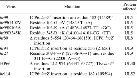
Virus Mutation Protein
affected
hr99 ICP6::lacZbinsertion at residue 182 (14589)c UL5
hr99G102V Residue 102-G3V (14829-T3A) UL5 hr99K103A Residue 103-K3A (14826–14827-TT3GC) UL5 hr99R345K Residue 345-R3K (14100–14101-CG3TT) UL5 hr80 Dresidues 5–554 (20464–188150), ICP6::lacZ
insertion
UL8
hr94 ICP6::lacZ insertion at residue 536 (21656) UL9 hr27 Residue 309-F3Y (22336-A3T) and residue
311-E3G (22330-A3G)
UL9
HP66 Dresidues 212–974 (63441–65727), TK::lacZd
insertion
UL30
hr114 ICP6::lacZ insertion at residue 182 (109594) UL52
a
See Materials and Methods for references. b
lacZ gene under control of the HSV-1 ICP6 promoter.
c
Sequence coordinate (24). d
lacZ gene under control of the HSV-1 thymidine kinase (TK) promoter.
on November 9, 2019 by guest
http://jvi.asm.org/
at2708C, thawed rapidly at 378C, and sonicated for 1 min on ice. The superna-tants were clarified, and the titers on permissive cells were determined as follows:
hr99 on the UL5-expressing cell line L2-5 (39), hr80 on the UL8-expressing cell
line SL8 6-6 (32a), and hr114 on the UL52-expressing cell line BL-1 (13). Indirect immunofluorescence. For transfection-superinfection experiments, 1.03106
Vero cells were transfected with 8mg (total) of plasmid DNA as described above for the transient complementation assay. Cells were grown on coverslips, adsorbed for 1 h with 10 to 20 PFU of the desired virus per cell, and incubated for 5.5 h postadsorption in the presence or absence of ACG (100mM) or PAA (400mg/ml). When indicated, cells were labeled with 1 mM BrdU for 15 min prior to fixation. Cells were fixed in 37% buffered formaldehyde (Sigma) diluted 1:10 in phosphate-buffered saline (PBS [pH 7.4]) for 30 min and perme-abilized with 1.0% Triton X-100 for 10 min. If cells had been labeled with BrdU, they were treated with 4 M HCl for 10 min. Cells were reacted with primary antibodies in 3% normal goat serum in PBS for 30 min. Anti-AU1 ascites were used at 1:1,000, anti-EE ascites were used at 1:500, anti-KT3 ascites were used at 1:500, anti-BrdU antibodies were used at 1:50, and 3-83 was used at 1:1,000 as indicated. Cells were reacted with secondary antibodies in 3% normal goat serum in PBS at a 1:200 dilution for 30 min. Coverslips were mounted in glycerol gelatin containing 2.5% DABCO to retard bleaching.
Imaging.Cells stained for immunofluorescence were imaged on a Zeiss Ax-iovert 135 laser scanning microscope (confocal) equipped with a Zeiss3630 Plan Neofluar objective. Collected images were arranged and labeled with a Silicon Graphics Work station equipped with Adobe Photoshop 3.0.
RESULTS
Constructs encoding epitope-tagged UL5, UL8, and UL52
produce stable, functional proteins.
To facilitate studies of the
localization of the helicase-primase complex subunits during
infection, we added sequences to each subunit that encode
peptide epitopes which can be recognized by monoclonal
an-tibodies. To confirm that these constructs produced stable
pro-teins that could be detected immunologically, Vero cells were
transfected with expression clones, superinfected, and
pro-cessed for Western blot (immunoblot) analysis. Each of the
constructs produces stable protein that is of the expected
mo-lecular weight and which is recognized both by the monoclonal
antibody recognizing its epitope tag and by its cognate
poly-clonal antibody (data not shown).
Each of the epitope-tagged proteins was found to be
func-tional by two approaches. First, the tagged proteins were
ca-pable of complementing their respective null mutant viruses
for replication of an HSV-1 origin-containing plasmid (data
not shown). Second, each of the tagged constructs was tested in
a transient complementation assay. This assay assesses the
ability of the epitope-tagged proteins, expressed from the
HSV-1-inducible ICP6 promoter on a transfected plasmid, to
complement the yield of its cognate null virus. Vero cells were
transfected with plasmids which express either the native or
tagged version of each gene (UL5, UL8, or UL52) and
subse-quently were superinfected with their cognate null mutant
vi-ruses (Table 2). All constructs complemented their respective
null mutant viruses efficiently. The biological significance of
the differences in complementation indices between the tagged
and native proteins is unclear. We cannot rule out the
possi-bility that the addition of the epitope tags to the proteins might
subtly affect overall virus yields. However, it appears that each
is functional for the complementation of its cognate null
mu-tant virus.
Localization of epitope-tagged UL5, UL8, and UL52 in
in-fected cell nuclei.
UL29, UL30, UL42, and UL9 all localize to
replication compartments (14, 30, 31). Olivo et al. were unable
to detect a subnuclear localization of UL5, UL8, and UL52
with polyclonal antisera in an indirect immunofluorescence
assay, perhaps because of the low sensitivity of the antisera
they used (30). We reasoned that the members of the
helicase-primase complex should localize to these sites of viral DNA
synthesis since they are essential replication proteins. To test
this hypothesis, the epitope-tagged constructs described above
were used in an indirect immunofluorescence assay to examine
the localization of the helicase-primase complex in infected
nuclei.
Previous studies suggested that efficient translocation of the
helicase-primase complex into the nucleus required
coexpres-sion of all of its subunits (3). Indeed, Vero cells that were
transfected with p6AU1UL5 alone and superinfected with hr99
exhibited extensive cytoplasmic staining for the AU1 epitope in
an immunofluorescence assay (data not shown). Accordingly,
transfection experiments were designed to ensure that each of
the helicase-primase complex subunits was expressed to a
sim-ilar level. For instance, in order to study the localization of
AU1-UL5 in infected nuclei, cells were cotransfected with
p6AU1UL5, p6UL8, and p6UL52 and superinfected with hr99.
Cells on coverslips were stained for immunofluorescence with
the polyclonal antiserum 3-83, which recognizes UL29, and a
monoclonal antibody which recognizes the AU1 epitope of
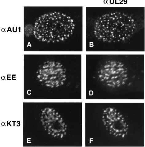
AU1-UL5 as described in Materials and Methods. Figure 1B
shows that UL29 localizes to typical replication compartments.
Figure 1A shows the AU1 staining pattern in the same cell.
The concordant patterns of staining demonstrate that
AU1-UL5 and UL29 colocalize in replication compartments. Within
the large globular replication compartments, substructures can
be seen as previously reported for UL29 (8). EE-UL8 and
UL52-KT3 were also shown to colocalize with UL29 in
repli-cation compartments in a similar manner. Figure 1C and D
illustrate the colocalization of EE-UL8 and of UL29,
respec-tively. Figure 1E and F demonstrate that UL52-KT3 and
UL29, respectively, colocalize in replication compartments.
To determine if the members of the helicase-primase
com-plex also colocalize with UL29 in prereplicative sites, these
experiments were repeated in the presence of PAA. Each of
the tagged proteins colocalizes with UL29 in prereplicative
sites (Fig. 2). Prereplicative sites are distributed throughout
the nucleus, whereas replication compartments appear to be
restricted in their distribution (compare Fig. 1 and 2). To
ensure that the epitope tag staining was not simply spurious
signal resulting from the overflow of fluorescein emissions into
the Texas red channel, cells were also stained with the
mono-clonal antibodies alone. The staining pattern for each was
typical of prereplicative sites (data not shown). In addition,
colocalization was also observed when the secondary antibody
fluors were switched (data not shown). In summary, the
epitope-tagged proteins proved to be useful reagents for
dem-onstrating that UL5, UL8, and UL52 colocalize with UL29
both in replication compartments and in prereplicative sites.
Furthermore, this is the first demonstration of
helicase-pri-mase complex localization to replication compartments and
prereplicative sites.
Prereplicative site formation in viral mutants which fail to
express UL5, UL8, UL52, or UL9.
UL29 is essential for
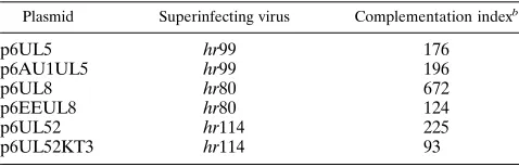
pre-TABLE 2. Complementation of hr99, hr80, and hr114 withnative and epitope-tagged constructsa
Plasmid Superinfecting virus Complementation indexb
p6UL5 hr99 176
p6AU1UL5 hr99 196
p6UL8 hr80 672
p6EEUL8 hr80 124
p6UL52 hr114 225
p6UL52KT3 hr114 93
aVero cells were transfected with the indicated plasmid and superinfected
with the indicated virus. The results of two experiments were averaged. bComplementation index for each virus is determined as the PFU of the null
mutant from cells transfected with the indicated plasmid/PFU of the null mutant from mock-transfected cells.
on November 9, 2019 by guest
http://jvi.asm.org/
[image:3.612.58.297.92.169.2]replicative site formation (7). However, it is not sufficient for
their formation; UL29 is diffusely distributed in cell nuclei
transfected with a UL29 expression clone (10) (data not
shown). We hypothesize that other essential replication
pro-teins may be necessary for prereplicative site formation. In
order to determine if the helicase-primase subunits or UL9 was
essential for prereplicative site formation, cells infected with
hr80, hr99, hr114, or hr94 (UL8, UL5, UL52, and UL9 null
mutant viruses, respectively) were examined for UL29 staining
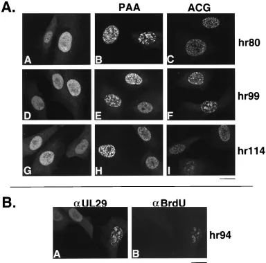
by indirect immunofluorescence. In cells infected with hr80,
hr99, or hr114, UL29 staining was diffuse and nuclear (Fig. 3A,
panels A, D, and G). When the infections were carried out in
the presence of PAA, greater than 50% of the cells contained
what appeared to be prereplicative sites (Fig. 3A, panels B, E,
and H; Fig. 3B, panel A). To demonstrate that the formation
of prereplicative sites was not due to an unusual effect of PAA
treatment, UL29 staining patterns were also examined in the
presence of ACG, another specific inhibitor of HSV
poly-merase. Similar results were obtained (Fig. 3A, panels C, F,
and I). Cells infected with hr94 behaved like cells infected with
the helicase-primase null mutants (described below). Thus,
UL29 is distributed diffusely in the nuclei of cells infected with
viruses that fail to express UL5, UL8, UL52, or UL9. In the
presence of PAA or ACG, however, UL29 localizes to
prerep-licative sites in cells infected with these mutants.
One of the characteristics of prereplicative sites formed in
drug-treated KOS-infected cells is that UL29 staining
colocal-izes with incorporated BrdU at sites thought to represent areas
of cellular replication (7). To determine whether the
prerep-licative sites described above also stain with BrdU,
mutant-infected cells were treated with BrdU for 15 min prior to
fixation and were doubly stained with a monoclonal antibody
which recognizes BrdU and with 3-83. In Fig. 3B, panels A and
B show the BrdU and 3-83 staining patterns, respectively, in a
group of cells infected with hr94 in the presence of PAA.
Although three cells are visible in Fig. 3, two exhibit diffuse
nuclear staining for UL29 and no BrdU staining, and one
exhibits colocalization of UL29 and BrdU incorporation in
prereplicative sites. Similar results were observed in KOS- and
the other null mutant-infected cells (data not shown). Thus
UL29 exhibits a prereplicative site staining pattern only in cells
which also incorporate BrdU. This is in contrast with previous
studies which concluded that some proportion of
KOS-in-fected cells treated with PAA contained prereplicative sites but
no BrdU staining (7). This discrepancy may reflect a difference
in staining conditions.
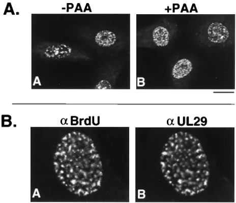
[image:4.612.57.299.75.322.2]A polymerase null mutant virus forms prereplicative sites in
the absence of PAA.
Prereplicative sites can be found in the
nuclei of cells infected with temperature-sensitive UL30 or
temperature-sensitive UL42 mutants at the nonpermissive
temperature or with a null mutant in UL30 (2, 14, 31). Thus,
inactivation of the polymerase complex with drugs or by means
of mutation leads to prereplicative site formation. To
deter-mine whether the prereplicative sites observed in cells infected
with a polymerase null mutant also stained with BrdU, Vero
cells were infected with HP66, which contains a
deletion/inser-tion mutadeletion/inser-tion in UL30, and stained with 3-83 and BrdU.
Pre-replicative sites were present in both the absence (Fig. 4A,
FIG. 1. Cotransfected-superinfected cells doubly labeled with 3-83 andmonoclonal antibodies recognizing epitope-tagged UL5, UL8, and UL52. Vero cells were cotransfected and superinfected as described in Materials and Meth-ods. Cells were fixed and permeabilized at 5.5 h postsuperinfection as described in Materials and Methods. (A and B) Vero cells were cotransfected with p6AU1UL5, p6UL8, and p6UL52 and superinfected with hr99. (C and D) Vero cells were cotransfected with p6UL5, p6EEUL8, and p6UL52 and superinfected with hr80. (E and F) Vero cells were cotransfected with p6UL5, p6UL8, and p6UL52KT3 and superinfected with hr114. (A, C, and E) Staining for rhoda-mine-conjugated goat anti-mouse secondary antibodies (a) reacting with, respec-tively, AU1 ascites, EE ascites, and KT3 ascites. (B, D, and F) Staining pattern of 3-83 as recognized by fluorescein isothiocyanate-conjugated goat anti-rabbit secondary antibody in the same cells. Marker bar, 15mm.
FIG. 2. Cotransfected-superinfected cells grown in the presence of PAA and doubly labeled with 3-83 and monoclonal antibodies recognizing epitope-tagged UL5, UL8, and UL52. Cells were cotransfected, superinfected, and processed as described in the legend to Fig. 1, except that superinfection was carried out in the presence of 400mg of PAA per ml. (A, C, and E) Staining for Texas red-conjugated goat anti-mouse secondary antibodies (a) reacting with, respectively, AU1 ascites, EE ascites, and KT3 ascites. (B, D, and F) Staining pattern of 3-83 as recognized by fluorescein isothiocyanate-conjugated goat anti-rabbit second-ary antibody in the same cells. Marker bar, 15mm.
on November 9, 2019 by guest
http://jvi.asm.org/
[image:4.612.317.553.410.650.2]panel A) and the presence (Fig. 4A, panel B) of PAA. BrdU
and UL29 colocalize in these sites (Fig. 4B, panels A and B,
respectively). This is the first demonstration of UL29 and
BrdU colocalization in cells infected with a polymerase null
mutant. We therefore conclude, in agreement with Bush et al.
(2), that the polymerase polypeptide itself is not necessary for
prereplicative site formation.
Viruses containing inactivating point mutations in UL5 and
UL9 helicase motifs fail to form prereplicative sites in the
absence of PAA.
The results presented above suggest that
in-hibition of the viral DNA polymerase results in the formation
of prereplicative sites. To determine if the inhibition of HSV
replication by other methods which do not involve the absence
of an essential replication protein also results in
prereplica-tive site formation, a number of viral mutants with subtle
mutations in UL5 or UL9 were examined. The UL5 gene
con-tains six well-conserved helicase motifs. Single-amino-acid
sub-stitutions of highly conserved residues in each of the motifs
result in the loss of function of the UL5 protein as assessed in
an in vivo replication complementation assay (38). Three of
these mutations were introduced into the HSV-1 genome, and
the resulting viruses are replication defective: hr99G102V and
hr99K103A contain single-amino-acid substitutions in helicase
motif I, and hr99R345K contains a single-amino-acid
substitu-tion in helicase motif IV (38). These mutants produce stable
mutant UL5 proteins that are capable of interacting with UL8
and UL52 in an immunoprecipitation assay, implying that their
global conformation is not grossly altered (38).
Vero cells infected with hr99G102V exhibit a diffuse nuclear
staining for UL29 (Fig. 5A), whereas hr99G102V-infected cells
treated with PAA exhibit a staining pattern for UL29 typical of
prereplicative sites (Fig. 5B). Cells infected with hr99K103A
and hr99R345K behave identically (data not shown). Similar
experiments were carried out with hr27, a viral mutant which
contains two amino acid substitutions in the conserved helicase
domain IV of UL9 (21). This mutant is replication defective
but produces a UL9 protein with the predicted molecular
weight. Vero cells infected with hr27 in the absence of PAA
show a diffuse nuclear staining for UL29 (Fig. 5C). However,
treatment of hr27-infected cells with PAA results in the
local-FIG. 3. UL29 localization in cells infected with viral mutants that fail to express UL5, UL8, UL52, or UL9. Vero cells were infected with 20 PFU of virus per cell and processed as described in Materials and Methods. (A) Cells were stained with 3-83 and fluorescein isothiocyanate-conjugated goat anti-rabbit secondary antibody. Cells were infected with the following mutants: panels A, B, and C, hr80; D, E, and F, hr99; G, H, and I, hr114. Infected cells in panels B, E, and H were treated with PAA, and those in panels C, F, and I were treated with ACG. Marker bar, 15mm. (B) hr94-infected cell doubly stained with antibodies (a) for UL29 and BrdU. Panels A and B show the same group of hr94-infected cells doubly stained, respectively, with 3-83 (fluorescein isothiocyanate-conjugated goat anti-mouse secondary antibody) and anti-BrdU (Texas red-conjugated goat anti-mouse secondary antibody). Marker bar, 15mm.on November 9, 2019 by guest
http://jvi.asm.org/
[image:5.612.114.498.72.452.2]ization of UL29 to prereplicative sites in greater than 50% of
infected nuclei (Fig. 5D). ACG treatment of cells infected with
the UL5 or UL9 helicase motif mutants resulted in
prerepli-cative site formation in a similar proportion of cells (data not
shown). In the presence of either drug, prereplicative sites
colabel with BrdU. We conclude that inhibition of viral DNA
synthesis by subtle mutation of UL5 or UL9 is not sufficient to
induce prereplicative site formation unless the viral
poly-merase is also inhibited.
DISCUSSION
Using epitope-tagged versions of UL5, UL8, and UL52, we
have demonstrated that the helicase-primase complex
colocal-izes with UL29 in replication compartments and prereplicative
sites in HSV-1-infected nuclei. With this observation, all seven
of the essential replication genes have now been shown to be
present in replication compartments. To determine if the
he-licase-primase complex and UL9 play a role in prereplicative
site formation, we examined UL29 staining patterns in cells
infected with various replication-defective viruses (see Table 3
for summary). The UL29 staining pattern of cells infected with
null mutants of UL5, UL8, UL9, and UL52 is diffuse nuclear.
In the presence of PAA and ACG, prereplicative sites
exhib-iting colocalized UL29 and BrdU staining were observed in
greater than 50% of the cells. The remaining cells exhibit
diffuse UL29 staining and no BrdU incorporation. One
expla-nation for the observation that prereplicative sites are not seen
in 100% of infected cells is that prereplicative site formation
requires cells to be competent to carry out cellular DNA
syn-thesis. This may imply a requirement for a natural or induced
progression into S phase, although we cannot rule out that
BrdU incorporation is not due to repair synthesis.
The fact that prereplicative sites can form in
mutant-in-fected cells treated with PAA or ACG suggests that their
formation may not require the UL5, UL8, UL52, or UL9
proteins. These observations are inconsistent with the idea that
prereplicative sites represent intermediates in the assembly of
the viral replication apparatus. However, it is possible that viral
polymerase inhibitors alter the cellular milieu such that
pre-replicative sites can form in the absence of viral factors that
would normally be necessary. Under this scenario, the fact that
UL29 does not localize to prereplicative sites in cells infected
with hr80, hr99, hr114, and hr94 unless polymerase inhibitors
are added suggests that these four replication proteins are
essential for prereplicative site formation (19a). However, our
observation that prereplicative sites do not form in cells
in-fected with subtle motif mutants of UL5 and UL9 in the
ab-sence of polymerase inhibitors implies that these viral proteins
are probably not essential for their formation.
[image:6.612.63.295.71.271.2]A more likely explanation for these results is that an active
viral polymerase complex prevents or inhibits prereplicative
site formation. Several mechanisms can be considered. Active
polymerase may affect or interact with some factor, viral or
cellular, which is necessary for prereplicative site formation.
An attractive candidate factor is UL29, which is essential for
prereplicative site formation (2, 7). Perhaps an active
poly-merase complex prevents UL29 from acting as a focus for
FIG. 4. UL29 localization in HP66-infected cells. Vero cells were infectedwith 20 PFU of virus per cell and processed as described in Materials and Methods. (A) Panels A and B show 3-83 staining as detected by fluorescein isothiocyanate-conjugated goat anti-rabbit secondary antibody in cells infected in the presence (1) and absence (2) of PAA, respectively. Marker bar, 15mm. (B) HP66-infected cell doubly stained with antibodies (a) for BrdU and UL29. Panels A and B show a cell nucleus doubly stained, respectively, with anti-BrdU (Texas red-conjugated goat anti-mouse secondary antibody) and 3-83 (fluores-cein isothiocyanate-conjugated goat anti-mouse secondary antibody). Marker bar, 15mm.
[image:6.612.315.555.91.219.2]FIG. 5. UL29 localization in cells infected with hr99G102V or hr27. Vero cells were infected with 20 PFU of hr99G102V (A and B) or hr27 (C and D) per cell and processed as described in Materials and Methods. Cells were stained with 3-83 and fluorescein isothiocyanate-conjugated goat anti-rabbit secondary antibody. Infected cells in panels B and D were treated with PAA, while those in panels A and B were not. Marker bar, 15mm.
TABLE 3. Prereplicative site formation in replication-defective viruses
Virus
Prereplicative site formation
2PAA 1PAA
KOS RCa 1
hr99 2 1
hr99G102V 2 1
hr99K103A 2 1
hr99R345K 2 1
hr80 2 1
hr94 2 1
hr27 2 1
hr114 2 1
HP66 1 1
a
RC, forms replication compartments.
on November 9, 2019 by guest
http://jvi.asm.org/
[image:6.612.60.296.504.672.2]prereplicative site formation or acts to disrupt these sites.
Spe-cifically, active viral polymerase may remove UL29 that is
associated with cellular single-stranded DNA. Our observation
that UL29 in prereplicative sites is always associated with
cel-lular DNA replication lends indirect support to the concept
that UL29 may be bound to single-stranded DNA.
Accord-ingly, when the polymerase complex is inactivated, UL29
would remain bound to single-stranded DNA, thereby forming
prereplicative sites. Interestingly, there is genetic evidence for
an interaction between UL29 and the polymerase complex (5).
Clearly, all of the factors and conditions necessary for
pre-replicative site formation have yet to be identified. Cells
trans-fected with UL29 alone only rarely display nuclear structure
when examined with anti-UL29 antibodies (11). In contrast,
nuclear structures form in cells cotransfected with UL5, UL8,
UL52, and UL29, which resemble prereplicative sites
(unpub-lished observations). This suggests that UL5, UL8, and UL52
can mediate UL29 localization in transfected cells. This
obser-vation may seem contradictory to the model presented above,
which suggests that UL5, UL8, and UL52 cannot be
consid-ered necessary for prereplicative site formation. However,
pre-replicative sites may form in two or more different ways. It is
possible that in the absence of an active helicase-primase
com-plex, another protein may be capable of mediating
prereplica-tive site formation. The presence of this ‘‘other factor’’ would
explain the formation of prereplicative sites in hr80-, hr99-, and
hr114-infected cells. One attractive candidate for the other
factor is UL9. Like UL5 in the helicase-primase complex, UL9
contains conserved helicase motifs that are essential for its
ability to catalyze origin-dependent DNA replication in vivo
(23). Furthermore, UL9 has been shown to directly interact
with UL29 (1). Perhaps either one of the HSV-1 helicases,
UL5 in the helicase-primase complex or UL9, could
indepen-dently mediate the localization of UL29 to prereplicative sites.
The putative requirement for one active helicase may reflect
the need to expose single-stranded DNA or may reflect
con-formational changes which occur upon interaction between
various members of the DNA synthetic machinery.
The possibility that cellular factors may play a role in
pre-replicative site formation and replication compartment
forma-tion should also be considered. Wilcock and Lane have
dem-onstrated that a number of host replication proteins, as such as
proliferating cell nuclear antigen and DNA polymerase
a
,
co-localize with UL29 both in prereplicative sites and in
replica-tion compartments (36). Currently, there is no in vitro system
for HSV-1 genome replication. This may indicate the need for
host factors in addition to the essential viral replication
pro-teins. Until other factors are definitively identified that
partic-ipate in prereplicative site formation, it will be difficult to
conclusively determine whether or not they represent
biologi-cally relevant subassemblies of replication proteins.
Neverthe-less, the study of these structures in infected cells and
trans-fection systems may prove to be a powerful tool for
understanding the process of HSV-1 DNA replication.
ACKNOWLEDGMENTS
We thank Steven Pfeiffer, David Knipe, and the members of our laboratory for critically reviewing the manuscript. We thank the fol-lowing individuals for plasmid construction: Ajay Malik, p6NBam; Rich Zhou, p6UL5119 and pUC19UL5; John Shanley, p6UL8; Ke-Feng Qin, EEUL8; Kevin Nawotka, UL52KT3 and pAPV, and Gor-don McLean, p6UL52, pGM3/UL52, and pGM7/UL8. We are grateful to David Knipe for providing the 3-83 antiserum and for communicat-ing results regardcommunicat-ing the behavior of UL29 in cells infected with hr80, hr99, hr114, and hr94. We are grateful to Donald Coen for providing HP66.
This investigation was supported by Public Health Service grant A121747. S.K.W. is the recipient of an American Heart Association-Genentech Established Investigator Award.
REFERENCES
1. Boehmer, P. E., and I. R. Lehman. 1993. Physical interaction between the herpes simplex virus 1 origin-binding protein and single-stranded DNA-binding protein ICP8. Proc. Natl. Acad. Sci. USA 90:8444–8448. 2. Bush, M., D. R. Yager, M. Gao, K. Weisshart, A. I. Marcy, D. M. Coen, and
D. M. Knipe.1991. Correct intranuclear localization of herpes simplex virus DNA polymerase requires the viral ICP8 DNA-binding protein. J. Virol. 65: 1082–1089.
3. Calder, J. M., E. C. Stow, and N. D. Stow. 1992. On the cellular localization of the components of the herpes simplex virus type 1 helicase-primase complex and the viral origin-binding protein. J. Gen. Virol. 73:531–538. 4. Challberg, M. D. 1986. A method for identifying the viral genes required for
herpesvirus DNA replication. Proc. Natl. Acad. Sci. USA 83:9094–9098. 5. Chiou, H. C., S. K. Weller, and D. M. Coen. 1985. Mutations in the herpes
simplex virus major DNA-binding protein gene leading to altered sensitivity to DNA polymerase inhibitors. Virology 145:213–226.
6. Crute, J. J., T. Tsurumi, L. A. Zhu, S. K. Weller, P. D. Olivo, M. D. Challberg, E. S. Mocarski, and I. R. Lehman.1989. Herpes simplex virus 1 helicase-primase: a complex of three herpes-encoded gene products. Proc. Natl. Acad. Sci. USA 86:2186–2189.
7. de Bruyn Kops, A., and D. M. Knipe. 1988. Formation of DNA replication structures in herpes virus-infected cells requires a viral DNA binding pro-tein. Cell 55:857–868.
8. de Bruyn Kops, A., and D. M. Knipe. 1994. Preexisting nuclear architecture defines the intranuclear location of herpesvirus DNA replication structures. J Virol. 68:3512–3526.
9. Digard, P., and D. M. Coen. 1990. A novel functional domain of an alpha-like DNA polymerase. The binding site on the herpes simplex virus poly-merase for the viral UL42 protein. J. Biol. Chem. 265:17393–17396. 10. Gao, M., J. Bouchey, K. Curtin, and D. M. Knipe. 1988. Genetic
identifica-tion of a poridentifica-tion of the herpes simplex virus ICP8 protein required for DNA-binding. Virology 163:319–329.
11. Gao, M., and D. M. Knipe. 1992. Distal protein sequences can affect the function of a nuclear localization signal. Mol. Cell. Biol. 12:1330–1339. 12. Goldstein, D. J., R. Toyama, R. Dhar, and R. Schlegel. 1992. The BPV-1 E5
oncoprotein expressed in Schizosaccharomyces pombe exhibits normal bio-chemical properties and binds to exogenous 16-kDa component of the vac-uolar proton-ATPase. Virology 190:889–893.
13. Goldstein, D. J., and S. K. Weller. 1988. An ICP6::lacZ insertional mutagen is used to demonstrate that the UL52 gene of herpes simplex virus type 1 is required for virus growth and DNA synthesis. J. Virol. 62:2970–2977. 14. Goodrich, L. D., P. A. Schaffer, D. I. Dorsky, C. S. Crumpacker, and D. S.
Parris.1990. Localization of the herpes simplex virus type 1 65-kilodalton DNA-binding protein and DNA polymerase in the presence and absence of viral DNA synthesis. J. Virol. 64:5738–5749.
15. Graham, F. L., and A. J. van der Eb. 1973. A new technique for the assay of infectivity of human adenovirus 5 DNA. Virology 52:456–467.
16. Grussenmeyer, T., K. H. Scheidtmann, M. A. Hutchinson, W. Eckhart, and G. Walter.1985. Complexes of polyoma virus medium T antigen and cellular proteins. Proc. Natl. Acad. Sci. USA 82:7952–7954.
17. Heilbronn, R., and H. zur Hausen. 1989. A subset of herpes simplex virus replication genes induces DNA amplification within the host cell genome. J. Virol. 63:3683–3692.
18. Hernandez, T. R., and I. R. Lehman. 1990. Functional interaction between the herpes simplex-1 DNA polymerase and UL42 protein. J. Biol. Chem. 265:11227–11232.
19. Knipe, D. M., D. Senechek, S. A. Rice, and J. L. Smith. 1987. Stages in the nuclear association of the herpes simplex virus transcriptional activator pro-tein ICP4. J. Virol. 61:276–284.
19a.Liptak, L. M., S. L. Uprichard, and D. M. Knipe. 1996. Functional order of assembly of herpes simplex virus DNA replication proteins into prereplica-tive site structures. J. Virol. 70:1759–1767.
20. MacArthur, H., and G. Walter. 1984. Monoclonal antibodies specific for the carboxy terminus of simian virus 40 large T antigen. J. Virol. 52:483–491. 21. Malik, A. K., R. Martinez, L. Muncy, E. P. Carmichael, and S. K. Weller.
1992. Genetic analysis of the herpes simplex virus type 1 UL9 gene: isolation of a LacZ insertion mutant and expression in eukaryotic cells. Virology 190: 702–715.
22. Marcy, A. I., D. R. Yager, and D. M. Coen. 1990. Isolation and character-ization of herpes simplex virus mutants containing engineered mutations at the DNA polymerase locus. J. Virol. 64:2208–2216.
23. Martinez, R., L. Shao, and S. K. Weller. 1992. The conserved helicase motifs of the herpes simplex virus type 1 origin-binding protein UL9 are important for function. J. Virol. 66:6735–6746.
24. McGeoch, D. J., M. A. Dalrymple, A. J. Davison, A. Dolan, M. C. Frame, D. McNab, L. J. Perry, J. E. Scott, and P. Taylor.1988. The complete DNA sequence of the long unique region in the genome of herpes simplex virus type 1. J. Gen. Virol. 69:1531–1574.
on November 9, 2019 by guest
http://jvi.asm.org/
25. McLean, G. W., A. P. Abbotts, M. E. Parry, H. S. Marsden, and N. D. Stow. 1994. The herpes simplex type 1 origin-binding protein interacts specifically with the viral UL8 protein. J. Gen. Virol. 75:2699–2706.
26. Nakamaru, H., T. Morita, and C. Sato. 1986. Structural organization of replicon domains during DNA synthetic phase in the mammalian nucleus. Exp. Cell Res. 165:291–297.
27. Nakayasu, H., and R. Berezny. 1989. Mapping replicational sites in the eucaryotic nucleus. J. Cell Biol. 108:1–11.
28. O’Donnell, M. E., P. Elias, B. E. Funnell, and I. R. Lehman. 1987. Interac-tion between the DNA polymerase and single-stranded DNA-binding pro-tein (infected cell propro-tein 8) of herpes simplex virus 1. J. Biol. Chem. 262: 4260–4266.
29. Olivo, P. D., and M. D. Challberg. 1990. Functional analysis of the herpes simplex virus gene products involved in DNA replication, p. 137–150. In E. Wagner (ed.), Herpesvirus transcription and its regulation. CRC Press, Boca Raton, Fla.
30. Olivo, P. D., N. J. Nelson, and M. D. Challberg. 1989. Herpes simplex virus type 1 gene products required for DNA replication: identification and over-expression. J. Virol. 63:196–204.
31. Quinlan, M. P., L. B. Chen, and D. M. Knipe. 1984. The intranuclear location of a herpes simplex virus DNA-binding protein is determined by the status of viral DNA replication. Cell 36:857–868.
32. Ruyechan, W. T., and A. C. Weir. 1984. Interaction with nucleic acids and stimulation of the viral DNA polymerase by the herpes simplex virus type 1 major DNA-binding protein. J. Virol. 52:727–733.
32a.Shao, L., J. D. Shanley, and S. K. Weller. Unpublished observations. 33. Weller, S. K. 1990. Genetic analysis of HSV genes required for genome
replication, p. 105–135. In E. Wagner (ed.), Herpesvirus transcription and its regulation. CRC Press, Boca Raton, Fla.
34. Weller, S. K., E. P. Carmichael, D. P. Aschman, D. J. Goldstein, and P. A. Schaffer.1987. Genotypic and phenotypic characterization of mutants in four essential genes that map to the left half of HSV-1 UL DNA. Virology 161:198–210.
35. Weller, S. K., K. J. Lee, D. J. Sabourin, and P. A. Schaffer. 1983. Genetic analysis of temperature-sensitive mutants which define the gene for the major herpes simplex virus type 1 DNA-binding protein. J. Virol. 45:354– 366.
36. Wilcock, D., and D. P. Lane. 1991. Localization of p53, retinoblastoma and host replication proteins at sites of viral replication in herpes-infected cells. Nature (London) 349:429–431.
37. Wu, C. A., N. J. Nelson, D. J. McGeoch, and M. D. Challberg. 1988. Iden-tification of herpes simplex virus type 1 genes required for origin-dependent DNA synthesis. J. Virol. 62:435–443.
38. Zhu, L., and S. K. Weller. 1992. The six conserved helicase motifs of the UL5 gene product, a component of the herpes simplex virus type 1 helicase-primase, are essential for its function. J. Virol. 66:469–479.
39. Zhu, L., and S. K. Weller. 1992. The UL5 gene of herpes simplex virus type 1: isolation of a lacZ insertion mutant and association of the UL5 gene product with other members of the helicase-primase complex. J. Virol. 66: 458–468.