JOURNALOFVIROLOGY,Oct.1970,p.463-469
Copyright @ 1970 American Society for Microbiology
Vol. 6, No. 4 Printed in U.S.A.
Sedimentation
Properties
of
Simian
Virus
40-Specific
Ribonucleic
Acid Present
in
Green
Monkey
Cells
During
Productive Infection
and
in
Mouse
Cells
Undergoing Abortive Infection
MALCOLM A. MARTINAND JANET C. BYRNELaboratory of Biology of Viruses, National Institute of Allergy andInfec.iousDiseases, NationalInstitutes of
Health, Bethesda, Maryland20014
Received forpublication23 June1970
The size distribution of polyribosome-associated simian virus 40 (SV40)
ribo-nucleic acid (RNA) was examined at various times after productive infection.
Eight hours after infection, virus-specific RNAwasdetectedin the 14to17Sregion
ofa sucrose gradient by deoxyribonucleic acid (DNA)-RNA hybridization; RNA
present in fractions sedimenting morerapidly did not react with SV40 DNA. At
successively later times, SV40 RNA was detected in more rapidly sedimenting
regions. By 24 hr,aportion of the SV40 RNAwasdetected in the 28S region,
sedi-menting slightly more rapidly than a MS2 RNA marker. Nuclear SV40 RNA,
prepared from cells 48 hr after infection, was distributed in more rapidly
sedi-menting regions of the gradient, peaking atabout 32to 34S. Some nuclear
virus-specific RNA could be detected in the 45 to50S region. During the abortive
infec-tion of mouse cells, the sedimentation profile of SV40 RNA was very similar to
that observed during the early phases of the lytic cycle.
Simian virus 40 (SV40)-specific ribonucleic
acid (RNA) has been detected in greenmonkey
cells undergoing lytic infection (1) as well as in
cellstransformedby thisagent (1, 11, 15). When
total cellularRNAis used tomonitorSV40 gene
activity during productive infection, it has been
shownthat50%oftheviralgenome istranscribed
(10).Noattempt was made at that time to
deter-mine whether this RNA was polycistronic or
consisted ofaseries ofgeneproducts,the sumof
which was
separately
transcribedfrom oneSV40deoxyribonucleic acid (DNA) strand. It has
been
previously
shownthatduring polyoma infec-tion 50%ofthe viral DNAis transcribed(9), and virus-specificRNA ispresent with sedimentationproperties consistent with polycistronic
trans-cription of one DNA strand-1.6 X 10⁶ daltons
(5). In both cases, DNA transcription was
monitored with radiolabeled total cellular RNA.
Virus-specific RNA associated with
cyto-plasmic polyribosomesis the RNA form whiCh is
actively involved in thesynthesis of the proteins
necessary for productive infection. For this
rea-son, we decided toexamine this species of
cyto-plasmic SV40 RNA atvarious times during the
lytic cycle. Ourresults indicatethatearlyin
infec-tion only
slowly
sedimenting RNA forms arepresent. Later, progressively larger SV40-specific
RNA molecules were observed, a portion of
which had sedimentation properties compatible
with the transcription of one SV40 strand.
MATERIALS AND METHODS
Preparation of SV40 DNA. Confluentmonolayers
of Vero (3) cells,growingin1-gal roller bottles,were
infected with small-plaque SV40 virus at a
multi-plicity of 0.25 to 0.50 plaque-forming units (PFU)/
cell.Cells weregenerallyharvested 6 to7 daysafter
infection,and thevirus was purified as previously
de-scribed (10). SV40 DNA was released by exposing
purified virus preparationsto1%sodium dodecyl
sul-fate (SDS) and 0.01 M ethylenediaminetetraacetic
acid (EDTA), pH 7.5, at 50 Cfor 30 min. The SDS
wasremoved byprecipitation withCsCl(1g/ml;18),
andthe DNAwascentrifuged inano. 40fixed-angle
Spincorotorfor 60hr at38,000rev/minat5C,in the
presence of ethidium bromide [200 ,ug/ml (14)].
SV40 DNAI wascollected withaPasteur pipette,and the ethidum bromide was removed by
chromatog-raphy on Dowex-50 equilibrated with one-tenth standard saline citrate (SSC).The DNA wasfurther
purified by gel filtration on G-100 Sephadex
equi-librated with0.1MNaCl.
Preparationof32P-labeledpolyribosomes fromcells
productively infected with SV40 virus. Confluent
monolayersof Verocellswereexposedtosmall-plaque
SV40 virus at a multiplicity of 25 to 50 PFU/cell.
Virus was allowedto adsorbfor 2 hrat 37C.
Phos-phate-free Eagle'sbasalno.2mediumwasaddedtothe
cells 16hrpriortotheaddition of
"2P-orthophosphate.
463
on November 11, 2019 by guest
http://jvi.asm.org/
MARTIN AND BYRNE
In one case (polyribosomes prepared from cells 8 hr afterinfection), the phosphate-free media was added 8 hrprior toinfection. The infected cells were labeled for 75 min in 50 ml of fresh phosphate-free media con-taining 50MuCiof carrier-free 32P-orthophosphate per ml. The cells were then harvested with 0.25% trypsin,
washed twice in phosphate-buffered saline and once with 0.01 Mtris(hydroxymethyl)aminomethane (Tris),
pH 7.4,0.01 MNaCl,and 0.0015 M MgCl2 [RSB (12)].
TheinfectedVerocellswereallowedtoswellin RSB
for5minat0Candlysed byDouncehomogenization
(12 strokes). Nucleiwere removed bysedimentation
at 5,000 rev/min for 10 min. Sodium deoxycholate
(0.5%), Brij 58 (0.5%; Atlas Chemical Industries,
Inc., Wilmington, Del.), and MgCl2 (0.07 M) were added, and the cytoplasmic fraction was kept in an ice-water bath for at least 90 min (6). Polyribosomes
werepellettedat17,000 X gfor 10 min,resuspended
in RSB, layered on 10 to 34% (w/v)sucrosegradients
containingRSB,andcentrifugedfor 90 minat23,000
rev/minat5 CinaSW 25.1 rotor.Fractionswere
col-lected bypuncturingthebottomof the tube, and the
polyribosomeswerepooledasindicated.
Preparationof 32P-labeledpolyribosomesfrom
unin-fected Vero cells. Vero cells (200,000cells/ml) were
added to1-galrollerbottlescontainingEagle'sbasalno. 2medium (400ml) supplementedwith 10% fetal bo-vine serum. When the cells had achieved
approxi-mately 75% oftheir final density, the medium was
changedto phosphate-free Eagle'sbasal no. 2. After 16 hr, thecellswereexposedtocarrier-free
32P-ortho-phosphate(50
IACi/ml)
in 50 ml of thephosphate-freemedia. Cellswere harvested 75 min later, and
poly-ribosomeswerepreparedasdescribed above. Preparation ofpolyribosome-associatedRNA.
Poly-ribosomeswerepooledand precipitatedwith2.5
vol-umesof95%ethanol.Thepolyribosome pelletwas
re-suspended in 0.1 MNaCl, 0.01 MTris (pH 7.5), and 0.001 M EDTAandexposedto0.5%SDSfor 2 hr. The
dissociated polyribosomes were layered on a 15 to
30% (w/v) sucrose gradient containing 0.1 M NaCl,
0.01 MTris (pH 7.5), 0.01 M EDTA, and 0.5% SDS
(SDSbuffer)andcentrifugedfor14hrat21,000rev/
min at 20 C. Exposure of the dissociated
polyribo-somes to60 Cfor5minpriortocentrifugation didnot
affect the sedimentation profile observed. Fractions
from thegradient were pooled and digested
sequen-tiallywith 25 ,ug ofelectrophoretically purified
pan-creatic deoxyribonuclease (Worthington Biochemical
Corp.) per ml and 50 ,ug of self-digested Pronase (Calbiochem) per ml, each for1 hratroom tempera-ture. The pooled fractions were then extracted with phenol, ethanol-precipitated in the presence of yeast carrier RNA (50,ug/ml), and resuspended in 0.2 to 0.3ml of one-tenthstrength SSC.
Preparationofnuclear RNA from infected Verocells.
Aconfluentmonolayerof Verocellsgrowing ina1-gal
bottle was infected with small-plaque SV40 at a
multiplicity of 25 to 50PFU/cell. Forty-eight hours
after infection,the cells were exposed to 1.0mCi of 3H-uridine (21Ci/mmole) in50mlof Eagle's basal no.
2mediumfor75min.Cellswereharvested and
frac-tionated asdescribed above. The nuclear pellet was
washed with phosphate-buffered saline, resuspended
in 0.1 M NaCl, 0.01 M sodium acetate, 0.005 M MgCl2, and0.5% SDS and digested with deoxyribonuclease
(100 ,g/ml) for 1 hrat room temperature. After a
phenolextraction at 60 C, the preparation was
pre-cipitatedwith2volumesof 95%ethanol,resuspended
inSDSbuffer, andcentrifugedasdescribed above. DNA-RNA hybridization. Small-plaque SV40 DNA I washeatedin lX SSC for 15 min and
im-mobilized on 50-mm nitrocellulose filters (type B6,
Schleicher &SchuellCo.,Keene,N.H.; reference4).
Nitrocellulose filters (7 mm), containing about 0.3
,Mg of SV40 DNA, were added toreaction mixtures containing 0.8 MNaCl, 0.001 M N-tris(hydroxymethyl)
methyl-2-aminoethanesulfonic acid (TES;
Calbio-chem, Los Angeles, Calif.), pH 7.5, 0.1% SDS, and labeled RNA in a volume of 0.25 ml. After a 16-hr incubationat 68C, the filters were exposed to pan-creatic ribonuclease (20,ug/ml) in2X SSC for 1 hr, extensively washed, dried, and counted.
RESULTS
Sedimentationproperties of SV40 RNA present
late in infection. To demonstrate that the
equiv-alent ofonestrandofSV40 DNA is transcribed during lytic infection (10), it was necessary to
prepare and pool RNA species made at various
times during the lytic cycle. The asynchronous
nature ofSV40 infection allowed preparation of
labeled polyribosomes at a time (48 hr after infection) when cells at various stagesof infection were certain to be present. Polyribosome-asso-ciated messenger RNA was isolated after
treat-ment with 0.5% SDS and sucrose gradient
centrifugation. Fractionswerepooled, purifiedas
described above, and added to DNA-RNA
hybridization reaction mixtures. Figure 1 shows the sedimentation properties of SV40-specific
RNApreparedfrom cells 48 hr after infection. It canbeseenthat,although SV40messenger RNA
is very heterogeneous in molecular size, a
sig-nificant proportion sediments in the region of
28S ribosomal RNA. The more rapidly sedi-menting SV40-specific RNAmolecules appear to
migrate slightly faster in the gradient than the
marker MS2 RNA [1.1 x 10⁶ daltons (16)].
Size distribution ofSV40 RNA at various times
during infection. At 8, 16, 24, and 48 hr after
infection with SV40, Vero cells were exposed to
32P-orthophosphate (50
XCi/ml)
for 75 min. Thepolyribosome pattern observed at each ofthese
timesis shown inFig. 2. Itshould be mentioned
that polyribosomes are virtually undetectable in
confluent uninfected Vero cells. After infection,
however, polyribosomes were readily detected,
reachinga peakat 16 hrandgradually declining
overthe next 32 hr. In eachcase, polyribosomes
wereprepared from 2 X 10⁸-infected Vero cells.
The RNAassociated with the 8-, 16-, and 24-hr
polyribosome preparations was purified as
des-464 J. VIROL.
on November 11, 2019 by guest
http://jvi.asm.org/
SEDIMENTATION OF SV40 RNA
IOOOr
800Sk
600
tL le
400H
200
C
FRACTIONJ NUJMBER
FIG. 1. Size distributioni ofSV40-specific RNA 48
hrafterinfection. 32P-labeled polyribosomes, prepared from Vero cells 48 hrafter infectioni, were mixedwith
H3MS2 RNA antdsedimented through a 15 to
30%10
(w/v) sucrose gradient containing SDS buffer. Frac-tions were collectedby puncturinzg the bottom ofthe
centrifuge tube andwerecheckedfor absorbantceat 260
tim.Samples ofeachfractionz wereprecipitatedwith 5% trichloracetic acidtodeterminetheposition oftheMS2 marker (0). Thefraciton2s were pooled in groups of thiree, purifiedasdescribed ini thetext,antdassayed for
the presentce ofSV40 RNA (-) by DNA-RNA
hy-bridization.
cribed above. It can be seen from Fig. 3 that
SV40 RNA ofincreasing sizeappearsduringthe
course of productive infection. At the earliest
time (8 hr), a portion of the SV40gene product
sediments in the 15to 17Sregion of thegradient
(Fig. 3A). RNA present in fractions which
sedi-mentedmorerapidly did not form stableduplex
moleculeswithSV40DNA.Eighthours later (16
hr after infection) SV40 messenger RNA was
detected in the 22 to 25S region of the gradient
(Fig. 3B). By24hr,aportionof thevirus-specific
RNAwasmeasurable inthe28Sribosomal RNA
region (Fig. 3C). The size distribution of the
polyribosome-associated SV40 RNA observed
24 hrafter infectionisverysimilartothatdetected
at48hr (Fig. 1).These experiments suggestthat
during the course of lytic infection the size
dis-tribution of cytoplasmic SV40 RNA molecules
gradually increases. Shortly after the time when
viral DNA synthesis occurs (24 hr), SV40 RNA
detectedintheregionof the 28S ribosomal RNA
[1.6 X 10⁶ to 1.9 X 10⁶ daltons (8, 13)] may
represent the complete transcription of a
poly-cistronicmessagefrom onestrand ofSV40 DNA
[1.5
X 106daltons(2)].
Figure4 shows that SV40-specific RNA could not be detectedin uninfected Vero cells. Actively
growing cells instead of confluent monolayers
were used in this experiment because of the low
level of polyribosomes present.
Resedimentation ofSV40-specific RNA. To rule
out the possibility that the SV40 RNA detected in the 28S region did not represent small RNA molecules that had diffused into the more rapidly
sedimenting regions of the gradient, the
experi-ment shownin Fig. 5 was carried out.
Polyribo-somes, labeled 48 hr after infection with
32p-orthophosphate, were prepared as described
5c l
8Hl
lI '>° 24 H
40 4
30
E
0
N
0
0
2.0-I 21 31 41 51 61 71
FRACTION NUMBER
FIG. 2. Sedimentationz profiles of polyribosomes
prepared from SV40-inzfected Vero cells. Vero cells, growingin
1-gal
rollerbottles,wereharvestedat8, 16, 24, and48hrafterinifection
withSV40,after
a75-mimz
exposureto32P-orthophosphate. Ineachcase,
approxi-mately2 X108cellswereharvested, andpolyribosomes
wereprepared asdescribedin the text. The
polyribo-somes were sedimented through a 10 to
34%7
(w/v) suicrosegradienit
containingRSB, as described inth1e
text, andfractions were collected bypuncturing
thebottomof
thle
centrifugetubes. Underthecentrifugation conditionsemployed, the 74S monosomepeak was lo-catedin theregionz
offractionzs
48-50. Polyribosomeswere pooledasfollows: 8hrafter infection,fractions
13-40; 16 hr after infection, fractions 5-39; 24 hr after
inifection,
fractions 12-39; 48hr after infection,fractions 13-39.
465
VOL.6, 1970
a w cr z a:
on November 11, 2019 by guest
http://jvi.asm.org/
[image:3.493.52.242.58.273.2] [image:3.493.255.447.136.484.2]MARTIN AND BYRNE
1.2 .6 above (Fig.
5A). The
polyribosome-associatedA. 1 RNAsedimenting inthe regionsofthe 28 or18S
.8 -4 ribosomal RNAspecies(Fig. 5B) wereseparately
collected and resedimented through sucrose.
.4 -.2.
Figure
5C
shows the results of thisexperiment
and indicates that the high-molecular-weight
0
° + .
.-.0.
. . ;1 -T fraction ofSV40-specific
RNA is distributed0 °
throughout
theregion
of the 28S ribosomal RNA.~i
B. f \ o This resultsuggests that, althoughSV40 RNA isw ~~~~~~~~~0
2- A Z 4 -20 heterogeneous in size, the high-molecular-weight
cl / \ / )n
virus-specific
RNAsedimenting
with the 28S10
° ribosomal RNA doesnotmerely
reflect thediffu-sion of smaller RNA
species
from moreslowly
___°____________ sedimenting regionsof thegradient.
a. K The
SV40
RNA sedimenting with the 28SI2
c.>ribosomal
RNA was never heated or purposely.Sf1.0 sheared prior to its addition to the DNA-RNA
I
/fWA hybridization mixture. Therefore, the
possibility
/ \J \
X~~~~~~
.5 exists that these RNA molecules may contain25
O
\*P1h. . ...0 2.50 10 20 30 40 50 60 70 40 A B 0
FRACTION NUMBER E 1.55
200
FIG. 3. Size distribution of SV40-specificRNA at8, a 53
16, and 24 hrafter infection. Polyribosome-associated 0
RNAwasprepared fromcells 8 hr(A), 16 hr(B),and I) 20 40 60 0 20 40 60
24hr (C)after infectionwithSV40asdescribedinthe C FRACTION NUMBER
text.Fractionsfrom15to30% (w/v) sucrosegradients 1.4
containing SDS bufferwere analyzed forSV40 RNA
(@) by
DNA-RNAhybridization.
0
0.5 1.0 r
0
30 O
z0
20 w )
I
03 .6
10 0
I0._ | | I _ 0 (A1oa%eiettoroieo 2-oyiooe
B 1s/
0.1 II
0
0.1 2.03~
0 10 20 30 40 50 60 70
I
~~~~~~~~~~~~~~~~~~FRACTION
NUMBERFIG. 5. Resedimentationof infectedVero cell RNA.
C-)~~~~~~Q5
~~~~~(A)
Zonal sedimentation profile of'2P-polyribosomescr
~
~
~ ~ ~ ~ ~~~~10
prepared from Vero cells 48 hr after infection with<
~~~~~~~~~~~~~S
V40
virus. (B) Fractions4-38fromthesucrosegradi-ir # t I \ent shown in (A) were pooled and preparatively
sedi-mented through a 15 to 30% (w/v) sucrose gradient
o
0 containing SDS buffer as described in the text. (C)0 10 20 30 40 50 60 70 Fractions 30-32 and 44-46 from the sucrose gradient FRACTION NUMBER shown in (B) were separately pooled, purified as
de-FIG.4. Assay of SV40 RNA in uninfected Vero scribed in the text, and resedimented through a 15 to
cells. 32P-polyribosomes were prepared from unin- 30% (w/v) sucrose gradient containing SDS buffer.
fected Vero cells that were80% confluent (A). Poly- Fractions of RNA originally sedimenting in the region
ribosomes were dissociated as described in the text of28S(0) or 18S (0) ribosomal RNA were analyzed
andfractionsweretestedfor SV40RNA (0) by DNA- for absorption (solid line) or virus-specific RNA (broken
RNA hybridization (B). line) as previously described.
466
J. VIROL.on November 11, 2019 by guest
http://jvi.asm.org/
[image:4.493.54.246.62.293.2] [image:4.493.253.448.265.504.2] [image:4.493.53.247.326.582.2]SEDIMENTATION OF SV40 RNA
covalentlylinked greenmonkeysequences.Ifsuch
host sequences were "sandwiched" betweenSV40
sequences on the same RNA molecules, the
resulting SV40 DNA-SV40 RNA duplex
mole-cules formed might still possess the necessary
stability to withstand the action of pancreatic
ribonuclease. This question was answered by
recovering the 32P-RNA that had reacted with
immobilized SV40 DNA and incubating it with
nitrocellulose filters containing SV40 and green
monkey DNA molecules. Table 1 demonstrates
that the labeled SV40 RNA molecules
sediment-ing in the region of the 28S ribosomal RNA contain no host sequences.
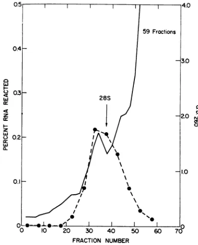
Size distribution of nuclear SV40 RNA. Since
polycistronicSV40 RNAapproaching the
molecu-lar size of one strand ofSV40 DNA could be
detected in thecytoplasmofinfectedVerocells, we
decided to characterize the virus-specific RNA
present in the nuclei of thesecells. Vero cells were
labeled with 3H-uridine (20,Ci/ml) for 75 min 48 hr after infection with SV40. Nuclear RNA was prepared as described above and analyzed
for virus-specific RNA after sedimentation in a
15 to 30% (w/v) sucrose gradient containing
SDS buffer. Unlabeled 28 and 185 ribosomal
RNA was simultaneously sedimented ina
separ-ate tube. In contrast with the results observed
earlier (Fig. 1, 3) withcytoplasmic RNA, nuclear
SV40 RNA was distributed in the more rapidly
sedimenting
regions of the gradient, peaking atabout 32 to 34S (Fig. 6). Some virus-specific
RNAcould be detected in the 45 to 50S region
TABLE 1. Rehybridization of the simian virus 40
(SV40) RNA sedimenting in the region of28S
ribosomal RNAa
DNA filter RNAreacting
SV40... 34.8 Greenmonkey... 0.0
Blank... 0.2
a Polyribosome-associated RNA present in
Verocells 48 hr after infection andsedimentingin
the region of 285 ribosomal RNA was prepared
as shown in Fig. 5. This RNA (180,000 counts/
min) was incubated with a nitrocellulose filter containing 8
1Ag
of SV40DNA. The filterwasex-tensivelywashedat68 C in4X SSC,and the
virus-specific RNA (532 counts/min) was eluted after
two 10-min exposures to0.01 M EDTA and0.1%
SDS at 95 C. The recovered SV40 RNA was in-cubated with three 7-mm nitrocellulose filters
containing 0.3 ,ug of SV40 DNA, 16 ,ug of green
monkey DNA, andnoDNA. After 16 hrof
incuba-tionat68C, thefiltersweretreated withribonuclease
and assayed forradioactivity as described in Fig. 2.
suggesting thatmolecules larger than unit length
may be transcribed in thenuclei of these cells.
Sedimentation properties of SV40 RNA during
abortive infection. Replication of SV40 does not
occurin mouse cells (nonpermissive). Some SV40
nonstructuralproteins(e.g.,Tantigen) associated
with the "early" period of lytic infection can be
detected after infection of these cells, but so called "late" viral functions, such as DNA replication
and thesynthesis ofcapsidproteins, are inhibited.
Since someoftheinitial biochemical events
asso-ciated with transformationoccurduring abortive
infection, experiments characterizing the SV40
RNA present in such mouse cells were carried
out. A confluent monolayer of mouse cells
[AL/N (17)] was infected with SV40 at a
multi-plicity of 400 PFU/cell. The infected cells were
labeled for 2 hr with 32P-orthophosphate 68 hr
after infection, andpolyribosomes were prepared as previously described. Figure 7 shows that a
portion of the SV40 RNA sedimented in the
region of the 18S ribosomal RNA, but none of
themorerapidlysedimenting virus-specificRNA
C
w
Ir
z cr
z
ac
w
a.
5.0
0
Wo m
0
.0
-0 10 20 30 40 50 60 705
FRACTION NUMBER
FIG.6. Size distribution ofSV40 RNA in the nuclei of infectedVero cells.Nuclei, labeled with 3H-uridine, werepreparedfrom Verocells48 hr after infection with
SV40asdescribedin the text. The nuclear RNA was
sedimentedthrougha15to30% (w/v)sucrosegradient
containingSDSbuffer,andfractionswereanalyzed for adsorptionat260nmand SV40 RNA.Purified28 and
18SribosomalRNAwascentrifugedinaseparatetube
andusedasmarkers.SV40RNA (0) wasdetected by
DNA-RNA hybridization.
I.,
59Fractions
-3.
28S
D.1
_-X
\\\
21.
2 ~~~~~~~~~~~~~I
0 ~~~0~
e0 11~~~0
VOL.6,1970 467
c
on November 11, 2019 by guest
http://jvi.asm.org/
[image:5.493.248.441.309.548.2] [image:5.493.45.239.407.503.2]MARTIN AND BYRNE
FRACTION NUMBER
FIG.7. Sedimentation profile of the SV40 RNA
presentinmousecells 68hrafter infection.A continuous
line (AL/N) ofmousecells wasinfectedwithSV40at amultiplicity of 400PFU/cell and exposed for90min to '2P-orthophosphate (200 ,uCi/ml) 68 hrafter
infec-tion. Polyribosome associated RNA was preparedas described in the text. Fractions from a 15 to 30% (w/v) sucrose gradient containing SDS buffer were analyzed forSV40 RNA (broken line) byDNA-RNA
hybridization.
observed lateinproductive infection (Fig. 1) was
detected. Infact, the size distribution of the SV40
RNAduring abortive infection is quite similarto
thatseenduring the early phases (Fig. 3A) of the
lytic cycle.
DISCUSSION
Ithas previously been shown thatduring lytic
infection 50% of SV40 DNAistranscribed (10).
Thoseresults, obtained fromDNA-RNA
satura-tion-hybridization experiments do not shed any
light on thephysical size of SV40-specific RNA.
Recent work with polyoma virus suggests that
RNA molecules are present during productive
infection having molecular weights equivalent to
one strand of circular polyoma DNA (5). Our
results indicate that by 24 hr after infection the
polyribosome-associated virus-specific RNA has
amolecularsizeapproaching one-half of the SV40
genome.Our data also show that thesize
distribu-tionof SV40 RNAgradually increases during the
course ofproductive infection. At 8 hr, for
ex-ample,wewereabletodetect RNAmoleculesin
the 5 x 10⁵to6 x 105dalton range.Progressively
largerSV40RNAwasseen at 16 and 24 hr after
infection,withlittle further change at 48 hr.
Alonietal. (1) haveshownthat "early" RNA
is present throughout the infectious cycle. An
additional species of SV40 RNA ("late") was
also detected after the onset of viral DNA
syn-thesis. Theslowly sedimenting virus-specificRNA
present at 8 hr (Fig. 3A) may represent those cistron(s) which are transcribed from DNA se-quencescoding for early functions. Late in infec-tion, the pattern of SV40 DNA transcription is
alteredsothatadditional regions of the viral DNA
are expressed, resulting in RNA molecules of
increasing size (Fig. 3B, 3C). We are currently
characterizing the slowly sedimenting RNA
presentearlyininfection and the
high-molecular-weight species found at 24 and 48 hr to see if
similar species of RNAare present in thesetwo
size classes.
Little is known about the physical properties
of newly transcribed animal viral RNA. The
SV40RNA detected in the nuclei of infected cells
sediments over a broad region of the sucrose
gradient (Fig. 6). Some of the RNA is present in
regions where moleculeshaving molecular weights
greaterthan 1.5 x 10⁶daltons would be expected
to sediment. This result is similar tothefindingsof
Lindberg and Darnell (7), who were able to
detectvirus-specificRNA in thenuclei of
SV40-transformed cells which sediment more rapidly
than 45S nuclear RNA. The presence of SV40
lytic RNA in the 45 to 50S region suggests that
SV40 gene products may be transcribed in
multiple units, resulting in molecules containing
more than one equivalent of viral RNA. Such
RNA molecules would have to be cleaved into
smaller functional units prior to attachment to
cytoplasmic ribosomes. We arepresently
charac-terizing the properties of nuclear SV40 RNA
during productive and transforming infection.
LITERATURE CITED
1. Aloni, Y.,E.Winocour, andL.Sachs. 1968. Characteriza-tion ofthesimian virus40-specific RNA invirus-yielding andtransformed cells.J. Mol. Biol. 31:415-429. 2. Crawford,L.V., andP.H.Black. 1964. Thenucleic acid of
simian virus 40. Virology 24:388-392.
3.Earley,E.,P.Peralta, andK.Johnson. 1967.Aplaque neu-tralization method forarboviruses. Proc. Soc. Exp. Biol. Med.125:741-747.
4. Gillespie, D., andS.Spiegelman. 1965.Aquantitative assay for DNA-RNA hybrids with DNA immobilized on a membrane. J. Mol. Biol. 12:829-842.
5.Hudson,J.,D.Goldstein,andR.Weil. 1970.Astudyonthe
transcriptionofpolyoma viralgenome. Proc.Nat.Acad. Sci. U.S.A. 65:226-233.
6. Levy, H.,andW.Carter.1968. Molecular basis oftheaction ofinterferon. J. Mol. Biol.31:561-577.
7. Lindberg, V., andJ.Darnell. 1970. SV40-specific RNA in the
468 J.VIROL.
on November 11, 2019 by guest
http://jvi.asm.org/
[image:6.493.57.249.54.291.2]SEDIMENTATION OF SV40 RNA
nucleus and polyribosomes of transformed cells. Proc. Nat. Acad. Sci. U.S.A. 65:108.9-1096
8. McConkey, E.,and J.Hopkins. 1969.Molecular weights of
some HeLa ribosomal RNA's. J. Mol. Biol. 39:545-550. 9. Martin, M., and D. Axelrod. 1969. Polyoma virusgene activ-ityduring lytic infection and in transformed animal cells. Science164:68-70.
10.Martin,M., and D.Axelrod.1969.SV40geneactivity during lyticinfection and inaseries ofSV40 transformedmouse
cells. Proc. Nat. Acad. Sci. U.S.A. 64:1203-1210. 11. Oda, K.,and R.Dulbecco. 1968.Regulationoftranscription
oftheSV40 DNA in productively infected and in
trans-formedcells. Proc. Nat. Acad. Sci. U.S.A. 60:525-532. 12.Penman, S., K. Scherrer, Y. Becker, and J. Darnell. 1963.
Polyribosomes in normal and poliovirus-infected HeLa cells and theirrelationshiptomessengerRNA.Proc. Nat. Acad. Sci. U.S.A. 49:654-661.
13. Petermann, M., and A. Pavlovec. 1966. The subunits and
structural ribonucleic acids of Jensensarcomaribosomes. Biochim. Biophys. Acta 144:264-276.
14. Radloff, R.,W.Bauer,and J.Vinograd.1967. A dye-buoyant-densitymethod for the detection and isolation ofclosed circularduplex DNA. Proc. Nat. Acad. Sci. U.S.A. 57: 1514-1521.
15. Sauer, J., and J. Kidwai. 1968. The transcription of the SV40
genome in productively infected and transformned cells. Proc. Nat. Acad.Sci. U.S.A. 61:1256-1263.
16. Strauss, J., andR.Sinsheimer. 1963. Purificationand
prop-erties ofbacteriophage MS2 and ofits ribonucleic acid. J.Mol.Biol. 7:43-54.
17. Takemoto, K. K., R. C. Y. Ting, H. L. Ozer, and P. Fabisch. 1968.Establishment ofacell line fromaninbredmouse
strain for viral transformation studies. J. Nat. Cancer Inst. 41:1401-1405.
18.Trilling, D., and D. Axelrod. 1970. Encapsidation of free host DNA bysimian virus 40. Science 168:268-271.
VOL.6, 1970 469