T E C H N I C A L N O T E
Open Access
A pink mouse reports the switch from red to
green fluorescence upon Cre-mediated
recombination
Heiner Hartwich
1, Somisetty V Satheesh
1and Hans Gerd Nothwang
1,2*Abstract
Background:Targeted genetic modification in the mouse becomes increasingly important in biomedical and basic
science. This goal is most often achieved by use of the Cre/loxP system and numerous Cre-driver mouse lines are currently generated. Their initial characterization requires reporter mouse lines to study thein vivospatiotemporal activity of Cre.
Findings:Here, we report a dual fluorescence reporter mouse line, which switches expression from the red fluorescent protein mCherry to eGFP after Cre-mediated recombination. Both fluorescent proteins are expressed from the ubiquitously active and strong CAGGS promoter. Among the founders, we noticed a pink mouse line, expressing high levels of the red fluorescent protein mCherry throughout the entire body. Presence of mCherry in the living animal as well as in almost all organs was clearly visible without optical equipment. Upon Cre-activity, mCherry expression was switched to eGFP, demonstrating functionality of this reporter mouse line.
Conclusions:The pink mouse presented here is an attractive novel reporter line for fluorescence-based monitoring of Cre-activity. The high expression of mCherry, which is visible to the naked eye, facilitates breeding and crossing, as no genotyping is required to identify mice carrying the reporter allele. The presence of two fluorescent proteins allows in vivo monitoring of recombined and non-recombined cells. Finally, the pink mouse is an eye-catching animal model to demonstrate the power of transgenic techniques in teaching courses.
Findings Background
Spatiotemporally restricted genetic modification in the mouse is becoming an indispensable tool in biological research [1]. Applications include analysis of essential genes, whose constitutive ablation results in embryonic/ perinatal lethality, discrimination between primary and secondary effects, or mapping cell connectivity and cell fate [2-4]. The importance of targeted genetic modifica-tion is underlined by a recent global initiative to gener-ate conditional alleles for all murine genes [5-7]. Most targeted genetic modifications make use of the Cre/loxP system. In this approach, a Cre-driver mouse line, expressing
Cre recombinase of the phage P1 under a tissue or cell-type specific promoter, is crossed to mice with a loxP flanked (“floxed”) allele, which is recognized by Cre [8].
Several Cre-driver lines have been generated and each of them requires initial characterization of the spatial and temporal Cre expression pattern [9]. A popular reporter line expresses LacZ from the ROSA26 locus upon Cre-mediated recombination [10], but its analysis is limited to fixed tissue. For in vivoobservation of Cre-mediated recombination, several fluorescent reporter lines have been generated [11-15]. However, only few dual-reporter lines are available with different labels of recombined and non-recombined cells. Some of them combine a fluorescent protein with an enzyme (alkaline phosphatase or LacZ) [16-18], others combine two dif-ferent fluorescent proteins [19,20]. Here we present a novel Cre-reporter line with high and ubiquitous expres-sion of two fluorescent proteins and which is easily recognized by its pink skin color.
* Correspondence:[email protected] 1
Department of Neurogenetics, Institute of Biology and Environmental Sciences, Carl von Ossietzky University, Carl-von-Ossietzky-Straße 9-11, 26129, Oldenburg, Germany
2Research Center Neurosensory Science, Carl von Ossietzky University
Oldenburg, Carl-von-Ossietzky-Straße 9-11, 26129, Oldenburg, Germany
reading frames for mCherry and eGFP, and the polya-denlation signal were released from the plasmid
pCAGGS_cherry_Intron_GFP by restriction with MssI
and Ecl136II (MBI Fermentas). The plasmid contains a
CAGGS promoter, followed by a loxP flanked
mcherry-pA cassette and a β-actin intron followed by the
sequence of egfp-pA (Figure 1A) The entire transgenic construct is flanked by FRT sites. After gel electrophor-esis in low melting agarose, the fragment was purified using agarase according to the manufacturer´s instruc-tions (MBI Fermentas). The final construct was dis-solved in microinjection buffer (5 mM Tris, 0.1 mM EDTA, pH 7.6). Pronucleus injection was carried out at the Transgenic Core Facility of the Max-Planck Institute of Molecular Cell Biology and Genetics at Dresden. Genotyping of the transgenic mice was performed using the primers 5´-CGTAATGCAGAAGAAGACCATGG-3´ and 5´-CCTGCTATTGTCTTCCCAATCC-3´. The Cre-driver lineEgr2Cre/+has been described previously [21].
All animals used for these experiments were main-tained on C57BL/6 J (Harlan Winkelmann, Borchen, Germany) and C57BL/6 N (Charles River, Sulzfeld, Ger-many) mixed genetic backgrounds. All protocols adhered to the German Animal Protection law and were approved by the local animal care and use committee (LAVES, Old-enburg). Protocols also followed the NIH guide for the care and use of laboratory animals.
Determination of transgene copy number
Transgene copy number was determined by semi-quantitative PCR, using the primers 5´-AAACATAACT-TCGTATAATGTATGCTATACG-3´ and 5´-CGTAATG-CAGAAGAAGACCATGG-3´ with the following conditions: denaturation for 5 min at 95 °C and 30 sec at 95 °C,
reflects the size difference between the transgenic con-struct and the diploid mouse genome of ~ 5 Gb.
Tissue preparation and fluorescence microscopy
Mice were killed by gassing with CO2 and the organs
immediately removed. Fluorescence of organs was either visualized with an UV lamp (Fluotest Original Hanau, Hanau, Germany) at 254 nm wavelength and a custom digital camera, or under an inverse microscope (Key-ence, Neu-Isenburg, Germany).
Perfusion, slicing and immunohistochemistry
Mice were anesthetized with 70 mg/ml chloral hydrate (1 ml/100 g body weight) and perfused transcardially with phosphate-buffered saline (PBS) (130 mM NaCl, 7 mM Na2HPO4, 3 mM NaH2PO4, pH 7.4), followed by
[image:2.595.62.539.631.696.2]Zamboni´s solution (2 % paraformaldehyde, 15 % picric acid in 0.1 M phosphate buffer, pH 7.4). The brain was removed from the skull and incubated overnight in 30 % (w/v) sucrose/PBS. Coronal brainstem sections of 30μm thickness were sliced using a microtome (MICROM GmbH, Walldorf, Germany), collected in 15 % (w/v) su-crose/PBS and washed twice in PBS and twice in PBST (PBS with 1 % (v/v) Triton X-100) (15 min each washing step). Sections were incubated for 1.5 h in blocking solu-tion (2 % bovine serum albumin, 10 % goat serum and 0.3 % Triton X-100 in PBS, pH 7,4). Sections were trans-ferred in blocking solution containing rabbit anti-GFP antibody (1:500, Invitrogen, Karlsruhe, Germany) and incubated overnight with agitation at 4 °C. After three washes with PBST (10 min each), sections were trans-ferred into carrier solution (1 % bovine serum albumin, 1 % goat serum and 0.3 % Triton X-100 in PBS, pH 7,4) and treated with the secondary antibody anti-rabbit
conjugated to Alexa Fluor 488 (1:500, Invitrogen). After 1.5 h with agitation at room temperature, slices were washed three times with PBS (10 min each) and mounted on glass slides with Vectashield Mounting Medium with DAPI (Vector Laboratories, Burlingame, USA). Photomicrographs were taken with an inverse microscope (Keyence) and the fluorescence was detected with the corresponding filter set.
In vivo slice preparation and imaging
Mice were killed by gassing with CO2and 150μm thick
living brainstem slices were cut with a vibratome (MICROM GmbH). During the preparation, slices were
incubated in 95 % O2and 5 % CO2aerated ACSF
solu-tion (118 mM NaCl, 1 mM NaH2PO4, 25 mM NaHCO3,
3 mM KCl, 1 mM MgCl2, 1.5 mM CaCl2, pH 7.4). After
preparation, brainstem slices were examined with a TCS SL confocal microscope (Leica, Nussloch, Germany) using 40-fold magnification and a PL FLUOTAR object-ive (40.0x/0.7 NA).
Results and discussion
[image:3.595.58.539.89.209.2]To generate a transgenic mouse line for in vivo cell labeling and the report of Cre-mediated recombina-tion, we used the plasmid pCAGGS_cherry_Intron_GFP (Figure 1). This plasmid expresses the red fluorescent
Figure 2The pink mouse. A) A transgenic mCherry and its wild-type littermate are shown at postnatal day (P)0. B, C) Comparison of the paws of a young-adult wild-type (B) and a transgenic (C) mouse. The pink colored paw of the transgenic mouse is clearly visible without optical instruments. D, E) An adult wild-type mouse with an opened body (D) and its transgenic littermate (E), which displays reddish organs.
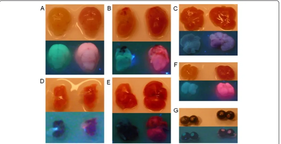
Figure 3Whole organ fluorescence under UV light.Different organs of wild-type (left side) and transgenic (right side) P0 mice are shown. Upper panels represent bright field images and lower panels represent fluorescence images of different organs.A) brain;B) heart;C) liver;
[image:3.595.58.542.437.685.2]protein variant mCherry under the CAGGS promoter (chicken β-actin promoter with CMV enhancer), which is active in almost all tissues [22]. Upon Cre-mediated expression, mCherry is deleted and eGFP expressed.
Pink colored mice
Pronuclear injection resulted in three founder mice. After crossing them back to wild-type mice, some of the young progeny of one founder had a pinkish skin
[image:4.595.59.539.91.345.2](Figure 2A), which made them easily distinguishable from littermates. The pink skin color was still observed in the paw of adult animals (Figure 2B,C) and inside the body (Figure 2D,E) in the absence of UV light. These data indi-cated an extraordinarily high expression level of mCherry in this founder line. Genotyping PCR confirmed the pres-ence of the reporter allele in these mice. The pink colored mice were fully viable and fertile, demonstrating, that the expression level of the mCherry protein had no toxic
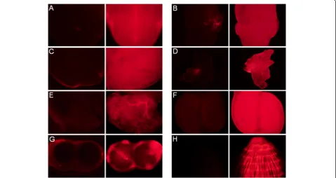
Figure 4Organ examination with the fluorescent microscope.Organs of P0 mice were examined under a fluorescent microscope to show the difference in the fluorescence of wild-type (left side) and transgenic mice (right side).A) brain;B) heart;C) liver;D) spleen;E) lung,F) kidney,
G) eye; andH) tail.
[image:4.595.59.540.527.686.2]effects. This phenotype was observed both in the first and the second filial generation, indicating a generational per-sistence and no silencing of the transgene.
Whole organ fluorescence
We next checked organs for fluorescence detection under UV light. The brain, heart, liver, lung, spleen, kidney, and eyes were taken from P0 mice and photographed in bright field and during excitation with UV light (Figure 3). Even without UV excitation, the organs of the transgenic mouse strain had a deeper shade of red than control lit-termates. After excitation with UV light, the difference between wild-type and transgenic mouse organs were also
clearly visible. The organs of the mCherry mouse were pink in contrast to those of the littermates. In agreement with these results, all organs tested of the mCherry mouse line gave an intense shade of red under fluorescence microscopy (Figure 4). These data demonstrated high expression of mCherry in all of the organs analyzed.
Copy number
[image:5.595.59.538.90.310.2]The high expression level of mCherry could result either from a high copy number of the transgene or from the integration site into the genome, thus representing a position effect. To estimate the copy number, we per-formed semi-quantitative PCR (Figure 5) on DNA from
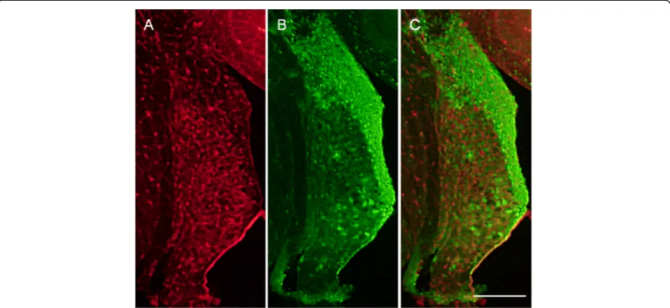
Figure 6Validation of Cre-mediated recombination.The cochlear nucleus from a reporter mouse crossed with anEgr2Cre/+driver line is
shown after fixation.A) Red fluorescence,B) green fluorescence,C) merged picture. The strong green fluorescence demonstrates successful recombination. Scale bar is 200μm.
Figure 7In vivo imaging of brainstem slices.Detection of non-recombined and recombined cells in a living brainstem slice from a reporter mouse crossed with anEgr2Cre/+driver line.A) Red fluorescence,B) green fluorescence,C) merged picture. The green fluorescence demonstrates
[image:5.595.62.538.537.694.2]as a Cre-reporter mouse line. Transgenic mice were therefore crossed with the Egr2Cre/+ Cre-driver mouse line [21], which expresses Cre in the auditory brainstem [23]. Fluorescent immunohistochemistry demonstrated expression of eGFP in the dorsal cochlear nucleus, a con-spicuous structure of the auditory brainstem (Figure 6). A minority of cells expressed mCherry and eGFP in parallel, as indicated by their orange/yellow appearance (Figure 6C). This indicates incomplete recombination of all 4 to 6 copies, which is likely caused by low Cre ex-pression in these cells. Despite this intermediate pheno-type, these data demonstrate that our novel dual reporter mouse line is well suited to monitor Cre expression. The mixed expression of mCherry and eGFP might even be exploited to determine promoter activities, using Cre-driver lines.
An important application of fluorescent proteins is their in vivo detection such as during electrophysio-logical characterization of neuronal subpopulations. We therefore analyzed whether red and green fluorescence labeled cells can be detected in vivo. Indeed, both non-recombined (red) and non-recombined (green) cells were easily recognized in unfixed, living brain slices (Figure 7). These data demonstrate the usefulness of our reporter mouse for in vivo monitoring and detection of recombination.
Conclusions
This novel pink Cre-reporter mouse line represents a versatile tool in transgenic mouse research. A great ad-vantage is the high expression level of the fluorescent protein mCherry, which allows easy recognition and sep-aration of animals harboring the reporter allele from lit-termates without further analysis. The presence of two fluorescent proteins is useful for parallel in vivo monitoring of recombined and non-recombined cells, in vivo recordings, and analyses of cellular connectivity. Finally, this mouse line is well suited for teaching purposes. Its pink color can be used to easily demon-strate the power of transgenic technologies and crossed with an ubiquitously expressing Cre-driver, the color would disappear.
Competing interests
The authors declare that they have no competing interests.
Received: 10 January 2012 Accepted: 8 June 2012 Published: 14 June 2012
References
1. Glaser S, Anastassiadis K, Stewart AF:Current issues in mouse genome engineering.Nat Genet2005,37:1187–1193.
2. Madisen L, Zwingman TA, Sunkin SM, Oh SW, Zariwala HA, Gu H, Ng LL, Palmiter RD, Hawrylycz MJ, Jones AR, Lein ES, Zeng H:A robust and high-throughput Cre reporting and characterization system for the whole mouse brain.Nat Neurosci,13:133–140.
3. Livet J, Weissman TA, Kang H, Draft RW, Lu J, Bennis RA, Sanes JR, Lichtman JW:Transgenic strategies for combinatorial expression of fluorescent proteins in the nervous system.Nature2007,450:56–62.
4. Koundakjian EJ, Appler JL, Goodrich LV:Auditory neurons make stereotyped wiring decisions before maturation of their targets.
J Neurosci2007,27:14078–14088.
5. Skarnes WC, Rosen B, West AP, Koutsourakis M, Bushell W, Iyer V, Mujica AO, Thomas M, Harrow J, Cox T, Jackson D, Severin J, Biggs P, Fu J, Nefedov M, de Jong PJ, Stewart AF, Bradley A:A conditional knockout resource for the genome-wide study of mouse gene function.Nature2011,474:337–342. 6. Collins FS, Rossant J, Wurst W:International Mouse Knockout Consortium,
A mouse for all reasons.Cell2007,128:9–13.
7. Collins FS, Finnell RH, Rossant J, Wurst W:A new partner for the international knockout mouse consortium.Cell2007,129:235. 8. Nagy A:Cre recombinase: the universal reagent for genome tailoring.
Genesis2000,26:99–109.
9. Nagy A, Mar L, Watts G:Creation and use of a cre recombinase transgenic database.Methods Mol Biol2009,530:365–378.
10. Soriano P:Generalized lacZ expression with the ROSA26 Cre reporter strain.Nat Genet1999,21:70–71.
11. Kawamoto S, Niwa H, Tashiro F, Sano S, Kondoh G, Takeda J, Tabayashi K, Miyazaki J:A novel reporter mouse strain that expresses enhanced green fluorescent protein upon Cre-mediated recombination.FEBS Lett2000, 470:263–268.
12. Srinivas S, Watanabe T, Lin CS, William CM, Tanabe Y, Jessell TM, Costantini F:Cre reporter strains produced by targeted insertion of EYFP and ECFP into the ROSA26 locus.BMC Dev Biol2001,1:4.
13. Luche H, Weber O, Nageswara Rao T, Blum C, Fehling HJ:Faithful activation of an extra-bright red fluorescent protein in "knock-in" Cre-reporter mice ideally suited for lineage tracing studies.Eur J Immunol
2007,37:43–53.
14. Diéguez-Hurtado R, Martín J, Martínez-Corral I, Martínez MD, Megías D, Olmeda D, Ortega S:A Cre-reporter transgenic mouse expressing the far-red fluorescent protein Katushka.Genesis2011,49:36–45. 15. Abe T, Kiyonari H, Shioi G, Inoue K, Nakao K, Aizawa S, Fujimori T:
Establishment of conditional reporter mouse lines at ROSA26 locus for live cell imaging.Genesis2011,49:579–590.
16. Lobe CG, Koop KE, Kreppner W, Lomeli H, Gertsenstein M, Nagy A:Z/AP, a double reporter for cre-mediated recombination.Dev Biol1999, 208:281–292.
17. Novak A, Guo C, Yang W, Nagy A, Lobe CG:Z/EG, a double reporter mouse line that expresses enhanced green fluorescent protein upon Cre-mediated excision.Genesis2000,28:147–155.
18. Badaloni A, Bonanomi D, Albieri I, Givogri I, Bongarzone E, Valtorta F, Consalez GG:Transgenic mice expressing a dual, CRE-inducible reporter for the analysis of axon guidance and synaptogenesis.Genesis2007, 45:405–412.
20. Stewart MD, Jang CW, Hong NW, Austin AP, Behringer RR:Dual fluorescent protein reporters for studying cell behaviors in vivo.Genesis2009, 47:708–717.
21. Voiculescu O, Charnay P, Schneider-Maunoury S:Expression pattern of a Krox-20/Cre knock-in allele in the developing hindbrain, bones, and peripheral nervous system.Genesis2000,26:123–126.
22. Okabe M, Ikawa M, Kominami K, Nakanishi T, Nishimune Y:'Green mice' as a source of ubiquitous green cells.FEBS Lett1997,407:313–319. 23. Han Y, Kaeser PS, Südhof TC, Schneggenburger R:RIM determines Ca2+
channel density and vesicle docking at the presynaptic active zone.
Neuron2011,69:304–316.
doi:10.1186/1756-0500-5-296
Cite this article as:Hartwichet al.:A pink mouse reports the switch from red to green fluorescence upon Cre-mediated recombination.BMC Research Notes20125:296.
Submit your next manuscript to BioMed Central and take full advantage of:
• Convenient online submission
• Thorough peer review
• No space constraints or color figure charges
• Immediate publication on acceptance
• Inclusion in PubMed, CAS, Scopus and Google Scholar
• Research which is freely available for redistribution