RESEARCH ARTICLE
Comparison of SYBR green I real-time
RT-PCR with conventional agarose gel-based
RT-PCR for the diagnosis of infectious bronchitis
virus infection in chickens in Morocco
Siham Fellahi
1,2, Mehdi El Harrak
3, Jens H. Kuhn
4, Ghizlane Sebbar
3, El Arbi Bouaiti
5, Khadija Khataby
2,
Ouafae Fassi Fihri
1, Mohammed El Houadfi
1†and My Mustapha Ennaji
2*†Abstract
Background: A rapid, sensitive, and specific molecular method for the diagnosis of infectious bronchitis virus (IBV) infection is important in curbing infectious bronchitis outbreaks in Morocco and other countries.
Methods: In this study, an easy-to-perform SYBR green I real-time reverse transcriptase polymerase chain reaction (RT-PCR) targeting the nucleocapsid gene of IBV was developed and compared with conventional agarose gel-based RT-PCR for the detection of IBV infection.
Results: We found that the SYBR green I real-time RT-PCR was at least 10 times more sensitive than the agarose gel electrophoresis detection method. The assay exhibited high specificity for IBV infection. All negative controls, such as Newcastle disease virus, infectious bursal disease virus, and avian influenza virus, were not detected.
Conclusion: The SYBR green I real-time RT-PCR test described herein can be used to rapidly distinguish IBV from other respiratory pathogens, which is important for diagnosis and control of infectious bronchitis outbreaks in Morocco. The test is a valuable and useful method as a routine assay for diagnosis of clinical IBV infection in commer-cial chickens.
Keywords: Coronavirus, Gammacoronavirus, IBV, Infectious bronchitis virus, SYBR green, Real time RT-PCR, RT-PCR
© 2016 Fellahi et al. This article is distributed under the terms of the Creative Commons Attribution 4.0 International License (http://creativecommons.org/licenses/by/4.0/), which permits unrestricted use, distribution, and reproduction in any medium, provided you give appropriate credit to the original author(s) and the source, provide a link to the Creative Commons license, and indicate if changes were made. The Creative Commons Public Domain Dedication waiver (http://creativecommons.org/ publicdomain/zero/1.0/) applies to the data made available in this article, unless otherwise stated.
Background
Infectious bronchitis (IB), an acute, highly contagious viral upper respiratory disease of chickens, is one of the most economically significant diseases hampering the intensive poultry industry worldwide. IB affects chickens of all ages, causing respiratory, reproductive, and renal manifestations [1]. Although control of IBV infection is primarily achieved through live attenuated vaccines, the
infection is difficult to contain because immunization with different serotypes of the virus do not necessar-ily cross-protect against other serotypes [2]. Within an infected poultry flock, quick and accurate detection of the presence of the virus is imperative to properly vac-cinate uninfected flocks. In addition, rapid differentiation of IBV infection from other upper respiratory tract dis-eases (e.g., avian influenza, Newcastle disease, infectious laryngotracheitis, avian mycoplasmosis) is important so that appropriate measures can be taken in a timely man-ner [3].
IB is a disease that negatively impacts the poul-try induspoul-try of developing countries. For instance, in Morocco, IB continues to be an uncontrolled problem [4–6] due to the lack of in-country diagnostic capabili-ties that can be performed quickly and interpreted easily
Open Access
*Correspondence: [email protected]
†Mohammed El Houadfi and My Mustapha Ennaji contributed equally to this work
2 Laboratory of Virology, Microbiology and Quality/Ecotoxicology & Biodiversity, Faculty of Sciences and Techniques-University Hassan II Mohammedia, PO Box 146, Quartier Yasmina-Mohammedia, 20650 Casablanca, Morocco
by local staff in potentially underequipped or otherwise challenging environments.
The causative agent of IB, infectious bronchitis virus (IBV), is a member of the species Avian coronavirus, genus Gammacoronavirus, family Coronaviridae [7]. IBV is an enveloped, positive-sense, single-stranded RNA virus (genome length = 27.6 kb) [8], expressing three major structural proteins: the nucleocapsid protein (N) surrounding the viral RNA, the membrane glycoprotein (M), and the spike glycoprotein (S) located on the surface of the viral envelope. The S protein contains two post-translational subunits, S1 and S2 [9].
Current diagnostic assays for IBV include virus iso-lation in embryonated eggs, tracheal organ culture, cell culture immunoassays, and molecular assays that detect viral RNA [10]. Virus isolation has been con-sidered to be the reference standard. However, such isolations are expensive and time consuming because several passages may be required to detect the virus. Immunoassays use IBV-specific monoclonal antibod-ies to detect the virus in direct or indirect fluorescent antibody and enzyme-linked immunosorbent assay (ELISA) formats. Although faster and simpler than virus isolation, immunoassays tend to lack specificity and sensitivity. None of these immunoassays detect all strains or types of IBV [11–13]. Molecular assays, such as reverse transcriptase-polymerase chain reaction (RT-PCR), for the detection of IBV are commonly used because highly specific and sensitive results can be obtained in a timely manner. Molecular assays detect viral RNA directly from a clinical sample or from virus isolated in a laboratory host system. When RT-PCR is used to amplify the spike glycoprotein (S) of IBV, the assay can be coupled with restriction fragment length polymorphism analysis or nucleic acid sequencing to identify serotypes of the virus [2, 10, 12–16]. More recently, many fluorescent probe-based real-time RT-PCR assays have been developed to detect IBV strains [2, 3, 9, 10, 15, 17]. Real-time TaqMan RT-PCR assays have been developed that amplify a fragment of the 5′ untranslated region of the IBV genome to detect turkey coronaviruses and IBV [17] or that target the N gene for IBV detection [9].
Unfortunately, performing most of these assays requires highly trained staff, a sophisticated infra-structure, or considerable monetary funds, and are therefore not necessarily viable options for develop-ing countries such as Morocco. SYBR green I-based RT-PCR assays have proven to be among the most effective tools in the rapid and differential detection of a variety of viral diseases such as avian influenza, Newcastle disease, and IB. These inexpensive and eas-ily performed assays are important to rapidly identify
the causative agent of any upper respiratory disease or changes in egg shell quality and egg production in chickens [17, 18].
However, an assay employing real-time RT-PCR with SYBR green I dye to target the N gene of IBV is lacking. Here we report the development of a real-time RT-PCR assay with SYBR green I dye for rapid detection of IBV viral RNA directly from Moroccan clinical samples. We also compared the assay with conventional RT-PCR and agarose gel electrophoresis to detect IBV PCR-amplified products.
Results Reproducibility
SYBR green I real‑time RT‑PCR
The cDNA IBV Beaudette strain (infectious bronchi-tis virus/G.gallus-wt/FRA/1995/Beaudette) was used to extract the standard control genomic RNA that then served as the source of cDNA template. Concentrations of the cDNA standard ranging from 10–106 copies/µl
were tested repeatedly (Fig. 1) by SYBR green-I-based real-time RT PCR. The results are reproducible through 10 cDNA copies/μl.
Conventional gel‑based RT‑PCR
The PCR targeted a sequence corresponding to a region of the IBV N protein. The obtained PCR products of 130 bp in length were separated on a 1.5 % agarose gel stained with ethidium bromide (Fig. 2). PCR products were detectable at a dilution of 100 cDNA copies/µl.
Specificity of SYBR green I real‑time RT‑PCR
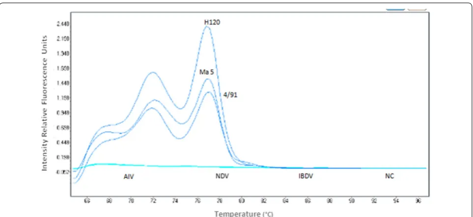
Melting peaks analysis on the PCR products of H120, MA5, and 4/91 vaccine strains and tenfold serially diluted cDNA (data not shown) did not indicate primer-dimers or nonspecific products. Specific amplification of
[image:2.595.305.540.546.654.2]the IBV target sequence was identified by the generation of a melt peak at 68 ± 0.20 °C. The specificity of the SYBR green-I real-time RT-PCR was 100 % since detectable fluorescent signals were not observed with the negative control (NC), avian influenza virus (FLUAV), infectious bursal disease virus (IBDV), and Newcastle Disease virus (NDV) nucleic acids. Only the IBV vaccine strain genetic material was detected (Fig. 3).
Sensitivity
SYBR green I real‑time RT‑PCR
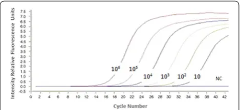
To determine the sensitivity endpoint of the assay, serial dilutions of cDNA at concentrations ranging from 1.25 to 100 copies/µl were analyzed. The real-time RT-PCR fluorescence curve derived from serially diluted standard concentrations (cDNA of H120 vaccine strain) indicates a high sensitivity of detection down to 100 copies/µl of cDNA (Fig. 4).
To determine the linearity of the reaction and the RT-PCR efficiency, the cycle threshold (Ct) values of indi-vidual dilutions were analyzed. The assay has a dynamic detection range that spanned 102–106 cDNA copies/µl.
Except for the undiluted cDNA, a strong inverse linear relationship was observed between the amount of input cDNA and the Ct values over six log10 dilutions (Table 1),
as indicated by a simple linear regression plot of the two variables (Fig. 5).
Conventional RT‑PCR
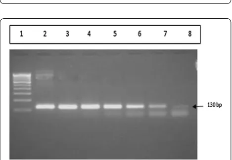
Results from a conventional RT-PCR using the IBV N gene confirmed that the obtained RT-PCR products were 130 bp in length, and the PCR products were detectable at a concentration of 100 copies/µl (Fig. 6).
Comparison of sensitivity and specificity of the SYBR green I real‑time RT‑PCR assay with conventional RT‑PCR
The lowest dilution of cDNA that SYBR green I real-time RT-PCR assay was able to detect was an order of mag-nitude higher than conventional RT-PCR assay (Table 1). Receiver operating characteristic curve analysis of Ct val-ues obtained from the SYBR green I real-time RT-PCR assay indicates the linearity of the reaction and the assay’s efficiency (Fig. 7). The assay has a maximum sensitivity and specificity over a detection range that spanned 104
cDNA gene copies/µl (Table 1).
Detection of IBV in tracheal swabs from clinical samples The optimized methods of SYBR green I real-time RT-PCR and conventional RT-RT-PCR were applied to clinical samples from broiler chickens showing clinical signs of IB, including coughing, sneezing, rales, and nasal dis-charge. From 34 tracheal swabs tested, all samples and positive controls were positive for IBV by SYBR green I real-time RT-PCR targeting the IBV N gene (data not shown and Fig. 8 for a subset of the total swabs tested). However, only 28 of 34 samples were positive for IBV by conventional RT-PCR (data not shown).
Intra‑ and inter‑assay variability
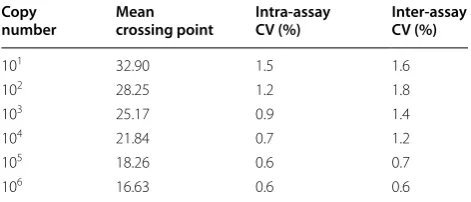
The SYBR green I real-time RT-PCR assay demonstrated high repeatability with coefficients of variation within runs (intra-assay variability) and between runs (inter-assay variability) that ranged from 0.6–1.5 and 0.6–1.8 %, respectively (Table 2).
One‑step RT PCR amplification of the IBV S1 gene and sequence analysis
For further confirmation of our results, we investigated whether tracheal samples that tested IBV-positive using SYBR green I real-time RT-PCR targeting the IBV N gene would also be positive using a one-step RT-PCR target-ing the IBV S1 gene. The IBV S1 gene of IBV commercial vaccine strains (H120, Ma5 and 4/91) and field strains “Moroccan 30” and “Moroccan 38” (randomly chosen from the 34 broiler chicken samples) were detected and amplified using a one-step PCR. The obtained RT-PCR product, 700 bp, corresponded to the predicted size (Fig. 9) [19]. The obtained sequences were aligned with those of IBV deposited in GenBank (H120 [M21970]; Ma5 [AY561713]; Italy 02 [AJ457137]; Italy 497/02-1 [DQ901377]; Ark/C6d [EU283056]; QX [AF193423]; strain 04/1991 [AF093794]; and D274 [X15832]), using the BLAST software. The results of sequenced field strains compared to IBV reference strains confirm that the amplified sequences corresponded to the IBV refer-ence strains [GenBank: KJ701019 and KJ701020]. Fig. 2 Agarose gel electrophoresis of conventional RT-PCR amplified
products from infectious bronchitis virus N gene. Electrophoresis of PCR amplification after serial dilutions of the IBV Beaudette strain in a 1.5 % agarose gel stained with ethidium bromide. Lanes 1 and
10 50-bp DNA ladder; lane 2 negative control; lane 3 108 copies/μl;
lane 4 107 copies/μl; lane 5 106 copies/μl; lane 6 105 copies/μl; lane 7 104 copies/μl; lane 8 103 copies/μl; lane 9 102 copies/μl. bp base pairs,
[image:3.595.57.292.88.193.2]Discussion
The use of PCR for diagnosis of viral diseases has increased to the point that the assay is now considered the gold standard instead of viral isolation [20]. Real-time PCR has catalyzed wider acceptance of PCR as a diagnos-tic tool because it is more rapid, sensitive, and reproduci-ble, and the risk of carryover contamination is minimized compared to conventional PCR [21].
Diagnosis of IB is commonly based on virus isolation in embryonated eggs, followed by immunological identi-fication of the isolates. This procedure is time consum-ing and requires use of specific polyclonal or monoclonal Fig. 3 Specificity of IBV N gene-based SYBR green I real-time RT-PCR. Amplification plot representing IBV (H120, Ma5, 4/91), FLUAV, IBDV, NDV, and negative control (NC). The blue curve shows the amplification plots of the IBV-positive samples while the turquoise curve shows the plots of the IBV-negative strains. FLUAV avian influenza virus A, IBV infectious bronchitis virus, IBDV infectious bursal disease virus, NDV newcastle disease virus
Fig. 4 SYBR green I real-time RT-PCR amplification plot derived from cDNA of IBV H120 vaccine strain. IBV infectious bronchitis virus, RT
-PCR reverse transcriptase-polymerase chain reaction
Table 1 Sensitivity and specificity of Sybr green I real-time RT-PCR in detection of cDNA from the IBV H120 vaccine strain compared to conventional RT-PCR agarose gel electrophoresis
CI confidence interval, Ct average cycle threshold, RT-PCR reverse transcriptase-polymerase chain reaction, + positive (P), − negative (N) cDNA concentration
(log10 copies/µl)
Detection method
SYBR green I real‑time RT‑PCR Conventional
RT‑PCR
Ct value Sensitivity Specificity P/N P/N
% CI % CI
101 36.31 0 0–69.5 100 19.3–100 − −
102 33.19 33.33 5.5–88.4 100 19.3–100 + −
103 26.59 66.67 11.6–94.5 100 19.3–100 + +
104 24.79 100 30.5–100 100 19.3–100 + +
105 21.90 100 30.5–100 50 8.2–91.8 + +
[image:4.595.58.540.86.307.2] [image:4.595.60.290.376.472.2] [image:4.595.58.541.562.717.2]antibodies, which may not be available. Moreover, some isolates could be mixtures of different serotypes of IBV that can confuse interpretation of results. RT-PCR tech-niques using RNA extracted from allantoic fluid and tra-cheal swabs from IBV-infected chickens are very efficient in the detection of IBV [22, 23]. However, conventional RT-PCR is time consuming, prone to error, and is less sensitive than real-time RT-PCR [3, 9, 24–26].
With the SYBR green I real-time RT-PCR assay that we developed, we compared the performance of this novel technique to conventional agarose gel-based RT-PCR in detecting specific IBV RT-PCR products. Based on seri-ally diluted cDNA standards, the SYBR green I real-time RT PCR detected down to 100 cDNA copies/µl of the IBV H120 vaccine strain genomic material. The SYBR green I real-time RT PCR assay targeting the highly conserved N
gene has a higher sensitivity than conventional RT-PCR in detecting IBV nucleic acids. Targeting the N gene has been postulated to be a better choice to improve the sen-sitivity of the PCR compared to the S1 gene, as the N pro-tein is abundant in infected cells [9, 27].
The specificity of our test was demonstrated by the absence of positive reactions with genetic mate-rial obtained from other RNA viruses, such as FLUAV, IBDV, and NDV (Fig. 3). The optimal melting peak of IBV amplicons strains is 68 ± 0.20 °C. No primer-dimer or nonspecific products were detected. These results are comparable with previously reported SYBR green I real-time RT-PCR assays established for coronaviruses and type-specific IBV multiplex SYBR green I real-time RT-PCR [24, 28].
SYBR green I real-time PCR and conventional RT-PCR were applied to 34 clinical samples obtained from Moroccan broiler chicken flocks showing clinical signs of IB. IBV N gene amplification with SYBR green I real-time RT-PCR detected all the IBV strains assessed, whereas only 28 of 34 clinical swabs tested were positive for IBV by conventional RT-PCR. The increased sensitivity of real-time RT-PCR compared to conventional RT-PCR might be due to detection of the fluorescent signal emit-ted by specific amplification products [24]. Therefore, the SYBR green I real-time RT-PCR assay developed here using Moroccan field cases could be an important tool for the screening of samples during IB-suspected cases as the assay is rapid, simple, efficient, highly specific, and sensitive.
Recently, real-time RT-PCR assays were described that can detect IBV either solely or in a multiplex set [24, 26,
28, 29]. However, none of these assays utilized the SYBR Fig. 5 Simple linear regression line of Ct values vs. log10 of tenfold
serial dilutions (106–10 copies/µl) of standard RNA genome of cDNA of H120 vaccine strain. Ct cycle threshold
Fig. 6 Agarose gel electrophoresis of conventional RT-PCR products from the N gene of IBV H120 strain. Amplified products were sepa-rated by electrophoresis on a 1.5 % agarose gel and stained with ethidium bromide. Lane 1 100 bp DNA ladder; lane 2 107 copies/μl;
lane 3 106 copies/μl; lane 4 105 copies/μl; lane 5 104 copies/μl; lane 6 103 copies/μl; lane 7 102 copies/μl; lane 8 10 copies/μl. IBV infectious bronchitis virus, RT-PCR reverse transcriptase-polymerase chain reac-tion
[image:5.595.306.538.86.262.2] [image:5.595.56.300.88.229.2] [image:5.595.57.292.263.425.2]green I chemistry for specific detection and quantifica-tion of IBV targeting the N gene. The main advantages of SYBR green I assays over other real-time PCR detection formats are: (1) SYBR green I is a low-cost fluorochrome; (2) SYBR green I assays are simpler to use, especially in regard to primer design and optimization procedures [25]; and (3) artifacts commonly observed in specific probes, particularly at amplification cycles beyond the 30th round, are minimal and can be ruled out by melting curve analysis. The assay described here is therefore ideal for establishing IB diagnostic capabilities in developing countries such as Morocco.
Conclusions
In conclusion, we report for the first time a one-step SYBR green I real-time RT-PCR protocol for IBV N gene detection. The assay is a sensitive, simple, efficient, and
cost-effective method that can be applied easily for labo-ratory diagnosis of IBV infections.
Methods Viruses
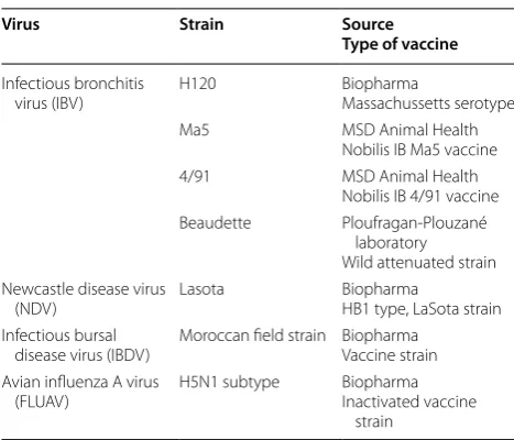
IBV was propagated in 9–11-day-old embryonated spe-cific pathogen-free chicken eggs as described previously [30]. The allantoic fluid was harvested 48 h post-inocula-tion and stored at −80 °C until RNA extraction. The dif-ferent viruses used in this study are listed in Table 3. Clinical standard samples
To validate the reliability of SYBR green-I-based real-time RT-PCR, the quantified IBV Beaudette strain was used as the standard control, and allantoic fluid with dif-ferent titers of the virus were obtained.
Tracheal swabs from IBV‑infected chickens
Tracheal samples were obtained from the Avian Unit, Agronomy and Veterinary Institute Hassan II, Rabat, Morocco. These samples were identified during rou-tine diagnostic efforts and stemmed from 34 commer-cial broiler chickens with clinical signs indicative of IB, including coughing, sneezing, rales, and nasal discharge. All samples had been confirmed to be IBV-positive by virus isolation, but serotypes were not determined. For this study, all samples were tested by SYBR green-I-based real-time RT-PCR and conventional RT-PCR. The swabs were suspended in 300 µl of phosphate-buffered saline, Fig. 8 Virus detection in tracheal swabs with SYBR green I real-time
RT-PCR. Tracheal swabs (34) were taken from broiler chickens show-ing clinical signs of infectious bronchitis. A subset of the results from 34 tracheal swabs using SYBR green I real-time RT-PCR are shown.
[image:6.595.307.541.86.239.2]NC negative control, RT-PCR reverse-transcriptase polymerase chain reaction
Table 2 Intra- and inter-assay reproducibility of SYBR green-I-based real-time RT-PCR
CV coefficient of variation Copy
number Mean crossing point Intra‑assay CV (%) Inter‑assay CV (%)
101 32.90 1.5 1.6
102 28.25 1.2 1.8
103 25.17 0.9 1.4
104 21.84 0.7 1.2
105 18.26 0.6 0.7
106 16.63 0.6 0.6
Fig. 9 Agarose gel electrophoresis of one-step RT-PCR amplified products of the IBV S1 gene. Commercial IBV vaccine (IBV H120, MA5, and 4/91) and field strains (“Moroccan 30” and “Moroccan 38”) were separated by electrophoresis on a 1.5 % agarose gel stained with eth-idium bromide. Lane 1 100 bp DNA ladder; lane 2 Moroccan 30 strain;
[image:6.595.60.289.88.252.2] [image:6.595.56.292.389.490.2]clarified by centrifugation at 1500×g at 4 °C for 15 min, and incubated at room temperature for 1 h. The virus-containing supernatants obtained from the tracheal swabs were used for direct extraction of viral RNA.
RNA extraction
IBV RNA and negative control RNA (NDV [183.6 ng/µl], IBDV [67.5 ng/µl], FLUAV [92.1 ng/µl]) were extracted from 140 µl of supernatant from allantoic fluid using the QIAamp Viral RNA or RNeasy Mini Kits (Qiagen, Valencia, CA, USA) according to the manufacturer’s instructions. Each RNA fraction was eluted in 50 µl of RNase-free water.
SYBR green‑I‑based real time RT PCR
SYBR green I-based real time RT-PCR was performed using a primer pair consisting of (a) downstream primer, AIBV-fr, targeting N gene nucleotide positions 811–832
(5’-ATGCTCAACCTTGTCCCTAGCA-3′); and (b)
upstream primer, AIBV-as, targeting N gene nucleotide
positions 921–941 (5′-TCAA-ACTGCGGATCA-TC
ACGT-3′, TIB Molbiol, Berlin, DE) as previously described by Meir et al. [9]. To minimize primer-dimer formation, primer set concentration and thermocycling conditions were both optimized (data not shown).
PCRs were performed on a Light Cycler (Roche Diag-nostics Ltd. Rotkreuz, CH). Each reaction was carried out using 5 µl of purified RNA and 20 µl of a reaction mixture, containing 0.5 µl of RT (50 U) (Applied Biosystems, Grand Island, NY, USA), 0.5 µl of RNase inhibitor (20 U), 0.5 µl of MgCl2 (50 mM), 2 µl of FastStart DNA Master Mix SYBR
green I (containing Taq DNA polymerase, SYBR green I, deoxynucleotide triphosphate mix [Life Technologies, Grand Island, NY, USA]), and 0.5 µl of the two primers
(final concentration of each primer is 10 µM) and enough nuclease-free water (Promega, Madison, WI, USA) for a final volume of 20 µL for each reaction. The thermal pro-file consisted of 10 min of reverse transcription at 50 °C, 10 min of hot-start enzyme activation at 95 °C, followed by 45 cycles of PCR at 95 °C for 10 s (denaturation), 58 °C for 20 s (annealing), and 72 °C for 30 s (elongation). To avoid cross contamination and sample carryover, pre- and post-PCR sample processing were performed in separate rooms. Plugged pipette tips were used to transfer all fluids to elim-inate aerosols. Amplified cDNA products were detected by melting curve analysis that consisted of 95 °C for 5 s and 65 °C for 60 s, and heated to 97 °C to measure continu-ous changes in fluorescence of SYBR green I. Following amplification, a melting curve analysis was performed with the Light Cycler instrument’s software (software release 1.5.0, version 1.5.0.39) according to the instructions of the manufacturer. Negative template control (diethyl pyrocar-bonate-treated water) and positive template controls (i.e., NDV, IBDV, FLUAV) were included with each PCR run. Each strain used was tested in triplicate.
Reproducibility of SYBR green‑I‑based real‑time RT‑PCR To evaluate reproducibility of the assay, quantification analysis of IBV cDNA was developed using the 34 clini-cal standard samples from Moroccan broiler chickens. cDNA of the genomic RNA of IBV Beaudette strain was used as the source of DNA templates. After quantifica-tion, IBV Beaudette strain cDNA was serially diluted (106–10 copies/µl) and used as a standard control. Three
separate dilution series were assayed in a single run to evaluate intra-assay variations, whereas inter-assay varia-tions were measured by testing each dilution in three sep-arate consecutive runs. The standard deviation (SD) was calculated using Light Cycler Software 480 version 1.5 (Roche Diagnostics Ltd. Rotkreuz, CH). The coefficient of variation (CV) was determined following the formula CV = (SD [Ct-value]/overall mean [Ct-value]) × 100.
Sensitivity of SYBR green‑I‑based real‑time RT‑PCR
The sensitivity of the SYBR green-I-based real-time RT-PCR assay was tested by the limiting dilution assay [31]. Genomic RNA from attenuated Massachusetts serotype H120 vaccine was then diluted to ≈100 (151.6 ng/μl), 20 (35.6 ng/μl), 5 (12.9 ng/μl), and 1.25 (0.7 ng/μl) copies/μl as previously described [32]. The tubes at each concentration were assayed using the forward and reverse primers. Sam-ples with Ct values of less than 32 were considered positive.
Conventional reverse transcriptase polymerase chain reaction
Conventional RT-PCR was performed in a thermal cycler (Techne, Staffordshire, UK), according to the instructions
Table 3 Viruses used in the study
Virus Strain Source
Type of vaccine
Infectious bronchitis
virus (IBV) H120 BiopharmaMassachussetts serotype Ma5 MSD Animal Health
Nobilis IB Ma5 vaccine 4/91 MSD Animal Health
Nobilis IB 4/91 vaccine Beaudette Ploufragan-Plouzané
laboratory Wild attenuated strain Newcastle disease virus
(NDV) Lasota BiopharmaHB1 type, LaSota strain Infectious bursal
disease virus (IBDV) Moroccan field strain BiopharmaVaccine strain Avian influenza A virus
[image:7.595.56.290.98.298.2]provided by the manufacturer. The RT-PCR was per-formed in one step, in a final volume of 20 μl. Briefly, the final volume consisted of 2.5 µl of buffer, 2.5 µl of MgCl2
(25 mM), 2.5 µl of deoxyribonucleotide triphosphates (10 mM), 9.7 µl of water, 0.5 µl of RNase inhibitor (20 U), 0.3 µl of reverse transcriptase (50 U), 0.5 µl of Taq poly-merase (Gold) (5 U), and 0.75 µl of each of the primers (forward and reverse) (10 µM).
One step, reverse transcription was performed at 48 °C for 30 min and 95 °C for 5 min. cDNA was ampli-fied through a 35-cycle PCR consisting of denaturation at 94 °C for 30 s, annealing at 52 °C for 30 s, and exten-sion at 72 °C for 30 s, with one final extenexten-sion cycle at 72 °C for 10 min. The amplified PCR products of the N gene were evaluated by 1.5 % agarose gel electrophoresis in Tris–borate–EDTA buffer stained with ethidium bro-mide (Promega).
Comparison of sensitivity and specificity of SYBR green I real‑time RT‑PCR with conventional RT‑PCR assay
The analytical sensitivity and specificity of the SYBR green I real-time RT-PCR assay were determined by test-ing sequential tenfold dilutions of the in vitro-transcribed RNA, which were obtained as previously described, in nuclease-free water (Promega).
One‑step RT‑PCR amplification of the IBV S1 gene
PCR amplification of the IBV S gene was carried out using commercial vaccines (H120, Ma5 and 4/91) and field strains, “Moroccan 30” and “Moroccan 38” from chickens with respiratory signs. Each 25 µl of PCR reac-tion mixture consisted of 2.5 μl of buffer (10X), 2.5 μl of MgCl2 (25 mM), 2.5 μl of deoxynucleotide
triphos-phates (10 mM), 0.75 μl of each primer (10 μM) (CK2 and S15mod) [19], and 9.7 μl of sterilized water. At the end, 0.5 μl of RNAase inhibitor (20 U/μl), 0.3 μl of RT (50 U/μl), 0.5 μl of Taq gold polymerase (ThermoFisher Sci-entific) (5 U/μl), and 5 µl of IBV RNA were added. This one-step RT-PCR reaction was carried out using Smart Thermal Cycler, (Smart Cycler Cepheid, Sunnyvale, CA, USA) following the protocol of 48 °C for 30 min, 95 °C for 5 min, 40 cycles of 95 °C for 30 s, 52 °C for 30 s, 72 °C for 30 s, and a final extension cycle of 72 °C for 10 min.
Nucleotide sequencing and sequence analysis
Following amplification, 20 μl of each S1 PCR reaction for “Moroccan 30” and “Moroccan 38” field strains and vaccine strains (H120, Ma5 and 4/91) were subjected to 1.5 % agarose gel electrophoresis (under 70 V for 30 min) and stained with ethidium bromide. The appropriate bands were excised and purified using the Gene Clean Kit (ExoSAP-IT, Affymetrix, Santa Clara, CA, USA) accord-ing to the manufacturer’s recommendations. Nucleotide
sequences were determined using the same primers (S15mod and CK2) [19] and the BigDye® Terminator v1.1 Cycle Sequencing Kit (Life Technologies, Grand Island, NY, USA). The second purification was performed by Big Dye X terminator Purification Kit.
Statistical analysis
The significant differences among the mean of the Ct values corresponding to each viral dilution were ana-lyzed by one-way analysis of variance (ANOVA) using Statistical Package for the Social Sciences (SPSS) Ver-sion 13 software (IBM, Armonk, NY, USA). A receiver operating characteristic curve statistical test was used to compare the sensitivity and specificity of the SYBR-green-I-based real-time RT-PCR over the conventional RT-PCR. A p value of less than 0.05 is considered statisti-cally significant.
Abbreviations
bp: base pairs; Ct: cycle threshold; ELISA: enzyme-linked immunosorbent assay; FLUAV: avian influenza virus; IB: infectious bronchitis; IBDV: infectious bursal disease virus; IBV: infectious bronchitis virus; M: membrane protein; N: nucleocapsid protein; NC: negative control; NDV: newcastle disease virus; RNA: ribonucleic acid; RT-PCR: reverse transcriptase-polymerase chain reaction; S: spike protein.
Authors’ contributions
MME conceived the study, and SF, MEH, MME participated in the design of the study. SF, KK, and MEH analyzed and interpreted data. SF, MEH, GS, and MME drafted and SF, MEH, and JHK edited the manuscript. EB participated in the statistical analyses. SF and KK participated in the molecular analyses. OFF participated in virus sequencing. All authors read and approved the final manuscript.
Author details
1 Unit of Avian Pathology, Agronomic and Veterinary Institute Hassan II, B.P. 6202, Rabat, Morocco. 2 Laboratory of Virology, Microbiology and Quality/ Ecotoxicology & Biodiversity, Faculty of Sciences and Techniques-University Hassan II Mohammedia, PO Box 146, Quartier Yasmina-Mohammedia, 20650 Casablanca, Morocco. 3 Laboratory of Molecular Biology-Society of Biological Products and Veterinary Pharmaceuticals (Biopharma), B.P. 4569, Km 2, Route de Casa, Rabat, Morocco. 4 Integrated Research Facility at Fort Detrick, National Institute of Allergy and Infectious Diseases, National Institutes of Health, B-8200 Research Plaza, Fort Detrick, Frederick, MD 21702, USA. 5 Laboratory of Epidemiology and Clinical Research. Faculty of Medicine, B.P. 1014, 4, Avenue Ibn Battouta, Rabat, Morocco.
Acknowledgements
We thank Laura Bollinger (Integrated Research Facility at Fort Detrick) for pro-viding Technical Writing Services in critically editing this manuscript on behalf of Battelle Memorial Institute.
Competing interests
The authors declare that they have no competing interests.
Sources of support
Higher Education of Morocco, University Hassan II Mohammedia-Casablanca, Mohammedia, Morocco, and the Society of Biological Products and Veterinary Pharmaceuticals (Biopharma), Rabat, Morocco, sponsored the molecular analyses and sequencing of the different samples.
Received: 1 February 2015 Accepted: 11 April 2016
References
1. Cavanagh D. Coronavirus avian infectious bronchitis virus. Vet Res. 2007;38(2):281–97.
2. Keeler CL Jr, Reed KL, Nix WA, Gelb J Jr. Serotype identification of avian infectious bronchitis virus by RT-PCR of the peplomer (S-1) gene. Avian Dis. 1998;42(2):275–84.
3. Callison SA, Riblet SM, Oldoni I, Sun S, Zavala G, Williams S, Resurreccion RS, Spackman E, Garcia M. Development and validation of a real-time Taqman PCR assay for the detection and quantitation of infectious laryn-gotracheitis virus in poultry. J Virol Methods. 2007;139(1):31–8. 4. Alarabi MAH: A field study of kidney disease among the broiler flocks in
Morocco and its relationship to infectious bronchitis virus. Ph.D thesis. Agronomic and Veterinary Institute Hassan II, Rabat; 2004.
5. El Bouqdaoui M, Mhand RA, Bouayoune H, Ennaji MM. Genetic group-ing of nephropathogenic avian infectious bronchitis virus isolated in Morocco. Int J Poult Sci. 2005;4:721–7.
6. El-Houadfi M, Jones RC, Cook JK, Ambali AG. The isolation and characteri-sation of six avian infectious bronchitis viruses isolated in Morocco. Avian Pathol. 1986;15(1):93–105.
7. King AMQ, Adams MJ, Carstens EB, Lefkowitz EJ, eds. Virus taxonomy: clas-sification and nomenclature of viruses: Ninth Report of the International Committee on Taxonomy of Viruses. San Diego: Elsevier Academic Press; 2012. http://www.ictvonline.org/index.asp?bhcp=1. Accessed 19 Sept 2014.
8. Lai MM, Cavanagh D. The molecular biology of coronaviruses. Adv Virus Res. 1997;48:1–100.
9. Meir R, Maharat O, Farnushi Y, Simanov L. Development of a real-time TaqMan RT-PCR assay for the detection of infectious bronchitis virus in chickens, and comparison of RT-PCR and virus isolation. J Virol Methods. 2010;163(2):190–4.
10. Kwon HM, Jackwood MW, Gelb J Jr. Differentiation of infectious bronchitis virus serotypes using polymerase chain reaction and restriction fragment length polymorphism analysis. Avian Dis. 1993;37(1):194–202.
11. Karaca K, Naqi S. A monoclonal antibody blocking ELISA to detect serotype-specific infectious bronchitis virus antibodies. Vet Microbiol. 1993;34(3):249–57.
12. Karaca K, Naqi S, Gelb J Jr. Production and characterization of monoclo-nal antibodies to three infectious bronchitis virus serotypes. Avian Dis. 1992;36(4):903–15.
13. Naqi SA, Karaca K, Bauman B. A monoclonal antibody-based antigen cap-ture enzyme-linked immunosorbent assay for identification of infectious bronchitis virus serotypes. Avian Pathol. 1993;22(3):555–64.
14. Jackwood MW, Yousef NM, Hilt DA. Further development and use of a molecular serotype identification test for infectious bronchitis virus. Avian Dis. 1997;41(1):105–10.
15. Kingham BF, Keeler CL Jr, Nix WA, Ladman BS, Gelb J Jr. Identification of avian infectious bronchitis virus by direct automated cycle sequencing of the S-1 gene. Avian Dis. 2000;44(2):325–35.
16. Cavanagh D, Davis P, Cook J, Li D. Molecular basis of the variation exhib-ited by avian infectious bronchitis coronavirus (IBV). Adv Exp Med Biol. 1990;276:369–72.
17. Callison SA, Hilt DA, Boynton TO, Sample BF, Robison R, Swayne DE, Jackwood MW. Development and evaluation of a real-time Taqman RT-PCR assay for the detection of infectious bronchitis virus from infected chickens. J Virol Methods. 2006;138(1–2):60–5.
18. Hairul Aini H, Omar AR, Hair-Bejo M, Aini I. Comparison of Sybr Green I, ELISA and conventional agarose gel-based PCR in the detection of infec-tious bursal disease virus. Microbiol Res. 2008;163(5):556–63.
19. Eterradossi N, Britton P: Avian infectious bronchitis. In: Biological Stand-ards Commission, editor. Manual of diagnostic tests and vaccines for ter-restrial animals. edn. World Organisation for Animal Health; 2013. http:// www.oie.int/fileadmin/Home/eng/Health_standards/tahm/2.03.02_AIB. pdf. Accessed 2 May 2014.
20. De Wit JJ. Detection of infectious bronchitis virus. Avian Pathol. 2000;29(2):71–93.
21. Mackay IM. Real-time PCR in the microbiology laboratory. Clin Microbiol Infect. 2004;10(3):190–212.
22. Cavanagh D, Mawditt K, Britton P, Naylor C. Longitudinal field studies of infectious bronchitis virus and avian pneumovirus in broilers using type-specific polymerase chain reactions. Avian Pathol. 1999;28(6):593–605. 23. Handberg K, Nielsen O, Pedersen M, Jorgensen P. Detection and strain
differentiation of infectious bronchitis virus in tracheal tissues from experimentally infected chickens by reverse transcription-polymerase chain reaction. Comparison with an immunohistochemical technique. Avian Pathol. 1999;28(4):327–35.
24. Acevedo AM, Perera CL, Vega A, Rios L, Coronado L, Relova D, Frias MT, Ganges L, Nunez JI, Perez LJ. A duplex SYBR Green I-based real-time RT-PCR assay for the simultaneous detection and differentiation of Massa-chusetts and non-MassaMassa-chusetts serotypes of infectious bronchitis virus. Mol Cell Probes. 2013;27(5–6):184–92.
25. Aldea C, Alvarez CP, Folgueira L, Delgado R, Otero JR. Rapid detection of herpes simplex virus DNA in genital ulcers by real-time PCR using SYBR green I dye as the detection signal. J Clin Microbiol. 2002;40(3):1060–2. 26. Cavanagh D, Gelb J: Infectious bronchitis. In: Saif Y, editor. Diseases of
poultry, 12 edn. Ames: Blackwell Publishing Professional; 2008. p. 117–30. https://www.himakahaunhas.files.wordpress.com/2013/2003/disease-of-poultry.pdf.
27. Spencer KA, Hiscox JA. Expression and structural analysis of infectious bronchitis virus nucleoprotein. Adv Exp Med Biol. 2006;581:133–8. 28. Escutenaire S, Mohamed N, Isaksson M, Thoren P, Klingeborn B, Belak S,
Berg M, Blomberg J. SYBR Green real-time reverse transcription-polymer-ase chain reaction assay for the generic detection of coronaviruses. Arch Virol. 2007;152(1):41–58.
29. Fan WQ, Wang HN, Zhang Y, Guan ZB, Wang T, Xu CW, Zhang AY, Yang X. Comparative dynamic distribution of avian infectious bronchitis virus M41, H120, and SAIBK strains by quantitative real-time RT-PCR in SPF chickens. Biosci Biotechnol Biochem. 2012;76(12):2255–60.
30. Villarreal LY, Brandao PE, Chacon JL, Saidenberg AB, Assayag MS, Jones RC, Ferreira AJ. Molecular characterization of infectious bronchitis virus strains isolated from the enteric contents of Brazilian laying hens and broilers. Avian Dis. 2007;51(4):974–8.
31. Taswell C. Limiting dilution assays for the determination of immunocom-petent cell frequencies. I. Data analysis. J Immunol. 1981;126(4):1614–9. 32. Chandriani S, Ganem D. Array-based transcript profiling and