A COMPARATIVE STUDY BETWEEN OPEN
AND LAPAROSCOPIC CHOLECYSTECTOMY
IN GALLSTONE DISEASE
M.S. GENERAL SURGERY
DEGREE EXAMINATION
Dissertation On
PART II
THE TAMILNADU DR.M.G.R. MEDICAL UNIVERSITY TIRUNELVELI MEDICAL COLLEGE HOSPITAL
ii
CERTIFICATE
This is to certify that this dissertation entitled. “A comparative study between open And Laparoscopic cholecystectomy In gallstone disease” is a bonafide record of the work done by Dr. S.Saravana Kumar under my supervision and guidance in the department of General surgery of Tirunelveli
Medical College. Tirunelveli during the period of his post graduate study from
may 2007 to march 2010. For the partial fulfillment of M.S (General surgery)
degree.
The Dean
Tirunelveli Medical College Tirunelveli.
Dr. S.Murugan, M.S.,
Professor of Surgery,
Department of Surgery,
Tirunelveli Medical College Hospital,
Tirunelveli.
Dr.J.Jeyakumar Sagayam M.S,
Professor and Head of the Department,
Department of Surgery,
Tirunelveli Medical College Hospital,
iii
ACKNOWLEDGEMENT
I am extremely thankful to our beloved Dean, Dr. A. Kanagaraj. M.D.,
for granting me permission to carry out this study in Tirunelveli medical
college.
It is an immense pleasure to acknowledge Professor. Dr.J.Jeyakumar
Sagayam M.S, Department of Surgery, who has given the moral support,
philosophical guidance and ever-available help to carry out this study.
With deepest appreciation and gratitude, I thank Prof. Dr. S.Murugan
M.S., , Prof. Dr.K. Parimalam M.S., My Unit Chiefs & Additional professor of Surgery.
And I thank the retired professors:
Prof. Dr. G. Thangaiah M.S., Prof. Dr. A. Chidambaram M.S., Prof. Dr.D. Balaji M.S., Prof.Dr. S. Ravindran M.S.,
Prof. Dr. P. Janakiram M.S.,
I also thank Dr. M.S.Varadarajan M.S., Dr. G.Nirmal Kumar M.S., and
Dr. Irene Aruna Edwin M.S ., assistant professors of my unit, for their moral
support.
Finally with grace of almighty God and with the cooperation of the
1
INTRODUCTION
Gastro-intestinal surgery has undergone a revolution in the recent
years by the introduction of laparoscopic techniques. The concept of
“keyhole surgery” created an immediate disparity between the potential of
the new technique and training of surgeons to perform it. Now modern
surgical methods are aimed at giving cure along with minimal invasive
techniques with patient in mind, safety never being compromised.
Cholelithiasis, which continues to be one of the most common digestive
disorders encountered, was traditionally being dealt by conventional or
open cholecystectomy. With the introduction of laparoscopic
cholecystectomy, the surgical community witnessed a revolution in basic
ideology and the importance of minimal access surgery.
Laparoscopic cholecystectomy (LC) has become so safe and easy
that it can be performed with much ease and safety because of better
magnification. Although LC has shown clear benefits in terms of
shortened hospital stay, less morbidity, mortality, a quicker return to work
and with cosmetic advantage, many questions regarding this procedure
remain unanswered, particularly relative to the gold standard procedure of
open cholecystectomy.
Some surgeons have suggested that the rates of serious
complications, particularly bile duct injury might be significantly higher in
2
Apart from the high costs of the equipment and the specialized
training that is mandatory for mastery of the technique, the procedure
inherently carries hazards and risks.
In a developing country like ours, where the medical costs and loss
of working days constitute major issues, could laparoscopic
cholecystectomy establish itself as a safe and cost effective alternative to
the open method?
In our study, we have made an attempt to compare the advantages
3
AIMS AND OBJECTIVES
The aim of this study is to compare conventional cholecystectomy
and laparoscopic cholecystectomy with respect to:
1. Duration of the procedure.
2. Post operative discomfort or pain.
3. Analgesic requirement.
4. Complications encountered.
5. Diet
6. Period of hospitalization.
7. Return to normal activity
4
REVIEW OF LITERATURE
Historical review:Most of the progress in the diagnosis and treatment of biliary tract
disease has been made in the last century, but gall stones and their
sequelae which cause most of the maladies date back to 1085-945 BC
having been discovered in the mummy of Priestess of Amen1.
• The first systematic data about the disease was published as “De
Medical Historic Mirabilis” by Marcellus Donatus1 in 1596.
• Zambeccari1 in 1636 performed cholecystectomy in a dog.
• The first cholecystectomy is credited to John Strong Bobbs1 on
June 15, 1867.
• Karl Langenbuch of Berlin performed first planned
cholecystectomy on July 15, 1882 using the aseptic technique of
Joseph Lister2.
Laparoscopy took its origin in 1901 when George Kelling
examined the abdominal cavity with an endoscope and named the
procedure as celioscopy. He used air through a puncture needle to
produce pneumoperitoneum.
• In 1929, Kalk introduced purpose designed instruments and was
the firs to advocate dual trocar technique which opened the way for
5
• In 1933, Fervers reviewed his experience with 50 patients and
recommended changing from room air to oxygen or carbon dioxide
as an insufflating agent.
• In 1938, Janos Veress developed his spring loaded needle, the
instrument of choice for creating pneumoperitoneum which
remains almost unchanged to the present day.
• In 1960, Professor Kurt Semm in Germany developed an automatic
insufflation device that monitored abdominal pressure and gas
flow. He also developed a number of endoscopic instruments
including thermo coagulation, angled lens, hook scissors, uterus
vacuum mobiliser and endo-loop applicator. He developed an
irrigation-aspiration apparatus with design modification to prevent
tube clogging and also popularized many laparoscopic procedures.
He also facilitated laparoscopic training by creating the pelvi-
trainer designed to demonstrate techniques required for operative
laparoscopy.
• Hassan proposed a method called “open technique” which provided
direct visualization of peritoneal cavity prior to trocar insertion.
This “Hassan technique” has greatly reduced the complication rate
associated with percutaneous or blind trocar entry into the
6
• The first ever laparoscopic cholecystectomy was performed by
Philip Mourret2 in Lyon in 1987 and Dubois performed it in Paris
in 1988.
• In 1991, Tehemton Udwadia performed the first laparoscopic
cholecystectomy in India.
Anderson et al3 in a study to compare the effectiveness of LC over
OC found that the hospital charge was $4070+ 297 for patients
undergoing LC and $5017 + 497 for patients undergoing OC. This
difference arose from the mean cost of in patient care which was $353 +
40 for LC patients and $1335+ 138 for OC patients. LC appeared to be
7
SURGICAL ANATOMY
Knowledge of relevant anatomy is important for the safe execution
of any operative procedure. Specifically, in the context of a
cholecystectomy, it has been recognized since long that misinterpretation
of normal anatomy as well as the presence of anatomical variations
contribute to the occurrence of major postoperative complications
especially biliary injuries. Such injuries in turn can cause significant
morbidity and occasionally even mortality. They are also one of the
commonest causes of litigation against abdominal surgeons in the
developed world. There is now a fair amount of data to suggest that the
acceptance of laparoscopic cholecystectomy (LC) as the standard
procedure, has led to an increase in bile duct injuries. This seems partly
related to the different anatomical exposure of the area around the
gallbladder especially the Calot's triangle during the laparoscopic
procedure as opposed to the open procedure.
Hence, it is important for biliary and minimally invasive surgeons
to appreciate basic anatomical facts as they apply to the performance of
cholecystectomy as well as understand from literature how anatomical
distortions or variations can contribute to complications. This review
attempts to address these issues. It is not an exhaustive description of
biliary anatomy but discusses anatomical facts that are of relevance to the
8 Gallbladder:
The gallbladder is a pear shaped organ situated in a fossa on the
liver undersurface. It may be partially or completely embedded within the
liver parenchyma, the so-called 'intrahepatic' gallbladder. This may create
difficulties in dissection and may increase the chance of intraoperative
injury to the liver. Although the main right pedicle is fairly deep in the
liver parenchyma, large portal, and hepatic venous branches traverse the
liver at a depth of around one cm from the gallbladder. Thus, a deep liver
tear during the dissection of the gallbladder off its fossa can occasionally
bleed profusely. Also, during the dissection it may be important to err on
the side of the gallbladder rather than the liver parenchyma.
The gallbladder is divided into a fundus, a body and a neck or
infundibulum. The 'Hartmann's pouch' an out pouching of the wall in the
region of the neck is recognized more as an outcome of pathology in the
form of dilatation or presence of stones. This pouch is variable in size but
a large Hartmann's pouch may obscure the cystic duct and the Calot's
triangle. This may be result of plain enlargement or due to adherence to
the cystic duct or bile duct. Thus a small cystic duct can get completely
hidden and traction on the gallbladder can lead to the bile duct looking
like the cystic duct. An exaggerated form of the same process is the
'Mirizzi's syndrome' in which a large stone in the Hartmann's pouch area
is either adherent to or erodes into the bile duct. This can create major
9 Cystic duct:
The cystic duct joins the gallbladder to the bile duct and is one of
the important structures needing proper identification and division during
a standard cholecystectomy. The cystic duct may run a straight or a fairly
convoluted course. Its length is variable and usually ranges from 2 to 4
cm. Around 20% of cystic ducts are less than 2 cm. Hence there may be
very little space to put clips or ligatures. True absence of the cystic duct is
extremely rare and if the duct is not seen is more likely to be hidden. The
cystic duct is usually 2-3 mm wide. It can dilate in the presence of
pathology (stones or passed stones). The normal bile duct is also around 5
mm and hence can look like a mildly dilated cystic duct. In general a
cystic duct larger than 5 mm (or the need to use a very large clip to
completely occlude the duct) should arouse a suspicion of mistaken
identity with the bile duct before it is clipped or ligated.
The cystic duct joins the gallbladder at the neck and this angle may
be fairly acute. Also the mode of joining may be smooth tapering or
abrupt. On the bile duct side its mode of union shows significant
variations. Since such variations are not uncommon it may not be safe to
try and dissect the cystic duct to its junction with the bile duct. It is
important to remember that even in the low insertion variety the cystic
duct rarely goes behind duodenum and therefore a ductal structure
10
Double cystic ducts are described but are exceedingly rare and therefore
two ductal structures entering the gallbladder should always be viewed
with suspicion. Also the cystic duct does not have vessels traveling on its
surface whereas the bile duct has such visible vessels.
Cystic artery and right hepatic artery
The cystic artery is a branch of the right hepatic artery (RHA) and
is usually given off in the Calot's triangle. It has a variable length and
enters the gallbladder in the neck or body area. The course and length of
the cystic artery in the Calot's triangle is variable. Although classically
the artery traverses the triangle almost in its center, it can occasionally be
very close or even lower than the cystic duct.
It usually gives off an anterior or superficial branch and a posterior
or deep branch. This branching usually takes place near the gallbladder.
When the point of dissection is very close to the gallbladder as in a LC or
the branching is proximal, one may have to separately ligate the two
branches. Also if the presence of a posterior branch is not appreciated it
can cause troublesome bleeding during posterior dissection.
The RHA normally courses behind the bile duct and joins the right
pedicle high up in the Calot's triangle. It may come very close to the
gallbladder and the cystic duct in the form of the 'caterpillar' or
'Moynihan's' hump. Although the incidence of this variation is variable it
11
as 50%. If such a hump is present, the cystic artery in turn is very short.
In this situation the RHA is either liable to be mistakenly identified as the
cystic artery or torn in attempts to ligate the cystic artery. The ensuing
bleeding in turn predisposes to biliary injury.
There are a fair number of other arterial variations of the cystic
artery also described. Many of these are unlikely to cause confusion if the
artery is divided very close to the gallbladder wall. There is a 2-15%
incidence of double cystic artery. Therefore it may be occasionally
necessary to ligate two arteries to the gallbladder. When the cystic artery
is given off not from the RHA but from other vessels like the common
hepatic artery or the left hepatic artery (2-5%) it crosses the bile duct
anteriorly and may be prone to injury. Also the superior mesenteric artery
may give off the cystic artery in which case it ascends to the gallbladder
below the cystic duct. An accessory or replaced RHA from superior
mesenteric artery which is a variation seen in almost 15% of individuals
the RHA courses thru the Calot's triangle (and therefore nearer the
gallbladder) and in turn has a shorter cystic artery.
Accessory and aberrant ducts:
There are a large number of accessory ducts described in the biliary
drainage network of the liver. These ducts are typically small and course
through the Calot's triangle (and therefore closer to the gallbladder)
12
confluence of the right and left duct at variable distances. Sometimes the
cystic duct may actually join the accessory duct.
Some of these ducts may drain substantial portions of the right lobe
of the liver, either one of the sectors (two segments) or a segment and
may in fact be the sole drainage of that part of the liver in which case they
are more precisely termed as 'aberrant' ducts. It has been suggested that
most such ducts are aberrant rather than accessory in which case it is even
more important to safeguard them. If such a duct is injured it can lead to
substantial biliary stasis or leak. The size of the duct may be an indirect
indicator of the amount of liver it drains. It has hence been recommended
that in case of injury if the duct is more than 3 mm it should always be
drained into a Roux loop. Alternatively one can perform a cholangiogram
through the duct to assess the amount of liver it drains as well as whether
it is accessory or aberrant. With increasing recognition of injury to such
ducts these have now been grouped into separate type in the recent
Strasberg classification of bile duct injuries.
Calot's triangle
This famous triangle was described as bound by the cystic duct, the
common hepatic duct and the cystic artery in its original description by
Calot in 1891. In its present interpretation the upper border is formed by
the inferior surface of the liver with the other two boundaries being the
13
RHA, the cystic artery, the cystic lymph node (of Lund), connective
tissue, and lymphatics. Occasionally it may contain accessory hepatic
ducts and arteries as discussed previously. It is this triangular space,
which is dissected in a cholecystectomy to identify the cystic artery and
cystic duct before ligation and division. In reality, it may be a small
potential space rather than a large triangle making the dissection of its
contents without damaging the bordering structures the most challenging
step of a cholecystectomy. In addition the space may be obscured and
shrunken by various mechanisms. The left (or medial) boundary of the
triangle formed by the common hepatic duct is the most important
structure, which needs to be safeguarded.
Laparoscopic Anatomy:
The different anatomical 'laparoscopic view' of the area around the
gallbladder especially the Calot's triangle does contribute to
misidentification of structures. The method of retraction during the
laparoscopic procedure tends to distort the Calot's triangle by actually
flattening it rather than opening it. Also, the reluctance to (or difficulty
in) performing a fundus first cholecystectomy during the laparoscopic
procedure as opposed to the open procedure also contributes to the same
lack of exposure of the Calot's triangle. Finally, the 'posterior' or 'reverse'
dissection of the Calot's triangle, which is popular during an LC, again
14
during this method may lead to further anatomical distortion. The
Rouviere's sulcus is a fissure on the liver between the right lobe and
caudate process and is clearly seen during a LC during the posterior
dissection in a majority of patients. It corresponds to the level of the porta
hepatis where the right pedicle enters the liver. It has hence been
recommended that all dissection be kept to a level above (or anterior) to
this sulcus to avoid injury to the bile duct. Also, this being an
'extrabiliary' reference point it does not get affected by distortion due to
pathology. Similarly, a clear delineation of the junction of the cystic duct
with the gallbladder along with the demonstration of a space between the
gallbladder and the liver clear of any other structure other than the cystic
artery (safety window or critical view) is also recommended as an
essential step to prevent bile duct injury.
Investigations to assess the anatomy:
Most cholecystectomies are performed after identification of
gallstone disease on ultrasound examination. Although on occasion an
ultrasound examination can predict gross distortions of anatomy like the
Mirizzi syndrome, in the usual case it does not throw any light on
anatomical relations. Thus knowledge of the specific anatomy in that
individual is not available to the surgeon preoperatively as a routine. If a
cholangiogram in the form of a magnetic resonance cholangio
15
cholangiopancreatography (ERCP) has been performed for some reason,
it may reveal anomalies like the presence of accessory ducts or a low
insertion of cystic duct.
Methods to assess anatomy during the surgery are perhaps more
relevant. The first and foremost (and perhaps the most reliable) is clean
dissection and accurate visual identification of the contents of the Calot's
triangle especially the cystic artery and duct. The role of a routine
intraoperative cholangiogram in delineating biliary anatomy and in turn
preventing misidentification has been a subject of a long and intense
debate amongst biliary surgeons but there is conflicting evidence on its
value. In reality most biliary surgeons do not perform a routine
intraoperative cholangiogram but use it selectively. Also a cholangiogram
may not delineate all aberrant ducts and does not provide any insight into
arterial anatomy.
This use of laparoscopic ultrasound for identification of structures,
laparoscopic Doppler for identification of arteries and the use of an
instrument called the tactile sensor probe. Some recent reports describe
innovative methods such as the injection of a dye called methelenum
coeruleum into the gallbladder which gives a blue color to the biliary
system and the introduction of a small optical fiber through ampulla of
vater which illuminates the entire biliary tree during the cholecystectomy
16
costly technology, are largely unavailable and have not been scientifically
validated. Thus, it seems that presently there is no good alternative to
meticulous dissection in a planned manner with precise identification of
17
Cholelithiasis
There has been marked rise of gallstone disease during the past
century. Prevalence rate varies from less than 1% (in Africa) from 38 %(
in Sweden).The prevalence in females is approximately twice that of
male. In India the prevalence is about 6% and incidence in north India is
significantly higher than south India (4).
Pathological features (5)
Bile has three major constituents:
• Bile salts (primary: cholic and chenodeoxycholic acids; secondary:
deoxycholic and lithocholic acids).
• Phospholipids (90% lecithin).
• Cholesterol.
Bile containing excess cholesterol relative to bile salts and lecithin
is predisposed to gallstone formation.
Types of gallstones:
• Pure cholesterol (10%). Often solitary, large (> 2.5cm), round.
• Pure pigment (bile salts; 10%). Pigment stones are of two types:
Black (associated with haemolytic disease)
Brown (associated with chronic cholangitis and biliary parasites).
18 Predisposing conditions:
• Increasing age.
• Female (pregnancy and use of the oral contraceptive).
• Obesity.
• Multiparity.
• Chronic haemolytic disorders (only for pigment stones).
• Long-term parenteral nutrition (alteration of bile constituents).
• Previous surgery (e.g. vagotomy or resection of the terminal ileum)
or disease involving the distal small bowel (e.g. Crohn's disease)
alteration of bile constituents.
Clinical features (common presentations)
• Biliary colic
• Intermittent severe epigastric and right upper quadrant; usually
associated with nausea and vomiting. Resolves after a few hours;
tenderness over gallbladder during acute episodes.
• Acute cholecystitis
• Severe continuous right upper quadrant pain; often radiates to right
flank and back associated with anorexia and pyrexia. Tenderness
19
Complications of acute cholecystitis include:
• Formation of an empyema or abscess of the gallbladder (rare)
indicated by high swinging fever and severe localized pain;
• Perforation with biliary peritonitis (very rare);
• Cholecystoenteric fistula formation (may lead to a gallstone
entering and obstructing the distal ileum
• Jaundice due to compression of the adjacent common bile duct by
swelling (Mirizzi syndrome).
Chronic cholecystitis:
A mucocele of the gallbladder or infection producing an empyema.
Diagnosis and investigations:
• Full blood count, Urea & Electrolytes, LFTs, blood culture, serum
amylase in acute presentations
• Abdominal X-ray. Only 10% of calculi are radio-opaque.
• Oral cholecystogram (Graham-Cole test): rarely used.
• Ultrasound procedure of choice. Identifies stones, determines wall
thickness, and assesses ductal dilatation.
• Hepatobiliary iminodiacetic acid (HIDA) scan: useful when
20 Surgical treatment:
Cholecystectomy:
Vast majority done laparoscopically. Often done as a day case.
This is the treatment of choice for all patients fit for General anaesthesia.
Indicated for:
• Patients with symptoms deemed to be due to gallbladder stones;
• Asymptomatic patients with gallbladder stones at risk of
complications (diabetics, porcelain gallbladder (15-20% associated
with carcinoma), history of pancreatitis, long-term
immunosuppressed).
Non-surgical treatments:
• Percutaneous drainage of gallbladder
• Done under ultrasound or Computed Tomography guidance.
• Used for empyema of the gallbladder in patients unsuitable for
emergency cholecystectomy.
• After resolution of the infection the calculi may be removed
percutaneously.
Dissolution therapy:
• Rarely used. Requires a functioning gallbladder, small stones.
• Problems: requires prolonged treatment, less than 70% response,
21
• Extracorporeal shock wave lithotripsy
• Hardly ever used. Risk of visceral injury and high risk of stone
recurrence.
22
SURGICAL PROCEDURES
Cholecystectomy:Indications6:-
• Cholelithiasis – with or without symptoms
• Acute or chronic cholecystitis – with or without stones
• Symptomatic gall bladder polyps
• Gall bladder carcinoma
• Torsion of gall bladder
• Traumatic rupture of gall bladder or cystic duct
• Biliary peritonitis – with or without demonstrable perforation
• Internal biliary fistula
• Gas in the gall bladder
• Non functioning gall bladder
Contraindications:
For open cholecystectomy:
• Asymptomatic gall stones or producing little trouble on poor risk,
aged and feeble patients
23 Laparoscopic Cholecystectomy :
Contraindications6: 1. Absolute:
• Peritonitis or cholecysto-enteric fistula
• Acute Pancreatitis
• Cholangitis
• Portal Hypertension
• Pregnancy
• Major bleeding disorder
• Carcinoma of gall bladder
• Morbid obesity
2. Relative:
• Prior abdominal surgeries
• Empyema gall bladder
• Common bile duct stones
24
TECHNIQUE OF OPEN CHOLECYSTECTOMY
Pre-Operative Assessment:After appropriate history taking and assessment of the patient’s
fitness for the procedure, patient is given prophylactic antibiotics either
with the premedication or at the induction time. Anti-thrombotic
prophylaxis is undertaken and a consent form is signed.
Operation:
The surgeon should have a perfect knowledge of anatomy with
congenital anomalies to minimize the complications.
Operation technique:
The patient is placed in the dorsal recumbent position. For obese patients
the table is placed in a slight reverse Trendelenburg position to aid the
downward displacement of liver.
Incision:
The Kocher’s right sub-costal incision is especially useful in
patients who are very obese or in whom the costal angle is wide. Vertical
incisions, either midline, right paramedian using a muscle slide technique
or a muscle split are also quite satisfactory.
The gall bladder is appropriately exposed and the packs are placed
on the hepatic flexure, duodenum and lesser omentum and retracted.
The gall bladder if found distended is emptied by aspirating bile using a
25 1. Retrograde cholecystectomy:
Here, the peritoneum overlying Calot’s triangle is placed on stretch
and divided close to the gall bladder wall. The fat on Calot’s triangle is
dissected to expose cystic artery and cystic duct. The cystic duct is
cleared down to CBD and the cystic artery is tied and divided. Then the
cystic duct is divided between ligatures.
2. Ante grade/ Fundus first method:
It is done when anatomy is not clear. Here mesentery of the gall
bladder is incised close to liver at a point above the neck of the gall
bladder and with finger dissection body and fundus of gall bladder is
detached from the GB fossa with minimum trauma to the liver tissue.
Then the cystic artery and cystic duct are approached and divided
between ligatures.
Modifications of fundus first method:-
• Espiner’s modification - is particularly suitable for thickened and inflamed gall bladder where dissection of GB from its bed is
carried out in the sub mucosal plane using diathermy. This obviates
the requirement to control the cystic artery and minimize the risk to
CBD.
• Lahey’s method- When GB is found to be inflamed and friable, no clamp or haemostat is applied. The medial peritoneal reflection of
26
finger is passed behind the GB in the layer of fascia over the liver
and swept upwards from its fossa. The body and fundus of gall
bladder is detached from the liver after which the cystic artery and
cystic duct are clearly displayed and dealt with.
After the gall bladder is extracted, haemostasis is secured and the
abdomen closed with or without drain.
Intraoperative problems:
It arises in the following situations:-
1. Narrow CBD- Here dissection is commenced close to the GB neck
and cystic duct is ligated at GB neck.
2. Moynihan’s hump- Prior to dividing cystic artery it is traced back
to its origin and properly identified.
3. Acute inflammation- Here fundus first method is preferable.
4. Portal hypertension- Here the chances of injury to liver are high
27
EQUIPMENTS AND INSTRUMENTS FOR
LAPAROSCOPIC CHOLECYSTECTOMY
Video laparoscopic surgery has been made possible by the major
advances in video technology. The combination of equipment and the
skills to use the equipment constitute the essentials in laparoscopic
surgery.
Essential Equipments: Light Source:
A high intensity light source such as Xenon with variable intensity
and a light filter provides adequate visualization of abdominal cavity at
various distances.
Fibre optic light guide cable:
A 5mm thick, 225cm long cable is desirable. A thick cable carries
more light and a long cable is more convenient and less likely to be
stretched.
Video camera:
To maximize the visualization of structure, single chip and viewing
camera having 480 lines/ inch resolution is the minimum requirement. It
is attached to the scope and cable hooked to a processor that transmits the
28
are expensive, but provide the best image. All cameras require white
balancing.
Laparoscope:
It is based on the Hopkins rod lens system. It is available in many
sizes, 10mm, 7mm, 5mm and the new 2mm. It may have 00 forward
views or 30/ 450 angled views. Telescope tips fog due to temperature
differences outside and inside the patient. This is aggravated by the cold
insufflation. Warming the telescope in warm water before use and
touching the tip to the liver surface avoids fogging.
High flow insufflator:
It is used to insufflate carbon dioxide to create pneumoperitoneum.
As a safeguard, it also monitors the intra abdominal pressure constantly to
stop the flow once 12mm Hg of pressure is achieved and also has
indicators for rate of flow and total volume of gas delivered. A rate of
8-10 L/min delivery is ideal but at least 6L/min is the minimum required.
Carbon dioxide is the standard gas used for creation of
pneumoperitoneum. It can be insufflated directly into the blood stream in
volumes up to 100L/min without serious metabolic effect. It suppresses
29 High resolution video monitor:
It should be capable of 480 Hz lines/ inch for one chip camera and
700Hz lines/inch for three chip camera. Monitors should be at least 13
inch in size for adequate visibility and must be grounded. For teaching
and documentation, printers and video recorders are invaluable.
Irrigation device:
A pressure of 300 mm Hg is usually used to irrigate the abdomen
-either manual or powered. The irrigation/ aspiration probes may have a
single channel for both these functions or separate channels. Heparin
1000U/L may be added to the irrigation fluid to minimize clot formation.
Electro-cautery:
It is used to dissect gall bladder from the bed and achieve adequate
haemostasis. It uses electrons to produce heat and to dissect and coagulate
tissues.
Instruments:
It includes highly specialized and innovative devices used to ensure
safety of the procedure.
Veress needle
It is used to insufflate abdomen. A metal sheath covers the needle
tip and retracts as the needle penetrates the abdominal wall and springs to
30
of abdominal organs during insufflation. It is connected to the tubing
from insufflator to establish pneumoperitoneum.
The Hassan trocar system is designed for use with open technique.
This approach is particularly useful in patients who have a previous
laparotomy and suspected adhesions near the site of proposed needle
insertion.
Trocars and cannulas:
Trocars for introduction of telescope and instruments are in two
sizes i.e.11mm and 5.5mm. The trocar consists of a metal tube with a
sharp conical or pyramidal tipped obturator. The outer surface of the
cannula has a dull finish to minimize reflection of light in the abdomen.
Gas escape is prevented by a flap gate or trumpet valve. All trocars have
stop cocks through which carbon dioxide can be insufflated or smoke
evacuated.
Retractors/ Graspers:
These are useful for grasping and retracting thick walled structures
or extracting gall bladder from the abdomen. They are 5.5mm in diameter
with jaws at the tip and handles with ratchets. They are inserted through
two lateral cannulas and retract gall bladder and fundus.
Dissectors and scissors:
These are used for dissecting tubular structures, passing ligatures
31
dissector has jaws bent at the tip. Hook scissors can cut and grasp tissues
with tip and pull them out. Straight scissors or micro scissors are used for
division of cystic duct and cholangio- catheter placement.
Occlusion clip applicators:
These come in 3 sizes- medium, medium large and large. These are
used to clip cystic artery and cystic duct.
Coagulators:
These are used to cut or coagulate. Hook or spatula is used for
32
TECHNIQUE OF LAPAROSCOPIC
CHOLECYSTECTOMY
Consent:
A fully informed written consent is taken informing about the
laparoscopic procedure, its complications and the need, if necessary for
conversion to open cholecystectomy. It also includes the cholangiography
and CBD exploration if planned.
Anaesthesia:
It is done under general anaesthesia with controlled ventilation and
monitoring of end tidal carbon dioxide and pulse oximetry. ETCO 2 is
most commonly used as a non invasive substitute for PaCO
2 in evaluating
the adequacy of ventilation. Appropriate measures are taken to prevent
DVT, respiratory complications and cardiac complications.
Position:
Patient is placed in supine position with 150 head tilt which
improves diaphragmatic function and respiratory status. Catheterization is
done if the bladder is found to be full.
Operating room set up:
Most surgeons utilize two video monitors, one on each side of the
operating table to facilitate visualization by both surgeons and assistant.
33
the first assistant stands to the patient’s right, and the laparoscopic video
camera operator stands to the left of the surgeon. In the French technique,
the patient’s legs are abducted and the surgeon stands between the legs.
Procedure:-
Creating pneumoperitoneum:
A transverse sub umbilical incision of around 1-1.5cm long is
made extending through the subcutaneous fat up to the rectus sheath.
The abdominal skin below the umbilicus is lifted up sharply using
an abdominal swab for a good grip. The shaft of the Veress needle is held
between the thumb and three fingers and the needle is gently inserted into
the abdomen at 450 angle pointed towards the pelvis with slow and
deliberate movement. After hearing two snaps (first- rectus sheath,
second- peritoneum), the needle is swung from side to side to ensure that
its movement is free and not restricted by adhesion. The ‘saline drop test’
and injection- aspiration of saline is done to confirm the needle tip in the
peritoneal cavity.
The gas insufflation tube is then attached to the needle hub and
insufflation started at 1-2L/min. A low intra-abdominal pressure
(<5mmHg) and a steady flow gas (0.5-1.5L/min) indicates the
34 Insertion of trocar and cannula:
The gas pressure is allowed to build up to 12-14mmHg before
introducing first blind trocar. The trocar and cannula is held in a way the
tennis racket is held with the index finger extended along the shaft
towards tip and hub of trocar resting over volar surface of wrist joint. It is
inserted at 450 angle pointed towards pelvis and with a rotatory movement
till the tip enters the peritoneum. A loss of resistance indicates entry into
gas filled peritoneal cavity.
The telescope is now inserted to view the peritoneal cavity and the
CO
2 gas tube from the insufflator is connected to the gas inlet cork.
The open technique for pneumoperitoneum is useful in the
presence of adhesions and in difficult cases. It avoids all the risks of
injury of the blind technique. A purse string suture is placed around the
Hassan’s trocar to reduce an air leak and fixed to the cannula.
The tip of the telescope is kept warm by dipping it in warm saline
or anti-fog solution or with povidone, which acts as a surfactant.
Other ports are introduced under vision:
• The right lateral 5mm port in the anterior axillary line – to hold the
grasper that grasps the fundus and pushes the gall bladder and liver
35
• The epigastric 10mm port – to support the various instruments used
by the surgeon. A reducer fitting into this port facilitates the
switching over between 10mm and 5mm instruments.
• The sub costal mid-clavicular 5mm port- takes the grasper that is
used to give counter-traction.
Separation of gall bladder adhesions:
The fundus of the gall bladder is held by the mid-axillary trocar and
retracted cranially. Through the mid clavicular trocar, the fundus is held
and retracted and adhesions separated from the gall bladder working
against the counter traction of the left hand forceps. Starting at the area
closest to the fundus the adhesions are gradually separated towards
Hartman’s pouch. This is proceeded till the entire body of gall bladder is
freed of all adhesions and Hartman’s pouch is clearly defined.
Dissection and skeletonisation of cystic duct and cystic artery—
Further dissection is commenced by division of the peritoneal fold
between Hartman’s pouch and liver. A posterior window is created at the
GB- cystic duct junction and continued medially clearing cystic duct.
Next anterior dissection is started and the cystic duct cleared. The cystic
artery is identified and both the structures are skeletonised from the
common hepatic duct and the branch of cystic artery to the cystic duct is
36
The bleeding points are pin point coagulated as they are seen. The
dissection field is flushed with normal saline and heparin. The dissected
cystic duct and cystic artery are clip occluded – two on the body side and
one at the neck of the gall bladder and duct divided close to the clip on
the specimen side. The artery is similarly divided.
Excision and extraction of gall bladder— with the cystic duct and cystic artery divided, traction is applied at the left hand at the neck of the
gall bladder and GB is dissected off the liver bed. The dissection is
started at the neck and worked towards the fundus using either sharp
division or with hook. The final fundus connection is undivided and gall
bladder is used for traction to examine the liver bed for any bleeding
which is then coagulated. After securing haemostasis, abdomen is
irrigated and sucked clean and carefully examined for fluid collection in
pelvis and sub diaphragmatic areas and aspirated.
A drain is inserted through the lateral trocar and positioned in the
sub hepatic region. With traction on the gall bladder, the peritoneal fold at
the fundus is carefully divided.
Once the gall bladder is completely freed, a grasping forceps is
introduced through the epigastric trocar; neck of the GB is grasped and is
drawn to the trocar sheath. The GB neck is drawn into the 10mm trocar
and is gradually extracted from the abdominal cavity with the gall
37
forceps and GB is extracted using firm rotatory movement. Peritoneal
cavity is re-examined and as much gas as possible is evacuated and
10mm trocar withdrawn.
Closure of incision:
Any prolapsing bowel or omentum is carefully reduced and the
sheath is sutured with vicryl/ prolene. All skin incisions are closed and
the drainage tube is connected to the bottle and covered with dressing.
The gall bladder is opened and examined and sent for histopathology.
Advantages and Disadvantages:
The advantages of LC over other therapies for gallstone disease are
multiple. Relative to traditional OC, postoperative pain and intestinal
ileus are diminished with LC. The small size of the fascial incisions
allows rapid return to heavy physical activities. The small incisions are
also cosmetically more appealing than is the large incision used during
traditional cholecystectomy. The patient can usually be discharged from
the hospital either on the same day or the day following operation, and
can return to full activity within a few days.11,12 These factors lead to
38
Advantages and disadvantages of lap. Cholecystectomy(9)
Advantages Disadvantages
Less pain Lack of depth perception
Smaller incisions View controlled by camera operator
Better cosmesis More difficult to control hemorrhage
Shorter hospitalization Decreased tactile discrimination
(haptics)
Earlier return to full activity
Potential CO2 insufflation complications
Decreased total costs Adhesions/inflammation limit use
Slight increase in bile duct injuries
There are, however, several potential disadvantages of LC.
Three-dimensional depth perception is limited by the two-Three-dimensional
monocular image of the videoscope, and the operative field of view is
usually directed by an individual other than the surgeon. It is more
difficult to control significant hemorrhage using laparoscopic technology
than in an open surgical field. There is also less tactile discrimination of
structures using laparoscopic instruments as opposed to direct digital
palpation during OC. CO2 insufflation to create the pneumoperitoneum is
39
caval flow and systemic hypercarbia with acidosis. Operative time is
generally longer than for the traditional open operation, particularly
during the early portion of the surgeon's experience. And finally, the
videoscopic technology and minimal access instrumentation are costly,
complex and continually evolving requiring the presence of appropriately
trained support personnel.
Complications of laparoscopic cholecystectomy Laparoscopic Cholecystectomy.(9)
Complications of LC
¾ Hemorrhage
¾ Bile duct injury
¾ Bile leak
¾ Retained stones
¾ Pancreatitis
¾ Wound infection
¾ Incisional hernia
Pneumoperitoneum related:
¾ CO2embolism
¾ Vaso-vagal reflex
¾ Cardiac arrhythmias
40 Trocar related:
¾ Abdominal wall bleeding, hematoma
¾ Visceral injury
¾ Vascular injury
Of all the potential complications, biliary injuries have received the
most attention. Most series quote a major bile duct injury rate of around
0.2% during OC, whereas the incidence of bile duct injuries during LC is
0.40% or higher.10 These injuries can cause major morbidity, prolonged
hospitalization, high cost and litigation.13,14 In addition to the surgeon's
experience and aberrant anatomy, a number of reports mention chronic
inflammation with dense scarring, operative bleeding obscuring the field,
or fat in the portal area contributing to the biliary injuries.15,16 The
classic biliary injury, however, occurs when the CBD or a right hepatic
duct is mistaken for the cystic duct and is divided between clips. Many
surgeons attribute this misidentification to the direction of traction of the
gallbladder, i.e., pulling the CBD and the cystic duct into alignment, thus
making them appear to be one. Other contributing factors to
misidentification are a short cystic duct, a large stone in Hartmann's
pouch (making retraction and display of the cystic duct difficult), or
tethering of the infundibulum to the CBD by acute or chronic
inflammation. If a bile duct injury occurs, an immediate repair should be
performed. When a bile duct injury is discovered in the postoperative
41
necessary to optimize management. There should be no hesitation in
asking for the help of a surgeon experienced in biliary repair.
Conversion:
Conversion to the open technique is a universal phenomenon. The
conversion rates vary according to the selectivity with which the surgeon
takes up cases for LC. The rates range from 2-15% and are higher in
acute cholecystitis. The following reasons were attributed for conversion
1. Dense omental and visceral adhesions
2. Post operative adhesions
3. Uncontrollable bleeding from liver and cystic artery
4. Obscure anatomy
5. Pneumoperitoneum related complications
6. Common bile duct injury
7. Obesity
8. Acute cholecystitis
9. Carcinoma gall bladder
10. Spillage of stones
11. Instrument and equipment failure
Post operative period:
Patient is kept nil by mouth for 24-36 hours and is discharged once
he/she tolerates orally. Patient is monitored for pain, fever, jaundice and
42
MATERIAL AND METHODS
The study subjects consisted of 81 patients with a diagnosis of
calculous cholecystitis that underwent cholecystectomy at Tirunelveli
medical college Hospital, Tirunelveli-11 from July 2007 to December
2009. The patients were interviewed for detailed clinical history
according to a definite proforma. All the patients were examined and
underwent routine blood investigations with LFT wherever necessary.
Abdominal USG was performed in all the cases.
Inclusion Criteria:
Patients with cholelithiasis proven by USG with at least one attack
of upper abdominal pain and considered fit for elective cholecystectomy
were included in the study.
Exclusion Criteria:
The patients with following conditions were excluded from the
study:
• History or investigations suggesting CBD stones.
• Patient’s underwent surgery for acute cholecystitis
• Patient’s who underwent surgery for complications like
43
Written informed consent was obtained from all the patients before
their enrolment in the study. The study protocol was approved by the
local ethical committee of this hospital.
All patients were kept nil by mouth overnight prior to surgery and
received antibiotic prophylaxis. Nasogastric tube was inserted depending
on individual basis and all patients were asked to empty the bladder prior
to entering the operating room.
Surgical Procedure:
All operations were performed by the consultant surgeon. All
operations were done under General Anaesthesia.
Open Cholecystectomy:
A sub costal muscle transection incision was used for open
cholecystectomy; the length of the incision was tailored to the individual
patient and kept to the minimum necessary to allow safe and adequate
access to the gall bladder. Dissection was started at Calot’s triangle and
proceeded antegradely towards the fundus. “Fundus first method” was
used in case of dense adhesions where anatomy of Calot’s triangle was
not clear.
Laparoscopic Cholecystectomy:
Laparoscopic cholecystectomy was performed with the operating
surgeon on the left side of the table and also by French set up. its up to
44
and by Hassan’s technique in some cases. It involved two 10mm and two
5mm trocars. Peritoneal cavity was visualized and any adhesions if
present were released. Calot’s triangle was visualized and dissection was
carried out by means of electrocautery and the cystic duct and artery were
secured with titanium clips. At the completion of the operation, a sub
hepatic drain was inserted as required in both the groups. Once the
patients were reversed from anaesthesia, they were shifted to recovery
room for observation for an hour and then shifted to the post op ward.
All patients were administered NSAID’s or opioid analgesics and
anti-emetics as required. Patients were allowed liquids once bowel sounds
returned. Patients were discharged from the hospital once they were fully
mobilized and able to tolerate a normal diet and pain relief was adequate.
Pain in the post op period was rated by each patient using a Visual
Analogue Scale (from 0 to 5). Patients were encouraged to resume work
and normal daily activity as soon as possible. Evaluation of return to
normal work and post op complications was made during an OPD
appointment 4 weeks after surgery.
Data was collected included patient’s demographics, laboratory
results, and operative findings, requirement for conversion to open
cholecystectomy, operating time (from incision to closure), operative
complications, and duration of post-operative pain, analgesic
administration and length of hospital stay along with post-operative
complications if any. The total cost incurred during hospitalization was
45
OBSERVATIONS AND RESULTS
This study was conducted during a two and half year period from
July 2007 to December 2009, a total of 81 cases of Cholecystectomy
were studied. Of these, 49 patients underwent laparoscopic
Cholecystectomy and 15 patients underwent open type and 5 patients had
to be converted to open type.
The results were:
46 Patient’s demographics:
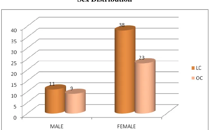
[image:49.612.130.483.376.594.2]1. Sex Distribution:- Table 1:
9 patients of OC and 11 patients of LC were males. Among OC
group 23 were females and among LC group 38 were females.
Sex Distribution
Sex LC OC
Male 11 9
47
0 5 10 15 20 25 30 35 40 45
21‐30 31‐40 41‐50 51‐60 >60
11
24
41
16
8
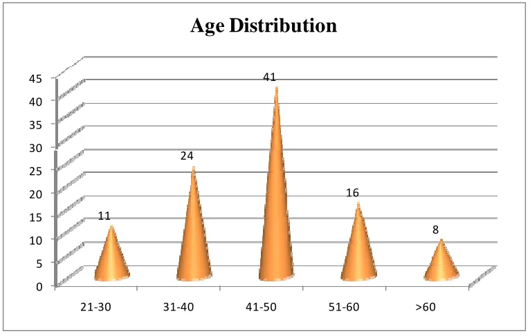
[image:50.612.115.498.395.638.2]Age Distribution
Table 2: Age DistributionAge No. Percentage
21-30 9 11
31-40 19 24
41-50 33 41
51-60 13 16
60 and more 7 8
48 0 10 20 30 40 50 60 70 80 90 100
Pain RUQ Vomiting Fever Dyspepsia Similar history
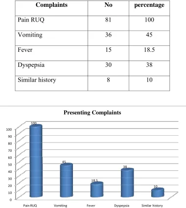
100 45 18.5 38 10 Presenting Complaints Presenting complaints:
All patients were presented with abd pain and others with
vomiting, dyspepsia and fever. Patients presented with jaundice were
[image:51.612.117.497.218.644.2]excluded from the study group.
Table 3: Presenting Complaints
Complaints No percentage
Pain RUQ 81 100
Vomiting 36 45
Fever 15 18.5
Dyspepsia 30 38
49
0 10 20 30 40 50 60 70 80
Solitary stone Multiple stones sludge 19.5
72.5
8
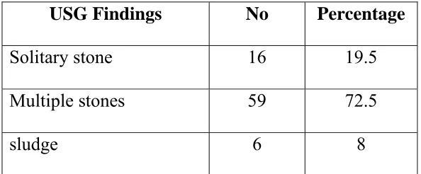
Sonographic findings Sonographic findings:
All patients the group underwent abdominal sonography. Solitary
stone was found in 16 patients of OC. Multiple stones were seen in 59
[image:52.612.158.455.218.341.2]patients.
Table 4: Sonographic findings
USG Findings No Percentage
Solitary stone 16 19.5
Multiple stones 59 72.5
50 0 0.5 1 1.5 2 2.5 3 3.5 4 4.5 5
VAS (Grades 0‐5) Analgesic used for (days)
2
3 3
5
Pain Score and analgesic
LC
OC
[image:53.612.104.510.136.572.2]Pain score and medication: Table 5: Pain Score and analgesic
LC OC p Value*
VAS (Grades 0-5)
(Range) Grade 2 (0-3) Grade 3 (1-5) p=0.024 (S)
Analgesic used for (days)
(Range) 3 (2-6) 5 (2-10) p=0.016 (S)
The VAS was median Grade3 in OC group as compared to median
Grade2 in LC group, p=0.024. The NSAID’s were used for more days in
OC group (median-5days) compared to LC group (median-3days),
51 0 1 2 3 4 5 6 7
Time to resumption of oral feeds (in days)
Duration of hospital stay ( in days)
Time taken to return to normal work (in weeks) Post operative recovery
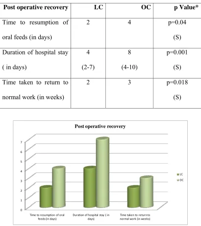
[image:54.612.101.511.92.552.2]LC OC Table 5: Post operative recovery
Post operative recovery LC OC p Value*
Time to resumption of
oral feeds (in days)
2 4 p=0.04
(S)
Duration of hospital stay
( in days)
4 (2-7) 8 (4-10) p=0.001 (S)
Time taken to return to
normal work (in weeks)
2 3 p=0.018
(S)
The duration of hospital stay was for a median period of 4days
(2-8days) in LC group and 7days (4-10days) in OC group. The difference
was statistically significant, p=0.001. It was more in OC group due to
increased pain, wound infection, injectable antibiotics used and less
mobilization due to pain and also due to our own custom of discharging
52
All patients who underwent LC were able to return to normal work
on an average of 2 weeks compared to 3 weeks in OC group. The
difference was statistically significant, p=0.018.
Post operative outcome:
Complications LC OC
Wound infection 2 6
Wound dehiscence - 1
Transient bile leak 3 2
53
DISCUSSION
Traditional cholecystectomy is an integral part of every surgical
training programme and is performed by most general surgeons. The
advent of laparoscopic cholecystectomy has created an excitement and a
flurry of activity in the medical community.
This study showed that morbidity rate is more with open
cholecystectomy than laparoscopic cholecystectomy. The open procedure
was associated with a shorter operating time (LC 60-160min and OC
40-135min). This is comparable with that of Trondsen7 and Porte8. As
experience is gained, an operating time of about 50 min can be achieved,
but this increases as other surgeons are trained or more challenging cases
are performed. This “learning curve” represents adapting to operating in
the 2-D screen, becoming familiar with the instrumentation and becoming
accustomed to the technique.
In this study, there were no major complications and several minor
ones. There was no peri-operative mortality and no CBD injury. The
complications observed were bile leak, stone spillage and blood loss
which were found to be comparable in both the groups. Drains were used
in both group but the difference was not found to be significant. Harris17
54
bleeding requiring transfusion (LC-1%, OC-2%)]. Other studies also
reported similar results 18, 19.
The conversion was necessary in 5 patients out of 49(10.2). Two
patient required conversion due to difficult dissection in view of acute
cholecystitis. Conversion rate was also found to be higher in acute cases
in other studies (0-45%)
The wound infection rate in this study was found to be less in
laparoscopic group being (4% in laparoscopic group versus 18% in open
group). This was due to the reduced size of the incision and lesser wound.
This also reduced the need for post operative antibiotics in the
laparoscopy group. Due to the severe wound infection and wound
dehiscence 1 patient in the OC group developed incisional hernia in the
follow up period. Harris 17 also noted 1 wound infection in 100 OC
patients and 0 in LC group.
Use of minimally invasive techniques in elective surgeries is
associated with a reduced inflammatory stress response with improved
pulmonary function and less hypoxia.
Reasons for conversion No
Acute cholecystitis( empyema GB) 1
Dense omental adhesions with fibrotic GB 2
Bleeding- obscuring the anatomy 1
55
The VAS was significantly less for LC group [Grade2 (median) for
LC and Grade3 (median) for OC; p=0.024]. Kum20 also found a mean
VAS score of 3.8 v/s 7.7 between LC and OC. The pain duration (median
2days for LC and median 4 days for OC patients; p=0.001) and the
duration of analgesics used (median 3days for LC and median 5days for
OC patients; p=0.016) also were significantly less in laparoscopic group
patients. This was due to the lesser incision size in LC. Other studies have
also shown similar results.21, 22, 23,24,25,26.
The two most beneficial aspects of LC are the short hospital stay
and the rapid recovery27. In this study, the median duration of hospital
stay was 4days for LC group and 7days for OC group. The difference was
found to be statistically significant (p=0.001). Porte8, Trondsen7 and
Lujan28 also found similar results. This was also confirmed in various
other series21, 12, 23,25,26,29.
The time taken to return to normal work was found to be more in
OC (median 3 weeks) compared to LC (median 2 weeks). It was
comparable to Schietroma26 who found the time taken were 4.4 days for
LC and 7.6days for OC patients. Other studies found that the duration of
sick leave was less in LC compared to OC30, 25.
56
CONCLUSIONS
Laparoscopic cholecystectomy is a considerable advancement in
the treatment of gall bladder disease. The advantages of laparoscopic
cholecystectomy are several:
¾ Technically, the dissection of the cystic artery and cystic duct is
very precise and bleeding is easily controlled with less peri
operative blood loss.
¾ LC is associated with less chances of wound infection and there is
no risk of wound dehiscence.
¾ The antibiotic usage in LC is comparatively lesser than that of OC.
¾ The degree of post operative pain and its duration is less.
¾ The amount of analgesic requirement is less in LC.
¾ LC patients tolerate oral feeds earlier and are mobilized faster.
¾ The duration of hospital stay is less and patients can be discharged
quickly from the hospital.
¾ Patients of LC group can resume their work earlier.
¾ The cosmetic advantage in LC is obvious.
Cholecystectomy remains a common operation. Laparoscopic
management of symptomatic gallstones has rapidly become the new
standard for therapy throughout the world. Many patients can now
undergo this operation in an ambulatory setting. There are numerous
57
physiological considerations will preclude the minimal access approach,
and conversion to an open operation in such cases reflects sound
58
BIBLIOGRAPHY
1. Glenn F, Grafe WR Jr. Historical Events in Biliary Tract Surgery.
Arch Surg 1966Nov; 93: 848-52.
2. David RR. Laparoscopic Cholecystectomy. In: Zinner MJ,
Schwartz SI, Ellis H, editors. Maingot’s Abdominal Operations
Volume2.10thedn. Connecticut: A Simon and Schuster Company;
1997.p.1855.
3. Anderson RE, Hunter JG. Laparoscopic cholecystectomy is less
expensive than open cholecystectomy. Surg Laparosc Endosc
1991Jun; 1(2):82-4.
4. ASI Textbook of surgery
5. Editors: McLatchie, Greg; Borley, Neil; Chikwe, Joanna Title:
Oxford Handbook of Clinical Surgery, 3rd Edition page- 296
6. Palanivelu C. Laparoscopic Cholecystectomy. In: Palanivelu C,
editor. CIGES Atlas of Laparoscopic Surgery. 2ndedn. New Delhi,
India: Vij JP; 2003.p.39.
7. Trondsen E, Riertsen O, Anderson OK, Kjaersgaard P.
Laparoscopic and open cholecystectomy: A prospective
59
8. Porte RJ, De Vries BC. Laparoscopic versus open
cholecystectomy: a prospective matched- cohort study. HPB Surg
1996; 9(2): 71-5.
9. Maingot's Abdominal Operations > Chapter 32. Cholecystectomy
(Open and Laparoscopic)
10.Strasberg S, Hertl N, Soper N. An analysis of the problem of
biliary injury during laparoscopic cholecystectomy. J Am Coll Surg
1995;180:101–125 [PubMed: 8000648
11.Barkun JS, Barkun AN, Sampalis JS, et al. Randomized controlled
trial of laparoscopic versus mini-cholecystectomy. Lancet
1992;340:1116–1119 [PubMed: 1359210]
12.Soper N, Barteau J, Clayman R, et al. Laparoscopic versus standard
open cholecystectomy: comparison of early results. Surg Gynecol
Obstet 1992;174:114–118 [PubMed: 1531160]
13.Cates J, Tompkins R, Zinner M, et al. Biliary complications of
laparoscopic cholecystectomy. Am Surg 1993;59:243–247
[PubMed: 8489086]
14.Asbun HJ, Rossi RL, Lowell JA, et al. Bile duct injury during
laparoscopic cholecystectomy: mechanism of injury, prevention,
and management. World J Surg 1993;17:547–552 [PubMed:
60
15.Soper NJ. Effect of nonbiliary problems on laparoscopic
cholecystectomy. Am J Surg 1993;165:522–526 [PubMed:
8480895]
16.Adams DB, Borowicz MR, Wootton FTI, et al. Bile duct
complications after laparoscopic cholecystectomy. Surg Endo
1993;7:79–83 [PubMed: 8395256]
17.Harris BC. Retrospective comparison of outcome of 100
consecutive open cholecystectomies and 100 consecutive
laparoscopic cholecystectomies. South Med J 1993Sep; 86(9):
993-6.
18.Hardy KJ, Miller H, Fletcher DR, Jones RM, Shulkes A, McNeil
JJ. An evaluation of laparoscopic versus open cholecystectomy.
Med J Aug 1994Jan17; 160(2): 58-62.
19.Porte RJ, De Vries BC. Laparoscopic versus open
cholecystectomy: a prospective matched- cohort study. HPB Surg
1996; 9(2): 71-5.
20.Kum CK, Wong CW, Goh PM, Ti TK. Comparative study of pain
level and analgesic requirement after laparoscopic and open
cholecystectomy. Surg Laparosc Endosc 1994Apr; 4(2): 139-41.
21.Hardy KJ, Miller H, Fletcher DR, Jones RM, Shulkes A, McNeil
JJ. An evaluation of laparoscopic versus open cholecystectomy.
61
22.Chan HS, Ha XF, Ooi PJ, Mack P. A prospective comparative
study between conventional and laparoscopic cholecystectomy.
Singapore Med J 1995Aug; 36(4): 406-9.
23.Buanes T, Mjaland O. Complications in laparoscopic and open
cholecystectomy: a prospective comparative trial. Surg Laparosc
Endosc 1996 Aug; 6(4): 266-72.
24.de Pouvourville G, Reibet-Reinhat N, Fendrick M, Houry S, Testas
P, Huguier M. A prospective comparison of costs and morbidity of
laparoscopic versus open cholecystectomy.Hepatogastroenterology
1997 Jan-Feb; 44(13): 35-9.
25.Hendolin HI, Paakonen ME, Alhava EM, Tarvainen R, Kempinen
T, Lahtinen P. Laparoscopic or open cholecystectomy: a
prospective randomized trial to compare postoperative pain,
pulmonary function and stress response. Eur J Surg 2000May;
166(5): 394-9.
26.Schietroma M, Carlei F, Liakos C, Rossi M, Carloni A, Enang GN
et al. Laparoscopic versus open cholecystectomy: An analysis of
clinical and financial aspects. Panminerva Med 2001Dec;
43(4):239-42.
27.Attwood SE, Hill AD, Mealy K, Stephens RB. A prospective
comparison of laparoscopic cholecystectomy versus open
62
28.Lujan JA, Parrilla P, Robles R, Marin P, Torralba JA,
Garcia-Ayllon J. Laparoscopic versus open cholecystectomy in the
treatment of acute cholecystitis: a prospective study. Arch Surg
1998 Feb; 133(2): 173-5.
29.Capizzi FD, Fogli L, Brulatti M, Boschi S, Di Domenico M, Papa
V et al. . Conversion rate in Laparoscopic cholecystectomy:
evolution from 1993 and current state. J Laparoendosc Adv Surg
Tech A 2003Apr; 13(2): 89-91.
30.Buanes T, Mjaland O. Complications in laparoscopic and open
cholecystectomy: a prospective comparative trial. Surg Laparosc
Endosc 1996 Aug; 6(4): 266-72.
31.Skandalakis' Surgical Anatomy > Chapter 20. Extrahepatic Biliary
INTRODUCTION
2. AIMS & OBJECTIVES OF STUDY
3. REVIEW OF LITERATURE
4. MATERIAL AND METHODS
5. OBSERVATION AND RESULTS
6. DISCUSSION
7. CONCLUSION
9. ANNEXURES
I ) Proforma