I
I
i
Ii
1,
Ii 1: I, ,,
A LIGHT AND ELECTRON MICROSCOPIC
STUDY OF CLARKE'S COLUMN
IN THE CAT.
by
MADELEINE JANE NICOL BSc. (Hons.).
Experimental Neurology Unit,
John Curtin School
of Medical Research
Australian National University.
A thesis submited for the degree of
Doctor of Philosophy
of the Australian National University.
ii I I 1 ,; I I I ' ii 11 I 1, I PREFACE
This thesis reports findings from several sen es of experiments investigating the morphology of DSCT neurones in Clarke's column, and the ultrastructural details of primary afferent terminations within Clarke's column. This work was commenced in January, 1984 in the John Curtin School of Medical Research.
A series of experiments on physiologically identified afferent inputs to Clarke's column was carried out in collaboration with Dr. R.E.W. Fyffe. The preparation of the initial experiments was shared equally between Dr. Fyffe and myself. Following each experiment, the histology and subsequent analysis of the tissue was all my own work. The results of this series of experiments are presented in Chapter 3, Sections II and III.
All other series of experiments described in this thesis were carried out in collaboration with Dr. B. Walmsley. These consisted of a series of experiments on the morphology of DSCT neurones which receive identified ankle extensor muscle input, and a series of experiments in which muscle afferents were physiologically identified and labelled, and their terminations in Clarke's column examined under the electron microscope. Again, responsibility for the preparation of the initial experiments was shared equally. The subsequent light and electron microscopic results were my own work, and are presented in Chapter 3, section I, Chapter 4 and Chapter 5.
A series of experiments was also carried out in collaboration with Dr. B. Walmsley and Dr. E. Wieniawa-Narkiewicz. The initial experiments and histology were performed by myself and Dr. Walmsley. In the first study, presented as Appendix I, the subsequent electron microscopy was performed by Dr. Wieniawa-Narkiewicz. The electron microscopy was shared by myself and Dr. Wieniawa-Narkiewicz in the second study, presented in Appendix IL
The following abstracts and papers have been published during the course of the present work:
'
I:
!,
I
Ii
i I
I II
Ii
1, 1, '
afferents and neurones 1n Clarke's column of the cat. The Journal of Neuroscience 5(8):2095-2106
Walmsley, B., Wieniawa-Narkiewicz, E. and Nicol, M.J. (1987) Ultrastructural evidence related to presynaptic inhibition of primary muscle afferents in Clarke's column of the cat. The Journal of Neuroscience 7(1):236-243
Wieniawa-Narkiewicz, E., Walmsley, B. and Nicol, M.J. Electron microscopy of the synaptic connection between primary afferents and DSCT neurones. Neurosci. Lett. (Suppl.) 15:S63
Walmsley, B. , Wieniawa-Narkiewicz, E., Nicol, M.J. and Tracey, D.J. (1984) Synaptic connections between primary muscle afferents and dorsal
spinocerebellar tract neurones of the cat spinal cord. Neuroscience
Abstracts, Vol X(l), No. 142.8
Walmsley, B., Wieniawa-Narkiewicz, E. and Nicol, M.J. (1985) The
ultrastructure of identifed primary afferent terminals in Clarke's column of the cat spinal cord. Proc. Aust. Physiol. Pharmacol. Soc. 16(2):237P
; 11 II I, 11 II 11 1, Ii ) ! ' I (,.,
ACKNOWLEDGEMENTS
The work contained in this thesis has been carried out in the Experimental Neurology Unit, J.C.S.M.R., and in the School of Anatomy, University of New South Wales. Financial support was provided by an Australian National University Graduate Scholarship, for which I am most grateful.
Firstly, I wish to thank my supervisor, Dr. R.E.W. Fyffe, currently at the University of North Carolina, for his support, encouragement and advice over the past 4 years. I value not only his teaching, but also his friendship very highly.
Thanks are due to Dr. S.J. Redman, Experimental Neurology Unit, J.C.S.M.R for providing continued financial support for these studies, and to Professor. M. Rowe for taking on the role of "local supervisor" over the
last year. I also wish to thank the School of Anatomy, U.N.S.W for allowing me to continue my studies in Sydney.
At the John Curtin School, I would like to thank Mr. Ken Collins, especially in his role of liason between me and the two departments,
Garry Rodda and Terrina Thompson for assistance in setting up the experiments in the Experimental Neurology Unit, and especially Lesley Maxwell and Elzbieta Wieniawa-Narkiewicz for teaching me serial
section electron microscopy. Also, I am grateful to Mr. Stuart Butterworth and his staff for their careful reproduction of the final figures.
At the University of New South Wales, I am deeply indebted to Mr. Collin Yeo, for his ready advice, and for his heroic achievement in
managing to keep all the machines running smoothly during the course
o_f my studies.
For critical reading and advice of this thesis I am grateful to Drs . B. Walmsley, R.E.W. Fyffe, and D.J. Tracey.
I,
l'i
Ii
!1
I
I,
I,
I
I:
1,
:
I
ii
I, Ii 11
I!
It Ii '
I ,
I
My greatest debt of thanks is due to my husband, Bruce, for his support, advice and encouragement throughout, and for his unfailing
sense of humour during the final frantic days. He has helped enormously in the production of the final figures, especially of the schematic drawings depicted in Chapter 4.
I! 1,
I
II I I I Ii " 1, :I' It I:ABSTRACT
A number of morphological studies have been carried out on Clarke's column in the cat spinal cord.
In one series of experiments, the location and morphology of physiologically identified dorsal spinocerebellar tract (DSCT) neurones in Clarke's column was investigated. DSCT neurones were intracellularly identified, labelled by injection of horseradish peroxidase, and reconstructed under the light microscope. The results of these experiments do not support previous proposals concerning a strict somatotopic location, nor a distinct morphology of DSCT neurones according to their location, or receptor type of their afferent input from the hindlimb.
A major ultrastructural study was subsequently undertaken. This study examined the ultrastructural features of an entire Ia afferent collateral branch within Clarke's column. The results of this study reveal a wide diversity in the structural features of the collateral branches and myelination, and of the synaptic boutons and the synaptic specializations contained within these boutons. The collateral exhibited nodes
synaptically specialized with a single
en passant
bouton, nodes specializedto include a series of
en passant
boutons, and terminal heminodes,without myelination. The synaptic specializations contained within the
boutons were found to exhibit a range of sizes and morphologies, including the presence of perforated contacts. These ultrastuctural features were interpreted as playing an important role in the shaping and
, I
111
ii
I
I
1,
11
11
11
I':
,,
I
An Ultrastructural Sonnet
In sombre beauty in her room she broods.
'Tis night - and all her pumps are deathly still
And thus she slumbers peacefully until
The morn, when unkind amperes end this interlude.
With steady beat, her motors wheeze and keen,
Industrious vapours drain the inner core
That Bohr's electrons shortly will explore
In headlong torrent downwards to her screen.
What truths does she uncover with her beam?
How much is artefact produced by man,
And how much really fits into the plan
Of nature? That believed is easily seen!
But even as she may promote confusion,
It
is at least an elegant illusion.
I!
TABLE OF CONTENTS
:I
CHAPTER ONE: BACKGROUND AND LITERATURE REVIEW
ll
I
' 1.1 Clarke's Column 1
'l
1.2 Morphological Cell types in Clarke's Column 2I
1.3 The Dorsal Spinocerebellar Tract
I
1.1 Ii
i) Cells of Origin of the D SCT 8
ii) Pathway of the DSCT 14
iii) Termination of the DSCT 17
!' 1.4 Afferent input to the cells of Clarke's column
:
' I
1,,
i) Physiological Studies of Afferent inputs to , I
1,
I
Clarke's column 19
ii) Presynaptic Modulation of Afferent Input 26
iii) Anatomical Studies of Afferent Inputs to
ii' the cells of Clarke's column 27
1.5 Synaptology of Clarke's column 32
I:
j I 1.6 Aims and Objectives 38
I CHAPTER TWO: MATERIALS AND METHODS
2.1 Preparation and Surgical procedures 40
I 2.2
Recording and Identification
I. Cells 42
II. Afferents 44
2.3 Staining 45
2.4 Perfusion 46
2.5 Histology 46
la I , 2.6 Light Microscopy
47
2.7 Electron Microscopy:
l
I. Fixation and Embedding 48
1,,
k II. Ultramicrotomy
50
III. Examination and Photography 51
IV. Reconstruction and Analysis 52
J
b
; I I, I,' I, I • a I 11 l ' L Ii
CHAPTER THREE: MORPHOLOGY, LOCATION AND SOMATOTOPY
OF DORSAL SPINOCEREBELLAR TRACT NEURONES IN CLARKE'S COLUMN
3.1 Introduction
3.2 Methods
3.3 Results
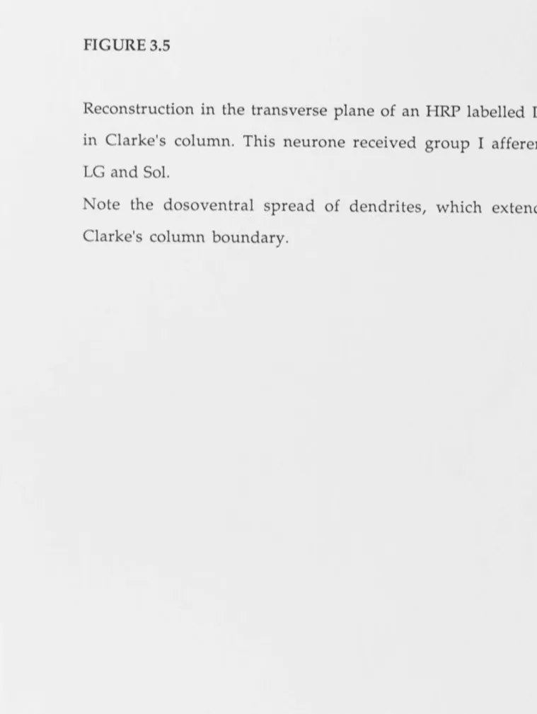
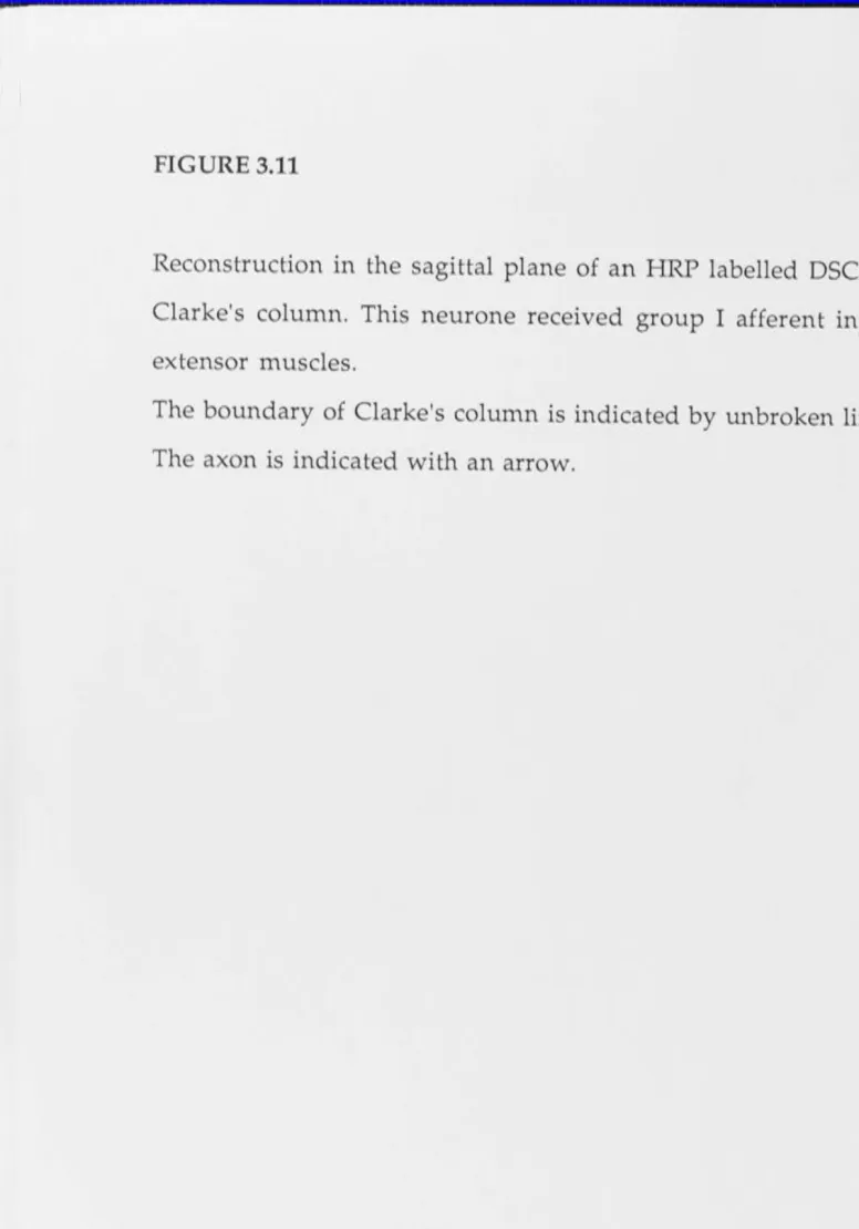
I. Location and Morphology of DSCT neurones
which receive group I afferent input from
ankle extensor muscles.
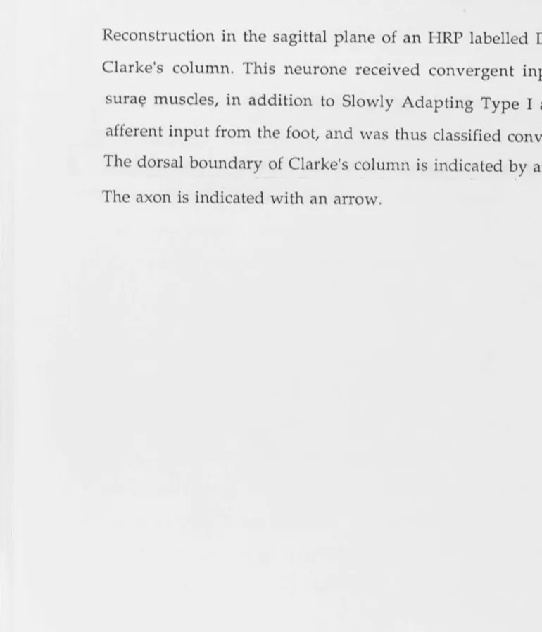
II. Morphology of DSCT neurones which receive
functionally identified input.
III. Cutaneous afferent input to
Clarke's column
3.4 Discussion
CHAPTER FOUR: A SERIAL SECTION ELECTRON MICROSCOPE
STUDY OF AN IDENTIFIED, HRP-LABELLED Ia COLLATERAL IN CLARKE'S COLUMN
4.1 Introduction
4.2Methods
4.3 Results
4.4 Discussion
CHAPTER FIVE: GENERAL DISCUSSION
REFERENCES
APPENDIX I
The ultrastructural basis for synaptic
transmission between primary muscle afferents
and neurones in Clarke's column of the cat.
APPENDIX II
Ultrastructural evidence related to
presynaptic inhibition of primary muscle
afferents in Clarke's column of the cat.
r
I
'
CHAPTER ONE
I
It
BACKGROUND AND LITERATURE REVIEW
I
I
I
11
I I
I!
i
I
la
It
,,
1,
I,
1
CHAPTER ONE: BACKGROUND AND LITERATURE REVIEW
1.1 Clarke's Column.
In 1851, Clarke first described a major nucleus which he called the
posterior vesicular columns, lying dorsolateral to the central canal and
extending the full length of the spinal cord in dogs, pigs, calves, sheep,
rabbits, cats, guinea pigs, frogs and man (Clarke, 1851). He later modified
his earlier finding to note that this nucleus dorsalis, or Clarke's column,
extends only from lower thoracic to mid-lumbar segments of the spinal
cord (Clarke, 1859).
Figure 1.1 shows a cross section of the spinal cord prepared by
Ramon y Cajal (1909), which clearly shows Clarke's column lying
dorsolateral to the central canal.
In his cytoarchitectonic atlas of the cat spinal cord, Rexed (1954)
suggested that Clarke's column arises at level Th2 in the newborn, and
Th3 in the adult. At the upper thoracic levels, the column consists of
individual cells or small cell groups. It expands to its fullest by segments
Th11-L3, and then diminishes caudally to end abruptly, always
terminating rostral to LS (Rexed, 1954; Matsushita, et al., 1979). Clarke's
column extends from Thl-L4 in the dog (Petras and Cummings, 1977),
and rhesus monkey (Petras, 1977), from Th1-L5 in the rat (Matsushita and
Hosoya, 1979) and from C8-L3 in the primate Galago senegalesis (lesser
bushbaby, Allbright and Haines, 1973). Clarke's column maintains a
similar position in the cord throughout its extent, dorsolateral to the
central canal, in a medial part of the grey matter corresponding to Rexed's
lamina VII (Mann, 1973), although in thoracic segments it is more lateral,
shifting dorsal and then dorsomedial as it extends caudally, and lying
'
I
FIGURE 1.1
Transverse section of the spinal cord prepared by Cajal (1909), in which
Clarke's column (Calonne de Clarke) is seen lying dorsolateral to the
central canal.
The neurones labelled "A" to the right of the figure are the large, complex
focal cells which comprise Clarke's column.
Afferent collaterals (labelled "B") from the dorsal columns are shown
terminating throughout the same region occupied by the focal cells.
(Reprinted from Cajal, 1909).
I
;I/
I
' I I Ill Ill I 11 ~ I ID, Ii 2
quite dorsal to the central canal 1n the lower thoracic/upper lumbar
segments (Rexed, 1954).
In cross section, the nucleus cervicalis cen tralis of the cervical cord
(demonstrated in the cat by Rexed, 1954) and Stilling's sacral nucleus of
the lower lumbar and sacral segments (demonstrated in the spider
monkey by Chang, 1951; and in the rhesus monkey by Petras, 1977) occupy
the same position in the cord as Clarke's column. Both have cellular
morphologies similar to that observed in Clarke's column, although
these are distinct nuclei, and are not continuous with Clarke's column.
The structure of Clarke's column has traditionally been described by
the following four distinctive characteristics: 1) a "close" nature, with
sharp restriction of the cells to the nuclear confines, 2) longitudinal
orientation of the cells and dendrites within the column, 3) a dorsal
funicular origin of collaterals to the column, 4) the axons of the main cells
travel to the dorsolateral fascisulus (DLF), giving rise to the dorsal
spinocerebellar tract (DSCT) (Rethelyi, 1968). Each of these characteristics
will be examined in greater detail, as each has bearing on the aims and
objectives of the present study.
The material presented in this chapter includes that published prior
to the onset of the present study, that is, up to and including 1984. Studies
which have been published during the execution of the present study are
discussed in the relevant experimental chapters.
.1.2 Morphological Cell types in Clarke's column.
In his description of Clarke's column, Clarke (1859) noted that the
constituent cells were characteristically large, of oval, pyriform, stellate or
fusiform shape, with longitudinal dendritic processes extending quite
some distance, intermingling with the processes of other large cells and of
small cells within the column, and occasionally leaving the boundaries of
Ill I 11 I II I! 3
the column. Lenhossek (1895, in Loewy, 1970) described two cell types
within the column, the focal and marginal cells, whose existence was
confirmed by Cajal (1909; see Figure 1.1).
Using Golgi techniques in the kitten, Rethelyi (1968) re-examined
the cytoarchitecture of Clarke's column, recognizing both the main (or
focal) cells, and the marginal cells, which he described as local
interneurones. The main cells clearly corresponded to the focal cells
described by Lenhossek (1895, in Loewy, 1970) and Cajal (1909), with large
spherical or elliptical cell bodies. Their dendrites extended 400-500 µm in
the rostrocaudal direction, but only 70-80 µm in the transverse plane, and
were never seen to leave the column.
Boehme (1968) described the architecture of the column in 1-10 day
old kittens using Golgi and Nissl staining techniques, basing the
categories on cell shape and size. Three cell types were described; 1) Large
bodied cells, rostrocaudally oriented, with long dendrites (500-1000 µm)
confined entirely to the column, 2) smaller bodied rostrocaudally oriented
cells with fewer trunk dendrites, and 3) large bodied cells whose dendrites
extended up to 1500 µm in length, prin1arily rostrocaudally, but also
extended ventrally into the intermediate nucleus. Boehme (1968) did not
describe the previously reported marginal cells as a separate category.
In 1970, Loewy proposed a classification scheme for the cell types in
Clarke's column of the adult cat. The classes were based on soma shape
and size, and number and orientation of the dendrites observed in Golgi
and Nissl stained tissue. The three cell types observed in the Golgi study
were 1) Type A cells, soma dia1neter 10-25 µm, with 2-5 main or trunk
dendrites which take a curvilinear course and have no preferred
orientation, 2) Type B cells, 25-50 µm in diameter, multipolar or fusiform
in shape, with 5-7 primary dendrites, and 3) Type C cells, oval to lenticular
cell bodies up to 135 x 60 µm, primarily rostrocaudally oriented, with 5-13
I I II I, ' 1, II
I I
I I
11
,I
4
main or trunk dendrites extending from the rostral and caudal poles.
These dendrites could extend up to 1000 µm, and were seen to leave the
column ventrally, laterally and dorsally. Three dendritic specializations
were described for Type C cells, a) dendritic spines, (although quite rare,
seen on less than 25% of the cells and restricted to the soma and proximal
dendrites), b) varicosities or beading along the dendrites, and c) small
branchlets, 3-90 µm in length. The axons of Type C cells take a lateral
course to the DLF, where they give rise to the dorsal spinocerebellar tract
(DSCT), and ascend ipsilaterally to tenninate as mossy fibres in the
anterior lobe of the cerebellum (Grant, 1962). The axons of type A and
Type B cells could not be followed. The Type C, or DSCT, neurones have
been studied extensively because of their large size and distinctive
character. However, in the cat they are not the most numerous of the cell
types within Clarke's column, being outnumbered 3:1 by the smallest type
A cells (Loewy, 1970). In the lesser bushbaby, type B and C cells
predominate in Clarke's column (Allbright and Haines, 1973).
The classification scheme proposed by Loewy (1970) appears to
adequately describe the cytoarchitecture of Clarke's column, and has since
been used almost exclusively to describe the column. Loewy (1970), in
accordance with Boehme (1968) did not include the marginal or border
cell group as Clarke's column cells, noting that most of the cells fitting
this description have their cell bodies and the majority of their dendrites
outside the column, and simply send a few of their dendrites into the
.column, as do other cells situated in the dorsal horn and intermediate
nucleus.
In summary, the cell types described in Clarke's column in the cat
and kitten are as folows: Type A cells are the smallest and most numerous
cell type. Type B cells correspond to the smaller bodied cells described by
Boehme (1968) and may account for some of the marginal cells described
I
1
I
Ii
Ii
II
1,1
II '
I
5
by Lenhossek (1895, in Loewy, 1970), Cajal (1909) and Rethelyi (1968) . Type
C cells (Loewy, 1970), the DSCT cells, are the largest cell type in the
column, and correspond to the two large cell types described by Boehme
(1968), and the main or focal cell types decribed by Lenhossek (1895, in
Loewy, 1970), Cajal (1909) and Rethelyi (1968). It has since been confirmed
that the same three cellular types are found in Clarke's column of the dog
(Petras and Cummings, 1977), rhesus monkey (Petras, 1977), lesser
bushbaby (Allbright and Haines, 1973) and rat (Matsushita and Hosoya,
1979).
Although Golgi studies have provided much information on the
cytoarchitectonic structure of Clarke's column, they are, as emphasized by
Boehme (1968), at best rando1n, providing no conclusive evidence on
total numbers or types of cells in the column. Nor do they offer any
information regarding the functional nature of the cells visualized. The
advent of micropipettes for intracellular recording, and relatively recent
advances in techniques for intracellular application of dyes and tracers
such as horseradish peroxidase (HRP) have greatly increased our ability to
correlate neuroanatomical and neurophysiological information (Snow et
al., 1976; Jankowska et al., 1976). It is now possible for individual cells to be
functionally characterized and subsequently identified histologically, thus
providing a direct correlation between physiological data and
morphological detail.
In 1981, Randie, Miletic and Loewy labelled DSCT neurones in
Clarke's column of adult cats using intracellular injection of HRP. Cells at
the L2 and L3 segments were identified as DSCT neurones by antidromic
activation of the DLF at the C2 level, and classified as receiving muscle,
cutaneous or convergent (cutaneous + muscle) afferent input. Randie et
al. (1981) recovered 19 intracellularly labelled DSCT neurones in Clarke's
I
11
6
dendritic complexity, and an attempt was made to correlate these
morphological groups with the three functionally based categories:
muscle, cutaneous or convergent input. On this basis, Randie et al. (1981)
determined that their largest cells, all of which received muscle input,
were consistent with Loewy's Type C cells (Loewy, 1970), with cell bodies
up to 40 x 140 µm, and 5-7 trunk dendrites extending up to 1250 µm
rostrocaudally (see Figure 1.2B). They noted that the dendrites were
smooth, with relatively few branch points. In the cases where the axon
was stained, no collaterals were observed. Thirteen of the nineteen
neurones recovered histologically received muscle input. However, this,
was regarded as sampling bias due to the larger size of the cell bodies of
these neurones. Randie et al. (1981) suggested that the DSCT neurones
which received convergent input had smaller cell bodies (up to 50 x 75
µm, see Fig. 1.2C), although their orientation, dendritic branching
patterns and axonal trajectory were very similar to that seen for cells
receiving muscle input. They showed that most of the cells receiving
purely cutaneous input at L3 were located outside Clarke's column,
although they recorded two cells which received purely cutaneous input
within Clarke's column. These two cells were not morphologically
similar, and one was believed to be a type B cell.
Houchin et al. (1983) subsequently labelled cells in Clarke's column
of the adult cat, identified as DSCT neurones through antidromic
activation of the DLF at C2, which received input from the sciatic nerve.
Subsequent histological recovery of the cells showed them to be extremely
morphologically complex, and very sin1ilar in their appearance. Cell
bodies were 70-100 x 30-70 µm, with long dendritic trees extending up to
3200 µm in the rostrocaudal plane (see Figure 1.2A). In the transverse
plane, the dendritic spread was much more closely restricted, with most
branches confined to the column, and only a few extending beyond. These
FIGURE 1.2
A. Reconstructions in the sagittal plane of HRP labelled DSCT neurones
which received group I afferent input following stimulation of the sciatic
nerve, showing large cell bodies, and complex, profusely branched
dendritic trees.
(Reprinted from Houchin et al., 1983).
B. Reconstructions in the sagittal plane of HRP labelled DSCT neurones
which received identified group I muscle afferent input.
(Reprinted from Randie et al., 1981).
C. Reconstructions in the sagittal plane of HRP labelled DSCT neurones
which received convergent (muscle + cutaneous) input.
(Reprinted from Randie et al., 1981).
The reconstructions in A, B and C have all been printed to exactly the
same magnification.
I
I
I
I
'
I
I!
I
I
I
A
.,
B
C
-
-
-
-, -, .r ..
II 1, 11 Ii II : I I I l 1 i I j : 1, I 7
cells exhibited 5-9 primary dendrites, all of which branched profusely and
were of extremely complex appearance. Houchin et al., (1983) also noted
the same three dendritic specializations described by Loewy (1970); spines,
branchlets and varicosi tes. The varicosi ties were subsequently examined
under the electron microscope, and found to contain grossly swollen
mitochondria, and signs of internal damage, suggesting that the beading
may be in part artefact. The computer aided reconstruction methods
employed by Houchin et al. (1983) allowed the HRP labelled DSCT cells to
be observed from any angle, and also provided statistical information
about the dendrites such as length, volume and surface area. The total
length of dendrites of any one cell was found to be approximately 35 mm,
slightly shorter than the total dendritic length of a motoneurone (Ulfhake
and Kellerth, 1981; Egger and Egger, 1982). The total volume and surface
area of DSCT neurone dendrites were also measured, and found to be
large, but not as large as those of the motoneurone. The axons of the cells
were well stained and were followed to the DLF (Houchin et al., 1983). No
recurrent collaterals were observed.
Figure 1.2 shows large type C cells from Clarke's column taken
from the studies of A) Houchin et al. (1983), B and C) Randie et al. (1981).
In view of the conflicting results presented in these two studies, the
morphology of DSCT neurones which receive identified input remains
inconclusive. In light of Golgi studies which showed the large cells of
Clarke's column to exhibit complex morphology with extensively
branched dendritic trees, the anatomical simplicity of the neurones
presented by Randie et al. (1981) (see Figure 1.2B and C) seemed quite
surprising. Their reconstructions illustrated far less morphological
complexity than those observed by (for instance) Boehme (1968) and
Loewy (1970). The subsequent finding by Houchin et al. (1983) that the
la II ,, I la I~ I I
I . I 1, 1, ii 1, Ii 1, I i II It [ , I 8
morphologically co1nplex with profusely branched dendritic trees (Figure
1.2A), led them to suggest that the HRP labelling achieved by Randie et al.
(1981) had not been entirely succesful, and that the cells were not
completely filled.
In summary, the morphology of functionally identified DSCT cells
in Clarke's column has not been properly resolved. Golgi studies provide
a limited picture of morphological detail, and provide no functional
information. Randie et al. (1981) intracellularly labelled DSCT neurones
which received identified input, but more recent evidence suggests that
the morphological details presented by them were not complete. This
means, therefore, that their proposal that the morphological
characteristics of DSCT neurones might indicate their function, and vice
versa, is not conclusive. Houchin et al. (1983) have presented convincing
evidence for the morphological complexity of DSCT neurones in Clarke's
column, but their functional identification of the cells was limited.
Clearly, more evidence is required on these issues concerning the
morphology and location of DSCT neurones which receive identified
muscle, cutaneous and convergent inputs from the hindlimb of the cat.
The following section explores the origin, pathway and termination
of the DSCT, and examines the relationship between the tract and the cells
of Clarke's column.
1.3 The Dorsal Spinocerebellar Tract
i) Cells of Origin of the DSCT
In 1876, Flechsig first described the connection between the nucleus
dorsalis (Clarke1
s column) and a myelinated fibre tract running in the
dorsolateral column which tenninated in the cerebellum, the dorsal
spinocerebellar tract (DSCT; in Petras and Cummings, 1977). Van
. I I I I I •• I I 1, 11 ' Ii
E I
I~ J II ' I 9
column cells showed signs of degeneration (in Petras and Cummings,
1977). Subsequent studies in which the DSCT was transected confirmed
this finding (see e.g. Sherrington and Laslett, 1903; Liu, 1956). Grant (1962)
showed that lesion of the spinal cord caudal to Clarke's column produced
no degeneration in the cerebellum, thus concluding that Clarke's column
was the site of origin of the tract. Until quite recently, there was general
agreement that the largest Clarke's column neurones were the sole cells
of origin of the DSCT (see Oscarsson, 1965,1973; Mann, 1973; Burke and
Rudomin, 1977 for reviews). This presumption has been challenged in
two different ways:
1) Petras and Cummings (1977) studying cerebellar projections from the
spinal cord in the dog, used retrograde HRP labelling and degeneration
techniques to show that in fact both the large and the medium sized
Clarke's column neurones (Types B and C of Loewy, 1970) projected to the
cerebellum. The axons of both types were shown to take a similar or
identical course, and almost all their projections were ipsilateral. Snyder
et al. (1978) in a retrograde labelling study of spinocerebellar neurones in
cat, rat and squirrel monkey found labelled small, medium and large cells
which projected ipsilaterally to the cerebellum, and suggested that the size
of the cell bodies in Clarke's column is not sufficient to indicate whether
or not they project to the cerebellum. Matsushita, Hosoya and Ikeda (1979)
conducted a similar experiment, injecting HRP into the cerebellum of the
cat and following its retrograde transport to the cells of the spinal cord.
They concurred with Petras and Cummings (1977) that both large and
medium sized Clarke's column neurones were cerebellar projecting.
Matsushita et al. (1979) also observed some labelling of oval cells on the
circumference of Clarke's column which they considered to be the
marginal or border cells described by Rethelyi (1968). (As noted
Ii 11 [ > II 1, I I > I > I 10
in fact located outside the boundary of Clarke's column, and that oval
cells within the boundary of Clarke's column may be medium sized (type
B) neurones. This suggestion received support from an observation by
Saito (1979), that within Clarke's column, the large and medium
neurones which label retrogradely on cerebellar injection of HRP are
located both centrally and marginally). In accordance with Snyder et al.
(1978), Matsushita et al. (1979) observed occasional labelling of small (type
A) cells. Thus it appears that cells other than the largest cells may
contribute to the DSCT.
2) In a study of supraspinal control of the DSCT, Hongo, Okada and Sato
(1967) recorded from DSCT units which received cutaneous and high
threshhold muscle afferent input. These cells were situated at spinal
segments L4-L6. Although this spinal level was considered caudal to the
extent of Clarke's column, these cells were identified as DSCT neurones
on the basis of antidromic activation from the ipsilateral anterior lobe of
the cerebellum. This suggests that cells of origin of the DSCT which
receive cutaneous and high threshold muscle input may lie outside
Clarke's column.
Prior to this study, suggestions that the cells of origin of the cutaneous
component of the DSCT lay caudal or lateral to Clarke's column had been
explained by assigning extra-Clarke's column cell types to ascending tracts
other than the DSCT, such as the spino-olivary or spinocervical tracts
(Grundfest and Carter, 1954). In 1959, Lundberg and Oscarsson, recording
DSCT axons at cervical levels, suggested that all units which received
muscle inputs were located in Clarke's column, but that some units
receiving group II and group III muscle input or cutaneous input from
receptors with large receptive fields could be located caudal to Clarke's
column. Units receiving input from cutaneous receptors with restricted
I
I:
I:
I
I ;
11
II
11
Clarke's column, and were not included as DSCT units due to lack of
activation on cerebellar stimulation. Oscarsson (1965) concluded that the
DSCT units receiving cutaneous input were not necessarily located caudal
to Clarke's column, but that there was no proof of their location within
the column, although the course and termination of their axons appeared
to be as for proprioceptive units.
Mann (1971) subsequently sampled a small group of neurones in
laminae III-VI in segments L6-S1, and was unable to record any units
driven by cerebellar sti1nulation. Kuno, Munoz-Martinez and Randie
(1973a,b) were able to record from cells in Clarke's column at L2 and L3
which received input from cutaneous receptors. However, they also
recorded from units which they classified as DSCT, receiving joint or
cutaneous input, whose cell bodies lay lateral to the boundary of Clarke's
column. Aoyama, Hongo and Kudo (1973) physiologically demonstrated a
group of cells whose properties resembled DSCT units situated in
segments LS and L6 in a location rese1nbling, but caudal to, Clarke's
column. These cells were monosynaptically activated by both group Ia and
lb muscle input, and their axons ascended ipsilaterally in the DLF. The
primary differences between these units and DSCT neurones in Clarke's
column included the predominance of extensor muscle input and
extensive convergence. Identification of cells recorded at segments LS and
L6 was based on antidromic activation of the DLF at Cl, thus projection to
the cerebellum was not unequivocally established (Aoyama et al., 1973).
Activation of the DLF at Cl is an adequate means of identification for
DSCT axons whose somata are subsequently determined to be located in
Clarke's column. However, as Oscarsson (1965) pointed out, three other
ascending tracts occupy regions of the DLF (Lundberg and Oscarsson,
1961), and therefore rigorous identification is required if a unit is to be
I
II
1:
Ii
12
column. Nevertheless, Aoyama et al. (1973) believed that their data
represented evidence of an uncrossed spinocerebellar tract with cells of
origin caudal to Clarke's column. It was not determined whether the cells
in LS and L6 represented a new subgroup of the DSCT or an alternative
spinocerebellar pathway.
Tapper, Mann, Brown and Cogdell (1975) recorded the response of
cells in spinal segments L4-L6, (caudal to Clarke's column) in laminae IV
and V in response to stimulation of cutaneous nerves of the hindlimb of
the cat. All units were tested for cerebellar projection. The location of the
neurones, and the extent of convergent input which they received,
suggested that they were the same cell group recorded previously by
Aoyama et al. (1973), who had tested their projection only as far as the Cl
level. Tapper et al. (1975) found no difference in axon al trajectory or
receptive field characteristics between the cells they recorded within, or
caudal to Clarke's column, suggesting that the only reason these cells had
not previously been identified was the prior assumption that all cells of
origin of the DSCT were located within Clarke's column. Tapper et al.
(1975) concluded that the definition of the dorsal spinocerebellar tract
should be based on axonal pathway and site of termination, and thus
expanded to include these cells situated caudal to Clarke's column.
Randie, Myslinski and Gordon (1976) examined mid-lumbar
segments for evidence of DSCT cell bodies outside Clarke's column. They
recorded from neurones in the dorsal horn at L3 and L4, which responded
to hair movement. These neurones had ipsilaterally ascending axons, and
some were identified as DSCT after activation from the cerebellum, while
some were assigned to the spinocervical tract. They concluded that all
DSCT neurones receiving muscle inputs, or cutaneous input from the
foot, were located in Clarke's column, but that those with receptive fields
'
•
j I i i J 1 I
~I
I I I 1, 13IV-VI, and that some of the cells included in this group were not DSCT,
but spinocervical tract cells.
The proposal by Tapper et al. (1975) that the definition of the DSCT
should be expanded to include the cells outside Clarke's column which
appeared to contribute to the tract, was subsequently contradicted by the
suggestions of Matsushita et al. (1979). From a study of cerebellar
projecting spinal cord neurones in the cat, Matsushita et al. (1979)
concluded that the term dorsal spinocerebellar tract was no longer
appropriate, and proposed its replacen1ent with three alternate terms,
each of which represented an individual fibre tract. These were: 1) the
Clarke's column-Spinocerebellar tract, comprising large and medium
sized neurones in Clarke's column from upper thoracic to mid-lumbar
segments, 2) the lamina V-Spinocerebellar tract from lower cervical to
lumbar segments, and 3) the medial lamina VI-Spinocerebellar tract in
the lumbar cord. A study by Matsushita and Hosoya (1979) suggested that
the ascending spinocerebellar neurones in the rat were similarly
organized, with cell bodies traced to Clarke's column, lamina V of
segments Ll-L3 and a group in the L6-caudal segments which appeared to
lie within Stilling's sacral nucleus. This same sacral nucleus had been
described in the spider monkey by Chang (1951) and more recently in the
rhesus monkey by Petras (1977), who concluded that the neurones of this
sacral nucleus were cytologically and topographically similar to those of
Clarke's column, and therefore consistent with Stilling's proposed
continuation of Clarke's column. In contrast, Snyder, Faull and Mehler
(1978) comparing spinocerebellar projecting neurones of the cat, rat and
squirrel monkey, found large multipolar neurones in the caudal and
sacral segments in the rat and monkey, which they concluded were
similar in position and orientation to Clarke's column, but not in
,:
i i , ij " a Ii I 1, I, I , II I I 14
be those described as Stilling's sacral nucleus and concluded that the
posterior vesicular columns described by Clarke (1851) constituted the
central cervical nucleus, Clarke's column and Stilling's sacral nucleus
described in continuity. Apart from heavy labelling of Clarke's column
neurones in all three animals, Snyder et al. (1978) also observed labelling
in cells immediately caudal, rostral and lateral to Clarke's column, which
had similar cytology and axonal projection to DSCT neurones, but could
not determine whether these were displaced Clarke's column neurones
or a distinct cell group.
Randie et al. (1981) recorded from DSCT units in L3 and L4 which
received input from hairy skin mechanoreceptors. Some of these units
were located outside the boundary of Clarke's column. A similar location
for such neurones was determined by Grant et al. (1982) in the kitten, who
also observed ipsilaterally projecting spinocerebellar cells caudal to
Clarke's column.
The location of functionally identified DSCT neurones remains
unclear. As a general guide, the DSCT is probably best described as
originating from the large and medium cells of Clarke's column, with
possible contributions from lamina V neurones in the cervical to lumbar
cord, and medial laminae V-VII neurones in segments LS and L6. Even
so, reports regarding the location of DSCT cells receiving cutaneous and
convergent inputs are largely conflicting (Mann, 1971; Kuno et al., 1973a,b;
Aoyama et al. 1973; Tapper et al., 1975).
ii) Pathway of the D SCT.
The axons of Clarke's column neurones, which constitute the main
body of the dorsal spinocerebellar tract, leave the column and travel
laterally to the DLF, in which they ascend as an uncrossed tract,
terminating in the ipsilateral anterior lobe of the cerebellum. Rethelyi
Ir
I
I
I I lmi ,1, I
i I Im I I I ll· l I II
I I I I
I " 1, II I " 11 15
(1968) suggested that the axons of the DSCT cells may have recurrent
collaterals which contacted other cells within Clarke's column. However,
in more recent intracellular labelling studies no such collaterals have
been observed (Randie et al., 1981; Houchin et al., 1983).
Yoss (1952) determined that in macaque monkeys, the DSCT axons
are located deep in the DLF at their most caudal extent, and shift dorsally
and peripherally as they ascend. This represents a simple stratification
pattern, with the longest fibres found at the periphery and the shortest
more medial. This same stratification has been described for the cat (Pass,
1933; Grant, 1962), with the longest fibres most dorsal at cervical levels.
This shift indicates that at more rostral segments, fibres are recruited to
the ventromedial aspect of the tract, which suggests that the DLF may be
topographically (or perhaps segmentotopically) organised. Yamamoto and
Miyajima (1959) proposed a medial to lateral arrangement of fibres from
cranial to caudal. Yamamoto and Miyajima (1959) may, however, have
been recording in a separate tract from the DSCT, as this finding is
inconsistent with earlier findings of dorsal and peripheral shift at rostral
segments (Yoss, 1952; Grant, 1962). Kitai and Morin (1962) saw no
evidence of somatotopic organization of the DSCT fibres at C2 based on
sensory modality, although Lundberg and Oscarsson had demonstrated a
somatotopic arrangement of the DLF fibres at upper lumbar and lower
thoracic levels (Lundberg and Oscarsson, 1961). Oscarsson (1965) noted
that neither Yamamoto and Miyaji1na (1959) nor Kitai and Morin (1962)
identified the fibres from which they recorded as belonging to the DSCT,
and that since three other ascending tracts occupy this region (Lundberg
and Oscarsson, 1961), they 1nay well have been recording responses from
other pathways.
Mann (1971) found that the DLF was quite inhomogeneous, with
I I
I
I
II
la I
I I I I Ir I I 11 II ' I I I I
I : 11 1, ,I i I Ill L 16
which is occupied by the spinocervical tract. Mann (1971) showed a
tendency for the fibres mediating cutaneous information to be located
more lateral and those mediating muscle information to be more medial,
but that there was considerable overlap. Within sensory modalities, DSCT
units receiving muscle, or muscle + cutaneous input were not shown to
be somatotopically organized. Units receiving cutaneous input from
receptive fields on the foot were recorded throughout the DLF, those with
input from receptors on the lateral or medial leg were found in the lateral
DLF and those with input from receptive fields on the trunk were found
more medial.
Hongo, Okada and Sato (1967) suggested that since somata of DSCT
cells receiving muscle input from foot and calf were located more
caudally, their axons should be found more dorsolateral than those of
cells receiving input from thigh muscles. This, however, was not found
(Mann, 1971). Since the cell bodies of the cutaneous component of the
DSCT were suggested to be located more caudal than those which received
muscle input, it was also suggested that muscle input cells would join the
tract at more rostral (and thus more ventromedial) aspects of the tract,
resulting in a more peripheral location of the axons mediating cutaneous
information. This, again, was not found to be the case (Mann, 1971).
Thus, although there is a tendency for DSCT fibres to shift dorsal
and lateral as they ascend, strict somatotopic arrangement of the fibres
based on sensory modality, or on receptor type, is not found, with
considerable intermingling of the DSCT and non-DSCT fibres within the
DLF.
Grant (1962) found the majority of the DSCT fibres to be quite large,
a finding expected due to the size of the contributing cell bodies. Van
Beusekom (1955) concluded that 7,000-10,000 fibres constitute the DSCT,
with 2% > 15 µm in diameter, 56% > 3um, and 17% < 1.5 µm, but Mann
II 11 11 l I ,I 11 II I 111 I 11 1, 1, 17
(1973) pointed out that van Beusekom sampled a region of the DLF,
which is inhomogenous, with DSCT and non-DSCT axons intermingled.
Mann (1973) suggested that the DSCT fibres were all> 3 µmin diameter.
iii) Termination of the DSCT.
DSCT fibres reach the cerebellum directly, without synaptic
interruption, but have been suggested to send collaterals to three other
sites. Cajal (1909) suggested that the lateral cervical nucleus might receive
input from DSCT collaterals. This was re-examined by Morin (1955) who
implicated the DSCT as a mediator of cutaneous information in a
spinothalamic pathway via collaterals to the lateral cervical nucleus.
However, this connection was denied by Brodal and Rexed (1953), and it is
now known that input to the lateral cervical nucleus comes largely from
the spinocervical tract. A possible cutaneous pathway via DSCT collaterals
to the inferior olivary nucleus was ruled out by Grundfest and Carter
(1954). Low et al. (1986) concluded that 92% of the afferent input to
nucleus Z is via DSCT collaterals, as a medullary relay for proprioceptive
information from the hindlimb to the sensory cortex. It thus appears that
nucleus Z is the only non-cerebellar recipient of DSCT input.
It is generally well agreed that on entering the cerebellum, DSCT
axons course through the white 1natter, sending collaterals to the folia
and becoming narrower before terminating as mossy fibres on the granule
cells (Brodal and Grant, 1962). Grundfest and Campbell (1942) recorded
responses to stimulation of the DSCT in the rostral vermis in the cat, with
some response in the declive and pyramids, but none in the cerebellar
hemispheres. Occasionally, inputs to the contralateral lobulis centralis
and folia of the culmen were recorded.
Yoss (1952) used degeneration techniques to determine a similar
' II
I,
I
11
II
I I
I I
18
the ipsilateral vermis, with some termination in the pyramids, declive
and uvula. He found no somatotopy to these terminations.
Morin, Lindner and Catalano (1957) found a direct connection to
the paramedian lobule of the posterior lobe for DSCT fibres mediating
tactile input.
Grant (1962) studied degenerating terminals of DSCT fibres in
cerebellar cortex after lesion of the tract in the cat. Lesion of the cord
caudal to Clarke's column caused no degeneration in the cerebellum,
leading to the conclusion that the cells of origin of the DSCT were situated
only in Clarke's column. Grant (1962) found that section of the DSCT
rostral to L3 always produced the same result, degeneration in the
anterior lobe (lobules I-V of Larsell, 1953), in lobule VI, VIIB, VIII, and IX,
the paramedian lobule and parafloccus, with almost the entire projection
confined to the ipsilateral side. Since these areas are representative of the
trunk and hindlimb, it was expected that some degree of somatotopic
organization of the terminations would eventually be found.
Grant (1962) reported that lesion of the DSCT did not result in any
degeneration in the cerebellar nuclei. However, Matsushita and Ikeda
(1970) using the same technique as Grant (1962) concurred with
Szentagothai (in Matsushita and Ikeda, 1970) that there is some nuclear
termination, concluding that DSCT fibres may terminate in nucleus
medialis, and nucleus interpositus, primarily ipsilaterally. Matsushita and
Ikeda (1970) suggested that these nuclear terminations are of collaterals
whose parent DSCT axons terminate in the cerebellar cortex.
Allbright and Haines (1973) found that the majority of DSCT fibres
terminate in the cortex of the vermal and paravermal functional zones of
the anterior lobe of the cerebellum in the lesser bushbaby, especially in the
central and culminate lobes, with some fibres terminating in the uvular
and pyramidal lobules of the posterior lobe.
I
I
! ' J ,, ., I I I II I• 1, ,,
i I
I
f :1
l
I~
n I
1,
19
The termination of DSCT fibres in the cerebellar areas which are involved
in control of movement in the trunk and hindli1nb thus lends support
to the proposed role of the DSCT in medi1ati~g information relating to posture, stance, balance and movement.
1.4 Afferent Input to the cells of Clarke's column .
The principal source of afferent input to the cells of Clarke's
column is from fibres ascending in the dorsal columns (Pass, 1933; Liu,
1956; Mann, 1973), although other inputs have been described from the
lateral columns (Boehme, 1968), descending fibres in the dorsal columns
(Liu, 1956) and from the cortex (Hongo et al., 1967). The DSCT receives
input from the hindlimb and trunk, almost exclusively from the
ipsilateral side of the body (Petras and Cummings, 1977). Afferent input to
the cells of Clarke's column has been investigated in great detail using
physiological, and to a lesser extent anatomical methods.
i) Physiological Studies of Afferent Inputs to Clarke's column.
Prior to 1958, when the first intracellular recordings from cell
bodies in Clarke's column were made (Curtis et al., 1958), the input to
Clarke's column neurones had been studied almost exclusively through
examining mass discharge or single unit responses in the tract axons
running in the DLF at the cervical and upper thoracic levels.
Grundfest and Campbell (1942) recorded responses from the DSCT
axons in the DLF on stimulation of tibial, peroneal and saphenous
nerves, concluding that the tract received input from the muscles of the
hindlimb, but that the response was mediated by a chain of
interneurones.
Lloyd and McIntyre (1950) reinvestigated the latency of the
I I 11 I 11 'I II 111 111 11 I I I
I
I
ii
20
input, and that since the conduction velocity through the pathway
averaged 109 m/ sec, saw no reason to assume interneurones in the
connection, claiming instead that DSCT neurones received monosynaptic
excitatory input from the hindlimb muscles, and conveyed stretch evoked
activity to the cerebellum. They also observed that while motoneurones
required considerable summation of afferent input to fire, DSCT units
could respond to a single input with transmission approaching 1-1, and
that the DSCT therefore acted solely as a relay pathway, with no
integrative function.
Lundberg's group carried out an extensive series of experiments to
determine the nature of information relayed in the tract. Laporte et al.
(1956a) recorded the mass discharge response in the DSCT on stimulation
of both skin and muscle nerves of the hindlimb of the cat, and found the
conduction velocity of each fibre type to average 100-110 m/sec. Grundfest
and Carter (1954) had previously dismissed the DSCT as a mediator of
cutaneous information, with the assertion that units relaying the
cutaneous impulses had their cell bodies caudal to Clarke's column.
Using single unit recording techniques, Laporte et al. (1956b) again
demonstrated DSCT units in the DLF which received cutaneous or
muscle inputs. They also demonstrated convergent excitation from more
than one muscle onto one DSCT cell, excitation and inhibition from more
than one muscle, and, occasionally, convergent excitation from the skin
and muscle nerves onto one cell.
On the basis of their own, and earlier studies, Laporte et al., (1956b)
divided the recorded units into two subgroups. They defined the classic
Flechsig's tract as the true DSCT, receiving group I muscle inputs and
making direct connection with the anterior vermis of the cerebellum. The
second subgroup received input from group II and III muscle afferents
and skin afferents, and represented an entirely different pathway, with
I
I
I
11 ,, 1, .\ I 1-1 11 [ 21
cells of origin outside Clarke's column, and a high probability of receiving
input from more than one receptor type. Thus the DSCT was considered
to be a purely proprioceptive pathway. Laporte et al. (1956b) found, in
accordance with Lloyd and McIntyre (1950) that DSCT cells did not require
the extent of summation required by the motoneurones to cause them to
fire. Laporte and Lundberg (1956) studied the response of DSCT neurones
to stimulation of stretch receptors in hindlimb muscles, demonstrating
the excitatory action of muscle spindle afferents (group Ia) to these
neurones. They concurrently tested discharges in other units of Flechsig's
fasciculus, recording units responsive to light touch, pressure or bending
of joints, or pressure on foot pads, which they attributed to an alternate
spinocerebellar pathway. Lloyd and McIntyre (1950) reported volleys from
both Ia and Ib muscle afferents in Clarke's column. Laporte and Lundberg
(1956) clearly demonstrated the existence of the group Ia responses in
DSCT units, but noted that the group I response in the DLF consisted of
two components. Subsequently, Lundberg and Oscarsson (1956) studied
the involvement of group lb inputs to the DSCT. After determination
that lb fibres arrived at the correct spinal levels to contact Clarke's column
DSCT neurones, and that the mass discharge recorded in the DLF
increased on recruitment of the lb fibre input, Lundberg and Oscarsson
(1956) proposed that the DSCT consisted of two functional subgroups, the
first receiving group Ia (muscle spindle primary afferent) and the second
lb (Golgi tendon organ) input. However, they then made the seemingly
contradictory observation that both Ia and lb afferents most probably
converge on the same cell, in which case such a division would seem
unwarranted.
Oscarsson (1957) determined that the course taken by Ia and lb
afferents to contact DSCT neurones was not identical. He found that as Ia
and lb afferents ascend in the dorsal columns, the lb afferents drop
II
I 11
1, I
II
22
collaterals to the grey matter at a more caudal level than the Ia afferents
from the same dorsal root. These lb collaterals then ascend in Clarke's
column for some distance before contacting DSCT neurones at the same
spinal level as the Ia collaterals, which drop to the grey matter in a
perpendicular manner at more rostral levels, contacting DSCT neurones
almost immediately. Oscarsson (1957) also determined that despite earlier
suggestions of Ia and lb convergence onto the same DSCT neurone
(Lundberg and Oscarsson, 1956), most units received either group Ia or lb
inputs. Oscarsson (1957) made the concurrent finding that group Ia and
group II muscle spindle afferents took a similar topographic course to
contact DSCT cells. This was of interest, as Laporte et al., (1956b) had
determined that the group II muscle responses recorded in the DLF were
attributable to pathways other than the DSCT.
In 1959, Lundberg and Oscarsson recorded from DSCT axons in the
DLF identified by antidromic activation from the cerebellum and a lack of
direct stimulation from the spinal cord caudal to LS. They defined three
groups of DSCT axons: 1) units monosynaptically activated by impulses in
muscle spindle afferents (group Ia and group II), 2) units activated
monosynaptically by group lb muscle inputs, and 3) a group activated by
inputs from group II and group III muscle receptors and skin or joint
receptors. All three types were regarded as DSCT axons, although not all
the axons responding to -the third group of afferent inputs were included
as DSCT, as some had their cell bodies caudal to Clarke's column and
were considered to constitute another pathway. A group of units activated
caudal to LS which received low threshhold cutaneous inputs from
receptors with restricted receptive fields, were also regarded as non-DSCT.
Yamamoto and Miyajima (1959) studied responses of the
spinocerebellar (including DSCT) axons at C2, recording units responsive
to touch, pressure or deep receptors. They found that the number of units
I
,,
.!
I
23
responsive to exteroceptive impulses approximately equalled the number
of proprioceptive units, and that hindlimb responses were twice as
frequently observed as forelimb. This suggested to Yamamoto and
Miyajima (1959) that the DSCT, classically considered a proprioceptive
pathway, was in fact functionally non-specific, with the implication that
the cerebellum was more complex than previously thought. However, as
noted above, many of the units probably belong to the SCT rather than the
DSCT.
Of the DSCT units recorded by Kitai and Morin (1962), 53%
responded to touch, 31 % to pressure, 2% to touch and pressure, and 14%
to joint input. They found tactile responses more numerous for the
forelimb, and pressure from the hindlimb . Joint units responded to one
joint only, and adjusted their firing frequency to the rate and degree of
displacement. Fields of touch and pressure receptors varied from very
small to quite large, with large fields suggesting a high degree of
convergence. Unfortunately, Kitai and Morin did not identify the fibres
from which they recorded as belonging to the DSCT, therefore these
results cannot be considered conclusive.
Jansen and Rudjord (1965) concluded from the response pattern of
DSCT fibres to muscle stretch that the cerebellum receives information
about the mechanical situation in the muscle from three receptor types.
Group Ia receptors provide a mixed signal about the muscle length and
speed of stretch, group II receptors provide information about muscle
length, and group lb on the degree of muscle contraction.
Cutaneous inputs to the cerebellum were reinvestigated by Mann
and Tapper (1970) who recorded from axons in the DLF identified as DSCT
by antidromic activation from the cerebellum. Four classes of neurones
with cutaneous input were identified: 1) those activated by rapidly
adapting receptor (such as hair) and slowly adapting Type I (SAI)
I
I I i ,, ,1 24
receptors, 2) those activated only by SA receptors, 3) those activated only
by rapidly adapting receptors, and 4) those activated by both cutaneous and
muscle receptors. They showed that DSCT neurones receive extensive
convergent input, but that a single presynaptic impulse could trigger a
post synaptic response. Mann (1971) subsequently found that, of units
recorded in the DLF (identified as DSCT by activation from the
cerebellum), 53% responded to deep or muscle input (of which many
responded to more than one, and up to four muscle groups), 19% to
cutaneous inputs, and 8% to convergent cutaneous + deep inputs. Both
Yamomoto and Miyajima (1959) and Kitai and Morin (1962) had shown
that more than half the recorded DSCT units responded to cutaneous
impulses. However, the fibres were not conclusively identified as DSCT
tract axons in either of the latter studies. Of the cutaneous units recorded
by Mann (1971), 30% responded to hair follicle afferent inputs, and were
rapidly adapting, 15% responded to slowly adapting units with large
receptive fields, and 54o/o responded to both slowly adapting and hair
afferent input. From these findings, Mann proposed that up to 37
afferents might converge on one DSCT neurone. Thus both 'unimodal'
and 'multimodal' units comprise the cutaneous as well as the muscle
components of the DSCT.
Although cutaneous units of the DSCT had been confirmed, Kuno
et al. (1973a,b) reexamined sensory inputs to the DSCT neurones on the
basis that the role of the DSCT as a relay for cutaneous information was
tenuous. Cutaneous volleys from the hind limb of the cat were found to
activate cerebellar mossy fibres and the DSCT was proposed as the
pathway for transmission of this information. However, limited numbers
of DSCT neurones in Clarke's column had been found to be activated in
response to cutaneous volleys. It was proposed that either 1) the number
of DSCT neurones in Clarke's column relaying cutaneous information