0022-538X/97/$04.0010
Copyrightq1997, American Society for Microbiology
Identification of a Sequence within U5 Required for Human
Immunodeficiency Virus Type 1 To Stably Maintain a
Primer Binding Site Complementary to tRNA
Met
SANG-MOO KANG, ZHIJUN ZHANG,ANDCASEY D. MORROW*
Department of Microbiology, University of Alabama at Birmingham, Birmingham, Alabama 35294
Received 28 June 1996/Accepted 23 September 1996
Initiation of reverse transcription of human immunodeficiency virus type 1 (HIV-1) occurs by extension from the 3*end of a cellular tRNA complexed to the primer binding site (PBS) located near the 5*end of the viral RNA genome. Although the PBSs for all naturally occurring HIV-1 viruses are complementary to the 3* -terminal 18 nucleotides of tRNA3
Lys
, we identified an HIV-1 virus which contained a PBS complementary to the 3*nucleotides of tRNAMet
; the PBS of this virus was not stable upon extended culture and reverted back to the wild type (S.-M. Kang, J. K. Wakefield, and C. D. Morrow, Virology 222:401–414, 1996). To further characterize the virus with a PBS complementary to tRNAMet
, a DNA fragment encompassing the PBS and U5 region from this proviral genome was substituted for the same region in the infectious HIV-1 proviral clone [named pHXB2(AC-Met)]. Three additional proviral genomes were also created: pHXB2(Met), which is isogenic with pHXB2 except for the PBS complementary to tRNAMet
; pHXB2(Met-AC-Met), which contains the PBS se-quence complementary to the 3*-terminal nucleotides and the sequence upstream of this PBS in U5 comple-mentary to the anticodon region of tRNAMet
; and pHXB2(Met-C-Met), which contains two G-to-C changes predicted to disrupt complementarity within the tRNAMet
anticodon region. Viruses derived from the trans-fection of these proviral genomes were infectious, although the appearance of the viruses was delayed compared to that of the wild-type virus. PCR amplification and DNA sequence analysis of the PBS regions from proviral genomes revealed that the PBSs from viruses derived from pHXB2(Met) and pHXB2(AC-Met) reverted back to the wild type by days 16 and 44 postcoculture, respectively. Two new, novel mutant viruses were identified among viruses derived from pHXB2(Met-C-Met) at day 35 postcoculture: one contained a PBS complementary to tRNA1,2
Lys
, while the second maintained a PBS complementary to tRNAMet
but contained a 26-nucleotide deletion in U5 upstream of the anticodon-complementary region. By day 125 postcoculture, the PBS in the virus from this culture had reverted back to the wild type, complementary to tRNA3
Lys
. In contrast, the viruses derived from pHXB2(Met-AC-Met) stably maintained a PBS complementary to tRNAMet
during the 125-day culture period examined. The results of these studies support the idea that HIV-1 can maintain a PBS complementary to alternative tRNAs provided that the appropriate complementarity exists between the U5-PBS region of the viral RNA genome and the tRNA molecule used to initiate reverse transcription.
An essential step in retrovirus replication is the reverse transcription of the viral RNA genome into a DNA interme-diate prior to integration into the host cell chromosome. The initiation of reverse transcription occurs near the 59end of the viral genome, designated the primer binding site (PBS), which is complementary to the 39-terminal 18 nucleotides of a cellu-lar tRNA used as a primer. The reverse transcription is carried out by a virally encoded enzyme, reverse transcriptase (RT), which extends from the 39OH of the tRNA primer bound to the PBS and copies the viral RNA genome to generate a double-stranded DNA copy of the viral genome (3, 5, 20, 21, 29, 31). During the synthesis of plus-strand DNA, the PBS is generated when RT copies the 39-terminal 18 nucleotides of the tRNA primer (30). As a consequence, the PBS in the integrated provirus genome reflects the 39-terminal 18 nucle-otides of the tRNA used to initiate reverse transcription.
For a given retrovirus, the tRNA used to initiate reverse transcription is highly conserved. For example, human immu-nodeficiency virus type 1 (HIV-1) or simian immuimmu-nodeficiency virus uses tRNA3
Lys; Mason-Pfizer monkey virus, visna virus,
and spumavirus use tRNA1,2
Lys; murine leukemia virus uses
tRNAPro; and avian leukosis virus (ALV) uses tRNATrp(19).
The mechanism by which the tRNA is selected from the cel-lular milieu is unknown. Previous results suggest that the RT of the Gag-Pol precursor protein in HIV-1 mediates the selection of tRNAs that are packaged into virus particles (14, 17). How-ever, the selective incorporation of the tRNA which is used as the primer does not completely explain the use of a single tRNA to initiate reverse transcription. For example, HIV-1 incorporates into virions tRNA3
Lys and tRNA 1,2
Lys as major
tRNA species, but only tRNA3
Lys is preferentially used to
ini-tiate reverse transcription (12). Early on, it was thought that complementarity between the PBS and the 39-terminal nucle-otides of a tRNA might determine the use of the tRNA to initiate reverse transcription. HIV-1 and ALV viruses contain-ing mutant PBSs correspondcontain-ing to the 39-terminal 18 nucleo-tides of alternative tRNAs could utilize different tRNAs to initiate reverse transcription (4, 16, 34, 35). However, a mon feature of those studies was that viruses with PBSs com-plementary to alternate tRNAs were not stable, and the PBSs of these viruses reverted back to the wild-type PBS upon ex-tended culture. Thus, it was suggested that regions of the viral genome outside the PBS sequence are involved in the prefer-ential use of the wild-type primer for the initiation of reverse transcription in retroviruses (1, 2).
Using enzyme and chemical probes, Isel et al. defined addi-tional regions in U5 of the HIV-1 viral RNA genome, which
* Corresponding author.
207
on November 9, 2019 by guest
http://jvi.asm.org/
interacts with primer tRNA3
Lys (10). One in particular was a
loop-loop interaction between the anticodon loop of tRNA3 Lys
and the A-rich loop located upstream of the PBS in the U5 region of the viral RNA. Support for this region playing a role in selection of the primer tRNA used to initiate reverse tran-scription has come from recent studies which demonstrate that proviruses which contained a PBS complementary to the 39 -terminal nucleotides of tRNAHisand a sequence upstream of
this PBS complementary to the anticodon loop of tRNAHisstill
maintained a PBS complementary to tRNAHisafter extended
culture (32, 36). Not all combinations of anticodon-comple-mentary sequences and PBSs, though, result in viruses which stably maintain PBSs complementary to alternative tRNAs. Recently, we found that proviruses which contain both the anticodon-complementary sequence and a PBS complemen-tary to tRNATrpreverted back to contain a wild-type PBS after
extended culture (13). From the analysis of the PBS of the virus from this culture, a provirus which contained a PBS complementary to tRNAMetwas found. This was a surprising
finding for several reasons. First, this was the only report of HIV-1 using tRNAMetto initiate reverse transcription. Second,
and more importantly, the virus had presumably mutated dur-ing culture to utilize tRNAMetto initiate reverse transcription.
Further analysis of this virus, then, might provide insights into the requirements for a virus to stably maintain a PBS comple-mentary to an alternative tRNA. In the present study, we have further characterized this novel HIV-1 virus which utilizes tRNAMetto initiate reverse transcription. We have constructed
additional mutant proviruses which contain both the antic-odon-complementary sequences and PBSs complementary to tRNAMet. The results of our studies demonstrate that viruses
require the anticodon-complementary sequence in U5 to main-tain a PBS complementary to tRNAMet; mutations within the
anticodon region-complementary sequence in U5 affect the maintenance of the PBS complementary to tRNAMet. The
results of these studies are discussed with respect to the mech-anisms by which the tRNA primer used to initiate HIV-1 reverse transcription is selected.
MATERIALS AND METHODS
Tissue culture.COS-1 cells were maintained in Dulbecco’s modified Eagle’s medium supplemented with 10% fetal calf serum at 378C and 5% CO2. SupT1
cells were grown in RPMI 1640 containing 10 to 15% fetal calf serum at 378C and 5% CO2.
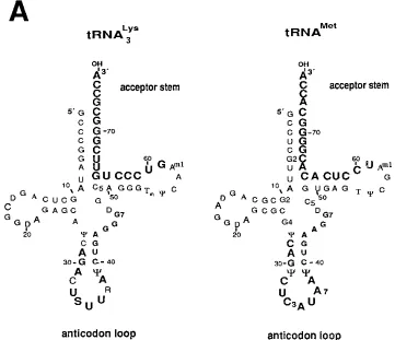
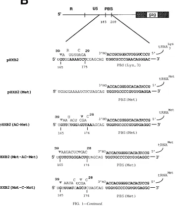
[image:2.612.126.487.71.382.2]Construction of mutant proviral genomes. General laboratory procedures were followed for DNA manipulation, plasmid preparation, and subcloning, essentially as described previously (18). The full-length molecular HIV-1 clone FIG. 1. Diagram of mammalian tRNAs and description of the mutations of the 39region of U5 (anticodon-complementary sequence) and the PBS region. (A) Cloverleaf structures of rabbit liver tRNA3
Lys
and HeLa cell tRNAMet
as described by Raba et al. (22) and Harada et al. (6), respectively. The nucleotides shown in large boldface type in the anticodon loop of tRNA are complementary to the sequences in U5 of the viral RNA genome. R, direct repeat sequences at the 59end of the viral RNA genome; U5, 59unique sequence; S, 5-methoxycarbonylmethyl-2-thiouridine; Y, pseudouridine; D, dihydrouridine; Tm, 29-O-methyl-5-methyluridine; Am1
, 1-methyladenosine; C5, 5-methylcytidine; G2, N2-methylguanosine; G4, N2, N2-dimethylguanosine; G7, 7-methylguanosine; T, 5-methyluridine. (B) The 59region
of the HIV-1 RNA genome is expanded to depict the locations of sequences having complementarity with tRNA as well as the expected base pairing between the sequence in U5 and the anticodon region of tRNA. A wild-type PBS (nucleotides 183 to 200) in pHXB2 was replaced with the PBS complementary to the 39-terminal 18 nucleotides of tRNAMet
[pHXB2(Met)]. A 200-bp fragment from a mutant with a PBS complementary to RNAMet
from pHXB2(Trp-AC) culture (13) was transferred to an infectious clone, HXB2, and the resulting construct was named pHXB2(AC-Met). An additional mutant proviral genome which contained sequences complementary to the anticodon region of tRNAMet
[pHXB2(Met-AC-Met)] was constructed. Finally, pHXB2(Met-C-Met), in which there are nucleotides which would disrupt the complementarity between the 39region of U5 in the viral RNA genome and the corresponding anticodon region of tRNAMet
, was constructed.
on November 9, 2019 by guest
http://jvi.asm.org/
pHXB2 (23, 24) was used to make mutant HIV-1 proviral genomes. To construct pHXB2(AC-Met), a DNA fragment encompassing the U5 and PBS regions was PCR amplified from an integrated provirus derived from the transfection of a proviral genome containing a PBS and the sequences complementary to the 39-terminal nucleotides and anticodon loop of tRNATrp, respectively (13). For
subcloning, an AflII-BssHII DNA fragment (nucleotides 64 to 255) was first cloned into a shuttle vector, pUC119(PBS), which contains an HpaI-to-PstI DNA fragment including the 59long terminal repeat, PBS, and leader region of the gag gene from pHXB2 (25); the resulting clone was named pUC119(AC-Met). The complete DNA sequence of the AflII-BssHII fragment was determined by se-quencing (26). pUC119(AC-Met) was used for the subsequent preparation of pUC119(Met-AC-Met) and pUC119(Met-C-Met); pUC119(Met) was prepared from pUC119(PBS) (25). Site-directed mutagenesis was carried out by two con-secutive PCRs (27). Megaprimers containing mutant sequences were produced by the first PCR with a primer in the U3 region of the 59long terminal repeat (59-TTGACAGCCGCCTAGC [nucleotides 8895 to 8910]) and a mutagenic oligonucleotide. The oligonucleotides for the mutants are as follows:
pHXB2(Met), 59-TCGCTTTCAATCCTCACACGGGGACCACTGCTAG-39;
pHXB2(Met-AC-Met), 59-ACCACTGCTACAGTCCCACAACACTGACTAA
A-39; and pHXB2(Met-C-Met), 59-ACCACTGCTAGAGCTGATAAACACTG ACTAAAA-39. The second PCR was performed to generate restriction enzyme recognition sites for cloning into pUC119(PBS) by using a megaprimer contain-ing mutant sequence and a primer downstream of the PBS (59-GCGCGCTTC AGCAAGCCG-39[nucleotides 262 to 245]). These mutagenic DNA fragments were digested with BglII and BssHII and were cloned into pUC119(PBS). All constructs were sequenced to verify that only the desired mutations in U5 and
the PBS were present. Each of the pUC119 plasmid constructs containing the mutations was digested with HpaI and BssHII, resulting in an 868-bp fragment encompassing the U5 and PBS. The 868-bp fragment was subcloned between the
HpaI and BssHII sites of pHXB2, and the resulting mutant proviral plasmids
were screened by restriction digestions and again verified by DNA sequencing (26).
DNA transfections and analysis of proviral gene expression.Transfection of proviral plasmid DNA into COS-1 cells by using DEAE-dextran was carried out as previously described (34). After 48 h posttransfection, the supernatant was collected and filtered through a 0.45-mm-pore-size syringe filter (Nalgene). The levels of p24 antigen were determined from two independent transfections by enzyme-linked immunosorbent assay (ELISA) (Coulter Laboratories).
Analysis of virus infectivity.To test for virus replication and infectivity, the various proviral constructs were transfected into COS-1 cells, which was followed 24 h later by coculture with SupT1 cells (53105), which support high-level
replication of HIV-1. After 48 h, the SupT1 cells were harvested by low-speed centrifugation (1,0003g) and further cultured with fresh medium and additional
SupT1 cells. The infected SupT1 cells were maintained by splitting 1:6 every 3 days. For cell-free infections at days 27, 35, 44, and 61 postcoculture, SupT1 cells (106cells per ml) were infected with equal amount of virus as measured by p24
antigen. After the virus was allowed to adsorb for 24 h, SupT1 cells were pelleted (1,0003g), washed with phosphate-buffered saline (pH 7.0), and further
[image:3.612.134.489.81.500.2]cul-tured. SupT1 cultures were monitored visually for the formation of multicell syncytia and at various time intervals were refed with SupT1 cells and medium. At the designated time intervals, samples of the culture supernatant were col-lected and analyzed for p24 antigen by ELISA (Coulter Laboratories). FIG. 1—Continued.
on November 9, 2019 by guest
http://jvi.asm.org/
PCR amplification and DNA sequencing of PBS-containing proviral DNA. Analysis of the proviral PBS sequences from infected SupT1 cells was carried out by using minor modifications of protocols previously described (33). Briefly, at the designated days postcoculture, genomic DNA was isolated from infected SupT1 cells by using the Wizard genomic DNA purification kit according to the manufacturer’s instruction manual (Promega). Approximately 2mg of cellular DNA was used to amplify integrated proviral DNA sequences comprising the U5 and PBS regions of the provirus genome by using two HIV-1-specific oligonu-cleotide primers, as follows: primer 1, 59-GCTCTAGACCAGATCTGAGCCT GGGAGCTC-39(nucleotides 17 to 38); and primer 2: 59-CGGAATTCTCTCC TTCTAGCCTCCGCTAGTC-39 (nucleotides 309 to 330) (the underlined nucleotides indicate the additional sequences at the 59end which correspond to
XbaI and EcoRI restriction endonuclease sites for subcloning of PCR products).
PCR-amplified DNA was digested with XbaI and EcoRI and then subcloned between the XbaI and EcoRI sites present in the polylinker region of pUC119 DNA or was directly ligated into the pCRII vector (Invitrogen). Following ligation, the DNA was transformed into competent Escherichia coli (DH5a) and plated on 1.5% Luria-Bertani agar medium containing isopropyl-b-D -thiogalac-topyranoside (IPTG) and 5-bromo-4-chloro-3-indolyl-b-D-galactopyranoside (X-Gal) (U.S. Biochemical). Following identification of colorless clones, the DNAs prepared from individual recombinant clones were sequenced by using the primer 59-GGCTAACTAGGGAACCCACTGC-39(nucleotides 42 to 63) (26).
RESULTS
Construction of HIV-1 proviral genomes with U5 and PBS mutations.In a previous study, we described the construction of a mutant proviral genome, pHXB2(Trp-AC), which con-tains a PBS complementary to the 39-terminal 18 nucleotides
of tRNATrpand an 8-nucleotide substitution at nucleotides 167
to 175 of the HIV-1 genome (167UCUGGAGU175)
comple-mentary to the anticodon loop of tRNATrp(13). Analysis of
the PBS sequences from proviral genomes derived from the transfection of pHXB2(Trp-AC) revealed that the majority, but not all, were complementary to a completely unexpected tRNA, tRNAMet, at 27 days following infection of SupT1 cells.
After further in vitro culture, though, we found that the PBSs of the viruses derived from pHXB2(Trp-AC) reverted back to be complementary to tRNA3
Lys. It was not possible to discern
the sequence of the reversion back to the wild-type PBS; that is, it was possible that the PBS complementary to tRNATrpfirst
mutated to be complementary to tRNAMetand then
subse-quently reverted to a PBS complementary to tRNA3
Lys. If this
was the case, we would expect the proviruses containing a PBS complementary to tRNAMetto be unstable. Alternatively, the
PBS complementary to tRNATrp might revert so as to be
complementary to either tRNAMetor tRNA 3
Lys; if this was the
case, we would expect that proviruses with a PBS complemen-tary to tRNAMetmight be stable. To address this question, a
DNA fragment encompassing nucleotides 64 to 255 from the provirus with a PBS complementary to tRNAMetwas cloned
into an infectious clone of HIV-1, pHXB2; this new construct, referred to as pHXB2(AC-Met), contains a complete HIV-1 provirus which has a PBS complementary to the 39-terminal 18 nucleotides of tRNAMet, an 8-nucleotide sequence (UCUGG
AGU) (nucleotides 167 to 174) complementary to the antico-don loop of tRNATrp, and two additional nucleotide mutations
(176AA177) between the PBS and the
anticodon-complemen-tary sequence (Fig. 1B).
Previous studies have shown that mutating a PBS comple-mentary to alternate tRNAs was not sufficient to produce a virus capable of maintaining the use of alternate tRNAs over multiple rounds of retrovirus replication (4, 16, 34, 35). To determine if this was also the case for a PBS complementary to tRNAMet, site-directed mutagenesis was used to replace the
wild-type PBS with sequences complementary to the 39 -termi-nal 18 nucleotides of tRNAMet[pHXB2(Met) (Fig. 1B)]. Since
the anticodon-complementary sequence in pHXB2(AC-Met) does not have base complementarity with the anticodon region of tRNAMet, an additional mutation in pHXB2(AC-Met) was
made so that the corresponding sequence complementary to the anticodon region of tRNAMetwas present; the resulting
construct was named pHXB2(Met-AC-Met) (Fig. 1B). Note that to avoid the insertion of a new AUG codon within the HIV-1 genome, the mutation was made to substitute a guanine for the adenine (GUG instead of AUG); complementarity with the anticodon region of tRNAMetwould still be facilitated by
virtue of a GzU base pair (28). Finally, to investigate the effect that a disruption of the complementarity between the sequence in U5 and the anticodon region of tRNA might have on the maintenance of a substituted PBS, pHXB2(Met-C-Met) was constructed, in which nucleotides cytosines 171 and 176 within this region (167UUAUCAGCUC176) were substituted for
guanosines; this mutation would disrupt the potential base pair with cytosines in the anticodon loop (39-38AAUACUCCGAC 28-59) of tRNAMet(underlined nucleotides) (Fig. 1B).
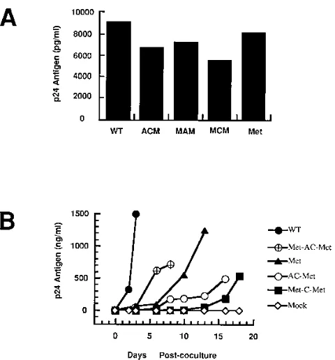
Replication potential of mutant proviral genomes.Since the 59region of the HIV-1 genome contains many cis-acting ele-ments, we wanted to determine whether the mutations in U5 and the PBS would have any effect on expression of HIV-1 proteins and on release of virions into medium upon transfec-tion. Plasmids containing the HIV-1 wild-type and mutant pro-viral genomes were transfected into COS-1 cells, followed by analysis of the culture supernatants for p24 antigen (Fig. 2A). Taking into consideration the variability inherent with trans-FIG. 2. Protein expression and infectivities of proviruses with mutations in
U5 and the PBS. (A) Release of virus from COS-1 cells transfected with the wild-type and mutant proviral genomes. COS-1 cells were transfected with the designated plasmids. At 48 h posttransfection, the culture supernatants were analyzed for released virus particles by quantitation of p24 antigen by ELISA (Coulter). The bar graph illustrates the mean values of p24 antigen detected from two independent transfections. WT, wild-type pHXB2; ACM,
pHXB2(AC-Met); MAM, pHXB2(Met-AC-Met); MCM, pHXB2(Met-C-Met); Met,
pHXB2(Met). (B) Kinetics of the appearance of infectious virus derived from transfection of wild-type and mutant proviral genomes. The plasmids containing the wild-type and mutant proviral genomes were transfected into COS-1 cells; this was followed 24 h later by coculture with SupT1 cells (53105). After 48 h
of coculturing, the SupT1 cells were then isolated by centrifugation, washed once, and further cultured with more uninfected SupT1 cells and fresh medium (day 0). At various intervals postcoculture, culture supernatants were collected and the p24 antigen was quantitated by ELISA (Coulter).
on November 9, 2019 by guest
http://jvi.asm.org/
[image:4.612.59.299.67.325.2]fection efficiencies, no significant difference between the amounts of p24 antigen released from cultures transfected with different viral genomes and the wild type was observed.
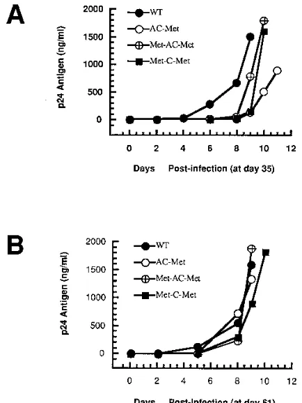
To determine the infectious potential of each of the mutant proviral genomes, transfected COS-1 cells were cocultured with SupT1 cells, which are susceptible to HIV-1 infection. At various times postcoculture, replication of both the wild-type and mutant viruses was monitored by visual inspection for syncytium formation and quantitated by levels of viral capsid (p24) antigen in the supernatants from the cultures (Fig. 2B). All of the viruses derived from mutant proviral genomes dem-onstrated a delay in virus production compared with that of the wild-type virus. The levels of supernatant p24 from viruses derived from pHXB2(Met) rapidly increased from day 10 post-coculture, reaching a level similar to that from wild-type virus at approximately 13 days postcoculture. The results of these studies demonstrate that the proviruses with the mutations in U5 and the PBS all gave infectious virus upon transfection, although the appearance of the viruses was delayed compared to that of the wild-type virus.
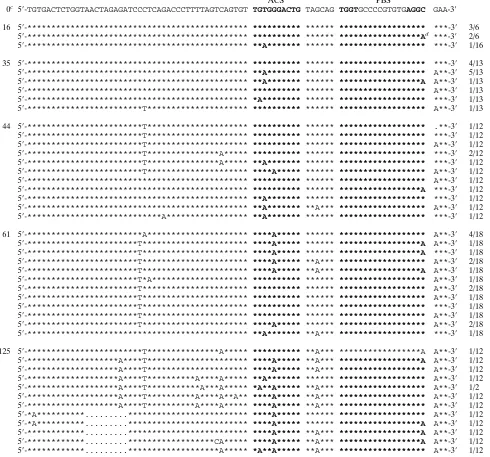
Analysis of the PBSs from viruses derived from pHXB2 (Met) and pHXB2(AC-Met).Since the PBS is generated when the first 18 nucleotides of the primer tRNA are copied during plus-strand strong-stop viral DNA synthesis of reverse tran-scription, analysis of the proviral PBS sequences will determine which tRNA species was used to initiate reverse transcription. We first analyzed the proviruses which were isogenic with HXB2 except for the PBS which was complementary to tRNAMet[pHXB2(Met)]. The viruses derived from pHXB2
(Met) at day 10 postcoculture maintained a PBS complemen-tary to the 39 nucleotides of tRNAMet, although additional
single-nucleotide substitutions were found around the PBS (data not shown). By 13 days postcoculture, the level of p24 antigen increased greatly (Fig. 2B). Analysis of PBS sequences from proviruses at day 16 revealed that the viruses were het-erogeneous (Table 1); two clones in which the PBS reverted back to be complementary to tRNA3
Lys were identified. Two
other clones contained a PBS complementary to tRNA3 Lys
in-serted upstream of a PBS complementary to tRNAMet, with 15
nucleotides of additional intervening sequence between the dual PBSs. We previously found proviruses with dual PBSs from viruses derived from a mutant proviral genome where the 39regions of U5 and the PBS were mutated to be complemen-tary to the anticodon region and the 39-terminal 18 nucleotides of tRNAPro, respectively (13); it is possible that this insertion
occurred as a result of aberrant template switching during reverse transcription (8). Additional analysis revealed a PBS complementary to tRNA1,2
Lys. By 35 days postcoculture, though,
all of the PBS clones isolated contained a wild-type PBS com-plementary to tRNA3
Lys.
In contrast to viruses derived from pHXB2(Met), viruses derived from pHXB2(AC-Met) contained the PBS comple-mentary to tRNAMet at 16 days postcoculture. Most of the
PBSs analyzed maintained an original input sequence up-stream of the PBS; two clones had mutations just upup-stream or downstream of the PBS (data not shown). By day 35 postcocul-ture, 6 of the 11 clones analyzed maintained a PBS comple-mentary to tRNAMet(Table 2). A point mutation was found at
nucleotide 201 (G to A) downstream of the PBS in all clones. Four of six clones with a PBS complementary to tRNAMethad
an additional mutation at nucleotide 171 (G to A) within the anticodon region-complementary sequence. Interestingly, the other clones (5 of 11) had a PBS complementary to tRNA1,2 Lys
rather than tRNA3
Lys. Since we did not detect a PBS
comple-mentary to tRNA3
Lys by day 35, we wanted to compare the
replication of the virus with that of the wild type. Culture supernatants at day 35 were collected, and equal amounts of virus as measured by the level of p24 antigen were used to infect SupT1 cells (Fig. 3A). The viruses still grew slowly com-pared to the wild-type virus, although the delay was not as long as that observed at earlier times postcoculture (Fig. 2B). Since tRNA1,2
Lysspecies were reported to be encapsidated into HIV-1
virion particles (12, 14), we wanted to determine if the rever-tant viruses with a PBS complementary to tRNA1,2
Lyswere
sta-TABLE 1. Partial sequences of the U5 and PBS regions of pHXB2(Met) mutant proviruses isolated after coculture
Day postco-culture
Sequencea Frequencyb
ACS PBS
0c 59-AGATCCCT CAGACCCTTT TAGTCAGT GTGGAAAA TCTCTA GCAG TGG TGC CCC GTG TGA GGA TTGAAAGCG-39
16 59-******** ********** ******** ******** ****** **** *** *** *** *** *** **Cd *********-39 1/8 59-******** ********** ******** ******** ****** ***A *** *** *** *** *** **C *********-39 2/8 59-******** ********** ******** ******** ****** ***A TGG CGCCCCA ACG TGC GGCe *********-39 1/8 59-******** ********** ******** ******** ****** *T** TGG CGC CCG AAC AGG GACf .********-39 1/8 59-******** ********** ******** ******** ****** ***A TGG CGCCCCG AAC AGG GACg *********-39 1/8
TGG CGC CCG AAC AGG GAC ACAAATCTCTAGCAAh
59-******** ********** ******** ******** ****** ***A *** *** *** *** *** **C *********-3* 2/8 35 59-******** ********** ******** ******** ****** **** TGG CGC CCG AAC AGG GACi .********-39 6/7 59-******** ********** ******** ******** ****** *T** TGG CGC CCG AAC AGG GAC .********-39 1/7
a
Asterisks indicate identity with the input sequence (day 0); boldface indicates nucleotides in ACS or PBS; dots indicate deletions. ACS, nucleotides located in the A-rich loop region in the HXB2 HIV-1 clone.
b
Frequency of the DNA sequence of the PBS region obtained from independent clones.
c
The sequence is the input sequence with the initial mutations in the PBS region (PBS complementary to tRNAMet
).
d
One nucleotide difference from the input PBS complementary to tRNAMet
(implying the presence of isoacceptor for tRNAMet
).
e
PBS complementary to the 39-terminal 18 nucleotides of tRNA1,2 Lys
(with one nucleotide insertion).
f
PBS complementary to the 39-terminal 18 nucleotides of tRNA3 Lys
.
g
PBS complementary to the 39-terminal 18 nucleotides of tRNA3 Lys
(with one nucleotide insertion).
h
Insertion of PBS complementary to tRNA3 Lys
and duplicated nucleotides (upstream sequence of PBS) before the PBS complementary to tRNAMet
.
i
One nucleotide deletion during reversion from PBS complementary to tRNA1,2 Lys
to PBS complementary to tRNA3 Lys
.
➛
on November 9, 2019 by guest
http://jvi.asm.org/
ble. By day 44 postcoculture, we identified PBSs in 2 of 11 clones complementary to tRNA3
Lys; a variety of mutations
up-stream of PBSs in most clones were also observed. Only two clones which maintained a PBS complementary to tRNAMet
were found. The remaining clones contained a PBS comple-mentary to tRNA1,2
Lys, accompanying adenine substitution
mu-tations within the anticodon loop-complementary sequence (169T to A and171G to A). By day 61 postcoculture, all of the
clones analyzed contained the wild-type PBS and also acquired the G-to-A substitution at nucleotide 171. As might be ex-pected, viruses analyzed at this time replicated similarly to the wild-type virus (Fig. 3B).
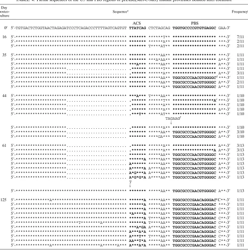
Analysis of PBSs of viruses derived from pHXB2(Met-AC-Met) and pHXB2(Met-C-pHXB2(Met-AC-Met). The delay in the reversion of the PBSs of viruses derived from pHXB2(AC-Met) to wild type convinced us to construct proviruses with modified U5 regions that might enhance the stability of the PBS. The most logical choice would be to construct a provirus with both the anticodon-complementary sequence in U5 and a PBS comple-mentary to the corresponding region of tRNAMet [pHXB2
(Met-AC-Met)]. The appearance of viruses derived from transfection of pHXB2(Met-AC-Met) was faster than that of the other mutant viruses (Fig. 2B). In all of the clones analyzed from day 16 postcoculture, the anticodon-complementary re-gion and PBS were complementary to tRNAMet(Table 3). A
single clone was found to have a T-to-A change at nucleotide 169 within the anticodon region-complementary sequence. Analysis at day 35 postcoculture revealed that all clones still
maintained a PBS complementary to tRNAMet. Two additional
mutations were observed most commonly: the T-to-A change at nucleotide 169 and a G-to-A change at nucleotide 201 just downstream of the PBS. We also isolated a single clone con-taining a substitution mutation of C to T at nucleotide 145, which is 38 nucleotides upstream of the PBS. The appearance of this virus was a little slower than that of the wild type when analyzed with samples from day 35 postcocultures (Fig. 3A). Surprisingly, it was found that all of the clones isolated at days 44 and 61 still maintained a PBS complementary to tRNAMet.
A substitution of C to T at nucleotide 145 was predominant at days 44 (7 of 12 clones) and 61 (15 of 18 clones). Two other prevalent point mutations were clear at day 61: a G-to-A change at nucleotide 171 (12 of 18 clones) within the anticodon region-complementary sequence and a G-to-A change at nu-cleotide 201 (15 of 18 clones) just downstream of the PBS. Viruses isolated at this time replicated similarly to the wild-type virus (Fig. 3B). Finally, analysis of the PBSs from the viruses derived from pHXB2(Met-AC-Met) at 125 days post-coculture revealed that the PBS was still complementary to tRNAMet(Table 3). Interestingly, no single dominant clone
was recovered; all of the clones had mutations in U5. Several adenine substitution mutations were found at the positions further upstream of the PBS (140G, 161C, and 179G to A).
Furthermore, clones which contained a 9-nucleotide deletion (133AACTAGAGA141) at the 42-nucleotide upstream position
[image:6.612.59.556.79.362.2]of the PBS were recovered (5 of 12 clones). Taken together, the results of these experiments demonstrate that mutation in
TABLE 2. Partial sequences of the U5 and PBS regions of pHXB2(AC-Met) mutant proviruses isolated after coculture
Day postco-culture
Sequencea Frequencyb
ACS PBS
0c 59-AGAGATCCCT CAGACCCTTT TAGTCAGTGT TCTGGAGT TAAA GCAG TGG TGC CCC GTG TGA GGC GAAAGCG-39
35 59-********** ********** ********** ******** **** **** *** *** *** *** *** *** A******-39 2/11 59-********** ********** ********** ****A*** **** **** *** *** *** *** *** *** A******-39 3/11 59-********** ********** ********** ****A*** **** **** *** *** *** *** *** **Ad A******-39 1/11 59-********** ********** ********** ******** **** **** TGG CGC CCA ACG TGG GGCe A******-39 5/11
44 59-********** ********** ********** ******** **** **** *** *** *** *** *** *** *******-39 1/11 59-********** ********** ********A* ******** **** **** *** *** *** *** *** *** A******-39 1/11 59-********** ********** ********** ******** **** *A** TGG CGC CCA ACG TGG GGC A******-39 1/11 59-********** ********** ********** ****A*** **** **** TGG CGC CCA ACG TGG GGC A******-39 1/11 59-********** ********** ********A* ****A*** **** A*** TGG CGC CCA ACG TGG GGC A******-39 1/11 59-********** ********** ********** **A*A*** **** **** TGG CGC CCA ACG TGG GGC A******-39 1/11 59-********** ********** ********** **A***** **** A*** TGG CGC CCA ACG TGG GGC A******-39 1/11 59-********** T********* *******A** **AA**** **** A**T TGG CGC CCA ACG TGG GGC A******-39 1/11 59-********** ********** .********* ***A**** **** **** TGG CGC CCA ACG TGG GGC A******-39 1/11 59-********** ********** ********** ****A*** **** **** TGG CGC CCG AAC AGG GACf *******-39 1/11 59-********** ********** *******G** ******** **** **** TGG CGC CCG AAC AGG GAC A******-39 1/11
61 59-********** ********** ********** ****A*** **** **** TGG CGC CCG AAC AGG GAC *******-39 8/15 59-********** ********** ********** ****A*** A*** **** TGG CGC CCG AAC AGG GAC *******-39 2/15 59-********** ********** ********** ****A*** **** **** TGG CGC CCG AAC AGG GAC A******-39 1/15 59-********** T********* ********** ****A*** **** **** TGG CGC CCG AAC AGG GAC *******-39 1/15 59-********** ********** ********** ***AA*** **** **** TGG CGC CCG AAC AGG GAC *******-39 1/15 59-***A****** ********** ********** ****A*** A*** **** TGG CGC CCG AAC AGG GAC *******-39 1/15 59-********** T********* A********* ****A*** A*** **** TGG CGC CCG AAC AGG GAC *******-39 1/11
aAsterisks indicate identity with the input sequence (day 0); boldface indicates nucleotides in ACS or PBS; the dot indicates a deletion. ACS,
anticodon-complementary sequences located in the 39region of U5 which were made complementary to the anticodon loop of tRNATrp. bFrequency of the DNA sequence of the PBS region obtained from independent clones.
cThe sequence is the input sequence with the initial mutations in the PBS region (PBS complementary to tRNAMet). dOne nucleotide difference from the input PBS complementary to tRNAMet(implying the presence of isoacceptor tRNAMet). ePBS complementary to the 39-terminal 18 nucleotides of tRNA
1,2 Lys. fPBS complementary to the 39-terminal 18 nucleotides of tRNA
3 Lys.
on November 9, 2019 by guest
http://jvi.asm.org/
nucleotides in U5 to be complementary to the anticodon re-gion of tRNAMetresulted in a virus which stably maintained a
PBS complementary to tRNAMet.
To further understand the role that the U5 sequences played in the virus maintaining a PBS complementary to tRNAMet, we
analyzed the effects that a change within the anticodon-com-plementary sequence would have on maintenance of the PBS. We altered nucleotides from G to C, which would disrupt base pairing with the anticodon region of tRNAMet
[pHXB2(Met-C-Met)] (Fig. 1B). The appearance of viruses derived from transfection of pHXB2(Met-C-Met) was slower than that of the other mutant viruses. By day 16 postcoculture, all clones analyzed maintained a PBS complementary to tRNAMet,
al-though none of the clones contained the exact input sequence. Additional mutations were found around the 39end of U5; a single substitution mutation at nucleotide 180 (C to T or A) was prominent. Two striking changes were observed in the clones isolated at 35 days postcoculture (Table 4). One was a 26-nucleotide deletion just upstream of the anticodon-comple-mentary region in U5. All clones having this deletion (4 of 11) maintained a PBS complementary to tRNAMetand also had
the same point mutation as at day 16 at nucleotide 180 (C to T or A). The other change was that in 4 of 11 clones the PBS was complementary to tRNA1,2
Lys. Additional mutations of one or
two nucleotides (C to A at nucleotide 180 or GC to AA at nucleotides 179 and 180) in the clones with the PBS comple-mentary to tRNA1,2
Lyswere found. Three clones analyzed at this
time maintained a PBS complementary to tRNAMet without
deletion of nucleotides in U5 and contained the same 2-nucle-otide substitutions (GC to AA) as those in clones with a PBS
complementary to tRNA1,2
Lys. Analysis of the virus replication at
day 35 revealed that the production of p24 antigen in the culture supernatant from virus derived from pHXB2(Met-C-Met) was delayed compared to that from the wild-type virus (Fig. 3A).
To further examine the stabilities of the PBSs of these vi-ruses derived from pHXB2(Met-C-Met), we analyzed the PBS sequences at days 44 and 61 (Table 4). No reversion to a PBS complementary to tRNA3
Lyswas detected. Most of the clones
with the deletions in U5 and the PBS complementary to tRNAMetcontained mutations of C to A at nucleotide 180 and
G to A at nucleotide 201. In contrast, the clones with a PBS complementary to tRNA1,2
Lys were heterogeneous within U5,
containing several substitution mutations within the anticodon region-complementary sequence and around the PBS at day 61. Interestingly, we did find that a GC-to-AA mutation at nucleotides 179 and 180 occurred in the viruses with a PBS complementary to tRNA1,2
Lys, which resulted in a stretch of four
adenine nucleotides (nucleotides 178 to 181); one of these mutations (C to A at nucleotide 180) was within the integration motif (CA, nucleotides 180 and 181), which is known to be required for integration of proviral DNA into the chromosome (15). Since the nucleotides within the anticodon-complemen-tary sequence in pHXB2(Met-C-Met) are not complemenanticodon-complemen-tary to the anticodon loops of both tRNA1,2
Lys and tRNAMet, we
wanted to determine the stability of the PBS after further extended in vitro culture. As might be expected, then, by 125 days postcoculture all of the PBSs recovered were wild-type PBSs complementary to tRNA3
Lys(Table 4). Additional
muta-tions within the anticodon-complementary sequence in U5 (a G-to-A change at nucleotide 173) and a G-to-C change at the 39end of the PBS were also found. The results of these studies demonstrate that mutations within the anticodon-complemen-tary region in U5 influence the stability of the maintenance of the PBS complementary to tRNAMet.
DISCUSSION
In this study, we have investigated the effect that mutations in the U5 region have on the selection of the tRNA primer used to initiate reverse transcription. For these studies, we reconstructed a series of HIV-1 proviral genomes in which the PBS was complementary to tRNAMet. The choice of tRNAMet
was based on a previous study in which we found HIV-1 viruses which contained a PBS complementary to tRNAMet(13). Our
starting construct contained a DNA fragment (nucleotides 64 to 255) from a mutant provirus containing a PBS complemen-tary to tRNAMet, which was found in a virus culture derived
from transfection of a plasmid containing a provirus with a PBS and an anticodon-complementary region complementary to tRNATrp(13). HIV-1 proviral genomes containing a PBS
complementary to tRNAMet[pHXB2(Met)] as well as proviral
genomes with mutant sequences located in U5 which are com-plementary to the anticodon loop of tRNAMet
[pHXB2(Met-AC-Met) and pHXB2(Met-C-Met)] were also created. Trans-fection of each of these constructs into cells resulted in the production of infectious virus. A DNA sequence analysis of the U5 and PBS regions from integrated provirus genomes re-vealed that some viruses derived from pHXB2(Met) reverted to have a wild-type PBS by day 16 postcoculture. In contrast, the viruses derived from pHXB2(AC-Met), pHXB2(Met-AC-Met), and pHXB2(Met-C-Met) still maintained a PBS com-plementary to tRNAMetat this time. Upon extended culture,
though, only the virus derived from pHXB2(Met-AC-Met) maintained a PBS complementary to tRNAMet.
[image:7.612.69.286.69.358.2]The results of our study are unique for several reasons. First, FIG. 3. Replication of the infectious virus. SupT1 cells were infected with
equal amounts (20 ng/ml) of virus isolated after 35 (A) and 61 (B) days of coculture. At the designated times postinfection, supernatants were removed and assayed for p24 antigen. The initial mutations in the PBS region are indicated.
on November 9, 2019 by guest
http://jvi.asm.org/
the isolation of a second HIV-1 virus which stably maintains a PBS complementary to an alternative tRNA complements our previous study in which we identified an HIV-1 which stably maintains a PBS complementary to tRNAHis (34). Although
tRNAMethas not been previously identified as a primer for
HIV-1 reverse transcription, the use of tRNAMetto initiate
reverse transcription is not without precedence, since most retrotransposons and plant pararetroviruses use an initiator
tRNAMetto initiate reverse transcription (19). The fact that
HIV-1 can stably use alternative tRNAs such as tRNAHisand
tRNAMethighlights the flexibility this virus has with respect to
some of the requirements for initiation of reverse transcrip-tion. Second, the fact that complementarity between the anti-codon-complementary sequence in U5 and the anticodon region of tRNAMetwas critical for maintaining a PBS
[image:8.612.63.553.120.577.2]comple-mentary to tRNAMetin mutant viruses adds further support
TABLE 3. Partial sequences of the U5 and PBS regions of pHXB2(Met-AC-Met) mutant proviruses isolated after coculture
Day post-culture
Sequencea Frequencyb
ACS PBS
0c 59-TGTGACTCTGGTAACTAGAGATCCCTCAGACCCTTTTAGTCAGTGT TGTGGGACTG TAGCAG TGGTGCCCCGTGTGAGGC GAA-39
16 59-********************************************** ********** ****** ****************** ***-39 3/6 59-********************************************** ********** ****** *****************Ad ***-39 2/6 59-********************************************** **A******* ****** ****************** ***-39 1/16
35 59-********************************************** ********** ****** ****************** ***-39 4/13 59-********************************************** **A******* ****** ****************** A**-39 5/13 59-********************************************** **A******* ****** *****************A A**-39 1/13 59-********************************************** ********** ****** ****************** A**-39 1/13 59-********************************************** *A******** ****** ****************** ***-39 1/13 59-************************T********************* ********** ****** ****************** A**-39 1/13
44 59-************************T********************* ********** ****** ****************** .**-39 1/12 59-************************T********************* ********** ****** ****************** ***-39 1/12 59-************************T********************* ********** ****** ****************** A**-39 1/12 59-************************T***************A***** ********** ****** ****************** ***-39 2/12 59-************************T***************A***** **A******* ****** ****************** ***-39 1/12 59-************************T********************* ****A***** ****** ****************** A**-39 1/12 59-********************************************** ********** ****** ****************** A**-39 1/12 59-********************************************** ********** ****** *****************A ***-39 1/12 59-********************************************** **A******* ****** ****************** ***-39 1/12 59-********************************************** **A******* **A*** ****************** A**-39 1/12 59-****************************A***************** **A******* ****** ****************** ***-39 1/12
61 59-************************A********************* ****A***** ****** ****************** A**-39 4/18 59-***********************T********************** ****A***** ****** *****************A A**-39 1/18 59-***********************T********************** ****A***** ****** *****************A ***-39 1/18 59-***********************T********************** ****A***** **A*** ****************** A**-39 2/18 59-***********************T********************** ****A***** **A*** *****************A A**-39 1/18 59-***********************T*A******************** ********** ****** ****************** A**-39 1/18 59-***********************T********************** ********** ****** ****************** A**-39 2/18 59-***********************T********************** ********** ****** ****************** A**-39 1/18 59-***********************T********************** ********** ****** ****************** ***-39 1/18 59-***********************T********************** ********** ****** ****************** A**-39 1/18 59-***********************T********************** ****A***** ****** ****************** A**-39 2/18 59-********************************************** **A******* **A*** ****************** ***-39 1/18
125 59-************************T***************A***** ********** **A*** *****************A A**-39 1/12 59-*******************A****T********************* ****A***** **A*** *****************A A**-39 1/12 59-*******************A****T********************* ****A***** **A*** ****************** A**-39 1/12 59-*******************A****T**********A****A***** **A******* ****** ****************** A**-39 1/12 59-*******************A****T***********A***A***** *A**A***** **A*** ****************** A**-39 1/2 59-*******************A****T**********A****A**A** ****A***** **A*** ****************** A**-39 1/12 59-*******************A****T**********A****A***** ****A***** **A*** ****************** A**-39 1/12 59-*A**********...************************* ****A***** ****** ****************** A**-39 1/12 59-*A**********...************************* ****A***** ****** *****************A A**-39 1/12 59-************...************************* ****A***** **A*** *****************A A**-39 1/12 59-************...******************CA***** ****A***** **A*** *****************A A**-39 1/12 59-************...*******************A***** *A**A***** **A*** ****************** A**-39 1/12
aAsterisks indicate identity with the input sequence (day 0); boldface indicates nucleotides in ACS or PBS; dots indicate deletions. ACS, anticodon-complementary
sequences located in the 39region of U5 which were made complementary to the anticodon region of tRNAMet. bFrequency of the DNA sequence of the PBS region obtained from independent clones.
cThe sequence is the input sequence with the initial mutations in the PBS region (PBS complementary to tRNAMet).
dOne nucleotide difference from the input PBS complementary to tRNAMet(implying the presence of isoacceptor for tRNAMet).
on November 9, 2019 by guest
http://jvi.asm.org/
for a role for this region in the selection of the tRNA to initiate reverse transcription. Viruses derived from pHXB2(AC-Met) and pHXB2(Met-C-Met), which had nucleotides in the 39 re-gion of U5 that would disrupt base pairing with the anticodon region of tRNAMet, did not stably maintain a PBS
complemen-tary to tRNAMet. Third, although the PBS in the virus derived
from pHXB2(Met-AC-Met) was stable, it appears that the
virus was still evolving after 125 days postcoculture. Additional mutations in and around the anticodon region-complementary sequence were observed in proviral clones derived from pHXB2(Met-AC-Met) after extended culture days. For exam-ple, a point mutation (T to A at nucleotide 169) in the region complementary to the anticodon region of tRNAMetwas
[image:9.612.61.554.74.571.2]iden-tified in a single clone at day 16. The number of clones with this
TABLE 4. Partial sequences of the U5 and PBS regions of pHXB2(Met-C-Met) mutant proviruses isolated after coculture
Day postco-culture
Sequencea Frequencyb
ACS PBS
0c 59-TGTGACTCTGGTAACTAGAGATCCCTCAGACCCTTTTAGTCAGTGT TTATCAG CTCTAGCAG TGGTGCCCCGTGTGAGGC GAA-39
16 59-********************************************** ******* ******T** ****************** ***-39 7/11 59-********************************************** ******* ******A** ****************** ***-39 2/11 59-********************************************** ******* T****AT** ****************** ***-39 2/11
35 59-********************************************** ******* *****AA** ****************** ***-39 1/11 59-********************************************** ******* ***A*AA** ****************** A**-39 1/11 59-********************************************** ***A*** *****AA** ****************** A**-39 1/11 59-*********************... .****** ******T** ****************** ***-39 1/11 59-*********************... .****** ******A** ****************** A**-39 3/11 59-********************************************** ******* *****AA** TGGCGCCCAACGTGGGGCd***-39 1/11 59-********************************************** ******* *****AA** TGGCGCCCAACGTGGGGC A**-39 2/11 59-********************************************** ******* ******A** TGGCGCCCAACGTGGGGC A**-39 1/11
44 59-********************************************** ***A*** T****AA** ****************** ***-39 1/10 59-*********************... .****** ******T** *****************Ae***-39 1/10 59-*********************... .****** ******A** ****************** ***-39 1/10 59-*********************... .****** ******A** ****************** A**-39 1/10 59-*********************... .***T** *****AT** ****************** ***-39 1/10
TAGAAGf
59-*********************... .****** ******A** ****************** ***-39 1/10 59-*********************... ******* *****AA** TGGCGCCCAACGTGGGGC A**-39 3/10 59-*********************... ******* ****GA*** TGGCGCCCAACGTGGGGC A**-39 1/10
61 59-*********************... .****** ******A** ****************** A**-39 3/13 59-*********************... .****** ******A** *****************A A**-39 3/13 59-*********************... ******* *****AA** TGGCGCCCAACGTGGGGC ***-39 1/13 59-*********************... ******A *****AA** TGGCGCCCAACGTGGGGC ***-39 1/13 59-*********************... A****** *****AA** TGGCGCCCAACGTGGGGC A**-39 1/13 59-*********************... A*****A A****AA** TGGCGCCCAACGTGGGGC A**-39 1/13 59-*********************... A*G***A A****AA** TGGCGCCCAACGTGGGGC ***-39 1/13 59-*********************... A*G*G*A A****AA** TGGCGCCCAACGTGGGGC A**-39 1/13
T
59-********************************************** ******* *****AA** TGGCGCCCAACGTGGGGC A**-39 1/13
125 59-**************A******************************* ******A *****AA** TGGCGCCCGAACAGGGACgC**-39 1/11 59-********************************************** ******A *****AA** TGGCGCCCGAACAGGGAC ***-39 1/11 59-********************************************** ******A *****AA** TGGCGCCCGAACAGGGAC C**-39 1/11 59-********************************************** *A****A *****AA** TGGCGCCCGAACAGGGAC ***-39 1/11 59-********************************************** ******A T****AA** TGGCGCCCGAACAGGGAC C**-39 1/11 59-**************************T********A********** ******A T****AA** TGGCGCCCGAACAGGGAC C**-39 1/11 59-********************************************** ***A*GA A****AA** TGGCGCCCGAACAGGGAC C**-39 1/11 59-********************************************** A***A*A *****AA** TGGCGCCCGAACAGGGAC C**-39 1/11 59-********************************************** A***T** T****AA** TGGCGCCCGAACAGGGAC C**-39 1/11 59-********************************************** AA**T*A *****AA** TGGCGCCCGAACAGGGAC C**-39 1/11 59-**************A********************A******A*** A***A*A *****AA** TGGCGCCCGAACAGGGAC ***-39 1/11
a
Asterisks indicate identity with the input sequence (day 0); boldface indicates nucleotides in ACS or PBS; dots indicate deletions. ACS, anticodon-complementary sequence located in the 39region of U5 which would be predicted to disrupt the complementarity with the anticodon loop of tRNAMet
.
b
Frequency of the DNA sequence of the PBS region obtained from independent clones.
c
The sequence is the input sequence with the initial mutations in the PBS region (PBS complementary to tRNAMet
).
d
PBS complementary to the 39-terminal 18 nucleotides of tRNA1,2 Lys
.
e
One nucleotide difference from the input PBS complementary to tRNAMet
(implying the presence of isoacceptor for tRNAMet
).
f
Nucleotides inserted (duplicated nucleotides upstream of PBS).
g
PBS complementary to the 39-terminal 18 nucleotides of tRNA3 Lys
.
➛
➛
on November 9, 2019 by guest
http://jvi.asm.org/
mutation increased at day 35 (6 of 13 clones); however, this mutation was found only in a single clone at day 61. Instead, point mutations at nucleotide 145 (C to T) 38 nucleotides further upstream of PBS at day 35 and at nucleotide 171 (G to A) within the anticodon-complementary sequence at day 44 became predominant by 61 days in culture. Coincidentally, at this time we found that viruses with a PBS complementary to tRNAMetreplicated similarly to the wild-type virus as
mea-sured by p24 antigen in the culture (Fig. 3B). It is not clear if these additional nucleotide changes in U5 provided an advan-tage for viruses to use tRNAMetas a primer. Previous studies
with ALV and HIV-1 reported that the genomic RNA from PBS mutant virions with slower growth early in culture was associated with less occupancy of the corresponding tRNA primer on the mutant PBS compared with that of the wild type (4, 35). In our case, it is possible that the nucleotide changes result in a virus which could utilize tRNAMetmore effectively
to initiate reverse transcription. In support of this idea, previ-ous studies by Isel et al. have demonstrated a complex struc-ture between the U5-PBS and tRNA3
Lysprimer, extending the
interactions of tRNA3
Lys with viral sequences 39 nucleotides
upstream of the PBS (10); interestingly, the C-to-T mutation at nucleotide 145 found predominantly at the later culture times is located at this position. Additional studies will be required to delineate the interaction between the U5-PBS and tRNAMet.
Further evidence for a complex RNA structure at the 59end of the RNA genome required for selection of tRNAMetand
maintenance of the PBS complementary to tRNAMetcomes
from the fact that the viruses derived from transfection of pHXB2(AC-Met) reverted back to a wild-type PBS. The pres-ence of a sequpres-ence in U5 complementary to the anticodon loop of tRNATrp, then, did not stabilize the maintenance of a PBS
complementary to tRNAMet. Thus, the reversion to a PBS
complementary to tRNA3
Lys by the virus with the
anticodon-complementary sequence in U5 and a PBS anticodon-complementary to tRNATrpwas most probably through a virus which contained a
PBS complementary to tRNAMet. The fact that the viruses with
the sequence in U5 complementary to the anticodon loop of tRNATrp and a PBS complementary to tRNAMethad
addi-tional mutations resulting in a stretch of three adenine nucle-otides [nuclenucle-otides 176 to 178 in pHXB2(AC-Met)] upstream of the PBS might have facilitated the reversion to wild type. That is, even subtle mutations within the anticodon-comple-mentary region influence the maintenance of an alternate PBS, as evidenced from the analysis of the viruses derived from pHXB2(Met-C-Met). In this case, the virus reverted to contain a PBS complementary to tRNA1,2
Lysor, even more dramatically,
resulted in a provirus which contained a deletion of 26 nucle-otides upstream of the anticodon-complementary region. The fact that few nucleotide changes could initiate a cascade of mutations resulting in the virus utilizing a different PBS is, to our knowledge, without precedence. Our results lend support to a growing appreciation that a complex RNA secondary structure which influences both the selection of the tRNA primer and initiation of reverse transcription exists in the 59 end of the viral genome. This region of secondary structure could, in fact, be extensive, since a recent study demonstrated that a single base pair change in the TAR region influences minus-stranded DNA synthesis (7).
One of the more interesting results of this study was the generation of a virus containing a PBS complementary to tRNA1,2
Lys as found in the day 35 cultures derived from
pHXB2(Met-C-Met) and pHXB2(AC-Met). Again, the use of tRNA1,2
Lys is not without precedence, since tRNA 1,2 Lys is the
primer for Mason-Pfizer monkey virus, visna virus, and spu-maviruses (19). More importantly, analysis of HIV-1 virions
revealed that tRNA1,2
Lys is packaged at levels comparable to
those for tRNA3
Lys(12). Previous studies from this as well as
other laboratories, though, have demonstrated that viruses with a PBS complementary to tRNA1,2
Lysrapidly reverted back
to wild type after limited in vitro culture (16, 34). We observed that viruses derived from pHXB2(AC-Met) contained a PBS complementary to tRNA1,2
Lysat day 35, but by day 61 the viruses
contained only a PBS complementary to tRNA3
Lys. Some of
viruses derived from pHXB2(Met-C-Met) maintained a PBS complementary to tRNA1,2
Lysfor over 61 days, although the PBS
eventually reverted back to wild type. It is not clear why the viruses with a PBS complementary to tRNA1,2
Lys derived from
pHXB2(Met-C-Met) were delayed in the reversion to a wild-type PBS. One of the major differences between the viruses with a PBS complementary to tRNA1,2
Lys derived from
pHXB2(AC-Met) and pHXB2(Met-C-Met) can be found within the U5 region. In the case of viruses from pHXB2(AC-Met), we noted the substitution of adenine nucleotides within the anticodon-complementary region, in some cases, creating a substitution of three adenines similar to the wild-type se-quence. In contrast, the viruses with a PBS complementary to tRNA1,2
Lys derived from pHXB2(Met-C-Met) had substitution
of GC to AA at nucleotides 179 and 180, which created a stretch of four adenine nucleotides. These base changes in combination with the anticodon-complementary sequence in U5 might have delayed reversion of the PBS of viruses derived from pHXB2(Met-C-Met) to wild type. The fact that the virus derived from pHXB2(Met-C-Met) could maintain a PBS com-plementary to tRNA1,2
Lys for the extended time in culture is
surprising. How the virus discriminates between tRNA1,2 Lysand
tRNA3
Lys, which are known to be encapsidated into the HIV-1
virions in similar amounts (12), is a major question. The sta-bility of additional tRNA-RNA template interactions might depend on particular base modifications found only in some tRNAs (9–11). The sequence comparison between tRNA1,2 Lys
and tRNA3
Lysshows that a single base differs in the anticodon
loop, two base pairs (tRNA1
Lys) or a single base pair (tRNA 2 Lys)
differs in the anticodon stem, and two base pairs and a single base differ in the TCC stem-loop; there is no difference in the D stem-loop (22). It is possible that these sequence differences between two tRNAs might allow the virus to discriminate tRNA1,2
Lysfrom tRNA 3
Lys. Further analysis of these mutant
vi-ruses will help to delineate the complex interactions during the initiation of reverse transcription.
ACKNOWLEDGMENTS
We thank John Wakefield and Yun Li for helpful comments, James Buescher for assistance with the p24 ELISA, and Dee Martin for preparation of the manuscript. C.D.M. acknowledges the continued support and encouragement of Mark A. Richardson.
Culture of HIV was carried out at the UAB AIDS Research Center Virus Core facility (AI-27767). The UAB AIDS Molecular Biology Core facility provided help with the PCR. This study was supported by PHS grant AI34749 from the NIH (to C.D.M.).
REFERENCES
1. Aiyar, A., D. Cobrinik, Z. Ge, M.-J. Kung, and J. Lies. 1992. Interaction between retrovirus U5 RNA and the TCC loop of the tRNATrpprimer is
required for efficient initiation of reverse transcription. J. Virol. 66:2462– 2472.
2. Aiyar, A., Z. Ge, and J. Leis. 1994. A specific orientation of RNA secondary structure is required for initiation of reverse transcription. J. Virol. 68:611– 618.
3. Baltimore, D. 1970. Viral RNA-dependent DNA polymerase. Nature (Lon-don) 226:1209–1211.
4. Das, A. T., B. Klaver, and B. Berkhout. 1995. Reduced replication of human immunodeficiency virus type 1 mutants that use reverse transcription primer other than the natural tRNA3Lys. J. Virol. 69:3090–2097.
5. Gilboa, E., S. W. Mitra, S. Goff, and D. Baltimore. 1979. A detailed model
on November 9, 2019 by guest
http://jvi.asm.org/
of reverse transcription and tests of crucial aspects. Cell 18:93–100. 6. Harada, F., M. Matsubara, and N. Kato. 1984. Stable tRNA precursors in
Hela cells. Nucleic Acids Res. 12:9263–9269.
7. Harrich, D., C. Ulich, and R. B. Gaynor. 1996. A critical role for the TAR element in promoting efficient human immunodeficiency virus type 1 reverse transcription. J. Virol. 70:4017–4027.
8. Hu, W.-S., and H. M. Temin. 1990. Retroviral recombination and reverse transcription. Science 250:1227–1233.
9. Huang, Y., A. Shalom, Z. Li, J. Wang, J. Mal, M. A. Wainberg, and L. Kleiman.1996. Effects of modifying the tRNA3
Lys
anticodon on the initiation of human immunodeficiency virus type 1 reverse transcription. J. Virol. 70:4700–4706.
10. Isel, C., C. Ehresmann, G. Keith, B. Ehresmann, and R. Marquet. 1995. Initiation of reverse transcription of HIV-1: secondary structure of the HIV-1 RNA/tRNALys,3
(template/primer) complex. J. Mol. Biol. 247:236– 250.
11. Isel, C., R. Marquet, G. Keith, C. Ehresmann, and B. Ehresmann. 1993. Modified nucleotides of tRNALys,3
modulate primer/template loop-loop in-teraction in the initiation complex of HIV-1 reverse transcription. J. Biol. Chem. 268:25269–25272.
12. Jiang, M., J. Mak, A. Ladha, E. Cohen, M. Klein, B. Rovinski, and L. Kleiman.1993. Identification of tRNAs incorporated into wild-type and mutant human immunodeficiency virus type 1. J. Virol. 67:3246–3253. 13. Kang, S.-M., J. K. Wakefield, and C. D. Morrow. 1996. Mutations in both the
U5 region and primer binding site influence the selection of the tRNA used for initiation of HIV-1 reverse transcription. Virology 222:401–414. 14. Kleiman, L., S. Caundry, F. Boulerice, M. A. Wainberg, and M. A. Parniak.
1991. Incorporation of tRNAs into normal and mutant HIV-1. Biochem. Biophys. Res. Commun. 174:1272–1280.
15. Lafemina, R. L., P. L. Callahan, and M. G. Cordingley. 1991. Substrate specificity of recombinant immunodeficiency virus integrase protein. J. Virol. 65:5624–5630.
16. Li, X., M. Johnson, E. J. Arts, Z. Gu, L. Kleiman, M. A. Wainberg, and M. A. Parniak.1994. Effects of alterations of primer-binding site sequences on human immunodeficiency virus type 1 replication. J. Virol. 68:6198–6206. 17. Mak, J., M. Jiang, M. A. Wainberg, M.-L. Hammarskjold, D. Rekosh, and L.
Kleiman.1994. Role of Pr60 gag-pol in mediating the selective incorporation of tRNALys
into human immunodeficiency virus type 1 particles. J. Virol. 68:2065–2072.
18. Maniatis, T., E. F. Fritsch, and J. Sambrook. 1982. Molecular cloning: a laboratory manual. Cold Spring Harbor Laboratory, Cold Spring Harbor, N.Y.
19. Marquet, R., C. Isel, C. Ehreshmann, and B. Ehreshmann. 1995. tRNA as primer of reverse transcriptase. Biochimie 77:113–124.
20. Peters, G., and J. E. Dahlberg. 1979. RNA-directed DNA synthesis in the Moloney murine leukemia virus: interaction between the primer tRNA and the genome. J. Virol. 31:398–407.
21. Peters, G., and C. Glover. 1980. tRNAs and primer of RNA-directed DNA
synthesis in mouse mammary tumor virus. J. Virol. 35:31–40.
22. Raba, M., K. Limburg, M. Burghagen, J. R. Katze, M. Simsek, J. E. Heck-man, U. L. Rajbhandary, and H. J. Gross.1979. Nucleotide sequence of three isoaccepting lysine tRNAs from rabbit liver and SV40 transformed mouse fibroblasts. Eur. J. Biochem. 97:305–318.
23. Ratner, L., A. Fisher, L. Linda, J. Agodzinski, H. Mitsuya, R.-S. Liou, R. C. Gallo, and F. Wong-Staal.1987. Complete nucleotide sequences of func-tional clones of the AIDS virus. AIDS Res. Hum. Retroviruses 3:57–69. 24. Ratner, L., W. Haseltine, R. Patarca, K. J. Livak, B. Starcich, S. F. Josephs,
E. R. Doran, J. A. Rafalski, E. A. Whitehorn, K. Baumeister, L. Ivanoff, J. S. R. Petteway, M. L. Pearson, J. A. Lautenberge, T. S. Papas, J. Ghrayeb, N. T. Chang, R. C. Gallo, and F. Wong-Staal.1985. Complete nucleotide sequence of the AIDS virus, HTLV-III. Nature (London) 313:277–284. 25. Rhim, H., J. Park, and C. D. Morrow. 1991. Deletions in the tRNALys
primer binding site of human immunodeficienct virus type 1 identify essential re-gions for reverse transcription. J. Virol. 65:4555–4564.
26. Sanger, F., S. Nicklen, and A. R. Coulson. 1977. DNA sequencing with chain-terminating inhibitors. Proc. Natl. Acad. Sci. USA 74:5463–5467. 27. Sarker, G., and S. S. Wimmer. 1990. The “megaprimer” method of
site-directed mutagenesis. BioTechniques 8:404–407.
28. Sugimoto, N., R. Kierzek, S. M. Freier, and D. H. Truner. 1986. Energetics of internal GU mismatches in ribonucleotide helixes. Biochemistry 25:5755– 5759.
29. Taylor, J. M. 1977. An analysis of the role of tRNA species as primers for transcription into DNA of RNA tumor virus genomes. Biochim. Biophys. Acta 473:57–71.
30. Taylor, J. M., and T. W. Hsu. 1980. Reverse transcription of avian sarcoma virus RNA into DNA might involve copying of the tRNA primer. J. Virol. 33:531–534.
31. Temin, H. M., and S. Mizutani. 1970. RNA-directed DNA polymerase in virions of Rous sarcoma virus. Nature (London) 226:1211–1213. 32. Wakefield, J. K., S.-M. Kang, and C. D. Morrow. 1996. Construction of a
type 1 human immunodeficiency virus that maintains a primer binding site complementary to tRNAHis
. J. Virol. 70:966–975.
33. Wakefield, J. K., H. Rhim, and C. D. Morrow. 1994. Minimal sequence requirements of a functional human immunodeficiency virus type 1 primer binding site. J. Virol. 68:1605–1614.
34. Wakefield, J. K., A. G. Wolf, and C. D. Morrow. 1995. Human immunode-ficiency virus type 1 can use different tRNAs as primers for reverse tran-scription but selectively maintains a primer binding site complementary to tRNA3
Lys
. J. Virol. 69:6021–6029.
35. Whitcomb, J. B., B. A. Orti-Conde, and S. H. Hughes. 1995. Replication of avian leukosis viruses with mutations at the primer binding site: use of alternative tRNAs as primers. J. Virol. 69:6228–6238.
36. Zhang, Z., S.-M. Kang, A. LeBlanc, S. L. Hajduk, and C. D. Morrow. Nucleotide sequences within the U5 region of the viral RNA genome are the major determinants for a human immunodeficiency virus type 1 to maintain a primer binding site complementary to tRNAHis
. Virology, in press.