“
COMPARATIVE STUDY OF RANSON’S VERSUS
APACHE II SCORING SYSTEMS IN PREDICTING THE
CLINICAL OUTCOME IN PATIENTS WITH ACUTE
PANCREATITIS
”
A DISSERTATION SUBMITTED TO THE TAMILNADU Dr. MGR MEDICAL UNIVERSITY
CHENNAI
In partial fulfillment of the Regulations For the award of the Degree of
M.S. (GENERAL SURGERY) BRANCH-I
DEPARTMENT OF GENERAL SURGERY MADURAI MEDICAL COLLEGE
CERTIFICATE BY THE GUIDE
This is to certify that the dissertation entitled “COMPARATIVE STUDY OF
RANSON’S VERSUS APACHE II SCORING SYSTEMS IN
PREDICTING THE CLINICAL OUTCOME IN PATIENTS WITH
ACUTE PANCREATITIS”
is a bonafide research work done byDr. VIVEK. M., M.S. Postgraduate student in the Department of General Surgery, Madurai Medical College & Hospital, Madurai, in partial fulfilment of therequirement for the degree of M.S. in GENERAL SURGERY.
Date: Place:
Madurai Dr.P. AMUTHA, M.S.,Professor,
Department of General Surgery, Madurai Medical College,
CERTIFICATE BY THE HEAD OF THE DEPARTMENT
This is to certify that the dissertation entitled“COMPARATIVE STUDY OF RANSON’S VERSUS APACHE II SCORING SYSTEMS IN PREDICTING THE CLINICAL OUTCOME IN PATIENTS WITH ACUTE
PANCREATITIS”is a bonafide research work done by Dr. VIVEK. M., M.S. Postgraduate student in the Department of General Surgery, Madurai
Medical College Madurai, under the guidance ofDr.P. AMUTHA M.S.,
Professor, Department of General Surgery, Madurai Medical College, GRH,
Madurai in partial fulfillment of the requirements for the degree of M.S. in
GENERAL SURGERY.
Date:
Place: Madurai PROF. Dr. D.MARUTHUPANDIAN MS
Professor and HOD of General Surgery, Department of General Surgery,
CERTIFICATE BY THE HEAD OF THE INSTITUTION
This is to certify that the dissertation entitled “COMPARATIVE STUDY OF RANSON’S VERSUS APACHE II SCORING SYSTEMS IN PREDICTING THE CLINICAL OUTCOME IN PATIENTS WITH ACUTE PANCREATITIS” is a bonafide and genuine research work carried out by
Dr.VIVEK. M., under the guidance of Dr.P. AMUTHA M.S., Professor, Department of General Surgery and HOD, Department of General Surgery,
Madurai Medical College, Madurai.
Date:
Place:Madurai PROF. Dr. D.MARUTHUPANDIAN MS
DEAN
DECLARATION BY THE CANDIDATE
I hereby declare that the dissertation entitled “COMPARATIVE STUDY OF RANSON’S VERSUS APACHE II SCORING SYSTEMS IN PREDICTING THE CLINICAL OUTCOME IN PATIENTS WITH ACUTE
PANCREATITIS” is a bonafide and genuine research work carried out by me under the guidance of Dr.P. AMUTHA , M.S., Professor, Department of General Surgery, Madurai Medical College, Madurai.
The Tamil Nadu Dr. M.G.R. Medical University, Chennai shall have the
rights to preserve, use and disseminate this dissertation in print or electronic format
for academic/research purpose.
Date:
Place:Madurai Dr. VIVEK M.
Postgraduate in General Surgery, Department of General Surgery,
ACKNOWLEDGEMENT
I am obliged to record my immense gratitude to Prof Dr. D. Maruthupandian MS,
Dean, Madurai Medical College Hospital for providing all the facilities to conduct the study.
I express my deep sense of gratitude and indebtedness to my respected teacher and guide
Dr. P.Amutha M.S., Professor and Prof. Dr. D. Maruthupandian MS, HOD, Department of General Surgery, Madurai Medical College, Madurai, whose valuable guidance and constant
help have gone a long way in the preparation of this dissertation.
I am also thankful to Assistant ProfessorsDr. T. Vanitha MS, Dr. A Suganya
MS, Dr. P. Vanitha MS DGO, Dr. S.Hemankumar MSfor their help.
I express my thanks to all Professors, Associate Professors, Assistant Professors, Staff
members of the Department of General Surgery and all my Postgraduates colleagues and friends
for their help during my study and preparation of this dissertation and also for their co-operation.
I always remember my family members, for their everlasting blessings and
encouragement.
Lastly, I express my thanks to my patients without whom this study would not have been
possible.
Date: Place:
Madurai Postgraduate in General Surgery,Dr. VIVEK. M.
CONTENTS
SL. NO. TOPIC Page No
1. INTRODUCTION 1
2. AIMS AND OBJECTIVES 2
3. REVIEW OF LITERATURE 3
4. MATERIALS AND METHODS 54
5. OBSERVATION AND RESULTS 56
6. DISCUSSION 76
7. CONCLUSION 83
8. SUMMARY 84
9. BIBLIOGRAPHY 86
10. ANNEXURE I - PICTURES 102
11. ANNEXURE II - PROFORMA 106
12. ANNEXURE III - MASTER CHART 110
1. INTRODUCTION
Acute pancreatitis is a common entity encountered during routine
surgical practice and it poses a great challenge to the treating surgeon. It is a
protean disease capable of wide clinical variation, ranging from mild
discomfort to severe consequences.
It is an inflammatory condition of the pancreas that is painful and at
times deadly. Despite the great advances in critical care medicine over the
past 20 years, the mortality rate of acute pancreatitis has remained at about
10%. Diagnosis of pancreatic problems is often difficult and treatments are
therefore delayed because the organ is relatively inaccessible. There are no
easy ways to see the pancreas directly without surgery, and available imaging
2. AIMS AND OBJECTIVES
Present study was aimed at analyzing patients admitted to
Department of General Surgery, Madurai Medical College with a diagnosis of
acute pancreatitis during the period between December 2015 and May 2017
with the following
OBJECTIVES:
To assess the severity of acute pancreatitis using Ranson’s scoring
system and APACHE II scoring system.
To compare these two scoring systems with respect to their
3. REVIEW OF LITERATURE
Thomas L Bollen et al (2012, April)2 did a comparative study of
radiological and clinical scoring systems in acute pancreatitis in 346
consecutive patients and found that CTSI (contrast-enhanced CT only)
demonstrated the highest accuracy but this was not statistically significant.
Hence he concluded that a CT on admission solely for severity assessment
in Acute pancreatitis is notrecommended.
Rawad Mounzer et al (2012, March)3 did a Comparative study of
existing clinical scoring systems to predict persistent organ failure in patients
with acute pancreatitis and found that the Glasgow score was the best
classifier at admission for predicting severity although all scoring systems
showed modest accuracy.
Fabre A et al (2012, Feb)4 studied 48 children with acute pancreatitis.
Ranson’s, Glasgow and CT severity index were calculated in all patients.
For Ranson’s score sensitivity was 56% and specificity was 85% for
predicting severity, compared to 80% and 86% respectively for CT severity
score. So he concluded that for paediatric cases of acute pancreatitis CT
severity index is best for predicting severity.
Zhang WW et al (2011, September)5, investigated the correlation
pancreatitis (SAP) and the clinical disease severity in 83 patients and found
that among the CT severity indexes, the score of extra-pancreatic
inflammation spread is superior and has good correlation with APACHE II
and Ranson’s scores.
Su Mi Woo et al (2011, May)6 prospectively studied 44 patients with
acute pancreatitis comparing serum Procalcitonin with Ranson’s, APACHE II,
Glasgow and Balthazar CT Severity Index scores in predicting severity of
acute pancreatitis and found that accuracy of serum procalcitonin was 77%
compared to 93% for Ranson’s score and 77% for APACHE II score. He
inferred Serum PCT was a promising simple biomarker and had similar
accuracy as APACHE II scores in predicting severity of acute pancreatitis.
Chavarri Herbozo CM et al (2011, Jan)7
compared hemoconcentration, APACHE II and Ranson’s as early predictors
of severity in patients with acute pancreatitis in Peru in 151 patients and
found area below the ROC curve of 0.89 and 0.68 for APACHE II and
Ranson’s scores respectively. Hence he concluded that Hemoconcentration
and Ranson’s score are not as useful as APACHE II score in predicting
severity in acute pancreatitis.
Ekrem Kaya et al8 (2007) prospectively studied 199 patients with acute
U/L, base excess > -5, CT severity index > 3 and APACHE score > 8 were
related to morbidity and mortality.
Yuk Pang et al9 (2006) prospectively studied 101 patients of acute
pancreatitis. Of these 11.9% patients had severe pancreatitis. Ranson’s,
APACHE II and APACHE O scores were performed in all patients on
admission as well as after 48 hours. AUC for the three scores on admission
were 0.549, 0.904 and 0.904 respectively. AUC for the same scores after 48
hours were 0.808, 0.955 and 0.951 respectively. So they concluded that
APACHE II is more accurate in predicting severity than Ranson’s score.
Addition of Obesity as a criterion did not improve the accuracy.
Masahiko Hirota et al10 (2006) did a review of literature about the
various severity scoring systems predicting severity in acute pancreatitis to
compare them with the new validated JPN scoring system. Examination of
the results of 1240 patients showed that the JPN score had almost the same
value for assessment as the APACHE II score and the Ranson’sscore.
Ting-Kai Leung et al11 (2005) reviewed 121 patients who underwent
helical CT within 48 hours after the onset of symptoms of a first episode of
AP between 1999 and 2003. They also reviewed Ranson’s and APACHE II
scores in the same patients and classified 85 patients (79%) as having mild
pancreatitis. They concluded that Balthazar computed tomography severity
index is superior to Ranson’s criteria and APACHE II scoring system in
predicting acute pancreatitis outcome.
Taylor SL et al12 (2005) did a retrospective chart review of 49
patients diagnosed as acute pancreatitis. They calculated Ranson’s, Glasgow,
MOSS and APACHE II scores in all patients. They studied if these scores
were predictive of patient outcome in the form of length of hospital stay.
They found that Glasgow and MOSS showed correlation with patient
outcome when APACHE II and Ranson’s did not, although authors did
agree that sample size was too small to change practice based on this study.
Chatzicostas et al13 (2003) prospectively studied 78 patients with acute
pancreatitis. Data pertinent to scoring systems were recorded 24 hours
(APACHE II and III scores), 48 hours (Ranson’s score) and 72 hours
(Balthazar computed tomography severity index) after admission. Statistical
analysis was performed by using receiver operating characteristic curves and
by comparing likelihood ratios of positive test (LRPT). LRPT were 2.4157
for Ranson’s, 4.0980 for APACHE II, 3.6670 for APACHE III score and
11.2157 for the Balthazar score. Balthazar Computed Tomography Severity
Index is Superior to Ranson’s Criteria and APACHE II and III Scoring
and APACHE scores perform slightly better with respect to organ failure
prediction.
Chatzicostas C et al14 (2002) prospectively studied 153 patients
with acute pancreatitis. Data pertaining to the scoring systems were
recorded 24 (the APACHE II scores) and 48 hours (the Ranson’s score)
after admission. Analysis was performed by using receiver operating
characteristic curves (ROC), area under6 curve (AUC), and by comparing
likelihood ratios of positive test (LRPT). AUC for Ranson’s was found to be
significantly larger than AUC for APACHE II and APACHE III scores (0.817,
cut-off > or = 3; 0.618, cut-off > or = 10; and 0,676, cut-off > or = 42
respectively). Ranson’s score achieved the highest sensitivity and the
lowest false-negative rate, but the positive and negative predictive values
and LRPT were of similar extent for all three scores. Ranson’s criteria proved to be as powerful a prognostic model as the more
complicated APACHE II and III scoring systems, butwith the disadvantage
of a Lankisch PG et al15 (2002) prospectively studied 326 patients with
a first attack of acute pancreatitis. The following parameters for the
severity of the disease were used: Atlanta classification, Ranson’s score,
Imrie score and Balthazar score (CT) in addition to APACHE II. In 74 (28%)
(overestimation of the disease), whereas the score was less than eightin
41 (64%) of 64 patients with necrotizing pancreatitis (underestimation).
Sensitivity was 36%; specificity was 72%; the positive predictive
value was 24%; and the negative predictive value was 82%. So they
concluded that APACHE II score is unreliable to diagnose
necrotizing pancreatitis.
Williams M et al16 (1999) retrospectively analysed 273 patients
with acute pancreatitis. Objective was to assess concordance between length
of stay as well as death, and Ranson’s criteria, APACHE III score and
modified Glasgow Coma score. APACHE III scores >30 at 96 hours, 5 or
more Ranson’s criteria, and a modified Imrie score of >3 predicted those
who died or had multiple complications. Those patients with combined
48-hr and 96-48-hr APACHE III scores of >60 either died or had hospitalizations of
>60 days. They found that magnitude of correlation between the length of stay
and the 96-hr APACHE III and modified Imrie is larger than that between
length of stay and Ranson’s criteria.
Paredes Cotoré JP et al17 (1995) prospectively studied 113 patients
with acute I scores were analyzed. Sensitivity of Ranson’s was 79% and
APACHE II was 86%. They concluded that APACHE II system was the best
Vesentini S et al18 (1993) Prospectively compared C-reactive
protein level, Ranson’s score and contrast-enhanced computed
tomography in the prediction of septic complications of acute pancreatitis
in 59 consecutive patients. Although all prognostic indices correlated
significantly with sepsis, multivariate logistic regression analysis showed that
the only variables predictive of the risk of subsequent sepsis were the
presence and extent of necrosis. So they concluded that CT severity index is
better than Ranson’s.
Roumen RM et al19 (1992) retrospectively studied 5 Scoring systems
for predicting outcome in acute hemorrhagic necrotizing pancreatitis in 39
patients. These included Ranson’s, Imrie, APACHE II, multiple organ failure
(MOF) and Sepsis Sensitivity Score (SSS). Sensitivity in prediction of death
was best with APACHE II score greater than 9 (96%) and Ranson’s score
greater than or equal to 3 (95%). Of the five scores, MOF greater than or
equal to 4 gave the best equilibration between sensitivity (73%) and
specificity (76%) and the strongest prediction of lethal outcome (80%).
They found that APACHE II scoring is best for grading the severity of
disease on admission to intensive care, while the MOF score is best for
monitoring the degree of organ dysfunction and the intensity of
Larvin M et al20 (1989) compared the value of
APACHE-II score with Ranson’s and Imrie scores in the evaluation and
monitoring of acute pancreatitis in 290 attacks. At 48 hour, APACHE-II
was most accurate, and correctly predicted outcome in 88% of attacks,
compared with 69% for Ranson’s and 84% for Imrie scores.
APACHE-II predicted 73% of pancreatic
collections at 48 hours, compared with 65% for Ranson’s and 58% for
Imrie scores. They concluded that APACHE II is best for monitoring the
progression of acute pancreatitis.
HISTORY
Earliest account of acute pancreatitis comes from the fatal illness of
Alexander the great.
Reginald Fitz presented his 1st landmark paper on acute
pancreatitis in 188921.
Opie (1873 -1971) proposed the common channel theory regarding
the pathogenesis of acute pancreatitis.
Comfort et al described the pathogenesis of alcohol induced
pancreatitis in1946.
Comfort and steinberg were 1st to describe hereditary
ANATOMY
The pancreas lies posterior to the stomach and lesser omentum in the
retro peritoneum of the upper abdomen. It extends from the medial edge of the
duodenal C loop to the hilum of spleen, lies anterior to the inferior vena cava,
Figure No 2–Blood supply of Pancreas
Arterial supply of pancreas is derived from celiac trunk and
superior mesenteric artery through splenic and pancreatico-duodenal arteries.
Venous drainage of pancreas drains into the portal, splenic and superior
mesenteric veins. Lymphatics follow the blood vessels to the
pancreatico-splenic nodes and pyloric lymphnodes, efferents of which drain into the celiac,
hepatic and superior mesenteric lymphnodes. Nerve supply of pancreas is
PHYSIOLOGY
Pancreas plays a vital role in the digestion and absorption of food from
the gut and plays an important role in glucose homeostasis. Humoral
control is by two hormones -- secretin and pancreozymin, liberated from
duodenum and proximal jejunum. Secretin induces watery alkaline secretion
rich in bicarbonate. Pancreozymin produces juice rich in enzymes namely
amylase, lipase,trypsinogen.
EXOCRINE FUNCTIONS
TRYPSIN PANCREATIC LIPASE
CHYMOTRYPSIN PHOSPHOLIPASE A2
ELASTASE COLIPASE
CARBOXYPEPTIDASE A & B RIBONUCLEASE AMYLASE
DEOXYRIBONUCLEASE
ENDOCRINE FUNCTIONS
ALPHA CELLS - GLUCAGON BETA CELLS
- INSULIN
DELTA CELLS - SOMATOSTATIN
ETIOLOGICAL FACTORS23
I. Toxic Alcohol
Organophosphorus and other toxic substances
II. Metabolic Hyperlipidemia Hypercalcemia
Venoms (scorpion, spiders) III. Mechanical Cholelithiasis
Congenital malformations Pancreas divisum Annular pancreas Anatomical variants:
Duodenal duplication Duodenal diverticulum Choledochal cyst Ampullary dysfunction Trauma
IV. Infections Virus : Mumps, Coxsackie A, HIV, CMV Bacteria : Mycobacterium tuberculosis Parasites : Ascaris
Others : Mycoplasma
V. Drugs Furosamide
Thiazide
6 Mercaptopurine Azathioprine Valproic acid Tetracyclin
Trimethoprim - sulfamethoxazole Metronidazole
Estrogen Isoniazid Sulindac
VI. Miscellaneous Vascular
Vasculitis Embolisms
Hypercoagulability Autoimmune disorders
Sjogren syndrome
Primary sclerosing cholangitis Celiac disease
Autoimmune hepatitis
Table No 1 - Etiology of acute pancreatitis Biliary pancreatitis
It is the most common cause of acute pancreatitis. It has been observed
that an episode of acute pancreatitis is frequently preceded by passage
of stone into duodenum. Stone can be retrieved from stools in roughly
90% patients with stone induced pancreatitis.
Various proposed mechanism for biliary pancreatitis are:
1. “Common channel theory” proposed by Opie24 -- Biliary stone gets lodged in the common channel between bile duct and pancreatic duct
which results in reflux of bile into pancreatic duct resulting in pancreatitis.
2. “Duct obstruction theory”-- Recent studies have shown that bile reflux is neither necessary nor sufficient to cause pancreatitis; hence duct
duct edema leads to duct obstruction and duct hypertension which in
turn triggers pancreatitis.
Alcohol induced pancreatitis
It is the most frequent cause for morphologically defined chronic
pancreatitis though it can also cause acute episodes. There is no threshold
rate of consumption below which acute pancreatitis doesn’t occur. It has been
observed that mean alcohol consumption in alcohol induced pancreatitis is
150 – 175 g/day. Mean duration for the same is 18+/-11 yrs for males and
[image:24.596.113.487.366.716.2]11+/-8 yrs for females26.
Various proposed mechanisms for alcohol induced pancreatitis are:
Alcohol induces spasm of sphincter of oddi resulting in ductal
hypertension.
Alcohol induces hypertriglyceridemia which leads to increased
production of free fatty acids which in turn are toxic to pancreatic acinar cells.
It also stimulates intrapancreatic generation of free radicals which injure
the acinar cells.
It reduces pancreatic blood supply by affecting microcirculation and
hence induces pancreatic ischemia.
It stimulates acinar cells to secrete pancreatic juice which is rich in
proteins.
a. Protein rich fluid leads to formation of protein plug leading to duct
obstruction.
b. There is secretion of enzymes which overwhelm the protective enzymes
leading to pancreatic auto digestion.
Idiopathic Pancreatitis
In about 20% cases of acute pancreatitis, no cause can be identified in
spite of extensive work-up. Probable mechanisms in these are:
Gall bladder sludge or microcrystals.
Sphincter of oddi dysfunction leading to ductal hypertension.
PATHOGENESIS
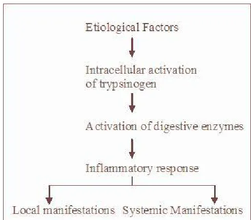
Exact mechanism is not known. Concepts which have been proposed are
based on the few experimental animal studies available. Most accepted
[image:26.596.174.419.204.419.2]mechanism is27
Figure No 4 - Mechanism of development of acute pancreatitis
Following are some of the known concepts in the pathogenesis of
acute pancreatitis:
1. Only 10% of alcohol abusers will develop the disease. Also every
individual with gall stone or hypercalcemia do not develop the disease.
Similarly severity of the disease varies from one patient to the other.
Reason behind all these is not yet known.
2. Acute Pancreatitis begins within the acinar cells as shown by
3. The exocrine pancreas synthesizes and secretes various digestive
enzymes like trypsinogen, chymotrypsinogen, lipase, amylase etc.
These get activated only in duodenum. Trypsin which is derived from
trypsinogen is the principal activator of all these enzymes. Even
normally a small proportion of trypsinogen gets activated
spontaneously inside the acinar cells. But the various protective
mechanisms present within pancreas wash out the activated Trypsin so
that there won’t be any damage to the gland. These include, Serine
protease inhibitor Kazal type 1 (SPINK1) Mesotrypsin Enzyme Y
1-antitrypsin, 2-macroglobulin28
4. Once these defensive mechanisms are overcome, there is intracellular
activation of enzymes which is also favoured by lysosomal
enzymes like catepsin B which lead to pancreatic self digestion.
5. Trypsin also activates other pathways, such as complement, coagulation
or fibrinolysis, extending the process outside the gland which is
responsible for systemic manifestation of the disease.
6. Occasionally this acute inflammatory process is associated with a
systemic inflammatory response syndrome (SIRS) mediated by
cytokines and pancreatic enzymes released in to general
circulation that may affect distant organs, giving rise to respiratory
alterations. Finally, a MODS may with vital risk of necrotic
tissue infection, a situation translocation of intestinal
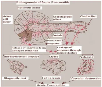
[image:28.596.135.461.200.478.2]pathogens plays an important role.29 occur where
Figure No 5-Pathogenesis of acute pancreatitis
7. Genetic factors implicated in pathogenesis of acute pancreatitis are
Cationic trypsinogen gene (PRSS1)
Cystic fibrosis transmembrane conductance regulator gene (CFTR)30 Polymorphisms in SPINK1
CLINICAL FEATURES Symptoms:
Located in upper abdomen[epigastrium / right hypochondrium]
Radiates to back
Abrupt in onset reaching to maximum level within hours
Very severe
Stabbing type
Constantly present throughout the episode
May be referred to shoulder because of pleuritic component
Typically relieved by leaning forward or lying down on one side with
drawing up of legs.
Nausea, vomiting and severe retching
Physical findings:
Typically pancreatitis patients are seen rolling around in the bed or
moving around trying to find the most comfortable position for pain relief
unlike those with hollow viscus perforation who will be lying still in the bed.
Per
Abdomen:-Tenderness either localized to epigastrium or diffuse all over abdomen
Guarding and rigidity
Absent bowel sounds due to paralytic ileus
Subcutaneous fat necrosis leading to subcutaneous tenderness and
edema.
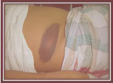
Umbilical area – Cullen’s sign
Loin – Grey Turner’s sign
[image:30.596.209.391.374.507.2] Groin – Fox’s sign
Figure No 6- Cullen’s sign
[image:30.596.206.389.590.708.2]Figure No 7-Grey Turner’s sign
General
examination:- Tachycardia / hypotension and tachypnea related to hypovolemic state.
Hyperthermia related to release of pro inflammatory cytokines.
Jaundice which may be a cause i.e., due to cholelithiasis or may be the
effect
o i.e., due to cholestasis or biliary obstruction secondary to compression by edematous pancreatic head.
Decreased breath sounds in basal lung fields secondary to atelectasis or pleural
effusion.
Diagnosis:-Diagnosing acute pancreatitis requires clinical, serological and imaging
correlation. Various serum markers used in the diagnosis and prognosis of
Laboratory Test
Time of onset
(hours) Purpose
Clinical observation / Limitations
Alanine
transaminase 12 to 24 Diagnosisand etiology Associated with gallstonepancreatitis; threefold elevation or greater in the presence of acute pancreatitis has appositive
predictive value of 95 percent pancreatitis.
Amylase 2 to 12 Diagnosis Most accurate when at least twice the upper limit of normal;
amylase levels and sensitivity decrease with time from onset of symptoms.
C-reactive
protein 24 to 48 Predictive ofseverity Late marker; high levelsassociated with pancreatic necrosis.
Interleukin-6 18 to 48 Predictive of
severity Early indication of severity Interleukin-8 12 to 24 Predictive of
severity Early indication of severity Lipase 4 to 8 Diagnosis Increased sensitivity in
alcohol-induced pancreatitis; more
specific and sensitive pancreatitis Phospholipase
A2
24 Predictive of
severity Associated with development ofpancreatic necrosis and pulmonary failure
Procalcitonin 24 to 36 Predictive of
severity Early detection of severity; highconcentrations in infected necrosis
Trypsinogen activation peptide
Within a
few hours Diagnosisand predictive of severity
Early marker for acute
pancreatitis and close correlation to severity
Among these, the two most important markers for diagnosis are serum
amylase and lipase.
Serum amylase
It is the most common serum marker used in the diagnosis of acute
pancreatitis.
It begins to elevate 2-12 hr after the onset of symptoms and remains
elevated for 3-6 days.
If it remains elevated for more than 1week it indicates development of a
complication.
Urinary levels remain elevated longer than serum levels.
Mechanism of hyperamylasemia in pancreatitis:
Older theory- Normally amylase is secreted from apex of acinar cells. In acute pancreatitis, it is secreted from basolateral surface. So it has better
access to lymphovascular system.
Recent theory-In acute pancreatitis, there is loss of cell to cell adhesions.
So amylase has better access to vascular system.
It is only diagnostic but has no prognostic value. So that even in severe
cases, there may be mild elevation. In 10% of cases of lethal pancreatitis,
Extrapancreatic sources of amylase
Salivary gland
Lung
Ovary
Prostate
False positive elevation of amylase levels
Acute cholecystitis
Intestinal ischemia
Hollow viscus perforation
Intestinal obstruction
False negative elevation of amylase levels
Acute on chronic pancreatitis
Severe pancreatitis with overwhelming necrosis
Acute pancreatitis due to hypertriglyceridemia
Macroamylasemia
It occurs in 0.5 % patients. In this condition, there is normal levels of
amylase. But amylase is bound to a high molecular weight protein. So it is not
excreted by kidneys. So levels remain elevated. But in this situation, urinary
Amylase to creatinine clearance ratio:
More reliable than the usual serum levels.
Mainly useful to differentiate actual elevation from macroamylasemia.
Amylase/Creatinine Clearance Ratio = Urine Amylase (U/L) x Serum
Creatinine (mg/dL) x 100% / Serum Amylase (U/L) x Urine Creatinine
(mg/dL).
Value greater than 5 % indicates acute pancreatitis.
Serum lipase
Advantage: More specific than amylase34.
Disadvantage: Levels remain elevated for one week. So not sensitive to
detect development of complications.
Other blood investigations
Increased haemoglobin, haematocrit, blood urea nitrogen and creatinine
due to hypovolemia.
Hypoalbuminemia secondary to fluid replacement with crystalloids.
Hyperbilirubinemia which may be a cause or effect of acute pancreatitis.
Hypochloremic metabolic alkalosis secondary to excessive vomiting.
Hypocalcemia secondary to
Hypoalbuminemia
Bone calcium does not respond to paratharmone
Associated hypomagnesemia
Hyperglycaemia due to
Associated Diabetes
Increased glucagonrelease
Increased catecholamine release
Imaging studies
A. X Ray abdomen:
Not much useful in diagnosis of acute pancreatitis. But may show
following signs due to paralytic ileus.
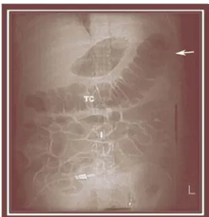
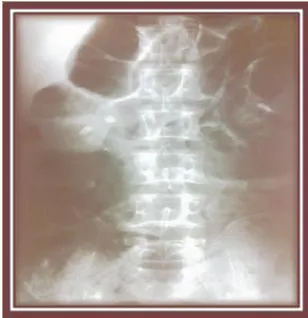
Sentinel loop sign
Colon cut off sign
[image:36.596.222.373.511.668.2] Renal halo sign
Figure No 10-Sentinel loop sign
B. USG abdomen
Limited value during acute episode because of presence of intestinal gas
shadows.
It can demonstrate
Biliary stone
Dilated pancreatic duct
Bulky edematous pancreas
Bulky edematous Pancreas
C. CECT
abdomen:-Investigation of choice for the diagnosis of acute pancreatitis as well as
detection of complications.
Features suggestive of acute pancreatitis are
Enlargement ofpancreas
Loss of peri pancreatic fat plane
Areas of decreased density
[image:38.596.207.388.369.529.2] Localized fluidcollection
Figure No 12-CECT abdomen showing edematous pancreas with peri pancreatic fat stranding
Main value of CT is in detection of pancreatic necrosis.
Normally on CT abdomen, viable pancreas enhances by > 50 HU on IV
such enhancement. Features suggestive of pancreatic necrosis on CECT
abdomen are:
Non enhancement of >30% parenchyma of pancreas.
Area of > 3cm of pancreas that does not enhance.
Ideal timing of CECT abdomen in cases of acute pancreatitis is
controversial because if it is done too early after diagnosis it may miss the
necrosis. So most authors prefer to do it 48 hours after the diagnosis.
[image:39.596.199.398.335.486.2]Sensitivity of CECT abdomen to detect necrosis at 4 days is 100%35.
D. MRI abdomen:
It is a reliable method of staging acute pancreatitis severity36.
Can predict prognosis of the disease, pancreatic duct disruption.
No advantage over CT abdomen.
Indications:
o Patients with renal dysfunction.
o Patients with allergy to contrast material.
Following table compares various imaging modalities used in
acute pancreatitis:
Table No 3-Comparison of various imaging modalities for diagnosis of acute pancreatitis37,38,39
Imaging technique Effectiveness
Contrast-enhanced computed
tomography 78 percent sensitivity and 86 percentspecificity for severe acute pancreatitis
Endoscopic ultrasonography 100 percent sensitivity and 91 percent specificity for gallstones
Magnetic resonance
cholangiopancreatography 81 to 100 percent sensitivity fordetecting common bile duct stones 98 percent negative predictive value and 94 percent positive predictive value for bile duct stones
As accurate as contrast-enhanced computed tomography in predicating severity of pancreatitis and
identifying pancreatic necrosis Magnetic resonance imaging 83 percent sensitivity and 91 percent
specificity of severe acute pancreatitis Transabdominal ultrasonography 87 to 98 percent sensitivity for the
COMPLICATIONS
Acute pancreatitis can range in severity from the most benign self
limiting conditions to the most severe cases which are associated with various
systemic manifestations which may end up in death of the patient.
Complications associated with acute pancreatitis can be classified into local /
regional/ systemic.
Local Fluid collections
Pancreatic ascites/pleural effusion Pancreatic pseudocyst
Pancreatic necrosis
Infected pancreatic abscess Haemorrhage/pseudoaneurysm
Regional Venous thrombosis Paralytic ileus
Intestinal obstruction
Intestinal ischemia/necrosis Cholestasis
Systemic Systemic inflammatory response syndrome Multiple-organ-dysfunction syndrome ARDS/pulmonary failure Renal failure Cardiovascular complications Hypocalcemia Hyperglycemia
Disseminated intravascular coagulopathy Protein calorie malnutrition
[image:41.596.72.518.293.736.2]Encephalopathy
ATLANTA symposium40 held in1992 has given the following definitions
for acute pancreatitis and its complications to maintain uniformity across
the world.
Acute pancreatitis An acute inflammatory process of the pancreas with variable involvement of other regional tissues or remote organ organ systems
Associate with raised pancreatic enzyme levels in blood and / or urine
Severity
Mild acute pancreatitis Associated with minimal organ dysfunction and uneventful recovery; lacks the features of sever acute pancreatitis. Usually normal enhancement of pancreatic parenchyma on contrast-enhanced computed tomography
Severe acute pancreatitis Associated with organ failure and / or local complications such as necrosis, abscess, or pseudocyst
Predicted severity Ranson’s Score>=3 or APACHE II Score >=8
Organ failure and systemic complications
Shock Systolic blood pressure <90mm hg
Pulmonary insufficiency PaO2<6mm Hg
Renal failure Creatinine>=177μmol/L or <=2mg/dL after rehydration
Gastrointestinal bleeding 500ml in 24 hours Disseminated
intravascular coagulation Platelets <=100,000/mm
3, fibrinogen<1.0g/L and
fibrin-split products>80μg/L Severe metabolic
disturbances Calcium <=1.87 mmol/L or <=7.5mg/dL
Local Complications
pancreatic parenchyma, typically associated with peripancreatic fat necrosis.
Non-enchanced pancreatic parenchyma>3cm or involving more than 30% of the area of the pancreas
Acute pseudocyst Collection of pancreatic juice enclosed by a wall of fibrous or granulation tissue, which arises as a result of acute pancreatitis, pancreatic trauma or chronic pancreatitis, occurring at least 4 weeks after onset of symptoms, is round or ovoid an most often sterile; when pus is present lesion is termed a “pancreatic abscess”
Pancreatic abscess Circumscribed, intra-abdominal collection of pus, usually in proximity to the pancreas,
containing little or no pancreatic necrosis, which arises as consequence of acute pancreatitis or pancreatic trauma. Often 4 weeks or more after onset Pancreatic abscess and infected pancreatic necrosis differ in clinical expression and extent of associated necrosis.
Table No 5- Atlanta definitions of acute pancreatitis and its complication Prognostic scoring system
Treatment of acute pancreatitis is mainly based on the severity. So it
is of prime importance to grade these patients into mild or severe. Hence
a number of scoring systems have been proposed. As can be seen with
their numbers none is considered to be the gold standard. Let us see the most
BISAP (Bedside Index for Severity in Acute Pancreatitis) Scoring System
BUN >25mg/dL
Impaired mental status (Glasgow Coma Scale Score<15) SIRS
SIRS is defined as two or more of the following 1) Temperature of <36 or > 38oC
2) Respiratory rate > 20 breaths/min or PaCO2 < 32 mm Hg 3) Pulse > 90 beats/min
4) WBC < 4,000 or > 12,000 cells/mm3or > 10% immature bands
Age> 60 years
Pleural effusion detected on imaging
One point is assigned for each variable within 24 hours of presentation and added for a composite score of 0 -5
Table No 6 -BISAP scoring system41
Ranson’s Scoring System
It is one of the most widely used scoring systems for acute pancreatitis.
First proposed in 197442.
Table No 7- Ranson’s scoring system
Total score of more than 3 indicates severity.
Main disadvantage is that it is possible to assess the severity only after 48
hours.
Glasgow Scoring System
It is a scoring system initially developed by Imrie in 197843.
It initially included 7 criteria to be measured within 48 hours of
admission.
[image:46.596.70.518.272.473.2] Later it was modified in 1984 to as mentioned below.
Table No 8 -Imrie [Modified Glasgow] scoring system
Total score of more than or equal to 3 indicates severity.
On Admission
Age >55 years old
WBC Count 15*10^9 cell/L
Blood glucose level >10mmol/L
Serum urea level 16mmol/L
PaO2 8kPa (60mmHg)
Within 48 hours
Serum calcium < 2mmol/L
Serum albumin <32g/L
LDH >600IU/L
APACHE II Scoring System
It means Acute Physiology And Chronic Health Evaluation44.
It is a physiological scoring system based on 14 criteria.
Total score of more than 8 indicates severity.
Advantages over other systems
o Severity can be assessed within 24 hours unlike others where 48 hours are required.
o Severity can be assessed continuously through out the clinical course of the disease.
o Prognosis can also be assessed after interventions like debridement.
Disadvantages:
Cumbersome
Not specific for pancreatitis
Modifications:
APACHE 345 - Here 5 additional criteria are taken into account to increase
the accuracy.
Atlanta Criteria for Severity of Acute Pancreatitis Severity Criteria Definition
Organ failure with one or more
Shock Systolic blood pressure < 90mmHg
Pulmonary insufficiency PaO2 < 60 mmHg
Renal failure Serum Creatinine level > 2mg/dL
after rehydration
Gastrointestinal tract bleeding 500 mL in 24 hours
Local Complications
Pancreatic Necrosis More than 30% of the parenchyma or
more than 3cm
Pseudocyst Collection of pancreatic juice
enclosed by a wall
Abscess Circumscribed collection of pus
containing little or no pancreatic
necrosis
Ranson’s Score >3
APACHE II Score >8
Balthazar CT Severity Scoring System
In contrast to the previous scoring systems, this is a radiological
scoring system based on CECT abdomen.
First developed by Balthazar et al in 198546, considered to be more
accurate than the clinical scoring system.
Grade CT Findings
A Normal
B Focal of diffuse enlargement of the pancreas, including irregularities of contour and inhomogeneous attenuation C Pancreatic gland abnormalities in grade B plus per pancreatic
inflammation
D Grade C plus a single fluid collection
[image:51.596.61.519.456.664.2]E Grade C plus 2 or more fluid collection and / or the presence of gas inor adjacent to the pancreas
Table No 11- Balthazar CT severity scoring system
Grades beyond “C”indicate severity.
CT Grade Assigned Score Percent necrosis Assigned Score
A 0 None 0
B 1 <30 2
C 2 30-50 4
D 3 >50 6
E 4
Score greater than 5 indicates severity.
Other prognostic factors:
These are used sometimes to assess the severity though their role in routine
clinical practice is not yet clear.
1. C Reactive Protein
2. IL 6
3. IL 8
4. Soluble IL 2 receptor
5. TNF Alfa
6. Trypsinogen Activating Peptide
7. Serum procalcitonin
MANAGEMENT
Management of acute pancreatitis is quite complicated and is associated
[image:53.596.78.490.160.599.2]with a lot of controversies in every aspect.
Treatment of acute pancreatitis involves 3 main components.
Initial management of the acute episode
Surgical management
Management ofcomplications
Management of acute episode: I. Fluid management
Most important initial step.
Fluid loss
o External – repeated vomiting / inadequate intake secondary to nausea
o Internal – fluid sequestration into areas of inflammation/ pulmonary parenchyma / soft tissues of the body
Fluid loss leads to hypovolemia, hemoconcentration and in severe
cases results in development of renal failure.
Fluid resuscitation in the range of 200 ml/hr may be required.
Close monitoring of cardiovascular and renal functions is a must.
Studies have shown that improper fluid sequestration can
increase the development of necrosis47.
II. Electrolyte management
imbalances which require appropriate management. These include:
Hypochloremic alkalosis secondary to repeated vomiting.
Hypoalbumenemia
Hypocalcemia
Hypomagnesemia
III. Pain management
Pain is considered to be the worst pain suffered by most individuals.
Patient controlled analgesia is preferred.
In most cases use of narcotics is required for adequate pain control.
Mepiridine and analogues are the preferred medications. Morphine needs to be avoided because at least theoretically it
aggravates biliary spasm.
IV. Role of Nasogastric tube
Previously Nasogastric aspiration was done routinely in all patients with the
assumption that it decreases the pancreatic stimulation. There are no
studies to support this concept. So placement of Nasogastric tube should be
individualized. It is indicated in those with
Severe vomiting to prevent aspiration pneumonia
Severe retching to prevent Mallory weiss tears
Paralytic ileus
Classical teaching – avoid enteral nutrition based on the concept that enteral
feeding leads to pancreatic stimulation and further aggravates pancreatic
injury.
Recent studies have shown that enteral feeding is feasible, safe
and even desirable48.
Advantages of enteral feeding over TPN
o Supports mucosal integrity and hence decreases septic complications
o Easier
o Lower complication rate
o Lower cost
Studies have shown that enteral feeding leads to
o Lower APACHE scores and CRP levels.49
o Lower septic and total complications.
To summarize, TPN is recommended in cases of severe acute
pancreatitis with paralytic ileus. Otherwise enteral feeding is recommended.
VI. Role of prophylactic antibiotics
Role of antibiotics is considered controversial.
Basis for the use of antibiotics.
Sepsis is the leading cause of death among patients with
extent of necrosis and duration of acute pancreatitis. Incidence of local
sepsis at 1 week is 29% and at 3 week is 71%50. Organisms commonly
implicated are E. coli, Klebsiella, streptococcus, Staphylococcus and
pseudomonas.
Disadvantages of routine use of antibiotics
o Development of multidrug resistant organisms
o Fungal super infection
Current evidence shows that use of prophylactic broad spectrum
antibiotics in patients with severe pancreatitis is associated with
decreased mortality51.
Current recommendation:
o Mild cases – no need for antibiotics
o Severe cases – antibiotics are recommended.
Duration of use of antibiotic: 1-4 weeks. Most authors
recommend it for 2 weeks52.
Commonly used regimens are Imipenem alone or Imipenem in combination
with Cilastatin and cefuroxime. Some have recommended use of anti fungals
like Fluconazole53.
Surgical Management
In the modern day practice things have changed, thanks to
better understanding of the natural history of the disease, basic
pathophysiology of pancreatitis and better anaesthetic facilities.
Indications for surgical intervention in Necrotizing Pancreatitis
1. Diagnostic uncertainty
2. Intra-abdominal catastrophe unrelated to necrotizing pancreatitis
3. Infected necrosis documented by FNA or extraluminal gas on CT
4. Severe sterile necrosis
[image:60.596.57.521.476.723.2]5. Symptomatic organized pancreatic necrosis
Table No 13 -Indications for surgery in case of acute pancreatitis
Surgical approach to the treatment of pancreatic necrosis Open surgery approaches Minimally invasive approaches
Pancreatic resection Minimally invasive approaches
Necrosectomy + wide tube drainage Laparoscopic necrosectomy
Necrosectomy + relaparotomy
(Staged reexploration)
Laparoscopic assisted percutaneous
drainage
Necrosectomy + laparotomy open
packing
Laparoscopic transgastric
Necrosectomy + drainage + closed
continuous lavage
Percutaneous necrosectomy and sinus
tract endoscopy
MRI- radiologically assisted
necrosectomy
Table No 14-Surgical options for pancreatic necrosis
The basic and universal aspect of necrosectomy is débridement of necrotic
peripancreatic and pancreatic tissue. As Necrosis is an ongoing process, it has
been addressed in a variety of ways, as follows:
Closed packing
Open drainage
Closed high-volume lavage of the lesser sac
Repeated, planned necrosectomy with abdominal wall closure
Necrosectomy and Closed Packing
The lesser sac is accessed through the base of the mesocolon. A thorough,
blunt necrosectomy is followed by packing with multiple, stuffed Penrose
drains; however, it is associated with a high incidence of intra-abdominal
abscess.
Necrosectomy and Open Drainage
An initial blunt necrosectomy is followed by marsupialization of the
unpacking and gentle irrigation are done.
Necrosectomy and Closed Lavage
It combines operative débridement (of only necrotic tissue to minimize
loss of pancreatic parenchyma) with subsequent high-volume lavage of the
lesser sac using a peritoneal dialysate.
Planned, Repeated Necrosectomy and Delayed, Primary Abdominal Wall Closure
The initial celiotomy and necrosectomy is followed by a planned,
repeated necrosectomy every 48 hours. The lesser sac is entered through
the gastrocolic ligament, and all areas of necrosis are unroofed and bluntly
débrided. Multiple soft drains are placed in both paracolic gutters and the
lesser sac. Subsequently, a nonadherent elastic drape or Adaptic gauze is
used to line the lesser sac, which is then packed with moist laparotomy pads.
Packing keeps the lesser sac open and readily accessible for repeated
necrosectomy. Temporary abdominal closure is obtained with a zipper.
Planned reoperation is scheduled at approximately 48-hour intervals until the
necrotic processes have been controlled or resolved.
They include laparoscopic necrosectomy, laparoscopic assisted
percutaneous drainage, laparoscopic transgastric necrosectomy, percutaneous
necrosectomy and sinus tract endoscopy, MRI– radiologically assisted
4. MATERIAL AND METHODS Source of Data
Patients admitted to Surgical wards in Madurai Medical College
Hospital, Madurai.
Method of Collection of Data
A time bound prospective study was conducted on patients
admitted with acute pancreatitis during the study period from December
2015 to December 2017. All the patients were subjected to detailed
clinical examination, laboratory investigations and radiologicalimaging.
Inclusion Criteria
Patients with confirmed diagnosis of acute pancreatitis based on
clinical / laboratory / radiological investigations.
Exclusion Criteria
Age less than 16 years; as physiological thresholds are calibrated for
adults.
Patients with acute on chronic pancreatitis.
Sample Size
After considering both inclusion and exclusion criteria, total
All the 100 patients were subjected to both Ranson’s and APACHE II
scoring systems. Scoring was done on admission/time of diagnosis and at
48 hours. The scores were compared with the clinical severity which
was graded according to Atlanta criteria and also compared with the clinical
outcome.
Methods of Statistical Analysis
Independent t test was used to examine differences in age; fisher’s
exact test for sex; and chi square test for etiology were used. Sensitivity,
specificity, positive predictor value, negative predictor value and accuracy
were calculated. A “p” value of less than 0.05 was considered to be
5. OBSERVATION ANDRESULTS
The study was conducted in Madurai Medical College Hospital.
Total number of patients studied were 100.
According to the Atlanta Criteria, 62 patients were classified as Mild
Sex Distribution of the Study Population
Sex Mild Severe Total
Male 56 36 92
[image:66.596.73.510.332.587.2]Female 6 2 8
Table No 16 – Sex Distribution
Figure No 19 – Sex Distribution
Of the 100 patients, 92 were Male (92.5 %) and 8 were Female (7.5%).
There was no statistical significance of Sex (p=0.545) on the severity of the
Etiology of Acute Pancreatitis
Etiology Mild Serve Male Female Total
Alcohol 65 23 88 0 88
Gall stones 6 2 0 8 8
[image:67.596.73.518.174.592.2]Idiopathic 2 2 4 0 4
Table No 17 – Etiology
Figure No 20 – Etiology
Out of 100 patients, 30 (74%) had Alcohol induced Acute Pancreatitis,
3 (8%) had Gall Stones induced Acute Pancreatitis and 7 (18%) had Idiopathic
Acute Pancreatitis. There was no statistical significance of Etiology (p=0.943)
Outcome of Patients No of patients without complicated No of Patients with complicated complicated
Local complications System complications Pseudo cyst Pancreatic necrosis Hemorrhagic pancreatitis SIRS
60 40 16 15 6 3
Table No 18 – Outcome of Patients
- Out of 100 patients
- 60% had uncomplicated outcome
- 40% of patients with any complication
- 6.4% of patients developed pseudo cyst
- 6% of patients developed Pan – Neurosis
Figure No 21 – Outcome of Patients
Out of 100 patients with acute pancreatitis, 25 patients (62.5 %) had an
15 patients (37.5 %) developed complications, of which 14 patients (93.4
%) developed local complications and 1 patient (6.6 %) developed
systemic complication. Of the local complications, 6 patients developed
Pseudo Cyst, 6 patients developed pancreatic necrosis, and 2 developed
hemorrhagic pancreatitis. The patient who developed systemic complication
(SIRS) had a fatal outcome.
Surgical intervention was performed in one patient. Exploratory
Outcome of patients based on different cut-off Ranson’s Score
Ransons score
Uncomplicated outcome
Complicated outcome
Local complications Syst complications Pseudo
cyst
Pancreatic necrosis
Hemorrhagic
pancreatitis SRS
<=3 39 3 0 0 0
>3 25 10 15 5 0
[image:71.596.67.546.138.400.2]>5 0 0 0 0 3
Figure No: 23 – Outcome for different Ranson’s Score
Out of 42 patients <+ 3
32.85% are uncomplicated
7.14 % are complicated
Out of 55 patients > 3
45.45% Are uncomplicated
18.18% are complicated – pseudo cyst
27.27% are complicated – Pan necrosis
9.09% are complicated – Hgic pancreatitis
Of the 25 patients (62.5 %) who had Ranson’s score of less than or
Pseudo Cyst. No patient in this group had Pancreatic Necrosis or any major
organ failure. There were no deaths in this group.
15 patients (37.5 %) had Ranson’s score of more than 3, one (6.6 %) of
them had an uncomplicated course and 14 patients (93.4 %) developed
complications, 13 had local complication and one had systemic complication.
One patient (2.5 %) had Ranson’s score more than 5 and developed
systemic complication (SIRS) and had fatal outcome.
Of the 25 Patients with Ranson’s Score < = 3, 96 % had an
uncomplicated mild course. The inference being Ranson’s Score < = 3 predicts
an uncomplicated outcome – mild acute pancreatitis.
Of the 15 Patients with Ranson’s Score > 3, 93.4 % developed
complications. The inference being Ranson’s score > 3 predicts a
Outcome of patients based on different cut-off APACHE II
Apache II Score
Uncomplicated outcome
Complicated outcome
Local complications Syst complications Pseudo
Cyst NecrosisPAN HemorrhagicPancreatitis SIRS
<=8 57 3 0 0 0
>8 4 6 9 2 0
[image:74.596.67.528.139.370.2]>12 1 6 6 3 3
Figure No 24 – Outcome for different Apace II Score
Apache II score <8Uncomplicated outcome were 57%. Local complications:
pseudo cyst were 5.26%.
Apache II score >8Uncomplicated outcome were 4%. Local
complications: pseudo cyst were 35.29%, pancreatitis necrosis were 55.97%,
Hemorrhagic pancreatitis were 11.17%.
Apache II score >12 Uncomplicated outcome were 1%. Local
complications: pseudo cyst were 33.3%, pancreatitis necrosis were 33.3%,
Of the 25 patients (62.5 %) who had APACHE II score less than or equal to 8,
24 patients (96 %) had an uncomplicated outcome. One patient (4 %)
developed Pseudo Cyst. No patient in this group had Necrosis or major organ
failure or death.
15 patients (37.5 %) had APACHE II score more than 8, one (6.6 %) of
them had an uncomplicated course and 14 patients (93.4 %) developed
complications, 13 developed local complications and one developed systemic
complication. Of the 7 patients who had APACHE II score more than 12, all
7 patients (100 %) developed complications.
Of the 25 patients who had APACHE II score < = 8, 96 % had
an uncomplicated outcome. The inference being APACHE II score < = 8
predicts an uncomplicated outcome -- mild acute pancreatitis.
Of the 15 patients with APACHE II score > 8, 93.4 %
developed complications. APACHE II score > 8 predicts a complicated
Mean of Ranson’s and APACHE II Score
Ranson’s Mean
Mild 2.40
Severe 4.53
[image:77.596.70.507.296.557.2]Over All 3.20
Table No 21 – Mean Ranson’s Score
APACHE II Mean
Mild 5.28
Severe 12.27
[image:78.596.72.507.294.559.2]Over All 7.90
Table No 22 – Mean APACHE II Score
Figure No 26 – Mean APACHE II Score
Ranson’s Score and APACHE II Score in severe acute pancreatitis
Prediction of severity by Ranson’s Score
Table No 23 –
Ranson’s
Score
Sensitivity Specificity PPV NPV Accuracy
>=3 100 56 57.69 100 72.5
>=4 93.33 96 93.33 96 95
>=5 53.33 100 100 78.1 82.5
Prediction of severity by Ranson’s Score
Ranson’s score of greater than or equal to 4 predicted 93% of severe
attacks and 96% of mild attacks with a PPV of 93.33 and NPV of 96 and
accuracy of 95.
Ranson’s score of greater than or equal to three predicted more
number of severe attacks (100%) but less number of mild attacks (56%) with
PPV of 57.69 and NPV of 100 and accuracy of 72.5.
Ranson’s score of greater than or equal to 5 predicted less number of
severe attack (53%) and branded more severe attacks as mild attacks.
Ranson’s score of greater than or equal to 4 had the best sensitivity,
Prediction of severity by APACHE II Score
Apache II
Score
Sensitivity Specificity PPV NPV Accuracy
>=8 100 80 75 100 35
>=9 93.33 96 93.33 96 95
>=10 86.66 100 100 92.6 95
>=11 80 100 100 89.2 92.5
Table No 24 – Prediction of severity by APACHE II Score
APACHE II score of greater than or equal to 9 predicted 93.33% of
severe attacks and 96% of mild attacks with a PPV of 93.33 and NPV of 96
and accuracy of
95. APACHE II score of greater than or equal to 10 also had the same
accuracy.
APACHE II score of greater than or equal to 8 predicted more
number of severe attacks (100%) but less number of mild attacks (80%)
with PPV of 75 and NPV of 100.
APACHE II score of greater than or equal to 11 predicted less
APACHE II score of more than or equal to 9 had the best
sensitivity, specificity and accuracy.
Prediction of Major Organ failure and Pancreatic collection by Ranson’s Score
Ranson’s Score
Sensitivity Specificity PPV NPV Accuracy
Pancreatic
Collection
93.33 96 93.33 96 95
Major
Organ
Failure
[image:81.596.66.516.235.467.2]100 64.1 6.66 100 65
Table No 25 – Prediction of organ failure & pancreatic collection by Ranson’s Score
The Ranson’s scores were very sensitive for prediction of systemic
complications (100%) but less sensitive for prediction of local
Prediction of Major Organ failure and Pancreatic collection by APACHE II
Score APACHE
II Score
Sensitivity Specificity PPV NPV Accuracy
Pancreatic
Collection
93.33 96 93.33 96 95
Major
Organ
Failure
[image:82.596.68.519.159.403.2]100 64.1 6.66 100 65
Table No 26 – Prediction of organ failure & pancreatic collection by APACHE II Score.
APACHE II scores showed higher sensitivity in the prediction of
systemic complications(100%) than in the prediction of local
Prediction of Severity by the two scoring Systems
Sensitivity Specificity PPV NPV Accuracy
Ranson’s
Score
93.33 96 93.33 96 95
APACHE
II Score
93.33 96 93.33 96 65
Table No 27 – Prediction of severity by Ranson’s and APACHE II scoring systems
As Sensitivity, Specificity, Positive Predictive Value, Negative
Predictive Value and Accuracy are found to be the same for Ranson’s and
APACHE II scores, Ranson’s scoring system is equally efficacious as
Hospital Stay
The mean duration of hospital stay was 6.60 days for mild cases .
The mean duration of hospital stay was 9.31 days for severe cases.
6. DISCUSSION
Acute Pancreatitis is an increasing common abdominal emergency.
Assessment of severity of acute pancreatitis is important for early
identification of patients who may benefit from additional supportive
and specific therapeutic procedures. Many different scoring systems have
been devised for the assessment of severity of acute pancreatitis, which are
divided into two types : The first type attempts to correlate laboratory and
clinical markers specific to pancreatitis with subsequent outcome and disease
severity, the most widely used in this group is Ranson’s Score. The
second type of scoring system is the application of non specific physiological
scoring system, which was originally created for use in general
population of critically ill patients like APACHE II scores.
Ideal predicting criteria should be simple, non-invasive, accurate and
quantitative; and the assessment tests should be readily available at the
time of diagnosis.
In this study we compare the classical and simple Ranson’s scoring
system with the more cumbersome APACHE II scoring system. We have
classified the severity of acute pancreatitis in this study based on the Atlanta
criteria.
In this study, acute pancreatitis was found 12 times more commonly in
match with the results of the study of Larvin et al where male is to female
ratio was 47:53 and mean age was 62 years.
In the present study alcohol was the etiological factor in 74 % of
patients and gall stones in 8 %, contrary to alcohol being 22 % and gall
stones 43 % in Larvin et al. The etiology had no significant influence on the
scores or the final outcome of acute pancreatitis, suggesting that once the
pathogenic mechanisms have initiated the disease, the course and outcome of
acute pancreatitis are not influenced by underlying etiological factors. Some
authors have published similar results as in the study by Su
Mi Woo et al6.
Out of the 40 cases in this study, 25 patients (62.5 %) had mild
acute pancreatitis and 15 patients (37.5 %) had severe acute pancreatitis. The
percentage of severe cases was higher in our study as compared to most of the
other studies. In the study by Larvin et al 20 % of all the cases were severe.
Mortality in our study was 2. 5 % and mortality in the study by Larvin et al
was 7.6 %. Mortality was less in our study.
In our study the mean Ranson’s and APACHE II scores calculated
during the first 48 hours showed significantly higher values for severe than
for mild cases of acute pancreatitis. The mean Ranson’s score in mild and
severe cases was 2.40 and 4.53 respectively. The mean APACHE II score
Comparing outcomes in patient groups based on a range of
Ranson’s and APACHE II scores, it was observed that complications like
Pseudo Cysts, Pancreatic Necrosis, major organ failure and deaths were more
common when Ranson’s score exceeded 3 and APACHE II scores exceeded 8.
Contrary to expectation Pseudo Cyst was observed in one patient whose
Ranson’s and APACHE II scores were 3 and 8 respectively. These patients
presented to hospital later than 48 hours after the onset of symptoms by which
time the severity of the attack has subsided and the recorded scores were
spuriously low. It can therefore be concluded that patients with Ranson’s score
more than 3 and APACHE II score of more than 8 are high risk patients.
In our study Ranson’s score of greater than 3 and APACHE II score of
greater than 8 had the highest sensitivity, specificity and accuracy for the
prediction of severity of acute pancreatitis.
In our study the Ranson’s and APACHE II scoring systems were
very sensitive for the prediction of systemic complications (100%) but less
sensitive for prediction of local complications (93.33%). This is comparable
to the study by Larvin et al, where the sensitivity to detect systemic
complications was higher (76%) than to detect local complications (73%).
In our study the sensitivity, specificity, positive predictor value,
negative predictor value and accuracy of Ranson’s and APACHE II scores are
Sensitivity Specificit PPV NPV Accuracy
Ranson’s 93.33 96 93.33 96 95
APACHE 93.33 96 93.33 96 95
Table No 28 – Accuracy of Ranson’s and APACHE II scoring systems
As sensitivity, specificity and accuracy of Ranson’s and APACHE II
scores are comparable in our study, Ranson’s is as powerful a prognostic