Herpes Simplex Virus 1 (HSV-1) and HSV-2 Mediate Species-Specific
Modulations of Programmed Necrosis through the Viral
Ribonucleotide Reductase Large Subunit R1
Xiaoliang Yu,aYun Li,aQin Chen,aChenhe Su,bZili Zhang,aChengkui Yang,aZhilin Hu,aJue Hou,aJinying Zhou,aLing Gong,a Xuejun Jiang,cChunfu Zheng,b,dSudan Hea
Cyrus Tang Hematology Center, Collaborative Innovation Center of Hematology, Jiangsu Institute of Hematology, the First Affiliated Hospital, and Collaborative Innovation Center of Hematology and Jiangsu Key Laboratory of Preventive and Translational Medicine for Geriatric Diseases, Soochow University, Suzhou, Chinaa; Soochow University, Institutes of Biology and Medical Sciences, Suzhou, Chinab; Cell Biology Program, Memorial Sloan-Kettering Cancer Center, New York, New York, USAc; Department of Microbiology, Immunology and Infectious Diseases, University of Calgary, Calgary, Alberta, Canadad
ABSTRACT
Receptor-interacting protein kinase 3 (RIP3) and its substrate mixed-lineage kinase domain-like protein (MLKL) are core
regu-lators of programmed necrosis. The elimination of pathogen-infected cells by programmed necrosis acts as an important host
defense mechanism. Here, we report that human herpes simplex virus 1 (HSV-1) and HSV-2 had opposite impacts on
pro-grammed necrosis in human cells versus their impacts in mouse cells. Similar to HSV-1, HSV-2 infection triggered propro-grammed
necrosis in mouse cells. However, neither HSV-1 nor HSV-2 infection was able to induce programmed necrosis in human cells.
Moreover, HSV-1 or HSV-2 infection in human cells blocked tumor necrosis factor (TNF)-induced necrosis by preventing the
induction of an RIP1/RIP3 necrosome. The HSV ribonucleotide reductase large subunit R1 was sufficient to suppress
TNF-in-duced necrosis, and its RIP homotypic interaction motif (RHIM) domain was required to disrupt the RIP1/RIP3 complex in
hu-man cells. Therefore, this study provides evidence that HSV has likely evolved strategies to evade the host defense mechanism of
programmed necrosis in human cells.
IMPORTANCE
This study demonstrated that infection with HSV-1 and HSV-2 blocked TNF-induced necrosis in human cells while these viruses
directly activated programmed necrosis in mouse cells. Expression of HSV R1 suppressed TNF-induced necrosis of human cells.
The RHIM domain of R1 was essential for its association with human RIP3 and RIP1, leading to disruption of the RIP1/RIP3
complex. This study provides new insights into the species-specific modulation of programmed necrosis by HSV.
N
ecrotic cell death characterized by the disruption of the
plasma membrane has been observed in a variety of
physio-logical and pathophysio-logical processes, including in mammalian
de-velopment, in tissue damage, and in pathogen infection (
1–3
).
Inhibition of apoptosis is known to facilitate programmed
necro-sis in cells. Proteins of the tumor necronecro-sis factor (TNF) family of
cytokines, including TNF-
␣
, TRAIL (TNF-related
apoptosis-in-ducing ligand), and FasL, are classic inducers of programmed
ne-crosis, also known as necroptosis (
4
). In TNF-
␣
-triggered
necro-sis, receptor-interacting protein kinase 1 (RIP1) (
5
) forms a
protein complex, called the necrosome (
6
), with
receptor-inter-acting protein kinase 3 (RIP3) (
7–9
) through the RIP homotypic
interaction motif (RHIM) domains of both proteins (
10
).
Deu-biquitination of RIP1 by cylindromatosis (CYLD) is required to
mediate necrosome formation and activation (
11
,
12
). Active
RIP3 subsequently phosphorylates its substrate, mixed-lineage
ki-nase domain-like protein (MLKL), to trigger membrane
localiza-tion of MLKL and downstream events for the induclocaliza-tion of
mem-brane rupture (
13–17
).
Additionally, the recognition of pathogen-associated
molecu-lar patterns by the Toll-like receptor (TLR) proteins triggers
pro-grammed necrosis. TLR3 and TLR4 specifically recognize,
respec-tively, viral double-stranded RNA (dsRNA) [or a synthesized
analog of dsRNA poly(I·C)], and bacteria lipopolysaccharide
(LPS), respectively (
18
). Activation of TLR3 and TLR4 by these
ligands induces the interaction of the Toll/interleukin-1 (IL-1)
receptor domain-containing adaptor inducing beta interferon
(IFN-

) (TRIF) with RIP3. TRIF, RIP3, and MLKL are all known
to be essential components in the regulation of TLR-mediated
necrosis (
19
,
20
).
Recent studies have revealed that programmed necrosis acts as
an effective mechanism to control viral replication and
pathogen-esis. Vaccinia virus (VV) is known to encode the caspase inhibitor
B13R (
21
,
22
) that confers the ability to block apoptosis. Infection
of vaccinia virus (VV) in mouse embryonic fibroblasts (MEFs)
sensitizes the cells to TNF-
␣
-induced necrosis (
7
). RIP3 knockout
mice exert reduced necrosis and succumb to VV infection (
7
). In
contrast, murine cytomegalovirus (MCMV) infection suppresses
both TNF receptor (TNFR)- and TLR3-mediated necrosis in
Received23 September 2015Accepted3 November 2015
Accepted manuscript posted online11 November 2015
CitationYu X, Li Y, Chen Q, Su C, Zhang Z, Yang C, Hu Z, Hou J, Zhou J, Gong L, Jiang X, Zheng C, He S. 2016. Herpes simplex virus 1 (HSV-1) and HSV-2 mediate species-specific modulations of programmed necrosis through the viral ribonucleotide reductase large subunit R1. J Virol 90:1088 –1095.
doi:10.1128/JVI.02446-15.
Editor:R. M. Sandri-Goldin
Address correspondence to Chunfu Zheng, [email protected], or Sudan He, [email protected].
Copyright © 2015, American Society for Microbiology. All Rights Reserved.
on November 7, 2019 by guest
http://jvi.asm.org/
mouse cells via the RHIM-containing viral protein M45/vIRA (
19
,
23
). M45/vIRA mutant MCMV triggers programmed necrosis by
inducing an interaction between RIP3 and the DNA-dependent
activator of IFN regulatory factor (DAI) (
24
). Unlike VV and
MCMV, herpes simplex virus 1 (HSV-1) infection naturally
acti-vates mouse RIP3 (mRIP3)/mMLKL-dependent necrosis in
mouse cells independently of TNFR, TLR3, and DAI (
25
,
26
).
During HSV-1 infection, RIP3 is activated by the assembly of a
complex with the RHIM-containing viral protein ICP6, the large
subunit (R1) of ribonucleotide reductase (RR), leading to MLKL
activation and necrosis of host cells (
25
,
26
). RIP3-deficient mice
showed severely impaired control of HSV-1 replication and
pathogenesis (
25
). Although HSV-1 is a common human
herpes-virus, it remains unclear precisely how HSV-1 modulates
pro-grammed necrosis in human cells. In the present study, we
dem-onstrate that HSV-1 and HSV-2 modulate programmed necrosis
by distinct mechanisms in murine cells and human cells, leading
to opposite consequences in these two species. Both HSV-1 and
HSV-2 trigger the formation of the mRIP3/mMLKL complex and
programmed necrosis in mouse cells. In human cells, in contrast,
HSV-1 or HSV-2 infection not only fails to activate programmed
necrosis but also effectively subverts TNF-induced necrosis. HSV
R1 is sufficient to prevent the recruitment of human RIP1 (hRIP1)
to hRIP3 and TNF-induced necrosis of human cells. Together, our
work reveals dual roles of HSV R1 in modulating programmed
necrosis via the RHIM-dependent activation or suppression of
RIP3 signaling in a species-specific manner.
MATERIALS AND METHODS
Reagents.Human TNF-␣recombinant protein and an Smac mimetic were generated as previously described (8). Z-VAD (benzyloxycarbonyl-Val-Ala-Asp-fluoromethylketone) was purchased from Bachem. Mouse anti-RIPK1 monoclonal antibody (MAb) was purchased from BD Biosci-ences (Shanghai, China). Rabbit anti-mRIP3 polyclonal antibody was purchased from ProSci (San Diego, CA). Mouse anti-VP16 MAb was purchased from Abcam (Shanghai, China). Anti-Flag MAb, anti-Myc MAb, antihemagglutinin (anti-HA) MAbs, and rabbit anti--actin poly-clonal antibody were purchased from Sigma (Shanghai, China). Rabbit anti-hRIP3 polyclonal antibody was generated as previously described (8). HSV-1 ICP6 (25) polyclonal antibody was generated as previously reported.
Cell culture and viral infection.HEK-293T cells, mouse fibrosarcoma L929 cells, and African green monkey kidney (Vero) cells were from ATCC. MEFs were isolated from day 14.5 to day 15.5 embryos. HEK-293T, L929, MEF, and Vero cells were cultured in Dulbecco’s modified Eagle’s medium (DMEM; Thermo Fisher). HT-29 cells were cultured in McCoy’s 5A culture medium (Invitrogen). All media were supplemented with 10% fetal bovine serum (FBS; Gibco) and 100 U/ml penicillin-strep-tomycin (HyClone). HSV-1(KOS), HSV-1 with a deletion of ICP6 (ICP6⌬HSV-1), and HSV-2 were grown in Vero cells.
Stable cell lines.HeLa-hRIP3 cells were generated as described previ-ously (27). HeLa cells stably expressing Tet repressor (HeLa-tetR stable cells) were generated after transfection with pcDNA6/TR (Invitrogen) and selection with 10g/ml blasticidin. HeLa-tetR stable cells were fur-ther transfected with a DNA plasmid expressing HA-Flag-hRIP3 and then selected with 1 mg/ml G418. HT-29 cells stably expressing a short hairpin RNA targeting RIP3 (HT-29-RIP3-shRNA cells) were generated as de-scribed previously (8).
Plasmids and siRNA oligonucleotides.cDNAs of ICP6 and ICP10 were amplified from total RNA of cells infected with HSV-1 and HSV-2, respectively, by reverse transcription-PCR (RT-PCR). ICP6 and ICP10 were cloned into the pCDNA3.1 plasmid. The ICP6 RHIM mutant (resi-dues 73 through 76 mutated to four alanine resi(resi-dues) and the ICP10
RHIM mutant were generated using a QuikChange Lightning site-di-rected mutagenesis kit (Stratagene). Mouse RIP3 and the MLKL small interfering RNAs (siRNAs) were synthesized by Shanghai GenePharma Co., Ltd. The following siRNA oligonucleotides were used: mRIP3, CCC GACGAUGUCUUCUGUCAA; mMLKL, GAGAUCCAGUUCAACG AUA; and negative-control siRNA, AACGUACGCGGAAUACUUCGA. Lipofectamine 2000 (Invitrogen) and INTERFERin (Polyplus) were used for the transfection of DNA plasmids and siRNA oligonucleotides, respec-tively.
Cell viability assay.Cell survival analysis was performed by measuring intracellular ATP levels using a Cell Titer-Glo Luminescent Cell Viability Assay kit (Promega) according to the manufacturer’s instructions. Data are represented as the means⫾standard deviations of duplicates. All experiments were repeated at least three times with similar results.
Western blot analysis and immunoprecipitation.The cells were har-vested and resuspended in lysis buffer containing 20 mM Tris-HCl (pH 7.4), 150 mM NaCl, 1% Triton X-100, 10% glycerol, 25 mM -glycerol-phosphate, 1 mM Na3VO4, 0.1 mM phenylmethylsulfonyl fluoride (PMSF), a complete protease inhibitor set (Roche), and a phosphatase inhibitor set (Sigma). After incubation on ice for 20 min, the total cell lysates were centrifuged at 20,000⫻gfor 20 min. The supernatant was then collected and used for subsequent Western blot or immunoprecipi-tation analysis. For Flag immunoprecipiimmunoprecipi-tation, cell lysates were incubated overnight with anti-Flag agarose beads (Sigma) at 4°C. Agarose beads were washed four to six times with lysis buffer, and the immunoprecipi-tants were eluted by the addition of a low-pH elution buffer (Pierce) or Flag peptide (Sigma). Myc pulldown was performed using anti-Myc aga-rose beads (Sigma) with a similar procedure. Finally, acid elution was neutralized by the addition of 1/20 volume of 1 M Tris-HCl (pH 9.4). Immunocomplexes were further examined by Western blot analysis.
RESULTS
HSV-2 as well as HSV-1 infection directly activates
RIP3/MLKL-dependent necrosis in mouse cells.
Our recent work has
demon-strated that HSV-1 activated RIP3-dependent necrosis in mouse
cells. We subsequently examined the effect of HSV-2 on
RIP3-dependent necrosis in MEFs. As shown in
Fig. 1A
and
B
, HSV-2
infection triggered necrosis in MEFs, and this cell death could be
blocked by the lack of RIP3. It is known that the addition of
TNF-␣
/Smac mimetic/Z-VAD triggers TNF-mediated necrosis in
MEFs. As expected, reducing the expression of mRIP3 or mMLKL
by RNA interference (RNAi) provided effective protection against
the cell death induced both by HSV alone and by HSV infection
plus TNF-
␣
/Smac mimetic/Z-VAD treatment (
Fig. 1C
and
D
). As
activated RIP3 is known to recruit its substrate MLKL, we further
examined whether HSV-2 infection could trigger the mRIP3/
mMLKL complex. As shown in
Fig. 1E
, the mRIP3/mMLKL
com-plex was apparently induced in response to infection of HSV-2 as
well as HSV-1.
HSV-1 or HSV-2 infection subverts necroptosis in human
cells.
As HSV-1 and HSV-2 are common human pathogens, we
further investigated the ability of HSV-1 and HSV-2 to induce
programmed necrosis in human colon cancer HT-29 cells, a
widely used cell model for the induction of programmed necrosis.
RIP3-dependent necrosis was determined by comparing the cell
survival rates between parental and RIP3 shRNA-expressing
HT-29 cells in response to HSV infection. To our surprise, HSV-1
or HSV-2 infection failed to induce RIP3-dependent necrosis in
HT-29 cells even at a high multiplicity of infection (MOI) of 5
(
Fig. 2A
). However, these cells underwent RIP3-dependent
pro-grammed necrosis in response to treatment with TNF-
␣
/Smac
mimetic/Z-VAD (
Fig. 2A
). The viral protein VP16 was detected in
the infected cells (
Fig. 2B
), indicating successful infection by
on November 7, 2019 by guest
http://jvi.asm.org/
HSV-1 and HSV-2. Strikingly, both HSV-1 and HSV-2 infection
exerted dose-dependent suppression of TNF-induced necrosis in
HT-29 cells (
Fig. 2C
and
D
). Consistently, both HSV-1 and HSV-2
infection resulted in the inhibition of TNF-induced necrosis in
HeLa cells ectopically expressing hRIP3 (HeLa-hRIP3) (
Fig. 2E
and
F
). Furthermore, we found that both HSV-1 and HSV-2
in-fection abolished TRAIL-induced programmed necrosis as well
(
Fig. 2F
). Taken together, these results suggest that both HSV-1
and HSV-2 interfere with the necroptotic signaling pathway in
human cells.
HSV R1 is required to disrupt necrosome formation during
necroptosis in human cells.
Since the R1 subunits of HSV-1 and
HSV-2 are RHIM-containing proteins (called ICP6 and ICP10,
respectively), we sought to characterize the role of the HSV R1
subunits in the suppression of TNF-induced necrosis of human
cells. We infected HT-29 cells with wild-type (WT) HSV-1 or an
ICP6 deletion mutant (ICP6
⌬
HSV-1). Compared to WT virus,
ICP6
⌬
HSV-1 lost the ability to block the necrosis induced by
TNF-
␣
/Smac mimetic/Z-VAD even though these viruses
repli-cated to similar levels (
Fig. 3A
and
B
). Moreover, we found that
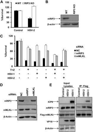
FIG 1HSV-2 as well as HSV-1 infection directly activates RIP3/MLKL-dependent necrosis in mouse cells. (A) Wild type (WT) and RIP3 knockout (KO) MEFs were infected with a control or HSV-2 at a multiplicity of infection (MOI) of 10 for about 20 h. Identical MOIs were used in MEFs in later experiments. Cell viability analysis was performed as described in Materials and Methods. (B) WT or RIP3 knockout MEF lysates were prepared and subjected to Western blot analysis. (C) L929 cells were transfected with a negative control (NC), mRIP3, or mMLKL siRNA oligonucleotide for 48 h. Then cells were treated as indicated for 15 h, and cell viability was determined. The HSV-1 MOI was 5. T, TNF-␣(10 ng/ml); Z, Z-VAD (10M). (D) L929 cells were transfected with the indicated siRNA oligonucleotides for 48 h. Then cell lysates were prepared and subjected to Western blot analysis. (E) MEFs stably expressing Flag- and HA-tagged mMLKL were infected with HSV-1 or HSV-2 for 6 h. Cell lysates were prepared for immunoprecipitation with anti-Flag agarose beads. The Flag-mMLKL immunocom-plex was then determined by Western blotting of the indicated proteins. IP, immunoprecipitation.
on November 7, 2019 by guest
http://jvi.asm.org/
[image:3.585.135.450.67.516.2]HSV-1 infection decreased the modification of hRIP3 and
re-sulted in the reduced phosphorylation of hMLKL following
treat-ment with TNF-
␣
/Smac mimetic/Z-VAD (
Fig. 3C
), indicating
that the activation of RIP3 was strongly attenuated in the WT
HSV-1-infected cells. In contrast, ICP6
⌬
HSV-1 failed to limit
the phosphorylation of both hRIP3 and hMLKL during
TNF-induced necrosis (
Fig. 3C
). As induction of the necrosome (the
RIP1/RIP3 complex) is critical for RIP3 activation during
TNF-induced necrosis, we examined necrosome formation in
HSV-infected HeLa-hRIP3 cells. As shown in
Fig. 3D
, HSV-1
and HSV-2 infections disrupted the formation of the
necro-some following TNF-
␣
/Smac mimetic/Z-VAD treatment,
whereas this complex was induced in cells infected with ICP6
⌬
HSV-1. These results demonstrate that the HSV R1 subunit is
critical for preventing necrosome formation during
necropto-sis in human cells.
FIG 2HSV-1 or HSV-2 infection subverts necroptosis in human cells. (A) HT-29 or HT-29-RIP3-shRNA cells were infected with HSV-1 or HSV-2 or treated with TNF-␣/Smac mimetic/Z-VAD (40 ng/ml TNF-␣, 100 nM Smac mimetic, and 20M Z-VAD) for 16 h, and then cell viability was determined. *,P⬍0.05 versus the control. (B) HT-29 or HT-29-RIP3-shRNA cells were treated with the indicated virus (MOI of 2.5). Cell lysates were collected at 6 h postinfection and then subjected to Western blot analysis. (C) HT-29 cells were infected with HSV-1 and HSV-2 at the indicated MOI. At 2 h postinfection, cells were treated with dimethyl sulfoxide (DMSO) or TNF-␣/Smac mimetic/Z-VAD for 16 h, and then cell viability was determined. *,P⬍0.05; **,P⬍0.001 versus the control. (D) HT-29 or HT-29-RIP3-shRNA cells were infected with HSV-1 at an MOI of 2.5. At 2 h postinfection, cells were treated with dimethyl sulfoxide or TNF-␣/Smac mimetic/Z-VAD for 48 h, and then cell viability was determined. (E) HeLa-hRIP3 cells were infected with the indicated virus. At 2 h postinfection, cells were treated with dimethyl sulfoxide or TNF-␣/Smac mimetic/Z-VAD for 16 h, and then cell viability was determined. HeLa-hRIP3 cell lysates were collected after treatment with HSV-1 or HSV-2 (MOI of 2.5). The expression of viral protein VP16 was detected by Western blotting using an anti-VP16 antibody. *,P⬍0.05; **,P⬍0.001 versus control (F) HT-29 cells were treated with the indicated virus (MOI of 2.5). At 2 h postinfection, cells were treated with dimethyl sulfoxide or TRAIL/Smac mimetic/Z-VAD for 16 h, and then cell viability was determined.
on November 7, 2019 by guest
http://jvi.asm.org/
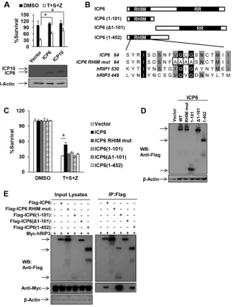
[image:4.585.113.469.64.517.2]Ectopic expression of HSV R1 is sufficient to block
TNF-in-duced necrosis of human cells depending on both RHIM and RR
domains.
We next investigated whether either ICP6 or ICP10 was
sufficient to abolish TNF-induced necrosis in human cells by
ec-topically expressing ICP6 or ICP10 in HeLa-hRIP3 cells. As shown
in
Fig. 4A
, expression of ICP6 or ICP10 efficiently reduced
TNF-induced necrosis. Moreover, we examined the roles of the RHIM
and RR domains of ICP6 by evaluating truncated forms of ICP6
lacking the N-terminal RHIM domain or C-terminal RR domain
in human cells (
Fig. 4B
). None of the truncated forms of ICP6,
including ICP6 consisting of amino acids 1 to 101 [ICP(1–101)],
ICP6 with a deletion of residues 1 to 101 [ICP6(
⌬
1–101)], and
ICP6(1– 452), was able to block TNF-induced necrosis although
their expression levels were similar to the level of full-length ICP6
in HeLa-RIP3 cells (
Fig. 4C
and
D
). Additionally, a RHIM mutant
form of ICP6, in which residues 73 to 76 were mutated to four
alanines (
Fig. 4B
), failed to affect TNF-induced necrosis in the
transfected HeLa-hRIP3 cells (
Fig. 4C
and
D
). Although
RHIM-containing truncated proteins such as ICP6(1–101) and ICP6(1–
452) retained the ability to interact with hRIP3, they failed to
influence necroptosis (
Fig. 4E
). These results indicate that both
the RHIM and the RR domains of ICP6 are required for its proper
function in modulating TNF-induced necrosis in human cells.
HSV R1 prevents the recruitment of hRIP1 to hRIP3.
Since
the RHIM domain of ICP6 is essential for the suppression of
TNF-induced necrosis of human cells, we examined the association of
ICP6 and ICP10 with hRIP1 and hRIP3. As shown in
Fig. 4E
and
5A
, hRIP3 was pulled down by either Flag-tagged ICP6 or ICP10.
We also found that ICP6 and ICP10 were pulled down by
Flag-tagged hRIP1 (
Fig. 5B
). Furthermore, we found that the RHIM
mutant form of ICP6 lost the ability to bind hRIP3 and hRIP1
(
Fig. 5A
and
B
). These findings suggest that HSV R1 has the
capacity to form complexes with both hRIP3 and hRIP1
through RHIM domains. We then investigated whether ectopic
expression of ICP6 and ICP10 could influence the recruitment
of hRIP1 to hRIP3. The hRIP1/hRIP3 complex was detected in
293T cells cotransfected with DNA plasmids expressing hRIP1
and hRIP3 (
Fig. 5C
). However, the formation of the hRIP1/
hRIP3 complex decreased in the presence of ICP6 (
Fig. 5C
).
The reduced hRIP1/hRIP3 complex level correlated with the
increased level of ICP6 (
Fig. 5C
). These results demonstrate
that HSV R1 is able to disrupt the binding of hRIP1 to hRIP3 in
an RHIM-dependent manner.
DISCUSSION
Initiation of programmed cell death (PCD) in host cells is a critical
strategy to prevent pathogen replication. Extensive studies have
shown that many pathogens encode apoptotic suppressors such as
caspase inhibitors to circumvent apoptosis, a major form of PCD
in mammals. Inhibition of apoptosis has been shown to facilitate
the activation of programmed necrosis, a process that is mediated
by RIP3 and MLKL. We recently demonstrated that
RIP3/MLKL-dependent necrosis is activated in HSV-1-infected mouse cells
through the recruitment of viral ICP6 to RIP3, a result supporting
the importance of programmed necrosis in the control of HSV-1
replication. In the current study, we demonstrate a negative
reg-ulation of programmed necrosis by both HSV-1 and HSV-2
infec-tion in human cells. The R1 subunits of HSV-1 and HSV-2 are
sufficient to disrupt TNF-induced necrosis of human cells.
Al-though HSV-2 as well as HSV-1 infection directly activates the
formation of the mRIP3/mMLKL complex in mouse cells, the
recruitment of HSV R1 with hRIP3 failed to trigger
hRIP3/hM-LKL signaling and also disrupted the binding of hRIP1 to hRIP3 in
human cells. Thus, this study uncovers dual roles of HSV R1 in
modulating programmed necrosis through either activation or
inactivation of RIP3 signaling in a species-specific manner.
Dur-ing the preparation of the present manuscript, similar work has
been published (
28
).
RIP3 plays a central role in the regulation of programmed
necrosis initiated by death ligands, TLR ligands, or viral
infec-FIG 3HSV R1 is required to disrupt necrosome formation during necroptosis in human cells. (A) HT-29 cells were infected with the indicated virus (MOI of 2.5). At 2 h postinfection, cells were treated with dimethyl sulfoxide (DMSO) or TNF-␣/Smac mimetic/Z-VAD for 16 h, and then cell viability was deter-mined. *,P⬍0.05; **,P⬍0.001 versus the control. (B) HT-29 cells were infected with the indicated virus (MOI of 2.5). Cell lysates were collected at 6 h postinfection and then subjected to Western blot analysis. (C) HT-29 cells were infected as indicated (MOI of 2.5). At 2 h postinfection, cells were treated with TNF-␣/Smac mimetic/Z-VAD for an additional 6 h. Then cell lysates were collected and subjected to Western blot analysis. (D) HeLa-hRIP3 cells were infected as indicated (MOI of 2.5). At 2 h postinfection, cells were treated with TNF-␣/Smac mimetic/Z-VAD for an additional 6 h. Cell lysates were collected and used for anti-Flag immunoprecipitation. The Flag-RIP3 immunocomplex was analyzed by Western blotting with the indicated antibody.
on November 7, 2019 by guest
http://jvi.asm.org/
[image:5.585.41.287.66.459.2]tion. The recruitment of an RHIM-containing protein to RIP3
is a crucial process for the activation of RIP3. For example,
TNFR- and TLR-mediated necrosis requires the formation of
the RIP1/RIP3 complex and the TRIF/RIP3 complex,
respec-tively. Moreover, the ICP6/RIP3 complex and the DAI/RIP3
complex are essential for HSV-1 and M45/vIRA mutant
MCMV-associated necrosis, respectively. Thus, RIP3 acts as a
cellular necrotic sensor in the recognition of RHIM-containing
proteins, leading to the activation of the MLKL substrate.
No-tably, RHIM-dependent modulation of RIP3 is utilized by
MCMV M45/vIRA to block programmed necrosis during viral
in-fection. The present study shows that HSV-1 ICP6 and HSV-2 ICP10
manipulate programmed necrosis of human cells through
RHIM-dependent suppression of RIP3 signaling. These findings provide
strong evidence that pathogens have likely evolved strategies to
mod-ulate the necrotic sensor RIP3 via disruption of the RHIM-dependent
activation of RIP3, leading to inactivation of the programmed
necro-sis responses in hosts.
FIG 4Ectopic expression of HSV R1 is sufficient to block TNF-induced necrosis of human cells depending on both RHIM and RR domains. (A) HeLa-hRIP3 cells were transfected with empty vector or an ICP6 or ICP10 DNA plasmid for 24 h. Cells were treated with TNF-␣/Smac mimetic/Z-VAD for an additional 36 h, and then cell viability was measured. Data represent the averages⫾standard errors for three independent experiments. *,P⬍0.05, versus results for the vector. Cell lysates were collected at 24 h posttransfection and subjected to Western blot analysis. (B) Domain structure of ICP6. Full-length ICP6 (amino acids 1 to 1137) contains the N-terminal RHIM domain and the C-terminal RR domain. ICP6(1–101) and ICP6(1– 452) contain the N-terminal 101 residues and 452 residues, respectively. ICP6(⌬1–101) lacks residues 1 to 101. The residues 73 to 76 of ICP6 were mutated to four alanine residues. (C and D) HeLa-hRIP3 cells were transfected with the indicated plasmids for 24 h. Cells were treated with TNF-␣/Smac mimetic/Z-VAD for an additional 36 h, and then cell viability was measured (C). Data represent the averages⫾standard errors for three independent experiments. *,P⬍0.05, versus results for the vector. Cell lysates were collected at 24 h posttransfection and subjected to Western blot analysis (D). (E) 293T cells were transfected with the indicated plasmids for 48 h. Cell lysates were collected and immunoprecipitated with anti-Flag agarose beads. The immunoprecipitate was analyzed by Western blotting (WB).
on November 7, 2019 by guest
http://jvi.asm.org/
[image:6.585.126.463.66.509.2]Interestingly, our work suggests that HSV R1 has an opposite
impact on programmed necrosis in mouse cells versus that in
human cells. Although HSV R1 is able to interact with both hRIP3
and mRIP3 through the RHIM domains, we found that the
re-cruitment of HSV R1 to mRIP3 directly activated the formation of
the mRIP3/mMLKL complex in mouse cells but not in human
cells. We found that the RHIM-dependent association of HSV R1
with hRIP3 or hRIP1 prevented the recruitment of hRIP1 to
hRIP3. Previous studies have shown that mMLKL is unable to
bind to hRIP3 and that mRIP3 cannot interact with hMLKL (
29
).
The phosphorylation of S227 in hRIP3 is critical for its interaction
with hMLKL, while the interaction between mRIP3 and mMLKL
requires the phosphorylation of the conserved S232 residue and
an additional T231 residue in mRIP3 (
14
,
29
). Although the
RIP3-MLKL interaction is functionally conserved for programmed
ne-crosis, differential sequences and phosphorylation sites of RIP3
control the species specificity of this RIP3-MLKL interaction. It is
tempting to speculate that HSV R1-mediated species-specific
modulation of programmed necrosis is determined by the
differ-ential manipulation of RIP3/MLKL signaling. Further
structure-based studies on HSV R1/RIP3 complexes are required to
under-stand the precise molecular mechanism for this species-specific
modulation.
ACKNOWLEDGMENTS
We thank Xiaodong Wang (National Institute of Biological Sciences [NIBS], Beijing, China) for anti-phosphor-MLKL antibody and Smac mi-metic. We also thank Zhigao Wang (University of Texas Southwestern Medical Center, Dallas, TX, USA) for HeLa-hRIP3 cells, Sandra K. Weller (University of Connecticut Health Center, Farmington, CT, USA) for the HSV-1 KOS strain and the ICP6 deletion mutant (ICP6⌬).
This work was supported by the National Basic Research Program of China (2013CB910102), the National Natural Science Foundation of China (grants 31222036 and 31471303), a Project Funded by the Priority Academic Program Development of Jiangsu Higher Education
Institu-FIG 5HSV R1 prevents the recruitment of hRIP1 to hRIP3. (A) 293T cells were cotransfected with the Myc-tagged hRIP3 plasmid and a plasmid expressing Flag-tagged ICP10 and the RHIM mutant form of ICP10 (RHIM mut). The residues from 64 to 67 of ICP10 were mutated to four alanine residues. At 48 h posttransfection, cell lysates were collected and used for anti-Flag immunoprecipitation. The Flag-hRIP3 immunocomplex was analyzed by Western blotting with the indicated antibodies. (B) 293T cells were cotransfected with the Flag-tagged hRIP1 plasmid and a plasmid expressing Myc-tagged ICP6, the RHIM mutant, or ICP10. At 48 h posttransfection, cell lysates were collected and used for anti-Flag immunoprecipitation. The Flag-hRIP1 immunocomplex was analyzed by Western blotting with the indicated antibodies. (C) 293T cells were cotransfected with plasmids as indicated. At 48 h posttransfection, cell lysates were collected for anti-Flag immunoprecipitation. The immunocomplex was subjected to Western blot analysis.
on November 7, 2019 by guest
http://jvi.asm.org/
[image:7.585.124.462.64.441.2]tions, Natural Science Foundation of Jiangsu Province (grants BK2012004 and BK2011287), the National Institutes of Health (1R01CA166413), and the Undergraduate Training Programs for Innovation and Entrepreneur-ship (201410285046).
FUNDING INFORMATION
National Basic Research Program of China provided funding to Sudan He under grant number 2013CB910102. National Natural Science Founda-tion of China provided funding to Sudan He under grant numbers 31222036 and 31471303. Natural Science Foundation of Jiangsu Province Grant provided funding to Sudan He under grant numbers BK2012004 and BK2011287. National Institutes of Health provided funding to Sudan He under grant number 1R01CA166413. Undergraduate Training Pro-grams for Innovation and Entrepreneurship provided funding to Sudan He under grant number 201410285046.
REFERENCES
1.Sridharan H, Upton JW.2014. Programmed necrosis in microbial patho-genesis. Trends Microbiol 22:199 –207. http://dx.doi.org/10.1016/j.tim .2014.01.005.
2.Han J, Zhong CQ, Zhang DW.2011. Programmed necrosis: backup to and competitor with apoptosis in the immune system. Nat Immunol12:
1143–1149.http://dx.doi.org/10.1038/ni.2159.
3.Moriwaki K, Chan FK.2013. RIP3: a molecular switch for necrosis and inflammation. Genes Dev27:1640 –1649.http://dx.doi.org/10.1101/gad .223321.113.
4.Degterev A, Huang Z, Boyce M, Li Y, Jagtap P, Mizushima N, Cuny GD, Mitchison TJ, Moskowitz MA, Yuan J.2005. Chemical inhibitor of nonapoptotic cell death with therapeutic potential for ischemic brain in-jury. Nat Chem Biol1:112–119.http://dx.doi.org/10.1038/nchembio711. 5.Holler N, Zaru R, Micheau O, Thome M, Attinger A, Valitutti S, Bodmer JL, Schneider P, Seed B, Tschopp J. 2000. Fas triggers an alternative, caspase-8-independent cell death pathway using the kinase RIP as effector molecule. Nat Immunol1:489 – 495.http://dx.doi.org/10 .1038/82732.
6.Declercq W, Vanden Berghe T, Vandenabeele P.2009. RIP kinases at the crossroads of cell death and survival. Cell138:229 –232.http://dx.doi.org /10.1016/j.cell.2009.07.006.
7.Cho YS, Challa S, Moquin D, Genga R, Ray TD, Guildford M, Chan FK.
2009. Phosphorylation-driven assembly of the RIP1-RIP3 complex regu-lates programmed necrosis and virus-induced inflammation. Cell137:
1112–1123.http://dx.doi.org/10.1016/j.cell.2009.05.037.
8.He S, Wang L, Miao L, Wang T, Du F, Zhao L, Wang X.2009. Receptor interacting protein kinase-3 determines cellular necrotic response to TNF-alpha. Cell137:1100 –1111.http://dx.doi.org/10.1016/j.cell.2009.05 .021.
9.Zhang DW, Shao J, Lin J, Zhang N, Lu BJ, Lin SC, Dong MQ, Han J.
2009. RIP3, an energy metabolism regulator that switches TNF-induced cell death from apoptosis to necrosis. Science325:332–336.http://dx.doi .org/10.1126/science.1172308.
10. Sun X, Yin J, Starovasnik MA, Fairbrother WJ, Dixit VM.2002. Iden-tification of a novel homotypic interaction motif required for the phos-phorylation of receptor-interacting protein (RIP) by RIP3. J Biol Chem
277:9505–9511.http://dx.doi.org/10.1074/jbc.M109488200.
11. Moquin DM, McQuade T, Chan FK.2013. CYLD deubiquitinates RIP1 in the TNF␣-induced necrosome to facilitate kinase activation and pro-grammed necrosis. PLoS One8:e76841.http://dx.doi.org/10.1371/journal .pone.0076841.
12. Hitomi J, Christofferson DE, Ng A, Yao J, Degterev A, Xavier RJ, Yuan J.2008. Identification of a molecular signaling network that regulates a cellular necrotic cell death pathway. Cell135:1311–1323.http://dx.doi.org /10.1016/j.cell.2008.10.044.
13. Zhao J, Jitkaew S, Cai Z, Choksi S, Li Q, Luo J, Liu ZG.2012. Mixed lineage kinase domain-like is a key receptor interacting protein 3 down-stream component of TNF-induced necrosis. Proc Natl Acad Sci U S A
109:5322–5327.http://dx.doi.org/10.1073/pnas.1200012109.
14. Sun L, Wang H, Wang Z, He S, Chen S, Liao D, Wang L, Yan J, Liu W, Lei X, Wang X.2012. Mixed lineage kinase domain-like protein mediates necrosis signaling downstream of RIP3 kinase. Cell148:213–227.http://dx .doi.org/10.1016/j.cell.2011.11.031.
15. Wang H, Sun L, Su L, Rizo J, Liu L, Wang LF, Wang FS, Wang X.2014. Mixed lineage kinase domain-like protein MLKL causes necrotic mem-brane disruption upon phosphorylation by RIP3. Mol Cell54:133–146.
http://dx.doi.org/10.1016/j.molcel.2014.03.003.
16. Cai Z, Jitkaew S, Zhao J, Chiang HC, Choksi S, Liu J, Ward Y, Wu LG, Liu ZG.2014. Plasma membrane translocation of trimerized MLKL pro-tein is required for TNF-induced necroptosis. Nat Cell Biol16:55– 65.
http://dx.doi.org/10.1038/ncb2883.
17. Chen X, Li W, Ren J, Huang D, He WT, Song Y, Yang C, Li W, Zheng X, Chen P, Han J.2014. Translocation of mixed lineage kinase domain-like protein to plasma membrane leads to necrotic cell death. Cell Res
24:105–121.http://dx.doi.org/10.1038/cr.2013.171.
18. Kawai T, Akira S.2011. Toll-like receptors and their crosstalk with other innate receptors in infection and immunity. Immunity34:637– 650.http: //dx.doi.org/10.1016/j.immuni.2011.05.006.
19. Kaiser WJ, Sridharan H, Huang C, Mandal P, Upton JW, Gough PJ, Sehon CA, Marquis RW, Bertin J, Mocarski ES.2013. Toll-like receptor 3-mediated necrosis via TRIF, RIP3, and MLKL. J Biol Chem288:31268 – 31279.http://dx.doi.org/10.1074/jbc.M113.462341.
20. He S, Liang Y, Shao F, Wang X. 2011. Toll-like receptors activate programmed necrosis in macrophages through a receptor-interacting ki-nase-3-mediated pathway. Proc Natl Acad Sci U S A108:20054 –20059.
http://dx.doi.org/10.1073/pnas.1116302108.
21. Dobbelstein M, Shenk T.1996. Protection against apoptosis by the vac-cinia virus SPI-2 (B13R) gene product. J Virol70:6479 – 6485.
22. Kettle S, Alcami A, Khanna A, Ehret R, Jassoy C, Smith GL. 1997. Vaccinia virus serpin B13R (SPI-2) inhibits interleukin-1beta-converting enzyme and protects virus-infected cells from TNF- and Fas-mediated apoptosis, but does not prevent IL-1beta-induced fever. J Gen Virol78:
677– 685.http://dx.doi.org/10.1099/0022-1317-78-3-677.
23. Upton JW, Kaiser WJ, Mocarski ES.2010. Virus inhibition of RIP3-dependent necrosis. Cell Host Microbe7:302–313.http://dx.doi.org/10 .1016/j.chom.2010.03.006.
24. Upton JW, Kaiser WJ, Mocarski ES.2012. DAI/ZBP1/DLM-1 complexes with RIP3 to mediate virus-induced programmed necrosis that is targeted by murine cytomegalovirus vIRA. Cell Host Microbe11:290 –297.http: //dx.doi.org/10.1016/j.chom.2012.01.016.
25. Wang X, Li Y, Liu S, Yu X, Li L, Shi C, He W, Li J, Xu L, Hu Z, Yu L, Yang Z, Chen Q, Ge L, Zhang Z, Zhou B, Jiang X, Chen S, He S.
2014. Direct activation of RIP3/MLKL-dependent necrosis by herpes simplex virus 1 (HSV-1) protein ICP6 triggers host antiviral defense. Proc Natl Acad Sci U S A111:15438 –15443.http://dx.doi.org/10.1073 /pnas.1412767111.
26. Huang Z, Wu SQ, Liang Y, Zhou X, Chen W, Li L, Wu J, Zhuang Q, Chen C, Li J, Zhong CQ, Xia W, Zhou R, Zheng C, Han J. 2015. RIP1/RIP3 binding to HSV-1 ICP6 initiates necroptosis to restrict virus propagation in mice. Cell Host Microbe17:229 –242.http://dx.doi.org/10 .1016/j.chom.2015.01.002.
27. Wang Z, Jiang H, Chen S, Du F, Wang X.2012. The mitochondrial phosphatase PGAM5 functions at the convergent point of multiple ne-crotic death pathways. Cell148:228 –243.http://dx.doi.org/10.1016/j.cell .2011.11.030.
28. Guo H, Omoto S, Harris PA, Finger JN, Bertin J, Gough PJ, Kaiser WJ, Mocarski ES.2015. Herpes simplex virus suppresses necroptosis in hu-man cells. Cell Host Microbe 17:243–251. http://dx.doi.org/10.1016/j .chom.2015.01.003.
29. Chen W, Zhou Z, Li L, Zhong CQ, Zheng X, Wu X, Zhang Y, Ma H, Huang D, Li W, Xia Z, Han J.2013. Diverse sequence determinants control human and mouse receptor interacting protein 3 (RIP3) and mixed lineage kinase domain-like (MLKL) interaction in necroptotic signaling. J Biol Chem 288:16247–16261. http://dx.doi.org/10.1074 /jbc.M112.435545.
on November 7, 2019 by guest
http://jvi.asm.org/