0022-538X/92/053183-05$02.00/0
Copyright
© 1992, American Society for
Microbiology
Phenotype-Associated
Sequence Variation in the Third Variable
Domain of the
Human
Immunodeficiency Virus
Type
1
gpl20 Molecule
RON A. M. FOUCHIER,
MARTIJNGROENINK,
NEELTJE A. KOOTSTRA, MATTHIJS TERSMETTE,HAN G. HUISMAN,
FRANK MIEDEMA,* ANDHANNEKESCHUITEMAKERDepartment of Clinical
Viro-Immunology, Central Laboratory of
theNetherlands
Red CrossBlood Transfusion
Serviceand
Laboratory
of
Experimental
and
Clinical
Immunology
of
the
University
of
Amsterdam, Amsterdam,
The Netherlands
Received11 December 1991/Accepted 30 January 1992
The
third variable
(V3) domain has beenimplicated
in determining the human immunodeficiency virus(HIV) phenotype, including fusion
capacity
and monocytotropism. Inalargesetof primaryH1Vtype1 (HIV-1)isolates,
V3 sequence analysis revealed thatfast-replicating, syncytium-inducing
isolates contained V3sequenceswith a
significantly
higher positive charge than
those of slow-replicating,non-syncytium-inducing
monocytotropic
isolates.It appeared that these differences
in chargecould be attributed
to highly variable aminoacid residues locatedoneither side
of
theV3
loop,midway between
thecysteine residues and the central
GPG motif. In
non-syncytium-inducing monocytotropic isolates,
theseresidues
werenegatively
charged
oruncharged,
whereas insyncytium-inducing nonmonocytotropic
isolates, either one orboth
werepositively
charged.
The substitutions atthesepositions
result in changes in thepredicted
secondary
structureof the V3loop.
Our data suggest that twoamino
acid residues
in thehighly
variable V3 domain
areresponsible for
phenotype
differences andpoint
toconformational differences
inV3
loopsfrom
phenotypically
distinctHIV-1
isolates.
Previously,
acorrelation between the biological
pheno-type of a human
immunodeficiency
virus type 1(HIV-1)
isolate and theclinicalcourseof
infection
wasdemonstrated(33).
Fast-replicating,
syncytium-inducing
(SI) variants
emergein 50% of
HIV-1-seropositive
individualspreceding
progression
toAIDS. Slow-replicating,
non-syncytium-in-ducing (NSI)
variants arepredominant in the asymptomatic
stage and
persist throughout all
stages of HIV-1 infection(33).
TheNSI
isolates ingeneral
aremuch moremonocyto-tropic
andprobably important
forpersistence, possibly by
forming
amajor
viral reservoir in HIV-1 infection(11, 25,
26). By using
a limited setof
viralisolates, regions
in theenvelope
gene have been delineated that are involved indetermining
hostrange, and in allcases,involvementof the V3 domain has beensuggested (4, 14, 20, 28, 36).
The V3 domain of HIV-1
gpl20
has been found to elicitneutralizing antibodies
as well as acytotoxic and helper
T-cellresponse in both humans and animals
(7, 12, 15, 16,
21, 23, 30).
As a consequence, this immunodominantprin-cipal neutralizing
determinant has become animportant
target for vaccine
development. Type
specificity
has been observed inbothantibody
andT-cellresponsesas aresult ofvariation in
V3
sequences(1, 21, 31),
which hashampered
development
ofan effectivevaccine for HIV-1.The aim of our
study
was toinvestigate
whether theprimary
orpredicted secondary
structure of the V3loop
correlates with differences in
biological phenotype
of HIV-1 variants. For this purpose, alarge panel
ofprimary
virusisolates and
biological
clones were used that were well defined for theircapacity
toinducesyncytia
andtoreplicate
in
primary
monocytes(26, 27, 33).
DNAwasisolated from infected cellsasdescribed before
(2).
V3sequenceswereamplified by
thepolymerase
chain* Correspondingauthor.
reaction with
primers
Aand H in the first and B and C in the secondreaction,asdescribedelsewhere (29).
Productswerepurified
with a Geneclean kit(Bio 101), and
both strands weresequenced directly
withprimers
B and Cby
thedideoxy
chain termination method withSequenase (USB),
both
according
to instructions from the manufacturers. InFig.
1 and2,
the derived amino acid sequencesof theV3
loops
of the virus isolates areshown,
aligned
with the consensus sequence of the isolates. The most extensivevariation is
observed
in theregions flanking
the GPG se-quence at amino acids 10 to 14 and 25 to 29. Inaddition,
sequencevariation ishigher
inthegroup of SInonmonocy-totropic
isolates than in the NSImonocytotropic
isolates.It has been
suggested
that the relativecharge
of the V3 domainmight
influence its function or the function of theentire
gp120 molecule,
forexample,
inbinding negatively
charged
counterpartstructures(3). Using
theCHARGEPROprogramfromPCGENE
(IntelliGenetics),
wecalculated thecharge
of theV3 domains atphysiological pH.
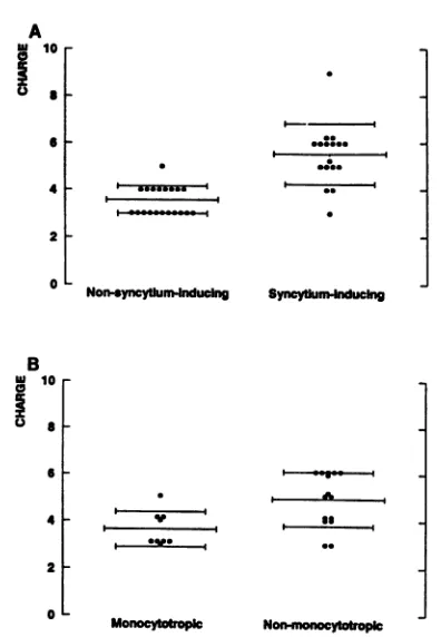
NSI isolates had lesspositively charged
V3 amino acid sequences than the SI isolates(3.6
+ 0.6[standard deviation]
versus5.5 +1.3,
P <0.0001) (Fig. 3A).
Similar results were obtainedwhen
comparing monocytotropic
andnon-monocytotropic
isolates
(3.6
+ 0.7versus4.9 +1.2,
P =0.007) (Fig. 3B).
To
investigate
whether substitution of neutral or acidicresidues for
positively charged
residuesrequired
fixedposi-tions,
statistical discriminantanalysis
wasperformed
afterassigning
numbers to amino acid residues based oncharge
(acid, neutral,
and basic residueswerenumbered1, 2,
and3,
respectively). By
use of thisanalysis,
NSI and SI HIV-1variants aswell as
monocytotropic
andnonmonocytotropic
HIV-1 variantswere
compared.
The serineandglutamic
acidresidues at
positions
11 and 28 of the consensus sequencediscriminatedbetween different
phenotypes
of virus isolatesonthe basis oftheir
primary
V3 structure. InNSIvariants,
the amino acid residue at
position
11 wasuncharged (in
3183
on November 9, 2019 by guest
http://jvi.asm.org/
3184 NOTES
Mono- 1 10 20 30 38
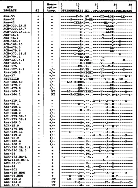
I80TI 8I ayto- _ t . ZII . PRYWBIDIQ
XSOLATE a troD CTPUN'KKI.NI._.010RAP1TE1XTZUDIIANC Ams-55 + _ ---x-a-.AV ---DI--ADZ----LK----Ams-32 + _ ---S--- R-HR
--N----l---Ams-42 + . ---IKRR-I--.---a---.
ACH-320.2A.5 + _ --- .---AAR
---ACH-320.2A.7 + _
---AARK---ACH-320.2A.1.1 + ---.. --.-.---AARi .---Ams-16.1 + _ ---
a-.---V----R---Ams-16.2 + _ ---a---v----R---ACH-168.7 + _ ---R---
----Q---N---ACH-168.10 + _ ---R---
----Q---N---ACH-479.5 + _ --- .--.-.---RR---ACH-479.6 + _ ---QG--- ---RR---ACH-373.38.5 + _ ---NI-RNHI--.---N---ACH-704.1 + ---RV.TM.
.---L---Ams-127.4.1 + ---RV.TH. .----VL---X----Ams-169.1 + -
---G-RIGHI----V---X---Ams-175 + - --- .Y-.. ---V--K-R---Ams-127.4.2 + +/- ---IV.TH. .----VL---K----Ams-169.2 + +/- ---G-HIGHI----V---K---Ans-37 + +/ ---RV.TL.. ----VW---K-Y-HTLVIIIB + +/- ---R-.R-QR---V-I-K-.
-NM---ACH-182.69 + +/- ---LSA-KIRNM.--.
.---A--K---X---ACH-479.7 + +/- ---Q. ---RR---ACH-479.8 + +/- ---QG-. Y-..---RR---Ams-165.1 + NT ---LS----RVNIKNHI----V---AV---R---Ams-164.1 + NT ---R-KINR-.--..----P--G-
.D-E-P----Y-Ams-119.1 -_ --- ---.
.A--S---A--A---K---Ams-96.1 - ---R---FG-.D----N-R
Ams-168.1 _ _ ---N ---A--D---ACH-168.1 _ +/ --- . --- A--D---ACH-168.2 _ +/1 _---A--D---ACH-373.38.3 _ +/ N---N---ACH-373.38.4 _ +/ ---N---N
Ams-169.3 - +/- ..
---A---ACH-15.9 _ +/- ---NM .
I---Q-V---Ams-176.BM - +/- --- N ---A---ACH-239.11 +/- ----S
---A--D---Ams-119.2 _ +/- --- A--S---A--A---K---Ams-96.2 _ +/- ---RG --- ---G-.D
---N---ACH-182.6 _ +/- ---. N
---A---R---Ams-168.2 _ +/- --- N --- A--D---ACH-320.2A.2.1 _ + --- K--- ---K-A--Q---ACH-320.2A.3 + ---0-.-M..---K- A--Q---Ams-24 - + ---PM ---D---ACH-172.Ba-L - + -I---
M---A---HTLVIIIB.Ba-L - + _____-__-.-.-_---
---ACH-63.11 - + ---N
---A---Ams-44 - + ---L..---K---D .---Ams-101 - +
---Ams-119.MDM - + ---R--.-- A --A- A---K
--Amis-96.MDM - + ----H----NRG ---
-T---Aas-165.2 - NT ---LS----ROV
---AV---Ams-164.2 - NT
---K-Q---FG-.D---N----Y-Ans-16.3 - NT
a---FIG. 1. Alignmentof V3 sequencesofdifferent isolateswiththe
consensussequence. Gapsareindicatedas
points;
similaritytotheconsensus sequence is shown as dashes. Thesyncytium-inducing
capacity
(SI)
andmonocytotropismof the viral isolatesareindicated(isolatesweretestedonmonocyte-derived macrophagesfromeight seronegative donors). Formonocytotropism, - represents
replica-tion in 0, + represents replication in
>4,
and +/- representsreplicationinthe monocytes of 1to3 of the donorstested; N.T.,not
tested. Amino acid residues in boldfacewereusedasclassification
variablesforstatisticalanalysis (Table 1).
general,
S
orG)
and the
residue
atposition
28
waseither
negatively charged (E
orD)
oruncharged
(A
orQ).
InSI
isolates,
either
one orboth amino acid residues
atpositions
11
and 28
werereplaced by
basic residues
(P
<0.00001;
Table
1). Comparison
ofmonocytotropic
and
nonmonocyto-tropic
isolates revealed
identical
variation,
albeit less
pro-nounced
(P
=0.0006;
Table
1).
Itshould be noted that in this
set of
isolates,
the occurrenceof
abasic residue
atposition
28 coincides
with aglycine
residue atposition
11in
allisolates.
NSI
monocytotropic
isolates with
aglycine residue
at
position
11could therefore be
anintermediate genotype in
populations
of viruses
during phenotype
conversions. Other
changes
in the V3
sequences
occurred in both groups of
virus isolates
oroccurred
infrequently
and
weretherefore
neither
predictive
nordiscriminative.
Incontrast to
the
findings
by
Westervelt
etal.
(34),
nodiscriminatory
value for
the
amino acid residues
atpositions
14and
24 wasobserved.
If
amino acid residues
11and 28
arethe
major
determi-nantsof
the viral
phenotypes,
comparable changes should be
present in
highly
related
yet
phenotypically distinct variants,
such
asthose present
insequential
isolates from
seropositive
Mono- 1 10 20 30 36
PATIBIIT ex tXono CTRPNNNIKI.HIL.GPGRAFYTTGEIIGDIRQAnC.
ACH-182 +/ --- .N-.---A--A---R---+ +/- ---LSA-KIRE.---A--I ---K---ACH-320 - + ---.- -M..- ---A--Q
---+ - a---0 --- R
ACs-16 - NT
---.---+ - _____-_____-_.--..--V----R---ACH-168 - +_ ---A--D---+ - ---R---.
--Q----N---ACH-479 ---- -.N
---Q---N---+ +_ ---.--
Q--.---ACH-373 _ +/- --- ---+ ---I-REHI
----N---Ans-169 _ + --- --.-
..A---A---+ - ---
G-IGHI---K--A---Ams-119 + ---R-.---.-.
---A---K---_ - ---R--.-A--S----A--A
---K----Ams-168 +/ ---- .N----R . .
---A--D---
N-..---A--D---Ans-165 NT ---LS----R0V.--.
.---AV---+ NT
---LS----RGVHIKHI----V---AV---R---Ams-164 _ NT ---K-Q--.--- ---FG-.
---N----Y-+ NT
---R-KIRRN--.--..----P--G-.D-E-P----Y-FIG. 2. Sequence alignment ofbiological clones with distinct
phenotypes obtained from the sameindividual.SeelegendtoFig. 1
fordetails.
individuals (33).
InFig.
2,
the V3 sequencesof
setsof
biological
clones obtained from 12 seropositive
individualsare
shown. In these
highly related viruses,
similarchanges
ofamino acid residues
atposition
11and/or
28 topositive
residues upon transition
fromNSI
toSI
can bedemon-strated. The
specific variation at positions 11 and 28 upon
transition suggests
animportant contribution
of these aminoacid residues
todetermining the
biological phenotype of
theisolates.
Whether
these amino acid
substitutions could
causechanges in the V3 secondary
structure wasinvestigated by
use
of the
method of
Garnier
(lOa) (PCGENE;
IntelliGenet-ics). Substitution of the residues at positions 11 and 28 with
basic residues appears
toinduce changes in secondary
structure at
the
tip of the loop (Fig. 4), indicating
that NSImonocytotropic and SI nonmonocytotropic isolates might
have
distinct V3
structures.The fusion
capacity-associated
sequence
variation in
theseviral isolates
points
to aprominent role for the V3 domain in
determining
this
biological property. However, analysis
ofchimeric
proviruses has revealed that V3 is involved in,
but notsufficient
for, determining monocytotropism
(5,
14, 28,
34). The fact that sequence variation related
tothe
monocy-totropic
and NSI
phenotypes
is
comparable could
be due toan
80%
overlap of these virus
populations (26).
Aproper
combination of
V3and other
functional domains involved in
determining monocytotropism appears
tolead
tothe
mono-cytotropic phenotype (14).
V3
sequences from the
highly
related
viral isolates
presented in
Fig. 2 point to a minor role
for
amino acid variation
atpositions
11and 28 in determining
monocytotropism (compare Ams-96, Ams-119, and
Ams-168).
If
the
correlation between V3 sequence and viral
pheno-type
is based
on astructure-function
relation, several
mech-anisms
canbe
suggested.
For
instance, the charge and
secondary
structureof
the V3
loop might influence binding
of the
virion,
resulting in altered syncytium induction and
infectivity.
Inaddition, processes subsequent to exposure of
J. VIROL.on November 9, 2019 by guest
http://jvi.asm.org/
[image:2.612.316.558.78.297.2] [image:2.612.65.302.79.400.2]A
I
'0r
a
-
61-4
0
B
u 10 a
6
4
2
0L
*,---oees---.
F
Non-syncy0um4nducing
0.
Syr#ytinh-uclng
M onocyoplc Nononcyttroplc
FIG. 3. Comparisonofthepeptide charges atpH 7.0as
calcu-lated byuseofthe CHARGEPROprogramfromPCGENE
(Intelli-Genetics). (A) Comparison on the basis of syncytium-inducing capacity; (B) comparisonon the basisofmonocytotropism. Note: multiple biologicalHIV-1clonesobtained fromoneindividual with
identicalcytotropismorsyncytium-inducing capacitywereexcluded
fromthisanalysis topreventabiasinthe results.
the V3 domain upon
binding
ofgp120
to CD4might
be influenced(24).
Site-directedmutagenesis
has indicated that the V3 domainplays
a role in thesyncytium-inducing
capacity
andinfectivity
of virusisolates(10,
32)
atthe level of viralfusogenic capacity.
In a recentstudy,
a limited number ofchimericproviruses
were constructed that dif-feredonly
in theregions
that contained the critical amino acid residues atpositions
11 and 28. These chimeras dem-onstrated correlated differences insyncytium-inducing
ca-pacity, pointing
to the functionalimportance
of these resi-dues(8).
Furthermore, syncytium-inducing capacity
has been demonstrated to bedependent
on V3 conformation(17).
Inaddition,
it has beensuggested
that V3 is cleavedby
proteases, which is aconformation-dependent
process(6,
PRINlhRYPND BYRUCYURU PREDICTED S300DILR PTDSTRUCTURE
CTRPNNNTRKSIHIGPGRAFYTTGEIIGDIRQAHC CTRPNNN IHIGPGRAFYTTGEIIGDIRQAHC
---_EECTTETEE lEREBECTC E H
---R--- ---C---_______________---R--- ---E---T----ZEIE ----_______________K - - - ---K--- _______--E----T ---R---K--- ---C--- T----1ZEE---H---RK--- ---C---Tr---- mm
[image:3.612.76.274.69.355.2]----FIG. 4. Predicted
secondary
structures of V3 sequences aftersubstituting
basic residuesatposition
11and/or28,asoccurring
in theprimary
virusisolates,
by
the method of Garnier(10a).
H,helix; C,coil; T,turn;E,extendedconfiguration.
PND,principal
neutral-izing
determinant.TABLE 1. Comparison of the charge of amino acid residues 11 and 28
Chargea(no. ofisolates) Isolates
Positive Negativeornone
Syncytium induction
SI 16 1
NSI 0 20
Tropism
Nonmonocytotropic 11 4
Monocytotropic 0 9
a Syncytium inductionandmonocytotropism werebothsignificantly corre-lated withcharge(P < 0.00001andP = 0.0006,respectively).
13).
Therefore, V3 secondary
structuremight be involved
in
determining the biological properties of the gp120 molecule.
The
Los Alamos V3
consensus sequence(19) is
derived
mainly from direct or singly passaged material. Since the
predominant phenotype
of the viral isolates involved in
persistent infection is
NSI
monocytotropic (25), one
mayexpect
the consensussequence
toresemble the V3
sequenceof
this virus
phenotype. Indeed, V3 sequences obtained from
the brain
andspleen
ofHIV-1-infected
individuals as well
asNSI
monocytotropic isolates show high homology to the
consensus
sequence
(9).
Thehigh
homology
tothe
consen-susV3 in
the NSI monocytotropic viral isolates
supportsthe
suggestion
thatthese variants
constitute the virus
tissuereservoir.
Since both
SI and NSI variants
canbe
transmitted
(22),
it
appears that the
uncompromised immune system is well
able toeradicate
orsuppress
fast-replicating SI variants upon
primary
infection, in
contrast toslow-replicating NSI
vari-ants, which may
notbe asimmunogenic, possibly by
virtueof their
monocytotropism. Because of the persistence of
theNSI
monocytotropic variants
(26), these variants should
be animportant target for vaccine development.
In
conclusion,
wehave demonstrated that sequence
vari-ation
atpositions 11 and 28 in V3 correlates with biological
variation in HIV-1. The functional
involvement of
envelope
proteins in processes such
asfusion is
thought
tobe
highly
dependent
onprotein
conformation, directly
orindirectly
determined
by V3 sequences
(17).
The
conformational
dif-ferences between HIV-1 variants may
result in differentialantibody
recognition,
relevant for induction of
neutralizing
antibody responses.
Indeed, differential
susceptibility
ofsequential
HIV-1isolates for neutralization with
autologous
sera
has been
demonstrated, pointing
todifferent
antigenic-ities of V3
(1). Moreover, it
wasshown that
naturally
occurring,
single-amino-acid-residue
substitutions affect the
antigenicity
of V3
(18,
35). Therefore,
it may be desirable
forefficacious
AIDS
vaccines to include V3sequences
fromboth the
persistent NSI
monocytotropic
variants
and thepathogenic
SI
nonmonocytotropic
variants
toinduce
neu-tralizing
antibodies and T-cell responses
toboth
biological
variants.
We thank Maarten Koot and PeterSchellekensforhelpful discus-sions and statisticalanalysisand TheoCuypers,Renevan
Lier,
and Ronald Plasterk forcritically
readingthemanuscript.
Thisstudywassupportedbygrants from theNetherlands Minis-try of Health. F.M. is a Senior Fellow of the
Royal
Netherlands Academy ofArtsandSciences.REFERENCES
1. Albert,J.,B.Abrahamsson,K.Nagy,E.
Aurelius,
H.Gaines,
G. Nystrom, and E. M.Fenyo.
1990.Rapiddevelopment
ofisolate-2
on November 9, 2019 by guest
http://jvi.asm.org/
[image:3.612.56.293.610.678.2]3186 NOTES
specific neutralizing antibodies after primary HIV-1 infection and consequentemergenceof virus variants which resist
neu-tralizationby autologous sera.AIDS 4:107-112.
2. Boom, R., C. J. A. Sol, M. M. M. Salimans, C. L. Jansen,
P. M. E. Wertheim-vanDillen,andJ.Van der Noordaa. 1991. A
rapid and simple method for purification of nucleic acids. J. Clin. Microbiol. 28:495-503.
3. Callahan,L.N., M.Phelan,M.Mallinson,and M. A. Norcross. 1991. Dextran sulfate blocksantibody binding totheprincipal neutralizing domain of human immunodeficiencyvirus type 1 without interferingwith gpl20-CD4 interactions. J. Virol. 65: 1543-1550.
4. Cheng-Mayer, C., M.Quiroga, J.W.Tung,D.Dina,andJ.A.
Levy. 1990. Viral determinants of human immunodeficiency
virustype 1T-cell ormacrophage tropism, cytopathicity, and CD4antigenmodulation. J. Virol. 64:4390-4398.
5. Chesebro, B., J. Nishio, S. Perryman, A. Cann, W. O'Brien,
I. S. Y. Chen, and K. Wehrly. 1991. Identification of human
immunodeficiency virus envelope gene sequences influencing
viral entry intoCD4-positiveHeLacells,T-leukemiacells,and
macrophages. J. Virol. 65:5782-5789.
6. Clements, G. J., M.J. Price-Jones, P. E. Stephens,C.Sutton,
T. F.Schulz,P. R.Clapham, J.A.McKeating,M.0.McClure,
S. Thomson, M. Marsh, R. Kay, R. Weiss, and J. P. Moore. 1991. The V3loopsof the HIV-1 andHIV-2surface
glycopro-teins contain proteolytic cleavage sites: a possible functionin viral fusion?AIDS Res. Human Retroviruses 7:3-16.
7. Clerici, M., D. R. Lucey, R. A. Zajac, R. N. Boswell, H. M.
Gebel,H.Takahashi, J.A.Berzofsky,and G. M.Shearer. 1991. Detection of cytotoxic T lymphocytes specific for synthetic peptidesofgpl60inHIV-seropositiveindividuals. J. Immunol. 146:2214-2219.
8. DeJong, J. J., J. Goudsmit,W. Keulen,B. Klaver,W. Krone,
M. Tersmette, and A. De Ronde. 1992. Human
immunodefi-ciencyviruses type 1 chimeric for theenvelopeV3 domainare
distinct in syncytium formation and replication capacity. J. Virol. 66:757-765.
9. Epstein,L.G., C.Kuiken,B. M.Blumberg,S.Hartman,L. R.
Sharer,M.Clement, andJ.Goudsmit. 1991.HIV-1 V3 domain variation in brain and spleen ofchildren with AIDS:
tissue-specificevolution within host-determined quasispecies.
Virol-ogy180:583-590.
10. Freed,E.O.,D.J. Myers,and R. Risser. 1991.Identification of the principal neutralizing determinant of human
immunodefi-ciencyvirus type 1as afusion domain. J. Virol. 65:190-194.
10a.Garnier, J.,D.J. Osguthorpe,and B. Robson. 1978.Analysisof theaccuracyandimplicationsofsimplemethodsforpredicting
the secondary structure of globular proteins. J. Mol. Biol. 120:97-120.
11. Gartner, S., P. Markovits, D. M. Markovits, M. H. Kaplan,
R.C. Gallo, and M. Popovic. 1986. The role of mononuclear
phagocytes in IHTLV-III/LAV infection. Science 233:215-219.
12. Goudsmit, J.,C.Debouck, R. H.Meloen,L.Smit,M.Bakker,
D. M. Asher, A. X.Wolff, C. J. Gibbs, and D. C. Gajdusek.
1988. Human immunodeficiency virus type 1 neutralization
epitope with conserved architecture elicits early type-specific
antibodies inexperimentallyinfectedchimpanzees. Proc.Natl.
Acad. Sci.USA 85:4478-4482.
13. Hattori, T.,K.Koito,K.Takatsuki,H.Kido, and N.Katanuma. 1989. Involvement of tryptase-related cellular protease(s) in
human immunodeficiencyvirus type 1 infection. FEBS Lett.
248:48-52.
14. Hwang,S.S.,T.J. Boyle, H. K. Lyerly, and B. R. Cullen. 1991. Identification oftheenvelope V3 loop as theprimary determi-nantof cell tropisminHIV-1. Science 253:71-74.
15. Javaherian, K.,A.J. Langlois,G. J. LaRosa, A. T. Profy, D. P.
Bolognesi, W. C. Herlihy, S. D. Putney, and T. J. Matthews. 1990. Broadly neutralizingantibodieselicitedby the
hypervari-able neutralizing determinant of HIV-1. Science 250:1590-1593.
16. Javaherian, K.,A.J. Langlois, C. McDanal, K. L. Ross, L. I.
Eckler,C. L.Jellis,A. T.Profy, J.R.Rusche,D.P.Bolognesi,
S. D. Putney, and T.J. Matthews. 1989.Principal neutralizing
domain of the humanimmunodeficiency virus type 1 envelope protein. Proc. Natl. Acad. Sci. USA 86:6768-6772.
17. Jones, I. M., and G. S.Jacob. 1991. Anti-HIVdrugmechanism. Nature(London) 352:198.
18. McKeating, J. A., and R. L.Willey. 1989. Structure and function ofthe HIVenvelope.AIDS 3:S35-S41.
19. Myers,G., A. B. Rabson, J. A.Berzofsky,T. F.Smith,andF.
Wong-Staal. 1990. Human retroviruses and AIDS. A
compila-tion and analysis of nucleic acid and amino acid sequences.
Theoretical Biology and Biophysics, Los Alamos National Laboratory, LosAlamos,N.Mex.
20. O'Brien, W. A., Y. Koyanagi, A. Namazie, J. Q. Zhao, A. Diagne, K. Idler, J. Zack, and I. S. Y. Chen. 1990. HIV-1
tropism for mononuclear phagocytes can be determined by
regions of gp120 outside the CD4-binding domain. Nature
(London)348:69-73.
21. Palker, T. J.,M. E.Clark,A.J.Langlois, T. J. Matthews,K.J. Weinhold, R. R.Randall, D. P. Bolognesi, and B. F. Haynes. 1988. Type-specific neutralization of the human immunodefi-ciency virus with antibodiestoenv-encodedsynthetic peptides.
Proc. Natl. Acad. Sci. USA 85:1932-1936.
22. Roos, M. T. L., J. M. A. Lange, R. E. Y. De Goede, R. A. Coutinho,P. T.A.Schellekens,F.Miedema, andM.Tersmette. Viralphenotype and immune response in primary human
immu-nodeficiencyvirus type 1 infection. J.Infect. Dis.,in press. 23. Rusche, J. R., K. Jahaverian, C. McDanal, J. Petro, D. L.Lynn,
R. Grimaila, A. J. Langlois, R. C.Gallo, L. 0. Arthur, P. J. Fischinger, D. P.Bolognesi, S.D. Putney, and T.J. Matthews. 1988.Antibodies that inhibit fusion of humanimmunodeficiency
virus-infected cells binda24-amino-acid sequence of the viral
envelope, gpl20.Proc. Natl. Acad. Sci. USA 85:3198-3202. 24. Sattentau, Q. J.,andJ.P. Moore.1991.Conformationalchanges
induced in the humanimmunodeficiencyvirusenvelope glyco-proteinby soluble CD4binding.J. Exp. Med. 174:407-415. 25. Schuitemaker, H., M. Koot, N. A. Kootstra, M. W. Dercksen,
R. E. Y. de Goede, R. P. van SteenwiJk, J. M. A. Lange, J.K. M. E.Schattenkerk, F. Miedema, and M. Tersmette. 1992.
Biological phenotypeof humanimmunodeficiencyvirus type 1 clonesatdifferent stages of infection: progressionof disease is associated with a shift from monocytotropic to T-cell-tropic
viruspopulations. J. Virol. 66:1354-1360.
26. Schuitemaker, H.,N.A.Kootstra, R. E. Y. deGoede, F. deWolf, F. Miedema, and M. Tersmette. 1991. Monocytotropic human
immunodeficiencyvirus type 1(HIV-1)variants detectable in all stages of HIVinfection lack T-cell linetropism and syncytium-inducing abilityinprimaryT-cell culture. J. Virol. 65:356-363. 27. Schuitemaker, H., N. A. Kootstra, M. Groenink, R. E. Y. De Goede, F. Miedema, and M.Tersmette. Differentialtropismof clinical HIV-1 isolates for primary monocytes and
promono-cytic-celllines.Submitted forpublication.
28. Shioda, T., J. A.Levy,and C.Cheng-Mayer. 1991.Macrophage
and T cell-line tropisms of HIV-1 are determined byspecific regionsoftheenvelopegpl20gene. Nature(London) 349:167-169.
29. Simmonds, P.,P.Balfe,C. A.Ludlam, J.0.Bishop, and A. J. L. Brown. 1990. Analysis of sequence diversity in hypervariable regions of the external glycoprotein of human
immunodefi-ciencyvirus type 1. J.Virol.64:5840-5850.
30. Takahashi, H., J. Cohen, A. Hosmalin,K. B.Cease,R. Hough-ten,J. L.Cornette,C. DeLisi, B. Moss, R. N. Germain, and J. A. Berzofsky. 1988. An immunodominant epitope of the human
immunodeficiency virus envelope glycoprotein gpl60 recog-nized by class I major histocompatibility complex molecule-restricted murine cytotoxic T lymphocytes. Proc. Natl. Acad. Sci. USA 85:3105-3109.
31. Takahashi, H., S. Merli, S. Putney, R. Houghten, B. Moss, R.
Germain, and J. Berzofsky. 1989. A single amino acid
inter-change yields reciprocal CTL specificities for HIV-1 gpl60.
Science 246:118-121.
32. Takeuchi, Y., M. Akutsu, K. Murayama, N. Shimizu, and H.
Hoshino. 1991. Hostrangemutantof humanimmunodeficiency
virus type 1: modification of cell tropism by a single point J. VIROL.
on November 9, 2019 by guest
http://jvi.asm.org/
mutation at the neutralization epitope in the env gene. J. Virol.
65:1710-1718.
33. Tersmette, M., R. A. Gruters, F. De Wolf, R. E. Y. De Goede, J. M. A. Lange, P. T. A. Schellekens, J. Goudsmit, J. G. Huisman, and F. Miedema. 1989.Evidence for a role of virulent humanimmunodeficiencyvirus(HIV) variants in the
pathogen-esisof AIDSobtainedfromstudiesonapanelofsequential HIV isolates. J.Virol.63:2118-2125.
34. Westervelt, P., H. E. Gendelman, and L. Ratner. 1991. Identifi-cation of a determinant within the human immunodeficiency
virus 1 surface envelope glycoprotein critical for productive
infection of primary monocytes. Proc. Natl. Acad. Sci. USA
88:3097-3101.
35. Wolfs,T. F. W., G.Zwart, M. Bakker, M. Valk, C. L. Kuiken, and J. Goudsmit. 1991. Naturally occurring mutations within HIV-1V3 genomic RNA lead to antigenicvariation dependent
on asingle amino acidsubstitution.Virology 185:195-205. 36. York-Higgins, D., C. Cheng-Mayer, D. Bauer, J. A. Levy, and D.
Dina. 1990. Humanimmunodeficiencyvirus type 1 cellular host range,replication,andcytopathicityarelinkedtotheenvelope region of the viral genome. J. Virol. 64:4016-4020.