Changes
in
Nuclear Basic Proteins During
Pseudorabies
Virus Infection
J. G. STEVENS, G. J. KADO-BOLL AND C. B. HAVEN
Departmentof Medical Microbiology and Immunology, Schoolof Medicine, University of California at LosAngeles, Los Angeles, California 90024
Receivedfor publication 6 January 1969
As apreliminary study to investigation of the possible role played by basic proteins in thegenetic regulation of virus-infected cells, acid-extractable proteins synthesized duringpseudorabies virus infection were investigated. The synthesis of histones was found to decrease in agradualmanner,andarrest was complete by 6 hr after
infec-tion. Five virus-induced acid-extractable proteins appeared in nuclei of infected
cells after 4hr ofinfection. Four of these proteins were virus structural proteins;
one was not. All these proteins contained tryptophan and,
therefore,
were not "classic" histones.In a number of systems, replication and
transcription of the host-cell chromosome is
specifically inhibited by virus infection (11).
In some instances, atleast, the synthesis of pro-tein is necessarytoachieve theseeffects.Whether
the proteins synthesized are themselves bound
to the chromosome and function as inhibitors
orwhether theyactin anindirect mannerisnot
clear, since, with the possible exception of the
adenovirus fiber and hexon proteins (10), an
inhibitor has never been isolated and
char-acterized.
Thefollowingresults ofothersledustobegin
a systematic
investigation
of basic proteins aspotential candidates for direct regulatory
mole-cules in cells infected with pseudorabies virus. First, several lines of evidence indicate that histones act directly as regulatory molecules in
mammalian cells (2). Second, in pseudorabies
virus-infectedrabbitkidney cells,itisknownthat
host-cell deoxyribonucleicacid
(DNA)
synthesisis completely inhibited by 7.5 hr after infection
inthesame nucleiinwhichviral DNAsynthesis
is proceeding at a rapid rate
(9).
This selectiveinhibitory process
(which
does not involvedegradation of cellular DNA into acid-soluble
material) requires protein synthesis (1). Lastly,
replication of the related herpes simplex virus
isprofoundlyaffectedbythepresenceorabsence
of the basic amino acids arginine, lysine, and histidine. Deletion ofarginine or histidine dras-tically inhibits viral replication, whereas the absence of lysine seems to
slightly potentiate
virus yields. Effects noted with single deletions of theseamino acidsaregreater than those notedforany of the 10 other amino acids in Eagle's basal medium (17).
The purpose of this initial report is to char-acterize some aspects of the synthesis of intra-nuclear basic proteins after pseudorabies virus
infection. It will be shown that the synthesis of
histones is completely inhibited by 6 hr after
infection and that five virus-induced proteins
which can be extracted with acid from nuclei
of virus-infected cells appear. Data concerning
the relationship of these five proteins to viral
structural proteins and to histones will be pre-sented.
MATERIALS AND METHODS
Cells. The RK13 rabbit kidney cell line was
gen-erously supplied by Nathlie Schmidt, California
Department of Public Health, Berkeley. Cells were
routinely grown in monolayer in Eagle's Minimal
Essential Media (MEM) (5), supplemented with
10% hypogammaglobulinemic calf serum in a
hu-midified atmosphere of5% CO2 andair. Stock
cul-tures were treated periodically with kanamycin
(Bristol Laboratories, Syracuse, N.Y.) and tylocin
tartrate(Grand IslandBiologicalCo., GrandIsland,
N.Y.) to control possible pleuropneumonia-like
organism (PPLO) contamination. Culturesprepared
by standard methods failed to detect PPLO in this celllineatanytime.
Virus. Pseudorabies virus was kindly supplied by
Albert Kaplan, Albert Einstein Medical Center,
Philadelphia. Itwassubsequently clonedthree times
on RK1,3 cells in this laboratory. Virus stocks were
prepared by infecting RK13 cells at a low-adsorbed
multiplicity (about 1 plaque-forming unit (PFU)/10
cells), afterwhich the cellsweremaintained in MEM
plus5%fetal calfserumuntilcytopathiceffectswere 490
on November 11, 2019 by guest
http://jvi.asm.org/
complete (usually 36 hr). At thistime, the cultures were frozen and thawed once (from -70to 37C),
debriswasremovedbycentrifugationat1,000X g for
15 min, and the supernatant fluid was stored at
-70 C.Titersofvirus stocksprepared bythismethod
were usually about 10' PFU/ml. Titrations were
performed on RKja cell monolayers in 2-oz French
square bottles by adsorbing 0.2 ml of the virus
dilution at 37 C for 1 hr. The overlay consisted of
MEMcontaining 0.225%NaHCO3,5%
hypogamma-globulinemic calf serum, and 0.6% Difco Special
Agar (Noble). After 24 to 36 hr of incubation at
37C,the overlaywaspouredoff,themonolayerwas
stained with crystal violet, and plaques were
enum-erated.
Media. Cells were grown in MEM plus 10%
hypogammaglobulinemic calf serum and virus was
replicated in MEM containing 5% fetalcalfserum,
since titers of virus in the hypogammaglobulinemic
serum wereconsistentlylowerthan infetalcalfserum.
As will be noted later, MEM deficient in selected
amino acidswasusedwhen thecorresponding
radio-active amino acidswereused inlabeling experiments.
Reagents and chemicals. L-Arginine-3H (specific
activity 300 to 1000 mc/mmole), L-arginine-14C
(specific activity 150to240mc/mmole), L-lysine-'H
(specific activity 300 to 500 mc/mmole), and
re-constituted 14C- and 3H-protein hydrolysates were
purchased from Schwarz Bio Research Inc., Van
Nuys, Calif. DL-Tryptophan (methylene-14C, 52
mc/mmole) was purchased from Amersham Searle,
DesPlaines,Ill. Allsera wereobtained fromHyland
Laboratories, LosAngeles,Calif.
Preparation of radioactively-labeled intranuclear basic proteins. Cells whichhad been labeled for the
appropriate length of time were removed from the
glass with a rubber policeman and centrifuged at
750X g for 5 min. Theresulting pelletwaswashed
once with phosphate-buffered saline (PBS), and
nuclei were prepared by amodification ofthe
tech-nique described by Penman (13). Preliminary
ex-periments showed thatRK13 cells could not be
effi-ciently broken by Dounce homogenization after
incubation inreticulocyte standard buffer. However,
ifthecellswereheld for 10minat4CintheTween
40-deoxycholate mixture used by Penman and then
homogenized in atight-fitting Dounce homogenizer,
all cells were broken, and the nuclei of uninfected
cells were preserved. Nuclei were centrifuged from
thissolution, washed oncewith PBS, andheldat 2C
for 30 min in 0.25N HClto extract basic proteins.
Usually, cells were briefly sonically treated at the
outset of this incubation to resuspend and break
nuclei. The suspension was then centrifuged at
100,000 X g for 60 min, dialyzed against 0.001 M
tris(hydroxymethyl)aminomethane (Tris) buffer at
pH 8.0, and madesuccessfully 2.5% with respect to
sodium dodecyl sulfate (SDS), 0.7 M with respect to urea, and 1.5 M with respect to 2-mercaptoethanol.
Themixture washeld at 37 C for 1 hr and dialyzed
for at least 14 hr against 0.01 M phosphate buffer
(pH 7.1)containing 0.1%SDS, 0.5 M urea, and 0.1%
2-mercaptoethanol (8). Finally, the sample was con-centrated to thedesired volume by placing the dialysis
bagindry Sephadex G-10.
Gel electrophoresiswasperformedon18-cm
poly-acrylamide gels containing 7.5% acrylamide, 0.73%
N,N-bis methylene acrylamide, 0.1% SDS, 1.0 M
urea, and 0.1 M phosphate buffer (pH 7.2). Electro-phoresis was for about 25 hr at room temperature and 2.5 v/cm. After electrophoresis, gels were frac-tionated into liquid scintillation vials using the Maizel autogeldivider (Savant Instruments, Inc.,
Hicksville, N.Y.).About 1001-ml samples were
ob-tainedfrom each gel. Bray's (3) solution(10ml) was
addedtoeachvialandthe radioactivedisintegrations
measured in a Mark I scintillation spectrometer
(Nuclear-Chicago Corp., Des Plaines, Ill). In some
experiments,nuclei were notisolated, butwholecells
wereextracted with 0.25NHCl.
Preparation and purification of radioactively
labeled virus. Radioactivelylabeled virus was pre-pared by first infecting monolayers in 16-oz pre-scription bottles with an adsorbed multiplicity of
about 1PFU/10 cells. Afteradsorption,theinoculum
wasreplacedwith 10 mlofMEMcontaining20%of
theusualconcentration ofarginine,2% dialyzed fetal
calfserum, and 1 ,uc of14C-arginine per ml. When
cytopathic effectswerecomplete (after about 36 hr),
the samples were subjected to Dounce
homogeniza-tionto break cells and nuclei andthentolow-speed
centrifugation (1,000 X g, for 15 min). Potassium
citrate was added to a concentration of0.3M, and
the samples were centrifuged at 12,000 X g for 10
min. Thesupernatantfluid waslayeredover7ml of 20% sucrose in water (w/w) containing 0.15 M
potassium citrate. This in turn was layered over 3
mlof a65% (w/w) solutionof sucrose inD20. The
samplewascentrifugedtoequilibriumat81,500 X g
in the SW-27rotorinacentrifuge(SpincoL-2-65B)at
4 Cfor 24 hr. At thistime,four bands were found in
thetube, three in the steep gradient formed by
dif-fusion between the 65% sucrose and 20% sucrose, and oneextendingtothetopof the tube. Band 1 was
sharp,wasplacedfardowninD20sucrose solution,
andrevealedfew discernible virusparticles when
ex-amined by thenegative-staining technique and
elec-tronmicroscopy.Band 2wasalsosharpandconsisted
mainly of "naked" particlesorparticleswith partial
membranes. Band 3 was more diffuse and, although
strict quantitative experiments were not performed,
wasfoundtocontain bothmoreparticlesandagreater
percentage ofparticleswith membranes than band 2. Band 4 wasextremelydiffuse, extendedtothe topof thetube, andwascomposedofcellular debris. Since band 3 contained the greatest number of
morpho-logically complete particles, in all experiments it was
used as the source of virus for further purification,
mainlyto remove extraneous membranous material.
After overnight dialysis against 0.01 M Tris buffer
(pH 7.3), 0.1M NaCl, and 0.001 M
ethylenediamine-tetraaceticacid, band 3 wassonically treated for 15
secandlayeredover7ml of20%sucroseinwater
con-taining the same buffer and other chemicals. This
had previously been layered over a cushion of 3 ml of65% sucrose inD20 (w/w). This preparation was centrifuged for 3 hr at 81,500 X g and 4 C in the SW-27 rotor. The virus particles collected on the
cushion, andaband ofdebris remained at the top of
the20% sucrose.The band containing virus particles 491
on November 11, 2019 by guest
http://jvi.asm.org/
wasagain dialyzedtoremove sucrose and sonicated for 15secand its puritywasassessed byperforminga
velocity sedimentation ina 5to50% (w/w) sucrose
gradient for 2.5 hrat52,200Xgand 4 Cin the SW-27
rotor. Radioactivity and infectivity move together indicating that the preparation is homogenous (Fig. 1). The width of the band is probably afunction of the
variation in size of the envelopes on this virus. The virus band taken from the cushion was dialyzed
against 0.001 MTris(pH 8.0)toremove sucrose,then
treatedwithsucrose,then treated with SDS,urea,and
2-mercaptoethanol and was electrophoresed in the same fashion as were the acid-extractable proteins
described earlier.
Two statements concerning the virus preparation should be made here. First, wehave been unableto
completely free the virus preparation from nonvirion-associated membranes without also stripping the virus particles of membranes. Thus, the preparation cannotbe consideredtobe highly purified. However,
we consider thepurification tobe adequate since (i) the preparation is homogenous upon velocity
sedi-mentation and (ii) in the preparation of all
radio-active, purified virus, cells wereinfected with a low
multiplicity (1 PFU/10 cells), and the radioactive amino acidswereadded immediately after adsorption.
Thus, radioactive host-cell proteinsweremade in 90%
of the cells during the first cycle of virus replication and could be expected to contribute to an impure
preparation. However,aswill be shown, only selected
700 1400
600_ 1200
500 1000
400 800.E
a-~~~~~~~~~~~
:) I1
I~ ~~
[image:3.484.65.252.349.591.2]FRACTION NUMBER
FIG. 1. Velocity sedimentation of purified
pseu-dorabies virus inaS to50% sucrosedensity gradient. Centrifugationwasfor2.5 hrat20,000rev/mininthe
S W-27 rotor of a Spinco ultracentrifuge at 4 C. Symbols: A,radioactivity (counts/min of 14C-arginine
permilliliter); 0,infectivity (PFU/ml).
proteins associatedwith virus infection appeared in the purified virus preparation. This argues strongly
against significantcontamination of the virus
prepa-rationwith nonstructural proteins.
Secondly, although all purified virus preparations
analyzedhere wereobtainedfrom band3and further
purified asindicated above, we have recentlyfound
that virus obtained from band 2 presents profiles
whichare atleastqualitatively identicaltothosefrom
band 3 after polyacrylamide-gel electrophoresis.
RESULTS
Differential arginine-histidine-lysine
require-mentforpseudorabies virus. First, it was essential to see if pseudorabies virus possessed the same differential arginine-histidine-lysine requirement whichTankersley (17) had previously shown for herpes simplex virus. To investigate this, RK13
cells in 2-oz French square bottles were
syn-chronously infected with virus which previously
had been passed through a Sephadex G-10 column to remove amino acids. At the end of a 1-hr adsorption period, the cells were washed three times with Hanks' balanced salt solution and complete MEM or MEM lacking arginine, histidine, or lysine was readded. At the end of 12hr [thedurationofthe one-stepgrowth cycles
in thissystem (Stevens, unpublishedobservations)],
the cultureswerefrozen and thawed once (from -70 to 37C) and titrated. Pseudorabies virus
requires arginine and histidine, but not lysine,
for replication (Table 1). Whereas there was
greaterthan a100-fold drop in the titer of virus replicated in MEM without arginine and
histi-dine, that replicated in MEM without lysine
demonstrated a titer equivalent to that in
com-plete MEM.
Acid-extractable, arginine-containing proteins
in virus-infected RK,3 cell nuclei. Using
radio-active arginine asthe label forproteins, we next
looked at the synthesis of proteins extractable with acid from virus-infected and noninfected
RKj3
cell nuclei. Initially, proteins made in theperiod from 4 to 8 hr after infection were
ex-amined.Thiswasdonesince Hamada andKaplan
(7) had shown earlier that both "early" and
"late"
viral-induced proteins were made inTABLE1. Pseudorabies virusreplication in the presenceand absenceofbasicaminoacids
Virus titer(PFU/ml) Medium
Expt1 Expt 2
MEM... 6 X 10⁶ 1.5 X 10⁷ MEM without arginine... 1.3 X 10⁴ 1 X 10l
MEM without histidine.. 6 X 10⁴ 3.5 X 10⁴ MEM without lysine...6.5 X 106 4 X 10⁶
on November 11, 2019 by guest
http://jvi.asm.org/
[image:3.484.268.462.558.646.2]significant amounts in this time period. Mono-layers of about 107 cells were synchronously infected with virus or sham-infected with MEM and 5% fetal calf serum. At the end of a 1-hr
adsorption period, the inoculum was replaced
with MEM and 5% fetal calf serum. After a
4-hrincubationat37 C, the mediumwaschanged
to MEMwith20% of the usual concentration of
arginine. In control cultures, this was
supple-mentedwith1 ,ucof14C-arginineperml, whereas infected cultures received 5 ,uc of 3H-arginine
per ml. After an additional 4 hr at 37C, the
monolayers were processedasdescribed in Ma-terials and Methods, the samples were pooled
and coelectrophoresed on a polyacrylamide gel.
As a histone marker, 100
jig
of purified calf thymus histones (a gift from DouglasFam-brough, California Institute of Technology)
was run on a companion gel. These proteins
had previously been treatedwith SDS, urea, and
2-mercaptoethanol in a fashion identical to the
treatment of nuclear extracts. After
electro-phoresis, this gelwasstainedwith amido schwarz
and destainedwith aceticacid andethyl alcohol
(6).The results of suchanexperimentareshown in Fig. 2. The positions of stained bands from the gel containing the purified histone
prepara-tions are marked under the radioactive peaks.
The following features of the electropherogram
aresignificant.Inthenoninfectedsample,several
[image:4.484.38.226.378.561.2]SAMPLE NUMBER
FIG. 2.Electropherogram of radioactive proteins extractedwith0.25NHCIfrom nuclei of pseudorabies
virus infected and noninfectedRK143cells. The labeling periodwasfrom4to8 hrafter infection. 14C- arginine
controlisrepresented byasolid line,virus-infected
3H-argininebyabroken line; peaksIthroughVrepresent
virus-inducedproteins, andpeaks A andC represent
histones. Experimental details for thisand subsequent
figures aregiven in the text.
radioactive peaks are evident. The bands
com-posed of histones aremarked A and C after the
convention of Robbins and Borun (14) who
analyzed histones from HeLa cells with similar
methods. According to them, the arginine-rich, slightly arginine-rich, and slightly lysine-rich histonesruntogetherinthe A group, whereasthe
very lysine-rich histone comprises the C peak.
By radioactive measurements, the lysine-rich
histone is not identifiable in this experiment.
Further evidence that these radioactive bands
arehistones andthat the lysine-rich histone can
be identified in these cells is presented below. As shown by others (14), nonhistone proteins
are also extracted with 0.25 N HCI. We do not
know thenature of these molecules, although it
has been suggested that at least some of them
are ribosomal proteins (2). In the acid extract
from infected nuclei, five major virus-induced peaks (tentatively designated I to V) with minorcomponents areevident. Although all the minor components were consistently found, they will notbe emphasized here. Some
prelimi-nary statements concerning peaks II throughIV
canbe made.Peak II inmostextractsconsistsof
2 components. This will become more evident
in laterfigures. Peak IIIelectrophoreseswith the main arginine-containing peak in uninfected cells and, from this experiment, may be
postu-lated merelytorepresentincomplete inhibition of the synthesis of this protein in infected cells.
However, subsequent experiments will show that
this is a viral structural protein. Peak IV is the major peak as determined by the magnitude of
arginine incorporation and also possesses a
minor component on the right side. The
sig-nificance of the two minor peaks in uninfected cells which coelectrophorese with IV is unclear.
Peak Vcannot bedifferentiated from the
lysine-richhistone in this experiment. Finally, itappears
that histone synthesis (ofthe A group at least) isinhibitedbyvirusinfection. This will be treated
in greaterdetail later.
The remainder of this communication will be devoted to experiments showing that (i) histone
synthesis is completely inhibited by
pseudo-rabies virus infection and that this inhibition is achieved in a gradual manner between 0 and 6
hr after infection, (ii) none of the proteins I
throughVisselectively induced earlyininfection, (iii) proteins I through IV are viral structural
proteins, but V most probably is not, and (iv)
proteins I throughVall containtryptophan and,
from this standpoint at least, are not "classic"
histones.
Histonesynthesisin pseudorabies virus-infected
cells. The previous experiment suggested that
histone synthesis was partially inhibited by 4 hr
493
on November 11, 2019 by guest
http://jvi.asm.org/
afterinfection, butfor severalreasons theresult was equivocal; for example, it might be argued that histones were selectively lost from
thedamaged isolated infected nuclei or were not
transported to nuclei of virus-infected cells.
Inaddition, proof that proteinV was nothistone C was lacking. To obviate these and other less important objections, infected cells were labeled
with 3H-lysine (5
Ac/ml)
and 14C-tryptophan (1 ,uc/ml) in tryptophan- and lysine-free MEM between 6 and 7hr after infection. Acid-soluble proteins were extracted from isolated nuclei ob-tained from control cells and from whole infected cells. Thesewerethentreated withchemicals and electrophoresed as before. In addition, a gel with "marker" histones was again tested. Figure3shows the result of such adouble-label
experi-ment in which the histones can be positively
identified in preparations made from control
cells. Clearly, the classification of A and C as histones is justified, since they electrophorese
with the purified marker histones and contain
significant amounts of lysine and only
back-ground amounts of tryptophan. In the
virus-infected sample (Fig. 4) thereare nolysine-rich,
tryptophan-poor peaks corresponding to the histone fractions; in fact, there is no
incorpora-tion oflysineatall.This factisconsistent with the results previously found (Table 1) for
pseudo-rabies virus, which does not require lysine for replication. From theseexperiments,weconclude that the synthesis of histones is completely in-hibited in pseudorabies virus-infected RK13
cellsby6hrafterinfection.
Because the previous experiment gave no in-dication of the kinetics of inhibition, we
[image:5.484.267.455.57.270.2]in-SAMPLE NUMBER
FIG. 3.Electropherogram of radioactive proteins extracted with 0.25 N HCI from nucleiofRK13 cells labeled with 14C-tryptophan (solid line) and3H-lysine
(broken line). PeaksA andCrepresenthistones.
1i
-4 +
[image:5.484.63.252.443.608.2]SAMPLE NUMBER
FIG. 4.Electropherogram of radioactive proteins extracted with 0.25 N HCI from pseudorabies
virus-infectedRK13cells. The labeling period was from 6 to
7 hr after infection. 14C-tryptophan (solidline);
3H-lysine (broken line). Peaks I through V represent
vir-us-induced proteins.
vestigated them by "prelabeling" cells for 12 hr
with 1 ,uc of 3H-lysine per ml of MEM
con-taining 20% of thenormal amount of lysine and 2% dialyzed fetal calf serum. Virus was
ad-sorbed, and complete medium was readded to all cultures. At various times (at 0, 2, 4, or 6 hr), this mediumwas removed fromindividual monolayers and replaced with lysine-freemedium
containing 1
Ac
of'4C-lysine
per ml. Monolayerscontaining about 107 cells were used for each
2-hrlabeling period. Thus, all cells were labeled
with 3H-lysine before infection, and individual
monolayers in the group were labeled during
2-hr intervals through 8-hr after infection;
prelabeling served as aninternal control. At the
end of each labelingperiod, cells wereprocessed
as usual for extraction of basic proteins, which
were subsequently analyzed on polyacrylamide gels. Electropherograms were prepared, and the
areas under histone peaks were integrated and
compared. The ratio of 14C to 3H was adjusted
to1 inthe 0 to 2 hr(baseline) sample, and ratios
ofisotopes in other intervals were corrected by
the same factor. Ratios of 14C to 3H were then
calculated for each successivesample. Theratios
fell to 0.43 in the 2 to 4-, 0.23 in 4 to 6-, and
<0.04 in the 6 to 8-hr labeling periods,
indi-catingthat there was arather smooth "shutoff"
in histone synthesis after infection. In addition,
inspection of the curves showed that no histone
fraction was inhibited to a degree greater than
anyother.
on November 11, 2019 by guest
http://jvi.asm.org/
Characterization of acid-soluble proteins in-duced by pseudorabies virus infection. As a first step in characterization, it was desirable to see
whether any of the virus-induced proteins (Fig. 2) were selectively induced early in infection. Cells were infected and labeled as in the ex-periment depicted in Fig.2 except thatthe label-ing period was between 0 and 4 hr after adsorption. Apparently, none of the peaks is significantly labeled in this time interval (Fig. 5).
From this it is concluded that none ofthe pro-teins isselectivelyinduced early in the infectious cycle. Additional experimentstodetermine when the individual peaks first appear are now in progress.
Next, the relationship of these proteins to
viral structural proteins was investigated. Here
4IC-labeled virus purified as described in
Ma-terials and Methodswastreated withSDS, urea,
and 2-mercaptoethanol just as for the
acid-extractable proteins and run with
acid-ex-tracted, 3H-arginine-labeled marker proteins
from the nuclei of infected cells labeled from 4 to 7 hr after infection. The results of a typical
experimentarepresentedinFig.6. Identity
can-not beunequivocally established by this type of
analysis, since thebasis ofseparationofpeptides
ismolecularweightonly (15).However,from the nature of the experiment, we can at least
tenta-tivelyconclude that,with theexception ofV, all
ofthemajor acid-extractable,arginine-containing
peptidesarevirusstructural
proteins.
Since only one amino acid was used as the
labelhere, it ispossible thatmore general
label-225 200
CM150
[image:6.484.246.436.60.259.2]--SAMPLENUMBER
FIG. 5. Electropherogram of radioactive proteins
extracted with0.25 NHClfrom nuclei ofpseudorabies
virus-infectedandnoninfected RKZY1 cells. The labeling periodwasfrom 0 to 4 hr after infection. Control,
14C-arginine (solid line); virus-infected, 3H-arginine (broken line). Peaks Aand Crepresenthistones.
3I
CPM
-17
5-105 70
-V.~~ ~
~~-10 20 30 40 50 60 70 80 90 100 SAMPLE NUMBER
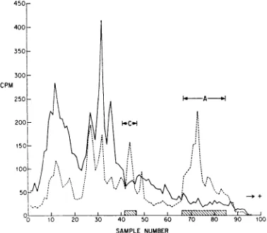
FIG.6.Comparison of pseudorabies virus radio-active structural proteins with proteins extractable with 0.25 N HCIfrom nuclei of virus-infected RK13 cells. Acid-extracted proteins, 3H-arginine(solidline);
virus structural proteins, 14C-arginine (broken line). Peaks I through V, virus-induced proteins.
ing of the proteins would allow us to better assess whetherpeakV waspresent inthe virion. Therefore, reconstituted protein hydrolysate
labeled with 3H or 'ICwas used as the label in
similarexperiments. When proteinslabeled with these materials were analyzed ongels, the label-ing patterns were similar to those with only arginine. Inaddition, itappearedthat peptide V
was relatively rich in arginine, since this peak
was diminished in electropherograms prepared
from proteins labeled with reconstituted protein
hydrolysate (Stevens, unpublished observations).
Thus, the presence or absence ofpeptide V in the virion could not beunequivocally established. However, the results of these experiments
sug-gest strongly thatpeptide Vis not a viral
struc-turalprotein.
Since the principal structural proteins in the virion could be extracted withacid from infected nuclei, the selectivityof the extraction procedure
was questioned. Nuclei isolated from infected
cellslabeledfrom4to 5.5 hrafter infectionwith
14Cj
or 3H-arginine were extracted with 0.25 NHCI (3H-arginine) or PBS ("4C-arginine). These
extracts were analyzed as usual on
polyacryl-amide gels. Results (Fig. 7) show that the acid-extraction procedure is only partially selective ascompared to extraction at neutralpH (peak V andmajor portions of II and IV are insoluble in PBS, the others are soluble to various degrees). Thus, some of the
acid-extractable
proteinsbe-have as ordinary proteins, which are soluble
3,
on November 11, 2019 by guest
http://jvi.asm.org/
[image:6.484.39.232.419.591.2]CPM 100
50-0 10 20 30 40 50 60 70 80 90 100
SAMPLE NUMBER
FIG.7.Electropherogram of radioactive proteins extracted with 0.25 N HCI or PBSfrom nuclei of
pseudorabies virus-infected cells labeled with
radio-active arginine between 4 and5.5 hr after infection.
'4C-labeled 0.25 N HCI extract (solid line);
8H-labeled PBS extract (broken line). Peaks I through
V represent virus-induced proteins.
under neutral conditions, and probably do not
haveanisoelectric pointatbasic pH.
Finally, although the results presented in Fig.
4 indicate that proteins I through V contained tryptophan and, therefore, did not fit the usual
definition forhistone (14), a tryptophan-arginine
double-labeling experiment was performed on
virus-infected cells to establish this
unequivo-cally. Virus-infected cells were labeled between
5 and 7 hr after infection in MEM without
arginine and tryptophan. Thiswas supplemented
with 5,cof 3H-arginineand5 ,ucof
14C-trypto-phanper ml.Acid-soluble proteinswereextracted
and runonpolyacrylamidegels. Allmajor peaks
do containtryptophan(Fig. 8).
DISCUSSION
Thesignificant results of this initial
investiga-tion maybe summarizedasfollows. (i) The
syn-thesis of histones in RK13 cells is completely
inhibitedby6hr after infection withpseudorabies
virus. (ii) Five major virus-induced
acid-extract-able proteins appear in nuclei of virus-infected
cells. Four are virus structuralproteins;onemost
probablyisnot.
Histone synthesis. If histones are involved in thespecific repression ofhost-cellDNAsynthesis
which occurs, one might expect them, either in
mass or in selected cases, to be either induced
tohigherlevels or inhibited aftervirus infection.
Thedata (Fig.4and5) showunequivocallythat
synthesis of all the "classic" histonesiscompletely
inhibited by 6 hr after infection, a conclusion reached independently by Shimono and Kaplan
(16).
Inaddition, the pulse-labeling studies indicate that this inhibition is gradual and generalfor all histone classes. It has been observed by Kaplan and Ben-Porat (9) that the inhibition of host cell DNA synthesis was complete soon after this time. A possible relationship between the in-hibition ofhost-cell DNA and histone synthesis in these experiments is suggested by the work of Robbins and Borun (14), who showed that synthesis of these macromolecules is specifically coordinated in normal HeLa cells. At present, we have no evidence for or against a specific interconnection in this virus-infected system. Note, however, that if there is a coordinate con-trolof DNA andhistone synthesis in the present system, it isspecifically related to host-cell DNA synthesis, since viral DNA synthesis is proceed-ing at a time when histone synthesis is completely inhibited.
Synthesis of other acid-extractable proteins. Finding five virus-induced acid-extractable pro-teins in the nuclei ofpseudorabiesvirus-infected cells led to a preliminary characterization of them. As with histones, they are extracted from cell nuclei with acid, and, since peptides electro-phorese mainly on the basis of size under these conditions (15), it may be concluded that protein Vis in the samegeneralsizerange as the
lysine-rich histone. However, allof thesepeptides differ
from the "classic" histones in that they contain
tryptophan (Fig. 8). Whether, like histones, any
of themis specificallycomplexed to DNA (viral,
cellular,orboth) iscurrently underinvestigation.
500r U
Nz 7
SAMPLE NUMBER
FIG.8.Electropherogram of radioactive proteins
extracted with 0.25 NHClfromnucleiof pseudorabies
virus-infected RKi3 cells labeled with 14C-tryptophan
(solidline) and3H-arginine (broken line) between 5
and 7 hr after infection. Peaks I through V
repre-sent virus-inducedproteins.
--j+
on November 11, 2019 by guest
http://jvi.asm.org/
[image:7.484.53.243.44.217.2] [image:7.484.262.452.402.595.2]It isclear from Fig. 5 that peptides I through
IV are virus structural proteins, whereas V
most likely is not. We are now attempting to
find where these proteins are located in the
virion. However, thefact thatIIIandIVmigrate
with peaks present before infection leads to the
interesting possibility that they may be host-derived proteins selectively produced after
in-fection.Although there is noprecedence for such
a view, this possibility becomes more tenable when it is recalled that themembrane of herpes
viruses contains host-cell antigens (12, 16) and
is derived from the inner nuclear membrane, which reduplicates during infection (4). Unless there is a large pool of such proteins in these cells, this implies that at least some host-cell membrane proteins continue to be made after infection.
Finally, peptide Vis, atpresent, the molecule of greatest interest to us, since, for several reasons, it appears to be the most likely
candi-date fora protein whichmight playadirect role
in inhibition of host-cell DNA replication.
Ex-periments concerned with further characteriza-tion of all these proteins and an assessment of
their possible role ingenetic regulation are now underway.
ACKNOWLEDGMENTS
This investigation was supported byPublic Health Service grantAI-06246 from the NationalInstitute ofAllergyand Infec-tiousDiseases,andbygrantsfrom the Life Insurance Medical ResearchFund, the American CancerSociety,and theCalifornia Institute for Cancer Research.
ADDENDUM IN PROOF
Wehaverecentlyfound(Stevens,unpublisheddata)
that peptides I andV are selectively boundto
chro-matin isolated 6 hr after infection.
LITERATURE CITED
1. Ben-Porat, T., and A. S.Kaplan. 1965. Mechanism of inhibi-tion of cellular DNA synthesis by pseudorabies virus. Virology25:22-29.
2. Bonner, J., M. E. Dahmus, D. Fambrough, R. C. Huang, K. Marushige, and D. Y. H. Tuan. 1968. The biology of isolated chromatin. Science 159:47-56.
3. Bray, G. A. 1961. Asimple efficient liquid scintillator for counting aqueous solutions inaliquid scintillation counter. Anal. Biochem. 1:279-285.
4. Darlington, R. W., and L. H. Moss IIL. 1968. Herpesvirus envelopment. J.Virol. 2:48-55.
5. Eagle, H. 1959.Amino acid metabolism in mammalian cell culture. Science 130:432-437.
6. Fambrough, D. M., F. Fujimura, and J. Bonner. 1968. Quantitative distribution of histone components in the pea plant. Biochemistry 7:575-585.
7. Hamada, C., and A. S. Kaplan. 1965. Kinetics of synthesis of various types of antigenic proteins in cells infected with pseudorabies virus. J. Bacteriol. 89:1328-1334.
8. Holowczak, J. A., andW. K. Joklik. 1967. Studies on the structuralproteins of vaccinia virus. L.Structural proteins ofvirions and cores. Virology 33:717-725.
9. Kaplan,A.S., and T. Ben-Porat. 1963. The pattern of viral and cellular DNAsynthesis in pseudorabies virus-infected cells in the logarithmic phase of growth. Virology 19: 205-214.
10. Levine, A. J., andH.S. Ginsberg. 1968. Role of adenovirus structuralproteinsin thecessation of host-cell biosynthetic functions. J. Virol. 2:430-439.
11.Luria, S. E., and J. E.Darnell,Jr.General virology, 2nd ed. John Wiley &Sons, Inc., New York.
12. Nii, S., C. Morgan, H. M. Rose, and K. C. Hsu. 1968.
Elec-tronmicroscopyofherpes simplexvirus. J. Virol. 2:1172-1184.
13.Penman, S. 1966. RNA metabolism in the HeLa cell nucleus. J. Mol. Biol. 17:117-130.
14.Robbins, E., and T. W.Borun. 1967. The cytoplasmic syn-thesis of histones in HeLa cells anditstemporal relation-ship toDNA replication. Proc. Nat. Acad. Sci. U.S.A. 57:409-416.
15. Shapiro, A.L.,E.Viinuela,andJ. V.Maizel.1967. Molecular weightestimationofpolypeptide chains by electrophoresis in S.D.S.-polyacrylamide gels. Biochem. Biophys. Res. Commun.28:815-820.
16.Shimono, H.,and A. S. Kaplan.1968. Histone synthesis in cellsinfectedwithavirulent or an oncogenic virus. Fed. Proc.27:615.
17.Tankersley, R. W., Jr. 1964. Amino acid requirements of herpessimplex virus inhuman cells.J. Bacteriol. 87:609-613.
18.Wildy, P., and D. H. Watson. 1962. Electron microscopic studiesonthe architecture of animalviruses.Cold Spring HarborSymp. Quant.Biol. 27:25-47.
on November 11, 2019 by guest
http://jvi.asm.org/