DISSERTATION ON
VALIDATING ALVARADO SCORING IN THE DIAGNOSIS OF ACUTE APPENDICITIS
M.S. DEGREE EXAMINATION BRANCH-I
GENERAL SURGERY
GOVERNMENT KILPAUK MEDICAL COLLEGE THE TAMILNADU DR.MGR MEDICAL UNIVERSITY
CHENNAI
BONAFIDE CERTIFICATE
This is to certify that this dissertation titled “VALIDATING ALVARADO SCORING IN THE DIAGNOSIS OF ACUTE APPENDICITIS” is the bonafide record work done by Dr. S.I.MOHAMMED SIKKANDER BASHA, submitted as partial fulfillment for the requirements of M.S. Degree Examinations Branch I, General Surgery, April 2014.
Prof.R.Kannan ,M.S Prof.P.N.ShanmugaSundaram,M.S Dissertation Guide and Unit Chief Head of the Department,
Department of General Surgery General Surgery,
Government Royapettah Hospital Kilpauk Medical College Hospital Kilpauk Medical College Chennai.
Chennai.
Prof.P.Ramakrishnan ,M.D,DLO DEAN
TAMILNADU DR MGR MEDICAL UNIVERSITY Chennai
DECLARATION
I, Dr. S.I.MOHAMMED SIKKANDER BASHA, solemnly declare that the dissertation submitted on the topic “VALIDATING ALVARADO SCORING IN THE DIAGNOSIS OF ACUTE APPENDICITIS” is a bonafide work done by me from Jan 2013 to December 2013, towards partial fulfillment of the requirements of M.S Degree examinations, General Surgery, April 2014.
Chennai Dr.S.I.MOHAMMED SIKKANDER BASHA
ACKNOWLEDGEMENT
I sincerely thank Prof.P.Ramakrishnan MD, DLO, the Dean, Kilpauk Medical College for granting me permission to carry out and successfully complete my dissertation work. I consider it a privilege to have done this study under the supervision and guidance of Prof.R.Kannan M.S, who was a constant source of inspiration and guidance.
Am indebted to Prof R.A.Pandyaraj MS,FRCS , Prof.R.V.Suresh MS , Prof. Affee Asma MS,DGO and Prof.V.Sathish ,M.S, for helping me through the process of completing my dissertation. Their invaluable advice has helped me complete the study on time.
I would also like to thank Dr. Rosy Adhaline Selvi MS, DGO, Dr.Princess Beulah MS, for their valuable support and encouragement throughout the period of study. I would like to thank all my colleagues Dr.Ahila, Dr.Vinodh , Dr. Abilash , Dr.Varun , Dr.Geetha , and the house surgeons Dr.Siddharthan , Dr.Sivaraman who were instrumental in bringing up this study.
LIST OF ABBREVATIONS USED
D.D. - Differential Diagnosis
HPE - Histopathological Examination USG - Ultra Sound
WBC - White Blood Cells RIF - Right iliac fossa
NMR - Nuclear Magnetic resonance
Hb - Haemoglobin
TLC - Total leucocyte count
ABSTRACT Introduction:
Acute Appendicitis is a common and sometimes confusing cause of acute abdomen at all age groups. Diagnosis of appendicitis can be difficult, occasionally taxing the diagnostic skills of even the most experienced surgeon. Despite increased use of USG, CT, the rate of misdiagnosis of appendicitis has remained the same (15.3%).
Objectives:
To review the usefulness of Alvarado score & to evaluate its feasibility and value as an aid in surgical decision making and to reduce the number of negative laparotomies.
Materials and Methods:
100 patients who were admitted to Government Royapettah Hospital from Jan 2013 to Dec 2013 with clinical suspicions of acute appendicitis were included in the study. The modified scoring system is based on 3 signs, 3 symptoms and 1 laboratory finding. The patient was classified as males, females and children (< 12 years). These were further grouped based on the scores 7 - 9, 5-6 and <5.
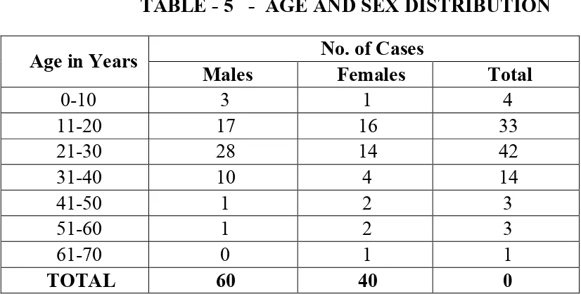
Observation & Results: In our study 52 were males,
Males with score 7-9 were 39 Females with score 7-9 were 14 Children with score 7-9 were 10 Males with score 5-6 were 08 Females score 5-6 were 18 Children Score 5-6 were 0
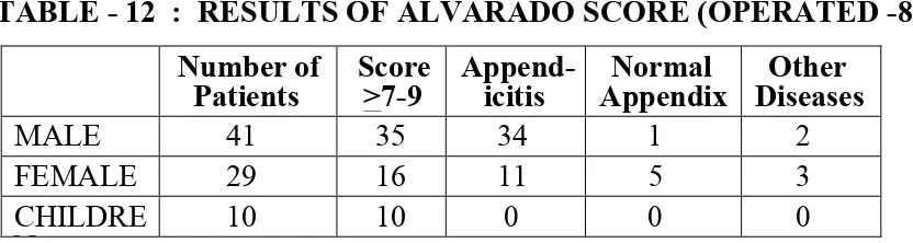
A total of 80 patients with score of 7-9 and 5-6 were operated. Among males with score of 7-9, 35 patients were operated and 34 were found to have inflamed appendix.
Females with score 7-9, 16 were operated and 11 were found to have inflamed appendix.
Conclusion:
Alvarado score significantly reduces the number of negative laparotomies without increasing the overall rate of appendicular perforation. It is very effective in Men & Children but diagnostic laparoscopy or ultrasonography is advised to minimize the high false negative rate in women.
Keywords:
TABLE OF CONTENTS
Sl.
No. CONTENTS Page No.
1. INTRODUCTION 1
2. AIMS AND OBJECTIVES 7
3. REVIEW OF LITERATURE 7
4. METHODOLOGY 73
5. RESULT 76
6. DISCUSSION 89
7. CONCLUSION 92
8. SUMMARY 94
9. ANNEXURE - I : BIBLIOGRAPHY 97 10. ANNEXURE - II :
PROFORMA
KEY TO MASTER CHART MASTER CHART
PLAGIARISM
LIST OF TABLES Sl.
No. CONTENTS Page No.
1. The Alvarado Score 5
2. Modified Alvarado Score 6
3. Various Positions of Appendix 16
4. Cases Operated 76
5. Age and Sex Distribution of cases 76
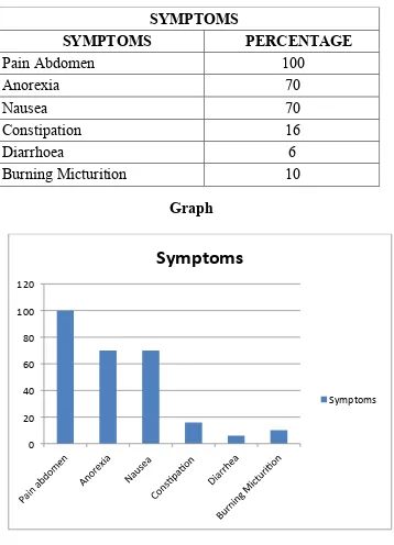
6. Symptoms 78
7. Site of Pain 79
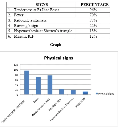
8. Physical Signs 80
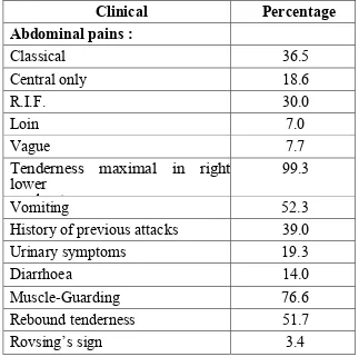
9. Symptomatology & Physical signs by Campbell and Mc Phail series
81
10. Total leucocyte count 82
11. Results of Alvarado Score (total cases in study) 84 12/12a Results of Alvarado Score (Operated cases) 85
13/13a Statistical Data Results 86
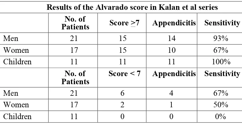
14. Results of Alvarado score in kalan et al series 87
15. Post operative complications 87
LIST OF FIGURES Sl.
No. CONTENTS Page No.
1. Anatomy of the Appendix and the Ileocaecal Region 14
2. Various Positions of Appendix 15
3. Histology of normal Appendix 19
4. Mc Burney’s Point 57
5. Steps in Open Appendectomy 60
6. Operative Photographs 72
INTRODUCTION
Acute appendicitis is acute inflammation of the appendix. It is a
common, sometimes confusing and often treacherous cause of acute
abdomen at all age groups. Of all the abdominal emergencies, acute
appendicitis heads the list of causes classified under acute abdomen.
It is not surprising that the diseases of appendix do not seem to
have a place in areas of active clinical investigation and one finds,
relatively few articles dealing with appendicitis. But no one in current
surgical practice can deny the fact that appendicitis still represents a
large portion of cases and they continue to baffle them by their
oft-deceptive presentations and sometimes may cause quite an amount of
morbidity and unnecessary mortality.
Acute appendicitis is commonly caused due to a variety of
reasons namely difference in dietary habits, food adulterations,
indulging in mixed diet habits, seasonal changes particularly colder
periods. Acute appendicitis is prevalent among males and females
irrespective of age factor but is noted in slightly large numbers among
males and rarely found in infancy and old age
The etiology of acute appendicitis is plenty among which
obstruction to lumen and infection play an important role.
The diagnosis of appendicitis is quiet difficult, occasionally
testing the diagnostic skills of even the most experienced surgeon.
similarly, the decision making in the management of patients with
inflammation or abscess of the appendix can be a task .Patients with
appendicitis first recognizes that they have a episode of pain which
is unique and then presents to the physician who diagnoses the
condition. Delay in diagnosis arise from errors on the part of the patient
or physician, and all delays complicate the disease.
The severity of acute appendicitis is in the frequency with the
peritoneal cavity which is infected from the same focus, either
by perforation or by transmigration of bacteria through the
appendiceal wall. The classic triad of a history compatible with acute
appendicitis, is pain at the McBurney’s point and leucocytosis has
diagnostic accuracy rate of less than 80 percent and even after the
invention of advanced radiological techniques such as
ultrasonography,computed tomography,magnetic resonant imaging or
radionuclide scanning are , the accuracy usually does not reach
more than or equal to 90 percent.
Patients presenting with acute right iliac fossa pain remain a
diagnostic challenge. Acute appendicitis is the most common
indication for surgery in these patients. After careful clinical
evaluations and observations surgical intervention is undertaken.
appendicitis and the prevalence of diseases that mimic it. Migrating
pain, and involuntary guarding and persistence or progression of
clinical signs are the main criteria favouring the operation.
The lifetime rate of appendectomy is 12% for males and 25% for
females, with almost 7% of the total population undergoing
appendectomy for acute appendicitis. The rate of appendectomy has
remained constant at 10 per 10000 per year. Appendicitis is almost
frequently found in their second to fourth decades of life with a mean
age of 31.3 years and a median age of 22 years. There is a mild male to
female predominance , M:F 1.2 TO 1.3:11,2
Despite an increased use of ultrasonography, computerized
tomographic scanning and laparoscopic techniques, the rate of
misdiagnosis of appendicitis has remained constant at a rate of
15.3%, at par with the rate of appendiceal rupture. The rate of
misdiagnosis of appendicitis is markedly high among women than
men,22.9 to9.3%. The negative appendicectomy rate for women with
reproductive age group is 23.2%, with the highest rates identified in
women among 40 to 49 years of age. The highest negative
appendicectomy rate is reported for a women who was older than 80
years of age.2,3,4.
The morbidity and mortality rates associated with acute
increases 15 fold and mortality rate is 50 folds greater. Appendiceal
perforation is a rare cause of tubal infertility (Mueller et al 1986).
The topmost priority and the aim of the surgeons should be to
avoid perforation at any cost . The cost of which is the high rate of
apendicectomy with histologically normal appendix, which is very
expensive, and it almost has a complication rate which is not much
lower after removal of a pathological appendix (Nase ET al 1980:
Arnborgnsson 1985) The surgery for negative appendix is associated
with the usual spectrum of immediate post-operative complication in
up to 15% of patients (Lewis et al 1975).Some patients may have
complications such as small bowel obstruction and incisional hernia.
A scoring system described by Alvarado5 was formulated to
track down the negative rate of appendicectomy with no significant
increase in morbidity and mortality.
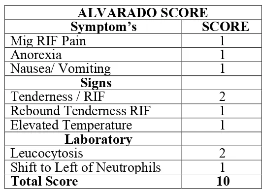
THE ALVARADO SCORE
Alvarado in 1986 formulated a scoring system for diagnosing
acute appendicitis. The scoring system developed by Alvarado has
[image:15.595.162.428.208.403.2]3 symptoms, 3 signs & 2 laboratory findings.
Table – 1
ALVARADO SCORE
Symptom’s SCORE
Mig RIF Pain 1
Anorexia 1
Nausea/ Vomiting 1
Signs
Tenderness / RIF 2 Rebound Tenderness RIF 1 Elevated Temperature 1
Laboratory
Leucocytosis 2
Shift to Left of Neutrophils 1
Total Score 10
According to the scoring system, patient who has a score of
1-4 were not considered probably to have acute appendicitis. Those
patients who has a score of 5-6 were considered to have a probable
diagnosis of acute appendicitis but not convincing enough to advice to
undergo immediate surgery and these were marked for further review.
Those patients who has a score of 7-8 were considered to have a
possible diagnosis of acute appendicitis & those patients who has a
score of 9-10 were considered to have an almost definitive diagnosis of
appendicitis and posted for surgery. The score can be varying on
re-examination. The lab finding of leucocytosis is defined as white
MODIFIED ALVARADO SCORE :
Alvarado Scoring was modified by M.Kalan, D.Talbat, W.J.
Cunliffe and A.J. Righ (1994). The modified Alvarado score
excludes one laboratory finding; the left shift to neutrophil maturation.
This laboratory parameter was excluded as it was not available on a
routine basis in the laboratories. The patients were therefore scored for
9 in contrast to 10 points.
Table – 2
MODIFIED ALVARADO SCORE
Symptom’s SCORE
Mig RIF Pain 1
Anorexia 1
Nausea/ Vomiting 1
Signs
Tenderness / RIF 2 Rebound Tenderness RIF 1 Elevated Temperature 1
Laboratory
Leucocytosis 2
Total Score 9
Other scoring systems recently introduced for diagnosis of
acute appendicitis are OHMANN, ESKELINEN, De-DOMBAL
score. The sensitivity and specificity of the OHMANN score in
diagnosing acute appendicitis is 63% and 93% the positive predictive
value was 77%. The sensitivity and specificity of
the ESKELINEN score in the diagnosis of acute appendicitis were 79%
and 85%. The positive predictive value was 65%. These scores had
different criteria for diagnosis, which make them less feasible in
AIMS AND OBJECTIVES
The aim of this study is to review and validate the use of
Alvarado scoring and to evaluate its feasibility and value as an
decision-making tool in cases of possible acute appendicitis in reducing
the number of negative laparotomies.
In this prospective study, 100 patients with a provisional
diagnosis of acute appendicitis admitted to Government Royapettah
Hospital, Chennai were studied.
The patients diagnosed to have acute appendicitis in view of
end score obtained by modified Alvarado scoring system were either
observed or operated according to the cut off point. The results so
obtained were studied in relation to the world literature available.
Results of operative measures, conservative measures, and
histopathological examination were reviewed.
HISTORICAL REVIEW
Review of ancient texts have various descriptions of surgery
being done for ailments mimicing like appendicitis,Claudius
Amyand, draws all credit for performing the first open
appendicectomy a surgeon at St Georges hospital in London.He
operated on a 11 year old boy in 1736 with scrotal hernia and fecal
fistula.Within the hernia sac, He found the appendix perforated by a pin.
Appendix was not considered as an organ which can cause
disease until the end of the 19th century. Royal Academy of medicine
in Paris witnessed the first paper presentation,by Louyer villermay
in 1824 and reported appendicitis on two autopsy specimens and
emphasized this condition to be important .
Villermay’s work was expounded by Francois Melier, a French
physician in 1827. He reported 6 autopsy cases of acute appendicitis
and was first to suggest the ante mortem recognition of appendicitis6.
This work was discounted by many physicians of the era, including
Baron Guillaume Dupuytren.
Dupuytren believed that inflammation of the caecum was the
main cause of pathology of the right lower quadrant . The term
typhlitis or perityphlitis was used to describe the right lower
quadrant inflammation. In 1839 a textbook authored by Bright and
Adisson titled elements of practical medicine described the symptoms
of appendicitis and identified the primary cause of inflammatory
process of the right lower quadrant 7Reginald Fitz, a professor of
pathologic anatomy at Harvard, is credited for coining the term
appendix. His landmark paper definitively identified the appendix as
the primary cause of right lower quadrant inflammation8. Initial
surgical therapy for appendicitis was primarily designed to drain the
right lower quadrant abscesses that occurred secondary to appendiceal
It appears that Hancock made the first surgical treatment of
appendicitis or perityphlitis without abscess in 1848. The first
published account of appendectomy for appendicitis was by
Kronlein in1886. Fergus in Canada, performed the first elective
appendectomy in 18836.
The greatest contributor to the advancement in the
treatment of appendicitis is Charles McBurney. In 1889 , he
published his landmark paper in the New York medical journal
describing the indications for early laparotomy for the treatment of
appendicitis. It is in this paper that he described McBurney’s point of
maximum tenderness9. Mc Burney subsequently published a paper in
1894 describing the incision that bears his name. However, Mc
Burney later credited Mc Arthur with first describing this incision 10.
Semm is widely credited with performing the first successful
laparoscopic appendectomy in 198211.The surgical treatment of
appendicitis is one of the great public health advancements of the last
150 years. Appendectomy for appendicitis is the most
commonly performed emergency in the world. Additionally,
appendicitis is a disease of the young, with 40% of the cases occurring
in patients between the ages of 10 and 29 years12. Fitz reported the
associated mortality rate of appendicitis to be at least 67% without
appendicitis is reported to be less than 1% 13. In US, more than
2,60,000 appendectomies are performed each year. It is assumed
that there is a 15% negative appendicectomy rate and a median life
expectancy of 80 years , development of surgical therapy of
appendicitis results in saving approximately 8 million lives per year in
the US alone.
In 1902, Sir Frederick Treves operated upon king Edward
III for appendicitis successfully few days before his coronation. It was
one of the most famous cases of appendicitis and did much to
popularize the operation. An attempt to sterilize the appendiceal stump
with chemicals or cautery became popular early and is still employed
by some surgeons. Increased understanding of the pathophysiology
of peritonitis, fluid resuscitation and antibiotic therapy in the 1940’s
decreased the mortality rate. In 1910 Albert Ocshner and James
Sherren advocated the conservative line of management for
appendicular mass.
In 1965, Brooke and Keller described radiologic signs in
acute appendicitis on plain X ray abdomen. In 1978 Gastro reported
3 examples of pneumoperitonium. Haker D A et al described the
use of laparoscopy in the diagnosis of acute appendicitis in young
women. Jeffrey et al gave an account of the role of ultrasound in the
diagnosis of acute appendicitis. He studied 250 cases of acute
elicited a sonographic Mc Burney’s sign of maximum tenderness on
probing.
Alvarado A in 1986 described Alvarado scoring as a practical
tool for diagnosis of acute appendicitis.
Puylaert in 1986 evaluated 60 patients with acute
appendicitis using graded compression with a higher frequency linear
array transducer and reported a specificity of 89%.
DEVELOPMENT OF APPENDIX
Appendix develops as an under developed distal end of the
caecum in the sixth week of intrauterine life. Appendix develops from
the post arterial segment of the midgut, along with caecum,
ascending colon and right two third of the transverse colon. Initially
a bud called caecal bud arises from the post-arterial segment very
near to the apex of the loop. The proximal part of the bud grows
rapidly to form the caecum but the distal part remains narrow and
forms the appendix.
Subsequently , the lateral or right wall of the caecum grows
much more rapidly than the medial wall. Thus the point of attachment
of the appendix comes to lie on the postero-medial aspect of the
caecum.
The caecum lies just below the liver and the ascending colon
iliac fossa and the ascending, transverse and descending parts of the
colon become distinct.
In the final stage, the duodenum, ascending colon and the
descending colon become retroperitoneal by the fusion of their
mesenteries with the posterior abdominal wall. But the mesentery of
the small intestine, transverse colon, sigmoid colon and appendix
remains.14,15.
CONGENITAL VARIATIONS 16
Congenital agenesis.
Collins collected 57 cases of true agenesis of appendix
Duplication or triplication
In 1968 Tinckler reported on operating on a triple appendix on a
12 months old male chinese child with other congenital anomalies.
Variations in positions.
Wall Bridge classified duplication of vermiform appendix as:
Type A
Single appendix and single caecum exhibiting partial duplication in
various degrees
Type B
Type B-1
Bud like - two appendix placed symmetrically on either side of the
Bauhin’s valve.
Type B-2
Taenia Colic type - one appendix from the usual site, and the other
from the caecum above the lining of taenia at varying distance from the
first.
Type C
Double caecum each bearing an appendix
VARIATIONS IN POSITION:
• Due to incomplete downward descent of caecum, the appendix
may remain in sub hepatic position.
• Due to overgrowth of ascending colon, appendix may sometimes
descend down to a pelvic position along the caecum.
• Due to incomplete or non-rotation of the midgut loop, appendix
may assume a position on the left side of the abdomen. This may be
associated with transposition of viscera.
• Caecum may have a long mesentery and may be mobile. Because
of its mobility, appendix may assume a variable position in the
ANATOMY 17
Fig : Anatomy of the Appendix and ileo-caecal region
Vermiform appendix is described as a narrow, vermion
(worm-shaped) tube arising from the posteromedial wall of caecum or
anywhere below the ileal end.It constantly arises from the site at which
the 3 teania coli converge. It has no constant anatomical position.
The three taenia coli merge into a complete longitudinal muscle layer
over the appendix. The anterior taenia is usually distinct and traceable
to the appendix, offering a guide to it.
Appendix differs from 2 -20cm in length, the average length is
about 9cm. Appendix being longer in children, may atrophy or reduce
POSITIONS:
[image:25.595.104.471.77.264.2]
Fig : Various Positions of Appendix
Treves described the following anatomical types comparing the
appendix with the face of the clock.
11’O Clock Para colic (lies on the sulcus in the lateral aspect of the
caecum).
12’O Clock retrocaecal (lies behind the caecum and may even be
totally or partially retrocaecal)
1’O Clock Pre-ileal
2’O Clock Post- ileal
3’O Clock Promontoric (the tip of the organ points towards the
promontory of the sacrum)
4’O Clock Pelvic (Appendix dips into the pelvis)
6’O Clock Subcaecal or mid inguinal
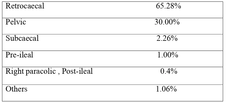
Incidence according to Wakely, following a study of 10,000 cases is as
Table - 3 : Positions of Appendix
Retrocaecal 65.28%
Pelvic 30.00%
Subcaecal 2.26%
Pre-ileal 1.00%
Right paracolic , Post-ileal 0.4%
Others 1.06%
MESENTERY OF APPENDIX.
The appendix has a complete peritoneal investment and a small
mesentery. This fold is derived from the left leaf of peritoneum and
is a continuity of the mesentery. It is triangular in form and is
attached along the whole length of the appendix.
BLOOD SUPPLY:
•Arterial:
Appendicular artery is a branch of lower division of the
ileocolic artery, runs behind the terminal ileum to enter the
mesoappendix a short distance from the appendicular base. Here it
gives off a recurrent branch, which anastomoses at the base of the
appendix with a branch of the posterior caecal artery.
The terminal part of the main artery lies on the wall of the
gangrene or necrosis. Variations are considerable. In nearly 50% of
the cases there is an accessory appendicular artery, a branch of
posterior caecal artery (after Seshachalam).
•Venous:
Appendicular vein is a radicle of the ileocolic vein. which drains
into the portal system.
•Lymphatics:
From the lymphatic follicles through the muscle wall drain into
nodes in the mesoappendix. These drain into the paracolic nodes lying
along the ileo-colic artery and then to the superior mesenteric group
NERVE SUPPLY:
• Sympathetic: Coeliac and superior mesenteric ganglia (T11, T12)
• Parasympathetic : Vagus.
Both these nerves form the plexus around the artery supplying the
appendix.
PARA- APPENDICEAL FOSSA:
Peritoneal folds near the base of the appendix are sometimes
found. Superior ileocaecal recess- opens medially and downwards just
above the terminal part of the ileum. It is bounded in front by the
vascular fold of the caecum, which contains the anterior caecal
caecum and ascending colon, Posteriorly by terminal ileum and its
mesentery.
Inferior ileocaecal recess opens downwards and medially
below the terminal ileum. Its anterior wall is formed by the
bloodless fold of Treves extending from the lower border of the
ileum to the caecum and anterior surface of the mesoappendix. Its
posterior wall is formed by mesoappendix.
Retrocaecal recess:- lies behind the caecum, bounded anteriorly by the caecum, posteriorly by parietal peritoneum, and on
each side by caecal folds of peritoneum.
SURFACE MARKING:
The base of the appendix corresponds to the Mc Burney’s
point.It is formed by the junction of lateral one-third and medial two -
third of the line joining the umbilicus with the anterior superior iliac
spine. It is only a surgical approximation with considerable variation.
LUMEN OF THE APENDIX:
It is a small canal which opens into the caecum through an
orifice lying below and a slightly behind the ileocaecal opening. A
semilunar mucosal fold forming a valve guards the orifice. The
Crypts are present but are not numerous. In the base of crypts
lie the special cells (kultschizsky cells) , which give, rise to
carcinoid tumours, and they can cause appendicitis.
HISTOLOGY18
Fig : Histology of Normal Appendix
The structure of the appendix is serosa , muscularis mucosa ,
sub mucosa , mucosa and lumen.
• Serosa: Is a complete covering except along the
mesenteric attachment.
• Muscularis layers : Longitudinal muscular fibres form a
complete uniformly thick layer, except over a few small areas where
both muscular layers are deficient leaving serosa and sub mucosa in
contact. At the base the longitudinal muscle thickens to form the
rudimentary taeniae. The circular muscle fibres form a thicker layer
separated by connective tissue.
• Sub mucosa: Contains number of lymphoid mass , causing the
Profused number of lymph tissue has promoted the description of
‘abdominal tonsil’ for the appendix.
• Mucosa: Is covered by columnar epithelial cells and attenuated
antigen transporting ‘M’ cells. Glands are few penetrating deeply into
lymphoid tissue. Lymphoid tissue in the lamina propria contains many
plasma cells with lymphocytes, eosinophils and leucocytes, mast cells,
macrophages are embedded in fibrocellular reticulum.
In many mammals, particularly herbivores the caecum and
appendix are large and constitute a highly important site of digestion
of cellulose by symbiotic bacteria.
ETIOPATHOGENESIS 19-25
Etiological factors are predisposing and exciting factors.
1. PREDISPOSING FACTORS :
• Age:- Commonest in the 20 t 30 years of age . Rare in infants and
old aged . In infants the lumen of the appendix is relatively large. In
older age group appendix undergoes involution commonly.
• Sex: Males preponderance common. Before puberty ratio is 1: 1 ,
post pubertal 2:1 till 25 years.
• Race and diet : Appendicitis being common in high
socio-economic status and certain communities, is rarely seen in low
who live on a diet abundant in cellulose are immune to the disease,
but when they adopt the diet of other civilizations they lose that
immunity. The severe gangrenous type of appendicitis is confined to
people having meat in their diet. Acute appendicitis occurs in life
long vegetarians and even in babies at the breast. Denis P Burkitt after
extensive research concluded that undue refining of dietary
carbohydrates is the most important causative factor.
• Social Status: more common among the upper and middle class
than in those belonging to the so called working class.
• Familial susceptibility: This unusual but generally accepted
fact can be accounted for by a hereditary abnormality in position
of organ, which predisposes to infection . Thus the whole family may
have a long retrocaecal appendix with comparatively poor blood
supply.
2. EXCITING FACTORS:
• Obstruction : Wangenstein and Bowers discovered obstruction in
72% of cases with acute suppurative appendicitis and in cases with
gangrenous appendicitis it was 100%. When obstruction sets in, the
lumen becomes distended, and the intraluminal pressure is rapidly
increased and the venous return is interfered, leading to the rupture of
the vessels, haemorrhage occurs when the wall is hypoxic and when
result it ends up in perforation. In an experimental study in dogs
Wangenstein and Bowers found that obstruction without infection
or infection without obstruction did not cause inflammation. In man it
appears that pressure distension is the exciting factor, bacterial invasion
of the injured wall being secondary. According to this view acute
appendicitis is a form of closed intestinal obstruction.
• Infection: It occurs as a secondary mechanism to mucosal
destruction. Cultures usually show a wide range of pyogenic
organisms. Commonly seen organisms are mixture of Esch.coli
(85%), Enterococcus (30%). Non-anaerobic bacteroids and Esch.coli
are the more frequent. Organisms that commonly infect are the
normal inhabitants of the appendiceal lumen. Blood spread from
thoracic infection, tonsils and other regions is quiet possibile, in some
circumstances there is history of preceding sore throat before the onset
of an acute attack.
PATHOLOGY 22,25
Acute appendicitis can present in various distinct forms. An
histologic criteria for diagnosis of appendiceal inflammation is
polymorphous leucocytic infiltration of muscularis mucosa.
CATARRHAL APPENDICITIS:
There is usually mild Inflammation and symptoms.
granular red membrane. There is congestion of subserosal vessels.
Mucosa and submucosa are inflamed. Mucosal ulcers will be seen
usually. There is no obstruction of the lumen.
• Micro: Scanty neutrophilic exudates seen all over the mucosa,
submucosa and muscularis.Scanty perivascular neutrophilic migration.
ACUTE SUPPURATIVE APPENDICITIS:
Inflamation is severer and purulent. Usually bottom of the crypts
gets infected first. From there, it spreads in to the loose submucosa
then muscularis,then to the vessels penetrating into the serosal layer.
• Gross: Lengthened and oedematous and erythematous
appendix, with dilated sub peritoneal vessels and fibrous or purulent
exudates on the surface. Yellowish spots are seen on the surface
pointing the formation of an abscess. Tip is usually edematous and
the entire pathology is less marked in the proximal and highly marked
in the distal part. A cause of obstruction is almost present and is
usually a lethal variant.
• Micro: There is congestion on all coats of the appendix with
oedema and infiltration of polymorphonuclear leucocytes, and the
mucosal membrane can exhibit little or nil infiltration. Ulcer
formation is usually seen as a result of mucosal necrosis and dead
lymphoid follicles approach the surface. Haemorrhage are seen
scattered in the core coat - acute haemorrhagic appendicitis.
At any stage of the inflammation perforation is prone, but is
ucommonly associated with gangrene. Mucosal ulcers may penetrate
the mucosal and serosa layers leading to perforation. A faeco-lith is
commonly present at the site of the perforation and plays a pivotal
part in its production. It can slip into the cavity of abdomen
occasionally . A full blown peritonitis is seen as a result of perforation
in to the peritoneal cavity.
GANGRENOUS APPENDICITIS.
It is an advanced stage of acute appendicitis. There is death
and putrefaction of tissues of the appendix either local or general, due
to interference with blood supply due to kinking or stricture of the
appendix or thrombosis of the vessel that is traversing in the lumen of
the appendix.
RECURRENT APPENDICITIS
Chronic inflammation of the appendix is usually a task for
the pathologist to diagnose. Chronic appendicitis per se does not
exist. Patients present when there is recurrence of the disease .
• Gross: oedema and fibrosis of the appendix lumen is seen and may
• Micro: Infiltration of mononuclear cells in to the wall,
especially in the submucosal layer and is often seen in association
with huge lymphoid follicles. Mucosa and submucosa are infiltrated
by large number of lymphocytes. Majority of the appendix labelled as
chronic appendicitis are almost usually examples of healing after an
acute attack.
APPENDICITIS OBLITERANS:
Atrophied and degenerated appendix with no possible
relation for inflammation of appendix.
EFFECT OF APPENDICITIS ON ILEOCAECAL REGION:
Inflammatory reaction in the ileocaecal region may lead to
oedema, acute adhesions and angulation of the ileum leading to
obstruction of the bowel, organic obstruction merging with paralytic
ileus.
CLINICAL FEATURES 22-27
The physical signs of acute appendicitis are not specific but
merely those produced by local peritoneal irritation in the right iliac
fossa.
SYMPTOMS:
Patient presenting with acute inflammation of appendix can
but it is not always so. Atypical presentations are not uncommon.
• Pain: In acute appendicitis, when there is inflammation of the
appendix and increase in the intraluminal pressure, the sympathetic
nerves are stimulated.
The “visceral” pain which is initially felt around the umbilicus
and lower epigastric regions, is a moderate to severe, diffuse pain.
Intermittent cramps can be felt occasionally. Because of the
embryonal origin of appendix, pain is usually confined to the midline.
This pain usually last for around 1 to 12 hours, and when the
inflammation extends and involves the serosal layer, the somatic
nerves of the peritoneum are stimulated and then a constant and
continuous pain is felt in the right iliac fossa- “Somatic Pain” In case of
obstruction, experience of colicky pain is common. In gangrenous
appendicitis pain is experienced in initial stages, and there is
destruction of nerve endings at a latestage. Once perforation occurs,
and infection is not controlled by local or general factors, constant
severe and generalized abdominal pain of diffuse peritonitis occurs.
• Atypical pain : appendix in malrotated gut, produces somatic
pain in left lower abdomen. A long appendix may also produce it
with its tip on the left part of the abdomen.
• Vomiting, nausea, and anoreia : Vomiting is usually seen in the
initial pain due to protective pylorospasm. Nausea is usually the
major complaint than vomiting as many patients do not experience .
In most of the cases dyspepsia is a common feature.Loss of
appetite or repulsion for food may be regarded as a lesser degree of
the same sensation and often of equal value in diagnosis. An individual
with previously good health when suddenly loses appetite and
complaints of pain abdomen needs to be carefully monitored.
The degree of nausea and the frequency of vomiting in the
early stage appear tobe depend on two factors - one the amount of
distension of the inflamed appendix, and two the reflux nervous
susceptibility for the patients. Vomiting is more prone to occur in
kids, or in patients in whom the alimentary tract is easily deranged.
It may be taken as an important general rule that the frequency and
severity of the vomiting at the onset of an attack of appendicitis
indicate the degree of distension of the appendix and consequently the
immediate risk to the patient that perforation may occur. Persistent
regurgitant vomiting occurs with diffuse peritonitis.
•Bowel disturbance: Constipation is common. Diarrhoea can occur
in pre or post ileal positions of the appendix because of the irritation of
the distal ileum. Pelvic abscess can irritate the distal gut leading to
•Urinary disturbance:Irritation of the ureters by the retrocaecal
appendix may give rise to pain mimicking right ureteric colic.
Increased frequency of micturition, hematuria or dysuria can occur
due to the irritation by the inflamed pelvic appendix.
SYSTEMIC MANIFESTATIONS:
• Fever: fever i.e is of low grade is not uncommon. When fever sets
in before the abdominal pain or is of high degree, the diagnosis is
questioned.Tongue is coated. Tachycardia and mild dehydration are
seen. In perforated appendix, full blown peritonitis results.
• Local signs: If the appendix is anteriorly placed, physical
signs are elicitable over the anterior abdominal wall. There may be
mild restriction of movement of the right lower abdomen with
respiration.
• Tenderness: is usually elicitable over the Mc Burney’s point
• Guarding and rigidity: Guarding is an protective involuntary
mechanism. True and false guarding must be sort out. Guarding is
commonly present in the right iliac fossa and lower abdomen. Rigidity
is seen when there is peritonitis.
• Muscular rigidity is seen when appendix comes in contact with
the muscle .
CLINICAL TEST DESCRIBED WITH REFERENCE TO APPENDICITIS.
• Mc Burney’s sign: Tenderness during palpation over Mc burney’s
point, where the appendiceal base is said to be situated.
• Rovsing’s sign: Pressure on the Lif produces pain over right lower
quadrant of abdomen. It was primarily thought to be due to shift of gas
in the colon into the caecum, distending it, and the inflammatory
phlegmon around. But Williams proved that this sign is positive in
inflamed lesions of any organ in right lower abdomen. This sign is
probably due to the shift of the coils of ileum from the left iliac
fossa to right iliac fossa, where there is local peritonitis .
• Blumberg’s sign: Eliciting rebound tenderness over Rif , after
deep palpation.
• Psoas test : It is because of irritation of the psoas muscle seen in a
inflammed retrocaecal appendix, patient lying on his left side when
extends his right thigh, exhibits pain.
• Cope’s obturator test: When the right thigh is flexed and
internally rotated, if the appendiceal inflammation is in contact with
obturator muscle, pain is felt over hypogastric region.
• Hyperasthesia in Sherren’s triangle : line adjoining the umbilicus,
abdominal wall or by stroking with a sharp object elicit this. Some
clinicians regard presence of hyperesthesia in Sherrens triangle as a
good guide in the diagnosis of acute appendicitis before perforation.
If, in such a case hyperesthesia disappears later on, it indicated
the bursting of the gangrenous appendix.
• Baldwin’s test: Finger locates the tenderest spot over flank,
compressing it slightly just enough to cause little pain. Patient is
adviced to lift his right leg few inches off the bed, holding the knee
stiff. When patient promptly complaints pain or drops the leg with pain,
test is considered positive. It indicated retrocaecal appendicitis.
• Pointing test: In acute appendicitis patient points to right lower
abdomen pain on coughing, pointing to the site of inflammation. Its
due to the irritation of parietal peritoneum by the inflamed organ.
• Auscultation: Bowel sounds are usually normal. Hyper peristaltic
sounds are heard when there is an element of intestinal obstruction.
Tinkling sounds are heard once paralytic ileus occurs secondary to
generalized peritonitis.
• Pelvic examination: Differential tenderness on the right side, is
significant in the cases of pelvic position of the inflamed organ. In case
VARIATIONS IN CLINICAL PRESENTATIONS ACCORDING
TO THE VARIOUS POSITIONS OF THE APPENDIX.
• Retrocaecal appendix: In this position, because of the
intervening caecum between the inflamed appendix and the anterior
abdominal wall, rigidity is not marked. Tenderness may be elicited
over the right flank, as also is the rigidity (Baldwin’s test). Psoas test
may be positive.
• Subcaecal appendix : Appendix is curled up below the caecum
and is in contact with the iliacus muscle and so, the extension of
the hip becomes painful due to spasm of the muscle, and pain is felt in
the hypogastrium.
• Pelvic appendix: When the appendix is almost entirely in the
pelvis, the clinical signs may be absent over the anterior abdominal
wall. Often, even the Mc Burney’s sign may be negative. Pelvic
examination detects tenderness on the right side. Obturator spasm
may be present rarely. Patient may have symptoms of strangury or
dysuria or tenesmus, due to irritation of the urinary bladder or rectum.
• Sub hepatic appendix : Due to its undescended position, the
clinical signs are referable to the right upper abdomen. A mild
degree of jaundice and very rarely hematemesis may occur due to its
• Retroperitoneal appendix: A retrocaecal appendix is totally
retroperitoneal organ. In these cases, tenderness over the abdomen may
be absent. Tenderness over the loin may be present. Hematuria is
known to occur in nearly 50% of these cases. Several complications
like retroperitoneal or sub diaphragmatic abscess, empyema, bronchial
fistula, psoas abscess, etc., are known to occur.
• Post ileal or retro mesenteric appendix: Because of its
position, inflammation of appendix in these cases is dangerous. Acute
inflammation does not give rise to the clinical symptoms or signs.
Irritation of the ileum leads to frequent bowel evacuations initially.
This is followed by paralytic ileus and distended coils may mask the
underlying appendicular mass or abscess. Tenderness on the right
iliac fossa is elicitable only on deep palpation. When the distended
coils sink down to the pelvis, they may irritate the large bowel and may
INVESTIGATIONS.22-24,26,28
TOTAL WHITE CELL COUNT:
A noticeable overlap is present between total leucocyte count
and neutrophil counts of healthy individuals and those with acute
appendicitis. Interpretation of neutrophil and leucocyte counts
together is important and significant than interpreting a single count.
It is clear that 80 to 85% of patients with appendiceal
inflammation will have a total WBC count of > 10,000/mm3. When
the WBC counts and neutrophilic counts are taken together, < 4% of
patients with inflamed appendix will have normal levels. Having
said,the WBC count is increased in 25 to 70% patients with other cause
of right iliac fossa pain. Leucocytes is increased with the duration of
the disease process, even a appendiceal perforation may present with a
normal wbc count. Others suggested that the wbc count and
neutrophilic count is very sensitive in children (Doraiswamy.N.V.1979).
In elderly patients with appendiceal inflammation the WBC
count has been markedly reported as being, either reliable (Berry.J :
Malt. 1984. Smithy 1984: Owens 1978: Burns 1985; Pelto Kallio. 1970)
or Unreliable (Law.WY. !985; Hubbell. 1961).
In addition an increase in the percentage of neutrophils THE
LEFT SHIFT with normal total wbc count supports the clinical
A raised WBC count has a high sensitivity for appendiceal
inflammation, it has low specificity and its value seem to be prompt
in a patient who has equivocal features of appendicitis.
URINE ANALYSIS29
Minimum albuminuria and few white blood cells in the urine
are seen in 20% of male patients with appendiceal inflammation.
Lacy, McDonald (1964) reviewed the records of 128 patients, who
underwent appendectomy for acute appendicitis and found
microscopic pyuria in 19% (1520 cells/HPF) and hematuria 5% (30
-50 cells/HPF). The incidence of urinary findings was more in
patients over 40years of age.
CHEST X RAYS:
Helps to rule out lung infections and overall assessment for
surgical fitness of the patient.
PLAIN X RAY ABDOMEN:
Good numbers of radiological signs are described. Brooks
and keller(1965) listed them as follows.
•Fluid level localised to the caecum and to the terminal ileum,
indicating local inflammation in right lower quadrant .
• Increased soft tissue density over right lower part of abdomen.
gall stones, or a calcified mesenteric nodes).
• Blurring of psoas shadow on the right quadrant of abdomen
• Pneumo appendix.
• Free intraperitoneal gas with perforated appendix.
• Deformed caecal gas shadow due to inflamed adjacent mass.
However there are no radiological signs that is pathognomonic, there
are certain signs, which may point towards diagnosis of acute
appendicitis. (Brooks D.W.1965. Soteropoulos C. 1958; Casper R.B.
1970). None of the above signs are specific to appendiceal
inflammation, they can be found in patients with other causes of
right iliac fossa pain and in many normal subjects.
Further more irradiation hazards, especially women of
reproductive age-group and childrens as well as the cost and over
loading of radiology departments make this investigation a tool with
low diagnostic score.
BARIUM ENEMA STUDY 30-32
Radiologic signs of acute appendicitis after barium enema.
• Persistently no visualization of the appendix (5 - 10% normal
appendix cannot be visualized)
• Pressure effects on the caecum
• Irritability of the caecum or ileum as demonstrated by fluoroscopy.
Barium enema is accurate in diagnosing acute inflammation but
may be technically unsatisfactory or nondiagnostic in some cases.
Other advantages are that barium enema also diagnoses other disease,
that can be confused with appendicitis when acute. Its disadvantage
lies in its relatively higher rates of faulty techniques and its
hazardous radiations. It can also lead to perforation.
ULTRASONOGRAPHY33,34
Newer studies with high resolution real time ultrasonography
have shown that visualization of a non-compressible appendix
appears to be a sensitive method of investigation. A normal
appendix is usually not visualized, or if visualized, it is
compressible. The varying echo-density of the lumen, with
thickening of the inflamed appendiceal wall gives a characteristic
sonographic picture, which is termed as “bulls eye” or “target”
Pearson (1988) described the ultrasonic appearance of acute
appendicitis as a non-compressible, peristaltic tubular structure with a
central dilated lumen surrounded by a inner echogenic mucosal layer
and an outer oedematous wall that shows few echoes. Ultrasonography
is highly attractive. The sensitivity ranges from75 to 89% and
retrocaecal appendices, early appendices, and perforated appendices.
Besides being highly specific in the hands of the expert
ultrasound has further advantage in excluding other diseases. Its main
disadvantage lies in the fact that it requires special equipment and
special expertise and it is difficult to use in the obese and fatty.
CT SCAN 35-37
Computed tomography can diagnose acute appendiceal
inflammation. It has been accurate for advanced cases and not very
accurate in early appendiceal inflammation. Such a highly
sophisticated and expensive apparatus can hardly be expected to be
used in day to day diagnosis of acute appendicitis.
LAPAROSCOPY 38,39
Laparoscopy with the attraction of being the only investigation
that can view the appendix directly. Negative laparotomy can be
obtained in as many as one quarter to one half of the patients by
using laparoscopy. The main drawback of laparoscope is that it is
invasive. It requires general anaesthesia. An other disadvantage being it
warrants special equipment and expertise.
DIAGNOSTIC PERITONEAL ASPIRATION OR LAVAGE:
The presence of pus or a leucocyte rich fluid in an elderly
which may confuse with acute appendicitis46.
C-REACTIVE PROTEIN
Measurement of CRP can increase the accuracy in diagnosing
acute appendicitis50.
DIFFERENTIAL DIAGNOSIS OF ACUTE APPENDICITIS
Diagnosis of appendicitis can be extremely difficult. It is wise to
consider carefully possible disease of the chest, the abdomen, the
pelvis, the genitourinary system, the central nervous system and the
spine16.
• Tonsillitis : In children abdominal colic may arise from swallowed
exudates (tonsil tummy)
• Pneumonia and Pleurisy: especially right basal gives rise to
right sided abdominal pain, but they are associated with increased
respiratory rate and the pain prevents deep inspiration. Pleural
friction or altered breath sounds on auscultation and a chest x-ray
may be helpful. The gall bladder, the duodenum, and the right
kidney are the viscera in anatomical proximity to the appendix and
inflammation of them or their surroundings may cause difficulty in
• Cholecystitis : The pain in cholecystitis is usually higher
than that of appendicitis, and there may be pain of a segmental nature
referred to right sub scapular region. There may be resonance of the
ascending colon over an inflamed, retrocaecal appendix. There is
never resonance in front of an inflamed gall-bladder, which is
usually on a plane anterior to the caecum, colon and appendix. In
very stout subjects and in patients with very rigid abdominal
muscles it may on occasion be almost impossible to diagnose
whether the appendix or gall-bladder is involved, unless the previous
history be clearly indicative of one or other condition .
• Perforated peptic ulcer: History of dyspepsia and very acute
onset of pain, starting in the epigastrium. The escaping contents
travel down the right Para-colic gutter and give rise to all the signs
of inflammation of the appendix. It may be possible to obtain a
typical duodenal or appendicular history. The initial shock at onset
is greater in the duodenal condition, and there will also be definite
right hypochondriac tenderness. Pain felt on top of the right shoulder
would be more in favour of a perforated duodenal ulcer. If there is
obliteration of liver dullness, in the absence of general abdominal
• Torsion of omentum: Torsion and strangulation of the whole or of
a portion of the omentum may simulate appendicitis. The part affected
is usually to the right of the midline and pain and tenderness will be
noted to the right of the umbilicus. If the affected fat becomes
adherent to the abdominal wall there may be superficial hyperesthesia.
Pain due to torsion of omentum is relieved when the patient lies down.
• Cyclical vomiting : The patient is an infant or young child and
there is similar history of previous attacks. Rigidity is absent and
acetone is found in the urine, but acetonuria may accompany starvation.
• Enterocolitis: History of epidemic diarrhoea and vomiting,
intestinal colic, but no localized tenderness. Post ileal appendicitis
may completely mimic this condition.
• Non-specific mesenteric lymphadenitis: The patient is usually a
child, is absolutely free of pain between the attacks, it lasts for a few
minutes. Shifting tenderness when the child turns on to the opposite
side, is present, is a convincing evidence.
• Tuberculous ileocaecal glands: Tuberculous ileocaecal glands
are easily mistaken for an inflamed appendix. They occur chiefly in
children, and cause slight tenderness, and may be a lump, in the right
iliac fossa. If the glands are fleshy and tend to undergo caseation
peritoneum, and the local signs will be increased by the presence of
greater local tenderness and possibly muscular rigidity.
Nausea, or vomiting may occur, but epigastric pain is not so likely
to be in evidence, and the typical symptom sequence will not be
obtained.
• Tuberculous mesenteric glands may be accompanied by an
irregular fever. Plain x-ray of the abdomen may reveal calcification in
the glands.
• Intestinal obstruction: Small bowel: Obstruction of the ileum,
accompanied by tenderness in the hypogastrium is frequently due to
adhesions caused by previous attacks of appendicitis. The adhesions
usually bind the end of the ileum down to the lateral walls of pelvis
or to bottom of pelvic pouch of peritoneum. The previous history of
appendicitis may be misleading.
Distinction is to be made by noting that in obstruction there is
greater acuteness of pain, which is of spasmodic nature and by
observing the frequency and character of vomit, which in
obstruction gradually becomes yellowish and finally feculent, a
change that never happens in appendicitis until extensive peritonitis
has developed. In intestinal obstruction the pain is seldom localized to
the right iliac fossa as in appendicitis but after distension has
obstruction the temperature is usually subnormal at onset, and does not
at any period become febrile as is usual in appendicitis. Frequency of
micturition or pain during the act may occur in appendicitis, owing
to irritation of the bladder.
Large Bowel: Obstruction of the large bowel causing hypogastric
symptoms is commonly due to carcinoma of the sigmoid or rectum, or
due to volvulus. In pelvic appendicitis the symptom sequence is fairly
constant and distention is not an early symptom. In both cases rectal
examination will reveal pelvic tenderness. In obstruction there may be
greater ballooning of the upper part of the rectum, whilst in
appendicitis there is often a tender lump on to the right side of pelvis,
the thigh rotation test may be positive. Fever is usually absent in
obstruction, and present in appendicitis.
• Regional ileitis: In its acute form is difficult to distinguish from
acute appendiceal inflammation unless a doughy mass of inflamed
ileum is felt. A history of diarrhoea suggests regional ileitis than
appendicitis.
• Carcinoma of the caecum: When obstructed may very
well mimic appendicitis in patients in the carcinoma age group.
• Meckel’s diverticulum: The symptoms of Meckel’s diverticulitis
without perforation are that of acute appendicitis and the diagnosis is
• Salpingitis: History of vaginal discharge, menstrual irregularities
and dysmenorrhoea or burning pain during micturition. The onset of
symptoms usually follows a menstrual period and the pain starts low
down and remains there. On rectal or vaginal examination the
enlarged tender fallopian tubes may be palpated.
• Ectopic gestation : In Right sided unruptured tubal pregnancy, the
signs are similar to acute appendicitis, except that the pain starting
on the right side remains there. Pain is intolerable and continues
unchanged till surgery, History of a amenorrhoea is present. The
cervix is soft and severe pain is felt cervix is moved on examination. In
tubal abortion, signs of intraperitoneal bleed dominates. After
sometime patient may complain of referred pain in the shoulder when
foot end is elevated for half an hour.
• Ruptured ovarian follicle: Occurs on fourteenth to sixteenth day
between a period, especially in early womanhood. The signs are similar
to those of early tubal abortion, but the history of a missed period and
soft cervix is absent. There is often history of such attacks and the
condition does not progress.
• Twisted right ovarian cyst: Pain is severe, often referred to the
loin and is made worse, when the patient rolls over. The pulse rate
rises while the temperature remains normal. Pelvic examination under
• Diverticulum of the caecum: Is Rare. When inflamed is
indistinguishable from acute appendicitis.
• Right ureteric colic: Pain commences in the loin and passes to
the groin. Urinary symptoms will be present.
• Tenderness in the right iliac fossa will be present, if colic is due
to stone. Pointing test is negative. Plain x-ray, urine microscopy,
ultrasound of the abdomen and IVP will help diagnosis.
• Right sided acute pyelonephritis: Usually associated and
preceded with increased frequency to micturate. The leading features
are fever, rigors, pyuria and tenderness confined to the loin.
• Herpes : Preherpetic pain in ninth, tenth and eleventh dorsal
nerves is localized similar to acute appendicitis. There is no shift and
is associated with hyperesthesia markedly. No small bowel symptoms.
• Tabetic crisis: Abdomen pain which is severe and vomiting usher
in the crisis. Other features of tabes confirm the diagnosis.
• Spinal conditions: Occasionally associated with acute abdomen
pain which is acute especially in children and the old age. These
include Pott’s disease, secondary carcinomatous deposits, senile
osteoporosis and myelomatosis.Pain is essentially due to compression
is rigidity of the lumbar spine and small bowel manifestations are not
present. Spinal x-ray will often be a guide.
OTHER CONDITIONS TO BE REMEMBERED:
• The abdominal crisis of porphyria: Characterised by violent
intestinal colic with constipation and passing of orange coloured
urine. Plain x-ray of the abdomen often displays short segments of
intestinal spasm with related gaseous distensions of the small and
large bowel.
• Diabetic abdomen: Severe abdominal pain and vomiting, which
occasionally precedes coma. The urine should be tested in every
abdominal emergency.
COMPLICATIONS OF ACUTE APPENDICITIS 22,40
LOCALISED PERITONITIS:
If the infection has spread to the entire thickness of the
appendiceal wall, there is local inflammation of peritoneum leading on
to local peritonitis. Until the infection gets to cease/control locally,
DIFFUSE PERITONITIS:
Peritoneal cavity in toto may get inflamed by one of the
following ways:
• Inflamed appendix acutely perforates or ruptures before the
localising factors can localise the infectious spread.
• Patients with very poor general conditions ,or in
immunocompromised patients.
• If organisms exhibit increased virulence the infection will spread
to the general peritoneal cavity.
APPENDICULAR MASS
A walled off appendiceal inflammation will result in the
formation of an appendicular mass. These patients usually have an
history of 4 to 5 days. Clinical features are usually surging
temperature with tachycardia.
There is a tender mass in the right iliac fossa that can often
also be palpated on rectal examination. Mass is fixed usually to
the posterior wall of the abdomen.
PATHOGENESIS:- Gradual process of inflammation in
appendix, provokes a fibrinoplastic reaction in the area surrounding it,
localises infection, before perforation has time to occur. This occurs
virulence of the organism is low. A inflammatory mass forms
comprising the appendix, surrounded by layers of omentum and
neighbouring loops of intestines, together with the sero-fibrinous
exudates. Some part of the mass is attached to the parietal peritoneum.
This mass usually undergoes spontaneous resolution under
favourable conditions. In about two days pus accumulates in the
center of the mass and the fibrins organize around to form an
appendicular abscess.
APPENDICULAR ABSCESS:-
Failure of the appendicular mass to resolve leads to the
formation of abscess.
CLINICAL FEATURES:- signs of increasing toxicity appear.
The mass and also the area of tenderness enlarge. Fluctuation is
elicitable from the tender bulge. Patient may have tenesmus or
strangury or dysuria if pelvic in position.
If the abscess appears on the right flank, signs appear
mimicking a classical perinephric abscess. The preileal abscess
irritates ileum leading to diarrhoea. Abscess spreads open into the
general peritoneal cavity causing diffuse peritonitis.
ILEAL OBSTRUCTION:
Paralytic ileus is common during the stage of inflammation.
MESENTERIC VEIN THROMBOSIS:
Appendicular vein thrombosis may progress and involve the
mesenteric vein. This might result in haemorrhagic infarction and
gangrene of the distal ileum, warranting resection.
PYELOPHLEBITIS AND LIVER ABSCESS:
Infection of the appendix can spread retrograde to the liver, due
to portal pyemia. This complication can occur during an acute
appendicitis or even aft